Review Article
Direct Control of Stem Cell Behavior Using Biomaterials and
Genetic Factors
Jeong-Kee Yoon,
1,2Mi-Lan Kang,
2Joo Hyun Park,
3Kyoung-Mi Lee,
2,4Young Min Shin,
2Jin Woo Lee,
2,4Hyun Ok Kim
,
5and Hak-Joon Sung
2,61Division of Cardiology, Severance Cardiovascular Hospital, Yonsei University College of Medicine, Seoul, Republic of Korea 2Severance Biomedical Science Institute, College of Medicine, Yonsei University, Seoul, Republic of Korea
3Department of Obstetrics and Gynecology, Yonsei University College of Medicine, Seoul, Republic of Korea 4Department of Orthopaedic Surgery, Yonsei University College of Medicine, Seoul 120-752, Republic of Korea 5Department of Laboratory Medicine, Yonsei University College of Medicine, Seoul, Republic of Korea 6Department of Mechanical Engineering, Georgia Institute of Technology, Atlanta, GA, USA
Correspondence should be addressed to Hak-Joon Sung; [email protected]
Received 2 December 2017; Revised 5 February 2018; Accepted 4 April 2018; Published 10 May 2018 Academic Editor: Açelya Yilmazer
Copyright © 2018 Jeong-Kee Yoon et al. This is an open access article distributed under the Creative Commons Attribution License, which permits unrestricted use, distribution, and reproduction in any medium, provided the original work is properly cited. Stem cells have recently emerged as an important candidate for cell therapy. However, some major limitations still exist such as a small quantity of cell supply, senescence, and insufficient differentiation efficiency. Therefore, there is an unmet need to control stem cell behavior for better clinical performance. Since native microenvironment factors including stem cell niche, genetic factors, and growth factors direct stem cell fate cooperatively, user-specified in vitro settings are required to understand the regulatory roles and effects of each factor, thereby applying the factors for improved cell therapy. Among others, various types of biomaterials and transfection method have been employed as key tools for development of the in vitro settings. This review focuses on the current strategies to improve stemness maintenance, direct differentiation, and reprogramming using biomaterials and genetic factors without any aids from additional biochemicals and growth factors.
1. Introduction
Stem cell therapy possesses signi
ficant advantages
com-pared to conventional cell therapy using mature cells, as
stem cells are more accessible and obtainable, are easy to
culture and expand, and enable avoiding graft-versus-host
rejection [1–3]. With such merits, stem cells have emerged
as a candidate for cell therapy since 1968 when bone marrow
transplantation surgery was conducted. Stem cells can
self-renew and further differentiate into specific lineages upon
stimulation. Among many kinds of stem cells, adult stem
cells, represented by mesenchymal stem cells (MSCs), can
be isolated or derived from many kinds of tissues and thus
possess similar but di
fferent properties from each other. In
a native microenvironment, MSCs are surrounded by stem
cell niches composed of extracellular matrix (ECM) and
growth factors. These microenvironment factors play
instructive roles in directing stem cell behavior such as
growth, lineage commitment, and stemness maintenance.
For clinical applications, stem cells have to be expanded
because only a limited number of cells can be extracted from
a tissue source. Moreover, when stem cells are expanded in a
series of exhausted in vitro culture, the e
fficacy of their
pro-liferation and di
fferentiation decreases due to a progressive
loss of stemness driven by senescence. To overcome such
problems, state-of-the-art technologies using biomaterials,
genetic factors, and growth factors which can mimic a native
microenvironment or improve stem cell behavior have been
employed recently. In conventional studies, various growth
factors or cytokines were pretreated to stem cells during
in vitro cultivation to induce a speci
fic direction of
differen-tiation for transplanting in a damaged tissue [4]. For
exam-ple,
fibroblast growth factor 2 (FGF2) has been reported to
enhance MSC proliferation [5, 6]. The pretreated cells with
Volume 2018, Article ID 8642989, 17 pages https://doi.org/10.1155/2018/8642989
growth factors, such as bone morphogenetic proteins
(BMPs) or transforming growth factor
β (TGF-β), can
pro-mote MSCs to differentiate into osteoblast or chondrocyte
in vitro and induce e
fficient bone formation and cartilage
regeneration compared to no treatment control [7
–10].
However, FGF2 treatment is not able to overcome cellular
senescence and the loss of di
fferentiation potential of MSCs
[11]. Moreover, because of the short half-life of growth
fac-tors, a large amount of growth factors is required to achieve
the goal, resulting in high cost. Also, direct injection of
growth factors may cause serious side effects such as
osteo-phyte formation, swelling, and synovial hyperplasia [9].
Because of such disadvantages of growth factor treatment,
applying biomaterials (e.g., natural, synthetic), biophysical
factors (e.g., ultrasound), or biochemical factors (e.g., gene
transfection) have emerged as alternative encouraging
strat-egies to control stem cell fate.
Here, we review the current strategies to control stem cell
fate using biomaterials, physiochemical factors, and genetic
factors (Figure 1) in the absence of growth factor treatment.
We
first reviewed the strategies for stemness maintenance
of adult stem cells using physiochemical factors (Table 1)
and biomaterials (Table 2). Next, we introduced various
types of biomaterials which can help adult stem cells to
induce differentiation into specific lineages (Table 3). Finally,
we reviewed genetic reprogramming methods for induced
pluripotent stem cells (iPSCs) (Tables 4 and 5).
2. Improvement of MSC Stemness Using
Biophysical Stimulation, Organic
Compounds, and Biomaterials
Adult stem cells, represented by MSCs, are considered as an
attractive agent for cell therapy because of their ability to
self-renew and di
fferentiate into various tissue cell types
[12, 13]. However, the cell number when isolated is not
usually sufficient for clinics. Therefore, a series of in vitro
expansion of stem cell is indispensable. As MSCs lose their
self-renewing ability and differentiation capacity during
sub-culturing, maintenance of stemness has become an essential
requirement for a successful stem cell therapy [14, 15]. Here,
we review biophysical stimulation (Table 1), organic
com-pound treatment (Table 1), and biomaterials (Table 2) as
major methodological factors to maintain mature and
homo-geneous di
fferentiation of stem cells [16, 17].
2.1. Biophysical Stimulation. Biophysical stimuli are one of
important factors to enhance the differentiation capability
of MSCs, for example, when a normal human cartilage was
continuously exposed to physical pressure, such as joint
load-ing. This stimulus went through cell membranes, thereby
playing a pivotal role in structural maturation of cartilage.
As another example, when MSCs were subjected to
low-intensity pulsed ultrasound (LIPUS) stimuli in vitro, the cells
di
fferentiated into chondrocytes. Furthermore, when
chon-drogenic di
fferentiation was induced in alginate,
LIPUS-stimulated MSCs were not dedi
fferentiated even though the
culture environment was not suitable for chondrogenic
differentiation [18]. Along the same line, when MSCs were
transplanted with PGA scaffold in a defect site post-LIPUS
exposure for a week, the tissue morphology was maintained
like an intact cartilage [19]. Another study demonstrated
that when MSCs were stimulated with ultrasound,
osteo-genic di
fferentiation was reduced compared to control MSCs
[20, 21]. This result suggests that biophysical stimulation has
signi
ficant effects on MSCs to keep the undifferentiated or
di
fferentiated status.
2.2. Biochemical Stimulation. MSCs express important
plu-ripotent factors including Oct4, Sox2, Nanog, and cMyc,
and these factors have been widely studied. However,
expres-sion of these factors reduces when MSCs undergo cell
senes-cence during a series of subculture [22–24]. To address this
issue, overexpression of pluripotent factors through lentiviral
transfection was studied, thereby enhancing the self-renewal
and di
fferentiation potential of MSCs [25]. In addition to
pluripotent factors, the telomere activity was found to reduce
Stem cell isolation
(i) Stemness maintenance (ii) Differentiation (iii) Reprogramming Hydrogel Bioinorganics Genetic factors Natural biomaterials
(ECM, decellularized tissue)
Topography
External stimuli (electric, ultrasound, etc.)
Figure 1: Strategies employing biomaterials and genetic factors to control stem cell fate. Stem cells can either maintain stemness, differentiate into specific lineages, or be reprogrammed to iPSCs.
during in vitro cultivation. Thus, sirtuin 1 (SIRT1: a class III
histone deacetylase protein) was treated to induce expression
of telomerase reverse transcriptase (TERT) [26]. Sirt1 is also
known as an important factor which regulates the lifespan,
aging, metabolic homeostasis, and age-associated senescence
of MSCs by controlling Sox2 acetylation [27].
In order to develop a better strategy to reduce cell
senescence or to improve stemness, organic compounds
are treated to MSCs to prevent the decrease of pluripotent
marker expression. For example, since resveratrol is an
antioxidant as well as Sirt1 activator, its treatment improved
the stability of Sox2 by preventing acetylation and
degrada-tion of Sox2 [27]. Moreover, sustained treatment of
resvera-trol during ex vivo expansion maintained self-renewal and
differentiation capacities from an early passage until a late
passage [28]. These results suggest that treating stem cells
with antioxidants can be a reliable option to maintain MSC
stemness during subculture.
Another major cause of cell senescence is intracellular
accumulation of reactive oxygen species (ROS) [29] (
“oxida-tive stress
”) which results in aging with end point apoptosis
of MSCs [30, 31]. Nuclear factor (erythroid-derived 2)-like
2 (NRF2) plays a vital role in defending against oxidative
stress at the cellular level. Therefore, conservation of NRF2
nuclear localization is important to overcome MSC aging
during subculture [32, 33]. A previous study reported that
treatment of t-BHQ, an antioxidant, increased translocation
of NRF2 into the nucleus and prevented cellular senescence
by regulation of the p53-Sirt1 axis, as p53 can suppress the
transcriptional activity of Sirt1 by binding to the Sirt1
pro-moter [34]. Due to such changes in cellular behaviors under
t-BHQ treatment, aged cells elevated the abilities for
self-renewal and osteogenic di
fferentiation [35]. Together, the
results suggest that antioxidant treatment is a promising
approach to reduce cell senescence especially in long-term
culture for a successful MSC therapy in vivo.
2.3. Biomaterials. The extracellular matrix (ECM) controls
stem cell fate (e.g., proliferation and di
fferentiation) through
integrin-receptor binding [36, 37]. Therefore, a series of
bio-materials have been employed due to user-de
fined tunability
of cell-matrix interaction (e.g., cell adhesion and
cytoskel-etal tension) as an arti
ficial matrix platform. In this part,
we introduce major types of biomaterials which have been
used to maintain or to enhance stemness of adult stem
cells, especially MSCs.
Biomaterials can be categorized into natural or synthetic
materials in general. Among natural biomaterials,
decellular-ized ECMs have been studied to control stem cell behavior
recently. For example, the ECM where naive human MSCs
(hMSCs) resided was decellularized and used for in vitro
cul-ture. This culture substrate was found to maintain stemness
of human or mouse-derived adult stem cells most likely
because it provided an in vivo-like stem cell niche [38
–41].
Also, decellularized tendon tissue was found to maintain
stemness and thereby promoted tenocyte differentiation of
human tendon stem cells (hTSCs) by providing an amiable
niche [42].
In addition to natural biomaterials, synthetic
biomate-rials have been recently designed to maintain or enhance
stemness. For example, encapsulation of MSCs into
hydro-gels can mimic the three-dimensional microenvironment of
Table 1: Maintenance of stemness using biophysical and biochemical stimulations.Type of stimulation Details of condition Type of cells Observation Ref.
Biophysical stimulation
Low-intensity pulsed
ultrasound (LIPUS) hMSCs
hMSCs differentiated into chondrocyte without dedifferentiation in nonchondrogenic differentiation
environments.
[18]
LIPUS hMSCs The transplanted cells diand regenerated defect sites of recipient cartilage.fferentiated into chondrocytes [19] Ultrasound hMSCs Ultrasound treatment enhanced fracture healing by
promoting osteogenic differentiation of hMSCs. [20] Fluidflow
Osteocyte, osteoblast, and hMSCs
Flow stimulation promoted recruitment, proliferation,
and differentiation of osteoprogenitor cells. [21]
Overexpression of genetical factor
SRY- (sex-determining region Y-) box 2 (SOX2)
Sirtuin 1 (SIRT1)
hMSCs Overexpression of Sox2 enhanced stemness of MSCs
during in vitro cultivation. [23]
hMSC Overexpression of SirT1 prevented age-associated
senescence of MSCs via Sox2 regulation. [26, 27] Octamer-binding
transcription factor 4 (Oct4) or pron. nanOg (Nanog)
hMSC Viral transfection of Oct4 or Nanog enhanced the
self-renewal and differentiation potential of MSCs. [24, 25]
Treatment of organic compound
Resveratrol hMSCs
Resveratrol treatment enhanced maintenance of the self-renewal and differentiation capacity of MSCs
during ex vivo cultivation.
[28]
Nuclear factor
erythroid-derived 2-like 2 (NRF2) hMSCs
Treatment of t-BHQ, the activator of NRF2, promoted self-renewal ability and osteogenic differentiation
via inhibition of p53 expression.
native tissues. Polyacrylamide, alginate/GelMA, or
pullulan-collagen hydrogels with low stiffness maintained stemness
because they helped with the maintenance of low
cytoskel-etal tension [43
–45]. Also, decreasing the cell
matrix-binding a
ffinity by reducing the Arg-Gly-Asp (RGD) density
is revealed to enhance stemness in poly(carboxybetaine)
hydrogels [46].
Besides, surface topography is a common method to
control cell behavior related to stemness. While MSCs
were cultured on nanopatterned poly(
ε-caprolactone) (PCL)
substrates with 120 nm pits in a square arrangement with
a centre-centre spacing of 300 nm, stemness was enhanced
[47]. On the other hand, nanotopography with an aligned
shape (polydimethylsiloxane, 250 nm in depth, 350 nm in
width, and with 700 nm pitch) did not enhance MSC
stem-ness compared to a nontopographic surface [48].
Polymeric surface coating serves as another promising
option to control stemness. Poly-L-lysine (PLL) is a widely
Table 2: Maintenance of stemness using biomaterials.Type of biomaterials Details of materials Type of cells Observation Ref.
Natural (nonsynthetic) Decellularized ECM of undifferentiated hMSCs hMSCs mASCs
Decellularized ECM of undifferentiated MSCs promoted self-renewal, colony formation, and stemness maintenance
of hMSCs.
[38–41] Decellularized
tendon tissue hTSCs
Decellularized tendon tissues enhanced self-renewal and
stemness maintenance of hTSCs. [42]
Hydrogel
Polyacrylamide gels
and PDMS stamps hMSCs
Low cytoskeletal tension was maintained by controlling substrate stiffness as cell spreading was restricted, thereby enhancing stemness. Polyacrylamide gels and PDMS stamps
were used to regulate biophysical parameters.
[43] Alginate/GelMA hydrogels hBMSCs and GMSCs
Compared to alginate hydrogels, alginate/GelMA hydrogels
maintained stemness due to decreased hydrogel stiffness. [44] Pullulan-collagen
hydrogel mBMSCs
Biomimetic hydrogel maintained stemness of mouse bone marrow-derived MSCs (mBMSCs) compared to tissue plate culture, resulting in enhanced viability after in vivo injection.
[45] RGD-modified
poly(carboxybetaine) hydrogel
hBMSCs
hMSCs formed 3D spheroids on the 5μM RGD substrate, and the stemness was well maintained compared to 5 mM RGD substrate, which enhanced osteogenic differentiation.
[46]
Topography
PCL hMSCs
A surface nanopattern with 120 nm pits in a square arrangement with a center-center spacing of 300 nm enhanced stemness of
hMSCs compared to theflat PCL surface.
[47]
PDMS hBMSCs
A PDMS nanopattern 250 nm in depth, 350 nm in width, and with 700 nm pitch decreased hBMSC stemness compared to
theflat surface control.
[48]
Polymeric surface coating
PLL-coated surface
hBMSCs PLL-coated surface improved proliferation but retarded the
replicative senescence of hBMSCs by increasing the S-phase. [49] hHSCs PLL substrates increased the total number of hHSCs while
stemness was maintained. [50]
PCL nanofiber hMSCs
Bone marrow collagen-mimetic PCL nanofiber matrices increased the expression of self-renewal factors and cell-cell
interaction markers in hMSCs.
[51]
PEG-PCL copolymer hMSCs
PEG-PCL copolymer exhibited moderate surface repellency and induced aggregation of hMSCs, which promoted stemness
and lowered intracellular ROS accumulation.
[52]
Nanofibrous scaffold
Emu oil-loaded
PCL/Coll nanofiber hASCs
Emu oil-loaded nanofibers with higher tensile strength enhanced the expression of stemness, proliferation, and cell
adhesion markers in hASCs compared to unloaded nanofibers. [53] Gelatin nanofiber hMSCs 3D culture of hMSC in a nanostructured electrospun gelatin
patch maintained stemness of hMSCs for 3 weeks. [54]
Chitosan
Chitosanfilm hASC
The chitosanfilm induced spheroid formation of hASCs with higher activities of self-renewal and colony formation, as well as significant upregulation of pluripotency marker expression.
[56]
Chitosanfilm + hypoxia hUCBMSC
The chitosanfilm promoted spheroid formation of hUCBMSC under hypoxia than normoxia. HIF-1 additionally induced
expression of stemness genes.
Table 3: Direct differentiation using biomaterials.
Property Type of
materials Differentiation Details of materials Comments Ref.
Composition
Scaffold
Chondrogenesis Cellulose/silk blend
Growing MSCs on a specific blend combination of cellulose and silk in a
75 : 25 ratio significantly upregulated expression of chondrogenic markers.
[120]
myogenesis
ECM-like porous scaffold of poly(3-hydroxybutyric
acid-co-3-hydroxyvaleric acid) (PHBHV)/ gelatin blends
PHBHV/gelatin constructs mimicking myocardial structural properties. [121]
Chondrogenesis/
osteogenesis Collagen-glycosaminoglycan
Collagen-chondroitin sulphate (CCS) scaffolds enhanced osteogenesis while collagen-hyaluronic acid (CHyA) scaffolds enhanced chondrogenesis.
[122]
Cardiomyogenesis Carbon nanotube/poly-L-lactide acid (PLA) nanofiber
The two-pronged carbon nanotube template provided a biomimetic electroactive cue, thereby directing
MSC differentiation.
[13]
Decellularized tissues
Chondrogenesis Cartilage extracellular matrix-derived particles (CEDPs)
Microtissue aggregates (BMSCs and CEDPs (263± 48 μm) cocultured in a
rotary cell culture system) showed a more rapid restoration of joint functions with superior cartilage repair compared to the control groups in vivo.
[3]
Osteogenesis
Calcium phosphate nanoparticles and demineralized bone matrix
(DBM) particles incorporated into injectable polyHIPE
PolyHIPE compositions with BMSCs promoted osteogenic differentiation through upregulation of bone-specific marker expression compared to a time
zero control.
[4]
Bioinorganics Osteogenesis 3D graphene foams (GFs)
3D GF culture platforms maintained stem cell viability and promoted
osteogenic differentiation. [123] Biomimetics Chondrogenesis Polyacrylate substrate functionalized
with RGD peptide
Biomimetic polyacrylate substrates can direct chondrogenic differentiation
of mMSCs, hMSCs, and mouse KSCs in the absence of exogenous TGF-bs.
[124]
Substrate
stiffness Hydrogels
Osteogenesis/ neurogenesis
Polyacrylamide (0.5~40 kPa) hydrogel substrate
MSCs on soft (~0.5 kPa) gels promoted expression of neurogenesis markers while
MSCs on stiff (~40 kPa) substrates elevated expression of osteogenesis markers. Transfer of MSCs from soft to stiff or stiff to soft substrates led to a
switch in the lineage specification.
[60] Osteogenesis/ chondrogenesis Methyl acrylate/methyl methacrylate (18–72 MPa) hydrogel substrate
Both chondrogenic and osteogenic markers were elevated when MSCs were grown on substrates with
stif fness < 10 MPa. MSCs on lower stiffness gels express elevated chondrogenesis markers while
MSCs on the higher stiff substrates express elevated osteogenesis markers.
[61]
Angiogenesis
Gelatin hydrogel conjugating enzymatically cross linkable hydroxyphenyl propionic acid
(GHPA)
GHPA as a promising soluble factor-free cell delivery template induced endothelial
differentiation of MSCs with robust neovasculature formation with favorable
host responses.
used polymer as a surface coating material, because it
improves proliferation but retards replicative senescence by
increasing the S-phase population of MSCs [49] and
hemato-poietic stem cells (HSCs) [50] in the cell cycle. Also, coating
the culture substrate with PCL nano
fibers which can mimic
the size and shape of collagen
fibers of ECM is also known
to maintain stemness [51]. As another approach,
poly(ethyl-ene glycol)-poly(
ε-caprolactone) (PEG-PCL) copolymers
were used to control surface repellency by altering the molar
percentage or chain length of PEG. This surface repellency
induced aggregation of hMSCs by upregulation of cell-cell
interaction proteins such as connexin-43, which further
increased stemness with a signi
ficant decrease in intracellular
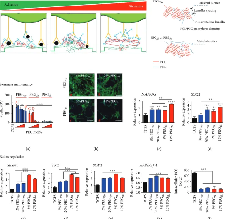
ROS accumulation (Figure 2) [52].
Furthermore, nano
fibrous scaffolds provided a 3D
microenvironment to stem cells and thus enhanced
stem-ness. For example, ASCs displayed an improved adhesion
capacity with high rates of bioactivity and proliferation when
cultured on emu oil-loaded nanofibers [53]. Mesenchymal
stem cells (MSCs) showed a superior differentiation capacity
towards typical mesenchymal lineages when grown in a
nanostructured electrospun gelatin patch [54]. Especially,
emu oil exhibited a free radical scavenging activity, thereby
enhancing stemness [55].
Finally, a chitosan
film induced spheroid formation and
triggered a cell-cell interaction of hASCs, thereby enhancing
stemness. After spheroid formation, the spheroid-forming
hASCs expanded efficiently, formed a colony, and
upregu-lated the expression of pluripotency marker genes compared
to the monolayer-cultured control condition [56, 57].
In conclusion, the aforementioned types and formats of
biomaterials were found to enhance or maintain stemness.
We summarize the three major mechanisms by which the
biomaterials enhanced or maintained stemness as follows:
(1) reduction of cytoskeletal tension by reducing matrix
stiff-ness, (2) spheroid formation by reduction of integrin-binding
sites and consequent promotion of cell-cell interaction, and
(3) antioxidative effects by radical scavenging activity. These
strategies have potential to e
ffectively improve stemness of
MSCs in various biomaterial formats.
3. Direct Differentiation of MSCs
Using Biomaterials
While most methods known to induce mesenchymal
line-age differentiation of MSC depend on exposure to one or
more soluble growth factors, a growing body of evidence
suggests that it is possible to control MSC di
fferentiation in
the absence of soluble factors. MSCs exhibit the ability to
di
fferentiate towards specific lineages through biomaterials
with modification of mechanical or biochemical properties,
matrix composition, topography, and surface stiffness. This
Table 3: Continued.Property Type of
materials Differentiation Details of materials Comments Ref.
Angiogenesis PEGylatedfibrin 3D matrix
Endothelial differentiation of MSC was induced by the 3D PEGylated
fibrin matrix. [64] Surface topograpy Film Neurogenesis/ myogenesis Micropatterned poly(lactic-co-glycolic acid) (PLGA) ultrathinfilm
Micropattering: microsize lanes of 20μm width separated by 40μm wide grooves on a PLGA ultrathinfilm (16.3 ± 1.5 μm)
[66]
Hydrogel Adipogenesis/neurogenesis
Hydrazine-treated polyacrylamide gel (circular and anisotropic
geometry)
Cells cultured in small circular islands show elevated expression of adipogenesis
markers while cells that spread in anisotropic geometries elevated expression of neurogenic markers.
[67]
Bioinorganics
Osteogenesis/
neurogenesis Graphene/electrical stimulation
Specific combinations of nonbiological inputs—material type, electrical stimulation, and physical patterns on graphene substrates regulated hMSC
lineage specification.
[70]
Osteogenesis Nanotubule-shaped titanium oxide surface
Small (30 nm diameter) nanotubes promoted cell adhesion without noticeable differentiation, whereas larger
(70 to 100 nm diameter) nanotubes elicited a dramatic stem cell elongation
(10-fold increased), which induced cytoskeletal stress and selective differentiation into osteoblast-like cells.
[71]
Osteogenesis Titanium substrate
Surface microstructure and surface energy from microstructured Ti substrate were able to direct osteogenic differentiation of mesenchymal stem cells.
approach would simplify the tissue engineering procedure
and be cost-effective. Here, we summarize recent studies
that employed such approaches to induce direct
differenti-ation of MSC via biomaterial technologies (Table 3).
3.1. Composition. Collagen and glycosaminoglycan (GAG),
major components of natural ECM, play a key role in
osteo-chondral regeneration. Hence, their combined (CG) sca
ffolds
have been used successfully in tissue engineering applications
for regeneration of cartilage and bone [57, 58]. A previous
study reported the effect of the composition and stiffness of
collagen and GAG scaffolds composed of chondroitin
sul-phate (CS) and hyaluronic acid (HA) on MSC differentiation
[59]. The study showed that the lowest stiffness (0.5 kPa) of
the CG scaffold facilitated chondrogenesis, while the stiffest
(1.5 kPa) scaffold induced osteogenesis. This was the first
evi-dence proving that osteochondral di
fferentiation of MSC
could be directed via sca
ffold composition using CG and
fur-ther enhanced by the GAG type. When cellulose, anofur-ther
abundant natural polymer, was blended with silk at di
fferent
compositions, growth and chondrogenesis of MSC were
Table 4: iPSC reprogramming and type of gene transfection.Type Advantages Disadvantages Transgene
expression Efficiency Ref.
Virus
Adenovirus Nonintegrative; infects
dividing and nondividing cells Low efficiency No 0.0001~0.01% [84, 85]
Lenti/retrovirus
Ease of handling with experience; medium–high
efficacy
Integration of foreign DNA into genome; residual expression
of reprogramming factors; controversy regarding tumor
formation Yes 0.1~1% [73, 125] Sendai virus Medium–high efficiency; nonintegrating; robust protein-expressing property;
wide host range
Involve viral transduction No 0.5~1.0% [88, 89]
Plasmid vector Episomal
Nonintegrative; simple to implement to laboratory setup; less time-consuming
Very low efficiency; the use of potent viral oncoprotein
(SV40LT antigen)
No 3–6 × 10 − 6 [87, 126]
Minicircle More persistent transgene
expression; lack bacterial origin Very low efficiency No 0.01% [127]
miRNA
Relative high efficiency; nonintegration; easily automated, making it an
exciting candidate for routine biomanufacture.
Requires high gene dosages and multiple transfections; daily transfection; controversy in reproducibility and mitigating
cost effectiveness No 1.4~2% [128, 129] PiggyBac transposons Elimination of insertional mutagenesis; no footprint upon excision; higher genome
integration efficiency
Inefficient excision, potential for genomic toxicity
Excision with
transposase 0.1~1% [80]
Protein
Free of genetic materials; direct delivery of reprogramming
factor proteins
Slow kinetics, low efficiency; difficulties in generation and purification of reprogramming
protein
No 0.005~0.001% [130]
Small molecules Ease of handling; no requirements
for reprogramming factors More than one target, toxicity No 0.3~0.5% [86] Table 5: iPSC reprogramming and donor cell type.
Donor cell type Ref.
Adipose-derived stem cells [131]
Amnioticfluid [132]
Blood cell cord blood stem cells [104]
B lymphocytes [133]
Bone marrow cells [134]
Cardiac myocytes [135]
Dental pulp [136]
Dermalfibroblasts [137]
Endometrial stromalfibroblasts [100]
Hematopoietic progenitor cells [138]
Hepatocytes [139]
Keratinocytes (from hair pluck) [101]
Pancreaticβ-cells [140]
promoted [60]. This was also the
first report demonstrating
the potential of cellulose to induce chondrogenic
differentia-tion of MSC.
Recent studies investigated whether chondrogenic
differ-entiation of MSC could be directed by biomimetic or
decellu-larized tissue-derived materials. Biomimetic polyacrylate
substrates functionalized with the RGD integrin-binding
peptide promoted chondrogenesis of MSC in the absence of
any soluble growth factors [61]. They suggested that the
amount of surface amine residues from the RGD peptide
was a key regulator to inducing the differentiation. Several
studies demonstrated that ECM components derived from
the cartilage promoted chondrogenesis of MSC [62–64]. In
all cases, however, the use of chondrogenic growth factors
was found to be essential for MSC chondrogenesis with
deposition of necessary matrix components. In addition, a
study reported that novel particles derived from natural
car-tilage ECM induced chondrogenic di
fferentiation of MSC
even in the absence of TGF-
β when the particles were used
as a cell carrier [65].
Calcium phosphate- (CaP-) based ceramics play a
sig-nificant role in bone repair due to their osteoconductive
300
Stemness maintenance
Redox regulation
200 A
PEG750 PEG2k PEG5k
B ⁎ ⁎⁎⁎⁎ C D 100 # cells/FO V 0 PEG mol% (a) (b) (c) (d) (e) (f) (g) (h) (i) T CPS PEG 750 PEG 2k Rela ti ve exp re ssio n 3 NANOG 2 1 0 T CPS 5% PEG 750 20% PEG 750 5% PEG 2k 10% PEG 2k ⁎⁎ ⁎⁎ ⁎⁎ ⁎⁎⁎⁎⁎⁎ SOD2 Rela ti ve exp re ssio n 3 4 5 SOX2 2 1 0 T CPS 5% PEG 750 20% PEG 750 5% PEG 2k 10% PEG 2k ⁎⁎ ⁎⁎ ⁎ ⁎⁎⁎ Rela ti ve exp re ssio n 6 SESN1 4 2 0 T CPS 5% PEG 750 20% PEG 750 5% PEG 2k 10% PEG 2k ⁎⁎⁎ ⁎⁎⁎ Rela ti ve exp re ssio n Rela ti ve exp re ssio n 4 TRX 2 3 1 0 2 3 1 0 T CPS 5% PEG 750 20% PEG 750 5% PEG 2k 10% PEG 2k T CPS 5% PEG 750 20% PEG 750 5% PEG 2k 10% PEG 2k ⁎⁎⁎⁎⁎⁎ Rela ti ve exp re ssio n APE/Ref-1 1.0 1.5 2.0 0.5 0.0 T CPS 5% PEG 750 20% PEG 750 5% PEG 2k 10% PEG 2k ⁎⁎⁎ ⁎⁎⁎ In tracell ula r R O S (RFU) 400 600 800 200 0 T CPS 5% PEG 750 20% PEG 750 5% PEG 2k 10% PEG 2k ⁎⁎⁎ Stemness Adhesion PEG750 PEG2k or PEG5k
PCL/PEG amorphous domains PCL crystalline lamellae Lamellar spacing Material surface Material surface PEG PCL
Figure 2: PEG chain length-dependent interactions with the PCL matrix enable stemness maintenance. Proper control of surface repellency by copolymerizing PEG2kwith PCL in a culture substrate form can improve stemness as cell-cell interaction increases relatively to cell-matrix interaction, thereby forming pseudo cell spheroids. Figure 2 is reproduced with permission from [52], American Chemical Society. All bars are mean± S.D.∗p < 0 05,∗∗p < 0 01,∗∗∗p < 0 001,∗∗∗∗p < 0 0001 relative to TCPS or as indicated between the lines.
potential for
filling lost bone volumes, when CaP
nanopar-ticles and demineralized bone matrix (DBM) were fabricated
as an injectable bone graft by incorporating polymerized
high internal phase emulsions (polyHIPEs). This injectable
bone graft was found to induce direct osteogenesis of MSC
[58]. On the other hand, our recent study investigated the
potential of 3D graphene substrates to induce spontaneous
osteogenesis of MSC without additional stimuli [59]. These
reports revealed that material-derived cues were able to
guide MSC differentiation to osteogenesis in the absence
of extrinsic biochemical inputs.
Proper regeneration of the myocardium is dependent on
scaffold properties and thus can be enhanced by mimicking
features of the myocardial ECM. A three-hydroxybutyrate
and 3-hydroxyvalerate (PHBHV)/gelatin construct
mimick-ing the myocardial ECM structure was developed to promote
cardiac di
fferentiation of MSC and cardiac resident cells
without any chemical stimulation [66]. This study
demon-strated that when specific physicochemical properties with
a microtopograph were produced to mimic the structural
and mechanical properties of myocardium in the PHBHV/
gelatin construct, myogenesis of the stem cells was promoted.
Electroconductive carbon nanotubes (CNT) demonstrated
an ability to induce myogenic differentiation of MSC in the
absence of additional stimuli [67]. Although the exact
mech-anism of this result is unclear, it was suggested that electrical
stimulation of MSC by culturing on the CNT-based
polylac-tic acid sca
ffold was a key factor to enhancing differentiation
to cardiomyocytes.
3.2. Substrate Stiffness. Among the biophysical cues that were
identified to regulate cell fate in static in vitro cultures,
stiff-ness of culture substrates was suggested
first as a key property
in several important studies [60–64]. An increased stiffness
of the culture substrate was found to induce osteogenic
dif-ferentiation of MSCs [60, 61]. The role of matrix sti
ffness in
directing lineage speci
fication of MSCs was examined on
the surface-charged methyl acrylate/methyl methacrylate
HO HO HO HO H2O2 HRP HO HO HO HO HO HO OH Nuclei 5% :0.005% 7% :0.005% 7% :0.01% 5% :0.005% + 7% :0.005% 7% :0.01% 5% :0.005% 7% :0.005% 7% :0.01% T CPS He ar t Mu sc le + controls Br ai n T CPS 5% :0.005% + 7% :0.005% 7% :0.01% TCPS mRN A exp ressio n (r el . t o co n tr o l) CD31 Flk1 Merge O O HO OH OH GHPA + H2O2 GHPA + HRP + cells OH OH OH
Injectable system Sprayable system
Vascular-endothelial Myogenin Myogenic 12 10 8 6 4 2 0 CD31 TCPS 5% : 0.005% 7% : 0.005% 7% : 0.01% ⁎ ⁎ ⁎ Flk1 In vitro EC marker expression MyoD NSE Neural Trk-A NFM NFL VEGFA Flk1 CD31 ANGPT1 ANGPT2 Tie1 Tie2 VE-cad vWF GAPDH ⁎ ⁎ ⁎
Figure 3: Gelation of GHPA by H2O2and horseradish peroxidase-catalyzed cross linking. In vitro endothelial differentiation of hMSCs in GHPA hydrogels. Figure 3 is reproduced with permission from [63], John Wiley and Sons. ∗ indicatesp < 0 05 in comparison to the control MSCs on tissue culture plate.
(MA/MMA) polymer substrate with varying elastic
modu-lus [60]. This study demonstrated that the substrate group
with lower sti
ffness induced chondrogenesis of MSCs while
the substrate with rigid stiffness induced osteogenic
specifi-cation of MSCs. Although its specific mechanism is unclear,
this study revealed that integrin
β1 played a critical role in
this process. Cells sense their mechanical microenvironment
via integrin-ligand interactions which form focal adhesions
and thereby regulate intracellular signaling [65]. Another
study supported this
finding [61] by showing that the soft
(
~0.5 kPa) substrate was effective in promoting
neurogen-esis of MSCs whereas the stiff (~40 pKa) one was effective
in promoting their osteogenesis. Switching the biophysical
microenvironment of MSCs from soft to stiff or stiff to
soft substrates led to rewiring the two directions of MSC
lineage specification.
(A) (B) (C) (D) (E) (F) (a) 400 300 200 iPS cell co lo nies/p la te 100 heiPS nfiPS p = 0.0032 0.4 0.5 0.3 0.2 % iPS co lo ny f or m at io n 0.1 heiPS nfiPS p = 0.003 (A) (C) (D) (B) (b) NANOG OCT4 10 8 6 hEM C1 4 2 10 8 6 hEM C2 4 2 10 8 6 4 2 10 8 6 4 2 10 8 6 hEM C3 4 2 30 nf Fo ld ind uc tio n Fo ld ind uc tio n ov er nf 20 10 30 20 10 d0 d4 d8 d0 d4 d8 10 8 6 4 2 (c)Figure 4: Pluripotency reprogramming of human endometrial cells (hEMC). hEMC-derived iPS (heiPS) showed higher expression of pluripotent markers compared to neonatalfibroblasts. Figure 4 is reproduced with permission from [100], Oxford University Press.
Although the differentiation potential of MSCs into
endothelial cells (ECs) remains unclear [68, 69], some studies
reported possible approaches to differentiate MSCs into ECs
[63, 64]. A previous study demonstrated that a 3D matrix
with tunable properties directed the di
fferentiation of MSC
towards vascular cell types [64]. We also applied in situ
cross-linkable gelatin hydrogels by conjugating
enzymati-cally cross-linkable hydroxyphenyl propionic acid (GHPA)
(Figure 3) [63]. The 3D culture of MSCs in these hydrogels
induced vasculogenesis both in vitro and in vivo. Our results
showed that GHPA hydrogels induced spontaneous
endo-thelial differentiation of MSC without any soluble factors.
3.3. Surface Topography. When cells are cultured on
bioma-terial substrates, surface topography is known as a key
reg-ulator of cell behavior. Several previous studies reported
that surface topographical cues induced direct lineage
spec-i
fication of MSCs [66, 67, 70–72]. Microfeatures (40 μm
line, 20
μm spacing, and 1 μm height) of fibronectin strips
printed on a poly(lactic-co-glycolic acid) (PLGA) thin
film
were found to direct linage commitment of MSCs [66]. In
this study, modi
fication of MSC morphology and cytoskeletal
arrangement on the patterned
film resulted in both neuronal
and myogenic lineages, even if myogenic differentiation was
dominant when the expression of functional proteins was
examined. Along the same line to direct MSC
differentia-tion, Lee et al. fabricated pseudo-3D microwells by
templat-ing a hydrazine-immobilized polyacrylamide gel displaytemplat-ing
inverse features of circular surface topography via PDMS
stamps (circular) [67]. As a result, small circular islands
induced an adipogenic phenotype of MSCs while anisotropic
geometries induced neurogenic di
fferentiation. Micro- and
nanostructured titanium surfaces were used as potential
topographical cues to induce osteogenic differentiation of
MSCs [71, 72]. The nanotubule-shaped titanium oxide
surface structures, which have 70 to 100 nm titanium oxide
nanotube arrays on them, induced cytoskeletal stress and
thereby directed osteogenic differentiation of MSCs [71].
It was also reported that osteoblastic di
fferentiation of MSCs
was induced on the microstructured titanium surface (
Ra =
3 22 μm) through α2β1 integrin-mediated interactions with
cocultured osteoblasts [72]. Our recent study determined
causative roles of topographical cues in directing lineage
speci
fication of MSC via patterned graphene surfaces with
additional evaluation of electrical stimulation as another
cue [70]. Our result showed that expression of
osteoprogeni-tor markers was increased by either (un)patterned graphene
substrate or electrical stimulation while the expression of
osteoblast makers was increased only when electrical
stimu-lation was applied together with the surface patterns.
4. Selection of Genetic Factor and Source Cell
Type for iPSC Reprogramming
Induced pluripotent stem cells (iPSCs) were introduced in
2006, which opened a new avenue for stem cell research
and regenerative medicine [73]. Obtaining an adequate
amount of stem cells is a major limitation for stem cell
ther-apy and research. Previously, classical methods employed to
induce pluripotency of somatic cells include somatic cell
nuclear transfer (SCNT) and cell fusion. However,
limita-tions associated with oocyte supply, low reprogramming
effi-ciency, and phenotypic abnormalities of the produced animal
o
ffspring still hamper the widespread distribution of these
classical methods [74].
Resident stem cells in various tissues were also studied as
a promising source of stem cells, but the lack of appropriate
markers to de
fine their phenotypes and their low
differentia-tion potential were considered as major hurdles for using
these cell sources. Hence, the breakthrough idea of
repro-gramming somatic cells with ectopic pluripotent markers
(Sox2, Oct3/4, Klf4, c-Myc, and Lin28) to eventually
rep-resent embryonic stem cell (ESC) characteristics was
undisputedly attractive [73]. However, it is still
controver-sial whether iPSCs possess the same pluripotency and
dif-ferentiation ability to ESCs. Donor cell-speci
fic epigenetic
signatures remain even after reprogramming and thus
gen-erate problematic variations from the expected quality and
characteristics of iPSCs in terms of homogeneity and the
potential for maturation in stem cell therapy [75, 76].
More-over, abnormalities created during the process of iPSC
repro-gramming, such as stablishing aberrant DNA methylation
patterns, were found to increase the heterogeneity in iPSCs
[77, 78]. Additionally, donor-specific genetic variations
fur-ther increase the heterogeneity of iPSC genetic pro
filing, such
as stablishing aberrant DNA methylation patterns.
4.1. Choice of Vectors for iPSC Reprogramming. Substantial
progress has been made in the methodologies to improve
the efficiency and efficacy of reprogramming somatic cells
to iPSCs in the past decade (Table 4). The type of vectors
used to overexpress ectopic pluripotency factors within the
target cells are classified into integrating DNA vectors and
nonintegrating DNA free vectors. Integrating vectors are
further subclassified into insertional vectors including viral
and linear DNA delivery systems whereas insertion-free
transgene vectors include PiggyBac transposon [79, 80] and
plasmid/episomal vectors. Recently, nonintegrating systems
involving direct protein or microRNA vectors as well as
var-ious small molecules are used for reprogramming of somatic
cells into iPSCs [81, 82]. Integrating DNA vectors represent
an early generation tool for reprogramming and are still
commonly used in experiments owing to their high
effi-ciency. Combinations of retroviral or lentiviral Sox2, Oct4,
Klf4, c-Myc, or Lin28 were most popularly used with or
with-out the use of transgene selection markers. Especially, these
first-generation viral vectors possess the potential for
ran-dom insertional mutagenesis, but their undeniable high e
ffi-ciency still renders them useful for a wide range of iPSC
research. Such random insertional mutagenesis contributes
to the unpredictability of iPSCs upon in vivo transplantation.
Viral promoter-driven
fluorescence and cre-LOX expression
systems have been used to track and control ectopic gene
expression but still generate insertional mutagenesis [83].
In order to overcome problems associated with mutagenesis
resulting from ectopic gene insertion, adenoviral, episomal,
or plasmid vectors were used as alternatives in the course of
developing the next generation of reprogramming methods
[84, 85]. However, although these alternatives were less
prone to mutagenesis associated with ectopic gene
integra-tion, the major hindrance was the poor transfection
effi-ciency, displaying e
fficiencies 1000–10,000 folds lower than
those of conventional viral vectors. Further improvement in
the reprogramming e
fficiency was achieved by applying
non-DNA methods using Sendai viral vectors, small
mole-cules, Lipofectamine, or miRNA transfections [86
–90]. For
example, cell membrane-penetrating proteins were tagged
with Oct4, Sox2, Klf4, and c-myc for intracellular
deliv-ery and cell reprogramming. Human immunodeficiency
virus transactivator of transcription (HIV-TAT) protein
or polyarginine-tagged pluripotency factors were used to
derive mouse and human iPSC lines. However, its low
reprogramming e
fficiency still remains as a major hurdle
to overcome [91]. Together, the aforementioned
noninte-grating methods are promising to signi
ficantly reduce
mutagenesis, but their reprogramming e
fficiencies need
to be enhanced further as the e
fficiencies are still considerably
lower (0.001%–) compared to integrating vectors (0.1%–1%).
Generating transgene-free iPSCs serves as an attractive
alternative because it can compensate for the low
transfec-tion efficiency. Additransfec-tion of small molecules including
his-tone deacetylase (HDAC) inhibitors and other epigenetic
modifiers have been reported as representative examples
[92, 93]. On the other hand, the number of ectopic vectors
could be reduced by introducing supplementary compounds,
where inhibitors of G9a histone methyltransferase could
replace either Sox2, Oct4, or c-Myc during
reprogram-ming of neural progenitor cells (NPCs) and
fibroblasts in
mice [94]. The TGF-
β receptor antagonist also significantly
increased the reprogramming efficiency and kinetics in
murine [95, 96] and human
fibroblasts [97].
It needs to be investigated further whether the type of
vectors used to induce pluripotency contributes to the
het-erogeneity of produced iPSC lines or not. When retrovirus,
Sendai virus, and episomal vectors were used for iPSC
gener-ation, di
fferent reprogramming strategies were applied to
obtain human iPSCs. As a result, very similar global gene
expression pro
files were displayed but subtle differences were
observed in the levels of gene expression, indicating that the
heterogeneity of produced iPSC lines resulted from clonal
signatures rather than the reprogramming method itself
[98]. However, even when iPSCs were generated from cells
of the same donor, characteristic aberrations in DNA
meth-ylation at the epigenomic level were shown to be dependent
on the choice of reprogramming factors [99].
As a summary, random DNA aberrations are most
notably caused by viral genome integration, leading to iPSC
heterogeneity and unpredictability. Thus, nonintegrating
systems should be primarily considered as a basic means
for differentiation strategies towards clinical applications.
4.2. Donor Cell Characteristics and Stemness. The origin and
quality of donor cells are also important factors to ensure
successful reprogramming results. In particular, easy access,
abundant quantity, and enough replenishment of donor cells
after harvesting should be considered when the target donor
source is selected.
Donor somatic cell-speci
fic transcriptional and epigenetic
signatures significantly contribute to the heterogeneity of
effi-ciency and efficacy in reprogramming and differentiation of
iPSCs (Table 5). The most widely used cell source for
repro-gramming into iPSCs is dermal
fibroblasts, most frequently
harvested from neonates as well as adults [73]. Keratinocytes
(a type of bone marrow cells), peripheral blood cells (a type
of CD34
+peripheral blood mononuclear cells), amniotic
fluid
cells, cord blood stem cells, endometrial stromal
fibroblasts,
and dental pulp cells have been reported as reliable sources
of somatic cells for reprogramming (Figure 4) [100–106].
Moreover, it has been reported, even in cells which were
orig-inated from the same donor but from different organs, that
the tissue-speci
fic epigenetic signatures affect the
heteroge-neity of e
fficiency and efficacy in reprogramming and the
di
fferentiation potential of iPSC lines. Such observations
were prominent in the early passages when the
reprogram-ming process is not yet complete [107
–109].
If stemness is enhanced, the negative effect of
tissue-specific epigenetic signature may be attenuated during the
process of serial passaging while losing the epigenetic
mem-ory sequentially. Characteristic DNA methylation patterns
are originated from the donor cells and thus can be tracked
in specific iPSC clones. Consequently, limitations in
stem-ness of iPSCs as opposed to the full pluripotency of
embry-onic stem cells are inevitable. Experimental techniques to
overcome the gap between the epigenetic memory of the
donor cell and the stemness of the derived iPSC lines have
been described in previous studies [110, 111]. One strategy
is to increase the iPSC passage number while another
approach is to introduce chromatin-modifying substances,
which diminishes the epigenetic memory and enhances
stemness. The process of acquiring pluripotency may not be
complete upon immediate silencing expression of exogenous
pluripotency factors but may continue for several rounds of
cell passaging. iPSCs exhibit considerable di
fferences in their
telomere length and the global pattern of transcription and
DNA methylation [110, 112, 113]. On the other hand,
transgenes are usually silenced in the process of
reprogram-ming by de novo methylation. When this process is not
fully accomplished, gaining the pluripotency of the
repro-grammed cells primarily relies on the exogenously
intro-duced factors. When the endogenous pluripotent genes
are halted from being fully expressed, these colonies are
defined as “partially reprogrammed.” Within such colonies,
pluripotency is frequently not fully acquired even after the
exogenous factors are eventually turned o
ff [114, 115].
Con-versely, when ectopic transgenes are not silenced and
exposed to residual activities or reactivation of the viral
trans-genes in the iPSC cells, tumor formation occurs as
demon-strated in chimera experiments [73]. Other potential causes
of epigenetic differences have also been attributed to either
aberrant or incomplete reprogramming or even by various
cell culture conditions [77, 116–119].
5. Conclusion
Although a growing body of evidence suggests stem cells as a
promising candidate for cell therapy in the position of
replacing somatic cells, the aforementioned issues regarding
senescence and low differentiation efficiency must be
addressed for successful clinical applications. In this review,
we introduced state-of-the-art methods which are currently
approached to improve e
fficiency and efficacy of stemness
maintenance, direct di
fferentiation, and iPSC
reprogram-ming, with the minimal use of expensive and side e
ffect-occurring growth factors. Biophysical stimulation, organic
compound treatment, genetic transfection, and various types
of biomaterials were employed to achieve the purposes. In
particular, the effects of matrix stiffness, improving cell-cell
interaction, and antioxidant treatment became a major
part of interest. Additionally, biomaterials with specific
composition, stiffness, and topography can serve as a
promising toolbox to guide direct di
fferentiation of stem
cells. Finally, several combinations or individual uses of
genetic factors to induce reprogramming of somatic cells
were introduced as a means of generating iPSCs. Pros
and cons of major reprogramming methods were
dis-cussed as well. Taken together, selection of biomaterials or
other external factors needs to be customized for
target-specific developments and application of stem cell therapy
towards successful clinical applications.
Conflicts of Interest
The authors declare that they have no con
flicts of interest.
Acknowledgments
This study was
financially supported by the Basic Science
Research Program through the National Research
Founda-tion of Korea (NRF) funded by the Ministry of Science,
ICT and Future Planning (NRF-2016M3A9E9941743 and
2017M3A9E9087117).
References
[1] O. Ringdén, M. Uzunel, I. Rasmusson et al.,“Mesenchymal stem cells for treatment of therapy-resistant graft-versus-host disease,” Transplantation, vol. 81, no. 10, pp. 1390– 1397, 2006.
[2] D. Polchert, J. Sobinsky, G. Douglas et al.,“IFN-γ activation of mesenchymal stem cells for treatment and prevention of graft versus host disease,” European Journal of Immunology, vol. 38, no. 6, pp. 1745–1755, 2008.
[3] R. Yanez, M. L. Lamana, J. Garcia-Castro, I. Colmenero, M. Ramirez, and J. A. Bueren,“Adipose tissue-derived mes-enchymal stem cells have in vivo immunosuppressive proper-ties applicable for the control of the graft-versus-host disease,” Stem Cells, vol. 24, no. 11, pp. 2582–2591, 2006. [4] E. J. Koay and K. A. Athanasiou,“Development of
serum-free, chemically defined conditions for human embryonic stem cell–derived fibrochondrogenesis,” Tissue Engineering Part A, vol. 15, no. 8, pp. 2249–2257, 2009.
[5] L. A. Solchaga, K. Penick, J. D. Porter, V. M. Goldberg, A. I. Caplan, and J. F. Welter,“FGF-2 enhances the mitotic and chondrogenic potentials of human adult bone marrow-derived mesenchymal stem cells,” Journal of Cellular Physiol-ogy, vol. 203, no. 2, pp. 398–409, 2005.
[6] S. Tsutsumi, A. Shimazu, K. Miyazaki et al.,“Retention of multilineage differentiation potential of mesenchymal cells during proliferation in response to FGF,” Biochemical and Biophysical Research Communications, vol. 288, no. 2, pp. 413–419, 2001.
[7] D. James, A. J. Levine, D. Besser, and A. Hemmati-Brivanlou, “TGFβ/activin/nodal signaling is necessary for the mainte-nance of pluripotency in human embryonic stem cells,” Development, vol. 132, no. 6, pp. 1273–1282, 2005.
[8] M. Sakaki-Yumoto, Y. Katsuno, and R. Derynck,“TGF-β fam-ily signaling in stem cells,” Biochimica et Biophysica Acta (BBA) - General Subjects, vol. 1830, no. 2, pp. 2280–2296, 2013. [9] H. M. van Beuningen, H. L. Glansbeek, P. M. van der Kraan, and W. B. van den Berg,“Differential effects of local appli-cation of BMP-2 or TGF-β1 on both articular cartilage com-position and osteophyte formation,” Osteoarthritis and Cartilage, vol. 6, no. 5, pp. 306–317, 1998.
[10] L. Z. Sailor, R. M. Hewick, and E. A. Morris, “Recombi-nant human bone morphogenetic protein-2 maintains the articular chondrocyte phenotype in long-term culture,” Journal of Orthopaedic Research, vol. 14, no. 6, pp. 937– 945, 1996.
[11] C. A. Hellingman, W. Koevoet, N. Kops et al., “Fibroblast growth factor receptors in in vitro and in vivo chondrogene-sis: relating tissue engineering using adult mesenchymal stem cells to embryonic development,” Tissue Engineering Part A, vol. 16, no. 2, pp. 545–556, 2010.
[12] A. De Becker and I. Van Riet, “Homing and migration of mesenchymal stromal cells: how to improve the efficacy of cell therapy?,” World Journal of Stem Cells, vol. 8, no. 3, pp. 73–87, 2016.
[13] H. M. Lazarus, S. E. Haynesworth, S. L. Gerson, N. S. Rosenthal, and A. I. Caplan,“Ex vivo expansion and subse-quent infusion of human bone marrow-derived stromal pro-genitor cells (mesenchymal propro-genitor cells): implications for therapeutic use,” Bone Marrow Transplantation, vol. 16, no. 4, pp. 557–564, 1995.
[14] S. P. Bruder, N. Jaiswal, and S. E. Haynesworth, “Growth kinetics, self-renewal, and the osteogenic potential of purified human mesenchymal stem cells during extensive subcultiva-tion and following cryopreservasubcultiva-tion,” Journal of Cellular Bio-chemistry, vol. 64, no. 2, pp. 278–294, 1997.
[15] K. Ksiazek,“A comprehensive review on mesenchymal stem cell growth and senescence,” Rejuvenation Research, vol. 12, no. 2, pp. 105–116, 2009.
[16] J. Lam, S. Lu, E. J. Lee et al.,“Osteochondral defect repair using bilayered hydrogels encapsulating both chondrogeni-cally and osteogenichondrogeni-cally pre-differentiated mesenchymal stem cells in a rabbit model,” Osteoarthritis and Cartilage, vol. 22, no. 9, pp. 1291–1300, 2014.
[17] F. Barry, R. E. Boynton, B. Liu, and J. M. Murphy, “Chondro-genic differentiation of mesenchymal stem cells from bone marrow: differentiation-dependent gene expression of matrix components,” Experimental Cell Research, vol. 268, no. 2, pp. 189–200, 2001.
[18] H. J. Lee, B. H. Choi, B. H. Min, and S. R. Park, “Low-inten-sity ultrasound inhibits apoptosis and enhances viability of human mesenchymal stem cells in three-dimensional algi-nate culture during chondrogenic differentiation,” Tissue Engineering, vol. 13, no. 5, pp. 1049–1057, 2007.
[19] K. Park, K. J. Cho, J. J. Kim, I. H. Kim, and D. K. Han, “Func-tional PLGA scaffolds for chondrogenesis of
bone-marrow-derived mesenchymal stem cells,” Macromolecular Biosci-ence, vol. 9, no. 3, pp. 221–229, 2009.
[20] F. Padilla, R. Puts, L. Vico, A. Guignandon, and K. Raum, “Stimulation of bone repair with ultrasound,” Advances in Experimental Medicine and Biology, vol. 880, pp. 385–427, 2016.
[21] R. T. Brady, F. J. O'Brien, and D. A. Hoey, “Mechanically stimulated bone cells secrete paracrine factors that regulate osteoprogenitor recruitment, proliferation, and differentia-tion,” Biochemical and Biophysical Research Communica-tions, vol. 459, no. 1, pp. 118–123, 2015.
[22] M. J. Go, C. Takenaka, and H. Ohgushi, “Forced expres-sion of Sox2 or Nanog in human bone marrow derived mesenchymal stem cells maintains their expansion and differentiation capabilities,” Experimental Cell Research, vol. 314, no. 5, pp. 1147–1154, 2008.
[23] D. S. Yoon, Y. H. Kim, H. S. Jung, S. Paik, and J. W. Lee, “Importance of Sox2 in maintenance of cell proliferation and multipotency of mesenchymal stem cells in low-density culture,” Cell Proliferation, vol. 44, no. 5, pp. 428– 440, 2011.
[24] T. M. Liu, Y. N. Wu, X. M. Guo, J. H. P. Hui, E. H. Lee, and B. Lim,“Effects of ectopic Nanog and Oct4 overexpression on mesenchymal stem cells,” Stem Cells and Development, vol. 18, no. 7, pp. 1013–1022, 2009.
[25] M. Ranzani, D. Cesana, C. C. Bartholomae et al.,“Lentiviral vector–based insertional mutagenesis identifies genes associ-ated with liver cancer,” Nature Methods, vol. 10, no. 2, pp. 155–161, 2013.
[26] H. Chen, X. Liu, W. Zhu et al.,“SIRT1 ameliorates age-related senescence of mesenchymal stem cells via modulating telo-mere shelterin,” Frontiers in Aging Neuroscience, vol. 6, p. 103, 2014.
[27] D. S. Yoon, Y. Choi, Y. Jang et al.,“SIRT1 directly regulates SOX2 to maintain self-renewal and multipotency in bone marrow-derived mesenchymal stem cells,” Stem Cells, vol. 32, no. 12, pp. 3219–3231, 2014.
[28] D. S. Yoon, Y. Choi, S. M. Choi, K. H. Park, and J. W. Lee, “Different effects of resveratrol on early and late passage mesenchymal stem cells through β-catenin regulation,” Biochemical and Biophysical Research Communications, vol. 467, no. 4, pp. 1026–1032, 2015.
[29] D. Harman,“Aging: a theory based on free radical and radi-ation chemistry,” Journal of Gerontology, vol. 11, no. 3, pp. 298–300, 1956.
[30] W. Zhu, J. Chen, X. Cong, S. Hu, and X. Chen,“Hypoxia and serum deprivation-induced apoptosis in mesenchymal stem cells,” Stem Cells, vol. 24, no. 2, pp. 416–425, 2006.
[31] S. Sart, L. Song, and Y. Li,“Controlling redox status for stem cell survival, expansion, and differentiation,” Oxidative Medicine and Cellular Longevity, vol. 2015, Article ID 105135, 14 pages, 2015.
[32] Y.-J. Surh, J. Kundu, and H.-K. Na,“Nrf2 as a master redox switch in turning on the cellular signaling involved in the induction of cytoprotective genes by some chemopreventive phytochemicals,” Planta Medica, vol. 74, no. 13, pp. 1526– 1539, 2008.
[33] H. Zhu, L. Zhang, K. Itoh et al.,“Nrf2 controls bone marrow stromal cell susceptibility to oxidative and electrophilic stress,” Free Radical Biology & Medicine, vol. 41, no. 1, pp. 132–143, 2006.
[34] S. Nemoto, M. M. Fergusson, and T. Finkel, “Nutrient availability regulates SIRT1 through a forkhead-dependent pathway,” Science, vol. 306, no. 5704, pp. 2105–2108, 2004. [35] D. S. Yoon, Y. Choi, and J. W. Lee,“Cellular localization of NRF2 determines the self-renewal and osteogenic di fferentia-tion potential of human MSCs via the P53–SIRT1 axis,” Cell Death & Disease, vol. 7, no. 2, article e2093, 2016.
[36] N. Z. Kuhn and R. S. Tuan,“Regulation of stemness and stem cell niche of mesenchymal stem cells: implications in tumor-igenesis and metastasis,” Journal of Cellular Physiology, vol. 222, no. 2, pp. 268–277, 2010.
[37] M. F. Brizzi, G. Tarone, and P. Defilippi, “Extracellular matrix, integrins, and growth factors as tailors of the stem cell niche,” Current Opinion in Cell Biology, vol. 24, no. 5, pp. 645–651, 2012.
[38] S. r. Pattabhi, J. S. Martinez, and T. C. S. Keller III, “Decellu-larized ECM effects on human mesenchymal stem cell stem-ness and differentiation,” Differentiation, vol. 88, no. 4-5, pp. 131–143, 2014.
[39] Y. Xiong, J. He, W. Zhang, G. Zhou, Y. Cao, and W. Liu, “Retention of the stemness of mouse adipose-derived stem cells by their expansion on human bone marrow stromal cell-derived extracellular matrix,” Tissue Engineering Part A, vol. 21, no. 11-12, pp. 1886–1894, 2015.
[40] R. Rakian, T. J. Block, S. M. Johnson et al.,“Native extracellu-lar matrix preserves mesenchymal stem cell“stemness” and differentiation potential under serum-free culture condi-tions,” Stem Cell Research & Therapy, vol. 6, no. 1, p. 235, 2015.
[41] B. Antebi, Z. Zhang, Y. Wang, Z. Lu, X. D. Chen, and J. Ling, “Stromal-cell-derived extracellular matrix promotes the pro-liferation and retains the osteogenic differentiation capacity of mesenchymal stem cells on three-dimensional scaffolds,” Tissue Engineering Part C: Methods, vol. 21, no. 2, pp. 171– 181, 2015.
[42] J. Zhang, B. Li, and J. H.-C. Wang,“The role of engineered tendon matrix in the stemness of tendon stem cells in vitro and the promotion of tendon-like tissue formation in vivo,” Biomaterials, vol. 32, no. 29, pp. 6972–6981, 2011.
[43] J. Lee, A. A. Abdeen, A. S. Kim, and K. A. Kilian,“Influence of biophysical parameters on maintaining the mesenchymal stem cell phenotype,” ACS Biomaterials Science & Engineer-ing, vol. 1, no. 4, pp. 218–226, 2015.
[44] S. Ansari, P. Sarrion, M. M. Hasani-Sadrabadi, T. Aghaloo, B. M. Wu, and A. Moshaverinia,“Regulation of the fate of dental-derived mesenchymal stem cells using engineered alginate-GelMA hydrogels,” Journal of Biomedical Materials Research Part A, vol. 105, no. 11, pp. 2957–2967, 2017. [45] K. C. Rustad, V. W. Wong, M. Sorkin et al.,“Enhancement of
mesenchymal stem cell angiogenic capacity and stemness by a biomimetic hydrogel scaffold,” Biomaterials, vol. 33, no. 1, pp. 80–90, 2012.
[46] H.-W. Chien, S.-W. Fu, A.-Y. Shih, and W.-B. Tsai, “Modu-lation of the stemness and osteogenic differentiation of human mesenchymal stem cells by controlling RGD concen-trations of poly(carboxybetaine) hydrogel,” Biotechnology Journal, vol. 9, no. 12, pp. 1613–1623, 2014.
[47] R. J. McMurray, N. Gadegaard, P. M. Tsimbouri et al., “Nanoscale surfaces for the long-term maintenance of mes-enchymal stem cell phenotype and multipotency,” Nature Materials, vol. 10, no. 8, pp. 637–644, 2011.