R E S E A R C H A R T I C L E
Open Access
The frequency of and risk factors for
osteoporosis in Korean patients with
rheumatoid arthritis
Joo-Hyun Lee
1, Yoon-Kyoung Sung
2, Chan-Bum Choi
2, Soo-Kyung Cho
2, So-Young Bang
3, Jung-Yoon Choe
4,
Seung-Jae Hong
5, Jae-Bum Jun
2, Tae-Hwan Kim
2, Jisoo Lee
6, Hye-Soon Lee
3, Dae-Hyun Yoo
2, Bo Young Yoon
1and Sang-Cheol Bae
2*Abstract
Background: The aim of this study was to investigate the prevalence of osteoporosis in rheumatoid arthritis (RA) patients and to analyze the risk factors in these patients using the KORean Observational study Network for Arthritis (KORONA) database.
Methods: Among the RA patients in the KORONA who were recruited between July 2009 and December 2011, postmenopausal women with bone mineral density (BMD) results within one year from the time of KORONA enrollment were included in this study. The baseline characteristics of patients in three groups, defined by BMD results, were compared. The BMD measurement rates and prevalence of osteoporosis in the study patients were calculated in accordance with age and gender subgroups. Multivariable logistic regression analysis was used to explore the association between osteoporosis and demographics and disease-related risk factors.
Results: Of 1322 postmenopausal woman patients with RA in whom BMD was measured within one year of study enrollment, 619 patients (46.8 %) were in the osteoporosis group (T-score≤ −2.5 SD). RA patients with osteoporosis had a higher frequency of previous fractures than those in other groups, especially fractures of the femur (p = 0.004) and wrist (p = 0.042). Advanced age (≥70 years; OR = 2.28, 95 % CI: 1.40–3.58), lower body mass index (<25; OR = 2.14, 95 % CI:1.52–3.02), longer disease duration (≥10 years; OR = 1.46, 95 % CI: 1.07–2.00), higher cumulative glucocorticoid dose (OR = 1.03, 95 % CI: 1.01–1.05), and higher Health Assessment Questionnaire score (OR = 1.37, 95 % CI:1.11–1.69) were independent risk factors for osteoporosis.
Conclusion: A large percentage (90.8 %) of RA patients enrolled in the KORONA cohort had osteoporosis and osteopenia. Nevertheless, BMD measurement rates in this population remained low, despite high risk groups of fractures.
Keywords: Osteoporosis, Rheumatoid arthritis, Frequency, Risk factors Background
Osteoporosis is a well-known extra-articular complica-tion in patients with rheumatoid arthritis (RA) [1]. It is more common in patients with RA than in the general population, due to active systemic inflammation, the use of corticosteroids, and lack of mobility [2, 3]. As a result, patients with RA are at increased risk of fractures,
an outcome that impairs quality of life and leads to mortality [4, 5].
The prevalence of osteoporosis in RA patients is re-ported to be approximately twice that in the general population [4]. The frequency of generalized osteoporosis in patients with RA ranges from 12.3 to 38.9 % in the lum-bar spine and from 6.3 to 36.3 % in the hip [4–6]. Above all, there is at least a two-fold increase in the risk of verte-bral fracture (VFs) in RA patients and a higher risk, up to six-fold, has been reported in patients with long-standing disease [6, 7]. However, studies on early RA have shown
* Correspondence:[email protected]
2Department of Rheumatology, Hanyang University Hospital for Rheumatic
Diseases, Seoul 133-792, Republic of Korea
Full list of author information is available at the end of the article
© 2016 Lee et al. Open Access This article is distributed under the terms of the Creative Commons Attribution 4.0 International License (http://creativecommons.org/licenses/by/4.0/), which permits unrestricted use, distribution, and reproduction in any medium, provided you give appropriate credit to the original author(s) and the source, provide a link to the Creative Commons license, and indicate if changes were made. The Creative Commons Public Domain Dedication waiver (http://creativecommons.org/publicdomain/zero/1.0/) applies to the data made available in this article, unless otherwise stated.
that VFs can be observed in the first year of the disease. As a result, approximately one-third of women with RA report a fracture within five years of follow up [8].
The risk factors for osteoporosis and osteoporotic frac-tures in RA patients are age, disability, low body mass index (BMI), previous non-vertebral fracture, long-standing disease, and glucocorticoids. In particular, regardless of additional risk factors, patients taking glucocorticoids should be screened for osteoporosis. Although aware-ness of osteoporosis by healthcare professionals has in-creased in recent years, it remains underdiagnosed and undertreated [9].
The increased risk of osteoporosis in RA patients is well reported [1, 2] and may be linked to differences in the dis-tribution and interactions of genetic and environmental factors [10, 11]. However, little information is available on the frequency of osteoporosis and bone mineral density (BMD) measurement rates in RA patients and the associ-ated risk factors in South Korea.
Thus, the aims of this study were to investigate the prevalence of osteoporosis in RA patients and to analyze the risk factors in these patients using the KORean Obser-vational study Network for Arthritis (KORONA) database [12], a large, nationally representative Korean RA-specific cohort.
Methods
Study population
KORONA was established in July 2009 by the Clinical Research Center for Rheumatoid Arthritis and funded by the Ministry of Health and Affairs, South Korea. RA pa-tients over the age of 18 who satisfied the 1987 American College of Rheumatology (ACR) classification criteria for RA [13] were recruited by rheumatologists from 23 centers across South Korea as part of KORONA. Among those patients, only postmenopausal women were enrolled in
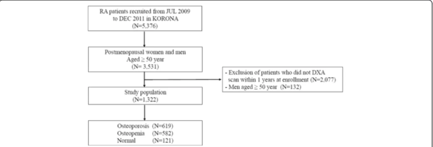
this study. A total of 3531 patients with RA were recruited between July 2009 and December 2011, and 1322 post-menopausal women whose BMD examination results were available within one year from the time of KORONA enrollment were included in this study (Fig. 1). The KORONA protocol was approved by the institutional review boards of Hanyang University Hospital and all participating hospitals, and informed consent was ob-tained from all patients before registration.
Data collection
Demographic and clinical features
All participants in this research completed an initial questionnaire to establish their demographic profile, medical history, and disease-specific outcomes. Disease activity was evaluated by physicians, and joint assessments were made by rheumatologists or well-trained health pro-fessionals. These evaluations, together with laboratory test results, prescription information (glucocorticoids—current and cumulative doses), and patient self-evaluation (history of previous fracture, fracture site, smoking, and alcohol intake), were incorporated into the KORONA database. Laboratory data to identify the characteristics of RA (anti-cyclic-citrullinated protein antibody (ACPA) and rheumatoid factor (RF)) were obtained, and disease activity was monitored using erythrocyte sedimentation rate (ESR) and C-reactive protein (CRP). Disease Activity Score 28 using erythrocyte sedimentation rate (DAS28-ESR) and Health Assessment Questionnaire (HAQ) results were used to assess health-related outcomes. The DAS28-ESR index combines information regarding the number of swollen and tender joints, as well as a measure of general health and acute phase response (ESR) [14]. The HAQ is a model of patient-oriented outcome assessment and is based on the following five patient-centered dimensions: disability, pain, medication effect, cost of care, and mortality [15].
BMD data collection
Only patients with BMD data available within one year from the time of recruitment were included in this study. BMD measurements were carried out with Holo-gic QDR (Waltham, MA, USA) and GE Lunar Prodigy (Madison, WI, USA) systems, using the manufacturers’ standard scan and positioning protocols. Trained techni-cians at each center performed the scanning according to a standardized protocol. Each center followed the manufacturers’ procedures to check the stability and ac-curacy of their equipment using regular measurements of European phantom. However, no cross-calibration was performed on any of the systems. The reference BMD values use Korean data provided by the manufacturers.
Diagnoses of osteopenia and osteoporosis were made using calculations based on the World Health Organization (WHO) T-score criteria (−1.0 to −2.5 SD and ≤ −2.5 SD) [16]. In addition, according to the recommendation of the International Society for Clinical Densitometry, osteopor-osis was diagnosed based on the lowest T-score among the three sites (lumbar spine, femoral neck, and total hip) [16].
Statistical analysis
Baseline characteristics were compared among three pa-tient groups (normal, osteopenia, and osteoporosis) defined
according to BMD results (Table 1). Chi-square, Kruskal-Wallis test and one-way ANOVA were used for categorical, skewed continuous and continuous variables, respectively. BMD measurement rates within one year from the time of enrollment were analyzed, and the frequency of osteopor-osis in the study patients was calculated for age subgroups (Table 2). Lumbar spine, femoral neck, and total hip T-scores in the enrolled RA patients were presented as the mean and standard deviation (SD) according to the patient’s age (Table 3). Finally, multivariable logistic regression analysis was used to explore the association between osteoporosis and the demographics and disease-related risk factors listed in Table 1 (Table 4). Some vari-ables (HAQ, DAS28-ESR, ESR and CRP) that were not normally distributed were log-transformed to approximate a normal distribution for analysis. All analyses were per-formed using SPSS software (version 17.0, SPSS Inc., IL, USA). Results were considered statistically significant when p values were less than 0.05.
Results
Demographic and clinical features
The demographic and clinical features of the RA patients are presented in Table 1. There were 1322 postmeno-pausal women with BMD results available within one year
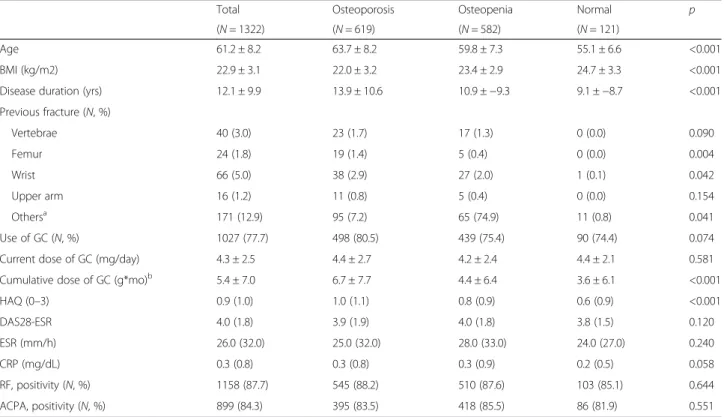
Table 1 Demographic and clinical features of study population
Total Osteoporosis Osteopenia Normal p
(N = 1322) (N = 619) (N = 582) (N = 121)
Age 61.2 ± 8.2 63.7 ± 8.2 59.8 ± 7.3 55.1 ± 6.6 <0.001
BMI (kg/m2) 22.9 ± 3.1 22.0 ± 3.2 23.4 ± 2.9 24.7 ± 3.3 <0.001
Disease duration (yrs) 12.1 ± 9.9 13.9 ± 10.6 10.9 ±−9.3 9.1 ±−8.7 <0.001
Previous fracture (N, %) Vertebrae 40 (3.0) 23 (1.7) 17 (1.3) 0 (0.0) 0.090 Femur 24 (1.8) 19 (1.4) 5 (0.4) 0 (0.0) 0.004 Wrist 66 (5.0) 38 (2.9) 27 (2.0) 1 (0.1) 0.042 Upper arm 16 (1.2) 11 (0.8) 5 (0.4) 0 (0.0) 0.154 Othersa 171 (12.9) 95 (7.2) 65 (74.9) 11 (0.8) 0.041 Use of GC (N, %) 1027 (77.7) 498 (80.5) 439 (75.4) 90 (74.4) 0.074
Current dose of GC (mg/day) 4.3 ± 2.5 4.4 ± 2.7 4.2 ± 2.4 4.4 ± 2.1 0.581
Cumulative dose of GC (g*mo)b 5.4 ± 7.0 6.7 ± 7.7 4.4 ± 6.4 3.6 ± 6.1 <0.001
HAQ (0–3) 0.9 (1.0) 1.0 (1.1) 0.8 (0.9) 0.6 (0.9) <0.001 DAS28-ESR 4.0 (1.8) 3.9 (1.9) 4.0 (1.8) 3.8 (1.5) 0.120 ESR (mm/h) 26.0 (32.0) 25.0 (32.0) 28.0 (33.0) 24.0 (27.0) 0.240 CRP (mg/dL) 0.3 (0.8) 0.3 (0.8) 0.3 (0.9) 0.2 (0.5) 0.058 RF, positivity (N, %) 1158 (87.7) 545 (88.2) 510 (87.6) 103 (85.1) 0.644 ACPA, positivity (N, %) 899 (84.3) 395 (83.5) 418 (85.5) 86 (81.9) 0.551
Values shown are median (interquartile range), mean ± standard deviation and frequency (percentage)
BMI bone mass index, BMD bone mineral density, GC glucocorticoid, HAQ health assessment questionnaire, DAS 28-ESR Disease Activity Score 28 using ESR, ESR erythrocyte sedimentation rate, CRP C-reactive protein, RF rheumatoid factor, ACPA anti-cyclic-citrullinated protein antibody
a
Nasal bone, clavicle, shoulder, scapula, rib, hand, pelvic bone, ankle, feet, coccyx
b
from the time of recruitment. The mean ages of these pa-tients in the osteoporosis, osteopenia, and normal groups were 63.7 (8.2) years, 59.8 (7.3) years, and 55.1 (6.6) years, respectively; the patients with osteoporosis were signifi-cantly older than those with osteopenia or normal BMD (p < 0.001). Disease duration in the study population was 12.1 ± 9.9 years (mean). The RA patients with osteoporosis were older (p < 0.001) and had longer disease duration (p < 0.001), lower BMI (p < 0.001), and higher cumulative dosage of glucocorticoid (p < 0.001) than the patients in the other groups. The osteoporosis patients also had a higher frequency of previous fractures than the patients in the other groups, especially fractures of the femur (p = 0.004) and wrist (p = 0.042). HAQ scores (median and interquar-tile range) were significantly higher [1.0 (1.1)] in the osteo-porosis patients than in the osteopenia patients [0.8 (0.9)] and the normal patients [0.6 (0.9)] (p < 0.001). Disease activity-related factors such as DAS28-ESR (p = 0.120), ESR (p = 0.240), CRP (p = 0.058), current smoking (p = 0.300), and alcohol intake (p = 0.067) did not differ among the three groups.
Frequency of osteoporosis based on BMD
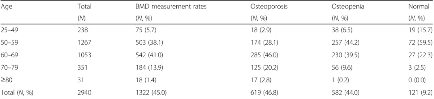
Among the 1322 patients with BMD data, 619 (46.8 %), 582 (44.0 %), and 121 (9.1 %) patients were in the osteo-porosis, osteopenia, and normal groups, respectively. The number of osteoporosis patients in the 60–69 years age group was the highest (n = 285, 46.0 %) (Table 2). In addition, osteoporosis occurred in 31.2 % in the lumbar spine, 29.9 % in the femoral neck, and 18.5 % in total
hip. The lumbar spine, femoral neck, and total hip T-scores in the entire cohort were analyzed according to each age subgroup. The femoral neck T-scores were lowest among the three sites in all age subgroups (Table 3).
Risk factors for osteoporosis
The multivariable logistic regression modeling attempted to use all demographic and disease-related factors asso-ciated with the outcome in Table 1. As a result, ad-vanced age (≥70 years; OR = 2.28, 95 % CI: 1.40–3.58), BMI < 25 (OR = 2.14, 95 % CI: 1.52–3.02), longer disease duration (≥10 years; OR = 1.46, 95 % CI: 1.07-2.00), higher cumulative dose of glucocorticoids (OR = 1.03, 95 % CI: 1.01–1.05), and higher HAQ score (OR = 1.37, 95 % CI: 1.11–1.69) were statistically significant inde-pendent factors associated with osteoporosis. However,
Table 2 The frequency of osteoporosis at either the femoral neck or lumbar spine BMD with RA patients
Age Total BMD measurement rates Osteoporosis Osteopenia Normal
(N) (N, %) (N, %) (N, %) (N, %) 25–49 238 75 (5.7) 18 (2.9) 38 (6.5) 19 (15.7) 50–59 1267 503 (38.1) 174 (28.1) 257 (44.2) 72 (59.5) 60–69 1053 542 (41.0) 285 (46.0) 230 (39.5) 27 (22.3) 70–79 351 184 (13.9) 125 (20.2) 56 (9.6) 3 (2.5) ≥80 31 18 (1.4) 17 (2.8) 1 (0.2) 0 (0.0) Total (N, %) 2940 1322 (45.0) 619 (46.8) 582 (44.0) 121 (9.2)
Table 3 T-scores in patients with RA according to age groups
Age Lumbar spinea Femoral neck Total hip
(N = 1317) (N = 1277) (N = 1266) 40–49 −0.9 ± 1.3 −1.2 ± 1.1 −0.6 ± 1.2 50–59 −1.6 ± 1.1 −1.6 ± 1.0 −1.1 ± 1.1 60–69 −1.9 ± 1.4 −2.1 ± 1.0 −1.6 ± 1.2 70–79 −2.1 ± 1.3 −2.5 ± 0.9 −2.1 ± 1.1 ≥80 −2.9 ± 0.9 −2.6 ± 1.0 −2.7 ± 0.9 Total −1.8 ± 1.3 −1.9 ± 1.1 −1.4 ± 1.2 a Average of T-score of L2-L4
Table 4 multivariale logistic regression analysis on risk factors of osteoporosis in RA patients Risk factors OR 95 % CI p Age < 70 yrs 1 ≥ 70 yrs 2.28 1.40–3.58 <0.001 BMI (Kg/m2) ≥ 25 1 < 25 2.14 1.52–3.02 <0.001
Disease duration (yrs)
< 10 yrs 1 ≥ 10 yrs 1.46 1.07–2.00 0.032 Cumulative dose of GC (g) 1.03 1.01–1.05 0.009 RF, Positivity 0.86 0.55–1.36 0.690 ACPA, positivity 1.00 0.65–1.52 0.718 HAQ score 1.37 1.11–1.69 0.003 DAS28-ESR 0.69 0.39–1.24 0.217 ESR (mm/h) 0.94 0.75–1.18 0.571 CRP (mg/dL) 1.01 0.89–1.16 0.853
OR odds ratios, CI confidence interval, BMI bone mass index, GC glucocorticoid, RF rheumatoid factor, ACPA anti-cyclic-citrullinated protein, HAQ health assessment questionnaire, DAS 28-ESR Disease Activity Score 28 using ESR, ESR erythrocyte sedimentation rate, CRP C-reactive protein
disease activity-associated factors (ESR, CRP, and DAS28-ESR), RF and ACPA showed no independent association with osteoporosis (Table 4).
Discussion
The aim of this study was to investigate the frequency of and risk factors for osteoporosis in Korean postmeno-pausal women with RA. The frequency of osteoporosis was 46.8 %. This frequency is higher in comparison to some other studies, which reported a prevalence of 22–24 % [17, 18]. However, a high prevalence (50 %) of osteoporosis was shown in a large Italian multicenter cross-sectional study that included 925 female RA patients [5]. In another
study of postmenopausal women and men ≥50 years of
age with seropositive RA in South Korea, 121 (52 %) of the 234 patients had osteoporosis [19]. This frequency was higher than that of the general population of females aged ≥50 years. In the Fourth Korea National Health and Nutri-tion ExaminaNutri-tion Survey database, it was estimated that in
the general Korean female population aged ≥50 years,
39.1 % had osteoporosis while 43.4 % had osteopenia [20]. Thus, the prevalence rate of osteoporosis in RA patients varies among studies. This diversity can be explained by the use of different age groups in the study populations, the menopausal status of the participants, and the use of glucocorticoids.
BMD measurement is required in all RA patients due to the increased risk of fractures. However, a significant percentage of RA patients with fracture risk do not undergo BMD testing despite regular visits to rheumatologists. Of the 3531 RA patients enrolled in our study, only 41.2 % had BMD data. According to a study by the Consor-tium of Rheumatology Researchers of North America (CORRONA), of the RA patients in the database with at least a one-year follow up, 45 % reported dual-energy X-ray absorptiometry (DXA) data upon study entry. Fur-thermore, of the 2717 RA patients with at least a one-year follow up without reported DXA data at enrollment in the CORRONA study, 297 (11 %) patients reported undergo-ing DXA durundergo-ing the first year of follow up [21]. Although the CORRONA study included patients <50 years of age, the BMD measurement rate was higher than in this study. The patients in this study had a lower BMD measurement rate despite being older at enrollment. This finding should motivate Korean clinicians to carry out awareness and health education programs in these patients to improve the prevention, diagnosis, and treatment of osteoporosis.
Risk factors for developing osteoporosis in patients with RA include not only the use of glucocorticoids, high disabil-ity scores, low body weight, and age, but also RF status; the frequency of osteoporosis and reduced bone mass is higher in RF (+) patients. In our study, older age (≥70 years), low BMI (<25), longer disease duration (≥10 years), higher HAQ score, and higher cumulative glucocorticoid
dose were significantly associated with osteoporosis. RF, ACPA, and the use of glucocorticoids were not significantly associated with osteoporosis (Table 1). Loss of BMD corre-lates with cumulative glucocorticoid dose, and a correlation has also been established between fracture risk and daily glucocorticoid dose [22]. A 30–50 % increase in fracture risk has been documented in patients receiving long-term glucocorticoid therapy in both cross-sectional and longitu-dinal studies [22, 23]. However, glucocorticoid therapy (used for controlling inflammation) had a positive effect on BMD in a placebo-controlled study conducted on RA pa-tients [24, 25]. This effect could contribute to variability of BMD changes in response to glucocorticoid therapy. In our study, the use of glucocorticoids was not associated with osteoporosis; however, cumulative glucocorticoid dose was significantly associated with osteoporosis. Independent of RA, risk factors such as older age and low BMI are consist-ent with those in the general population [26]. However, dis-ease activity (inflammation), lack of mobility, and treatment with corticosteroids, especially a high cumulative dose, are the main factors that increase the risk of osteoporotic frac-tures, on top of the background fracture risk based on ad-vanced age, low BMI, and female gender, among others [27]. In our study, cigarette smoking and alcohol consump-tion were investigated as environmental factors; however, these factors were not found to be significantly associated with osteoporosis.
Fracture risk is highest among women with osteoporosis. However, many fractures occur in women with BMDs above the WHO threshold of−2.5 In a large cohort study, 82 % of women who sustained osteoporotic fractures of the wrist or forearm, hip, rib, or spine within one year after per-ipheral BMD testing had T-scores greater than−2.5 [28]. In the current study, the number of osteopenic patients (44.8 %) was comparable to that of osteoporotic patients (44.9 %). Nevertheless, no guidelines have yet been estab-lished for osteopenia patients in South Korea. Therefore, regular BMD screening and fracture risk assessment (such as Fracture Risk Assessment (FRAX®) is needed in RA patients, the high risk group for fractures [28].
To the best of our knowledge, this is the first study to investigate the prevalence of osteoporosis in RA patients using a large nationwide cohort (KORONA) in Korea. The numerous centers, both physician-derived and patient-derived outcomes, well controlled data, and use
of a “real-world” cohort are the strengths of the study
[12]. This study has a number of limitations. First, we cannot exclude the possibility of patient selection bias, because the 23 South Korean centers participating in this study were tertiary referral centers. The patients enrolled in this study had higher disease activity and functional disability than individuals not enrolled in this study. Therefore, BMD measurement rates in this study cannot represent the real rate of DXA in our country.
Second, only patients with BMD data available within one year from the time of recruitment were included in this study. As a result, only about one-third of the population could be included in the analysis of risk factors for osteoporosis including BMD. Third, no phantom cross-calibration was performed among study centers. However, the centers were monitored with their local quality control phantoms and were found to be stable and calibrated to their manufacturer’s standards. Fourth, we used a cross-sectional study design; hence, it was not pos-sible to make any cause–effect inferences about the rela-tionship between RA characteristics and BMD. Therefore, further studies are needed to analyze the risk factors for osteoporotic fractures and anti-osteoporotic medication. Conclusions
We found that older age (≥70 years), low BMI (<25),
longer disease duration (≥10 years), higher HAQ score,
and higher cumulative glucocorticoid dose were signifi-cantly associated with osteoporosis. These findings were not markedly different from those of previous studies. A large percentage (90.8 %) of RA patients enrolled in the KORONA cohort had osteoporosis and osteopenia. BMD measurement rates in this population remain low, despite being a high-risk group for osteoporotic fractures.
Abbreviation
BMD:bone mineral density; DXA: dual X-ray absorptiometry; KORONA: KORean Observational study Network for Arthritis; WHO: World Health Organization.
Competing interests
The authors declare that they have no competing interests. Authors’ contributions
JHL: manuscript writing and data analysis. YKS, and SCB: study conception, design, and project management. CBC, SKC, and SYB: analysis and interpretation of the data. SJH, JBJ, THK, JL, HSL, and BYY: data collection, result interpretation. DHY and JYC: data collection and critical revision. All authors have read and approved the final manuscript for publication. Acknowledgments
This study was supported by a grant of the Korea Healthcare technology R&D Project, Ministry of Health and Welfare, Republic of Korea (A102065). Author details
1
Department of Rheumatology, Inje University Ilsan Paik Hospital, Goyang, Republic of Korea.2Department of Rheumatology, Hanyang University
Hospital for Rheumatic Diseases, Seoul 133-792, Republic of Korea.
3Department of Rheumatology, Hanyang University Guri Hospital, Guri,
Republic of Korea.4Department of Rheumatology, Catholic University of Daegu School of Medicine, Daegu, Republic of Korea.5Department of
Rheumatology, Kyung Hee University Hospital, Seoul, Republic of Korea.
6Department of Rheumatology, Ewha Womans University Mokdong Hospital,
Seoul, Republic of Korea.
Received: 3 September 2015 Accepted: 17 February 2016
References
1. Deodhar AA, Woolf AD. Bone mass measurement and bone metabolism in rheumatoid arthritis: a review. Br J Rheumatol. 1996;35:309–22.
2. Kleyer A, Schett G. Arthritis and bone loss: a hen and egg story. Curr Opin Rheumatol. 2014;26:80–4.
3. Ørstavik RE, Haugeberg G, Uhlig T, Mowinckel P, Kvien TK, Falch JA, et al. Vertebral deformities in rheumatoid arthritis: a comparison with population-based controls. Arch Intern Med. 2004;164:420–5.
4. Haugeberg G, Uhlig T, Falch JA, Halse JI, Kvien TK. Bone mineral density and frequency of oteoporosis in female patients with rheumatoid arthritis: results from 394 patients in the Oslo County Rheumatoid Arthritis register. Arthritis Rheum. 2000;43:522–30.
5. Sinigaglia L, Nervetti A, Mela Q, Bianchi G, Del Puente A, Di Munno O, et al. A multicenter cross sectional study on bone mineral density in rheumatoid arthritis. Italian Study Group on Bone Mass in Rheumatoid Arthritis. J Rheumatol. 2000;27:2582–9.
6. Guler-Yuksel M, Bijsterbosch J, Goekoop-Ruiterman YP, de Vries-Bouwstra JK, Ronday HK, Peeters AJ, et al. Bone mineral density in patients with recently diagnosed, active rheumatoid arthritis. Ann Rheum Dis. 2007;66:1508–12. 7. van Staa T, Geusens P, Bijlsma JW, Leufkens HG, Cooper C. Clinical
assessment of the long-term risk of fracture in patients with rheumatoid arthritis. Arthritis Rheum. 2006;54:3104–12.
8. Michel BA, Bloch DA, Fries JF. Predictors of fractures in early rheumatoid arthritis. J Rheumatol. 1991;18:804–8.
9. Compston J. Management of glucocorticoid-induced osteoporosis. Nat Rev Rheumatol. 2010;6:82–8.
10. Molokhia M, McKeigue P. Risk for rheumatic disease in relation to ethnicity and admixture. Arthritis Res. 2000;2:115–25.
11. Tobon GJ, Youinou P, Saraux A. The environment, geo-epidemiology, and autoimmune disease: Rheumatoid arthritis. Autoimmun Rev. 2010;9:A288–92. 12. Sung YK, Cho SK, Choi CB, Park SY, Shim J, Ahn JK, et al. Korean Observational Study Network for Arthritis (KORONA): establishment of a prospective multicenter cohort for rheumatoid arthritis in South Korea. Semin Arthritis Rheum. 2012;41:745–51.
13. Arnett FC, Edworthy SM, Bloch DA, McShane DJ, Fries JF, Cooper NS, et al. The American Rheumatism Association 1987 revised criteria for the classification of rheumatoid arthritis. Arthritis Rheum. 1988;31:315–24. 14. Wells G, Becker JC, Teng J, Dougados M, Schiff M, Smolen J, et al. Validation
of the 28-joint Disease Activity Score (DAS28) and European League Against Rheumatism response criteria based on C-reactive protein against disease progression in patients with rheumatoid arthritis, and comparison with the DAS28 based on erythrocyte sedimentation rate. Ann Rheum Dis. 2009;68: 954–60.
15. Bae SC, Cook EF, Kim SY. Psychometric evaluation of a Korean Health Assessment Questionnaire for clinical research. J Rheumatol. 1998;25:1975–9. 16. Report of a WHO Study Group. Assessment of fracture risk and its
application to screening for postmenopausal osteoporosis. World Health Organ Tech Rep Ser. 1994;843:1–129.
17. Gonzalez-Lopez L, Gamez-Nava JI, Vega-Lopez A, Rodriguez-Jimenez NA, Gonzalez-Montoya N, Aguilar-Chavez E, et al. Performance of risk indices for identifying low bone mineral density and osteoporosis in Mexican Mestizo women with rheumatoid arthritis. J Rheumatol. 2000;39:247–53. 18. Heberlein I, Demary W, Bloching H, Braun J, Buttgereit F, Dreher R, et al.
Prophylaxis and treatment of osteoporosis in patients with rheumatoid arthritis (ORA study). Z Rheumatol. 2011;70:793–8.
19. Yoon J, Kwon SR, Lim MJ, Joo K, Moon CG, Jang J, et al. A comparison of three different guidelines for osteoporosis treatment in patients with rheumatoid arthritis in Korea. Korean J Intern Med. 2010;25:436–46. 20. Kim KH, Lee K, Ko YJ, Kim SJ, Oh SI, Durrance DY, et al. Prevalence, awareness,
and treatment of osteoporosis among Korean women: The Fourth Korea National Health and Nutrition Examination Survey. Bone. 2012;50:1039–47. 21. Aizer J, Reed G, Onofrei A, Harrison MJ. Predictors of bone density testing in
patients with rheumatoid arthritis. Rheumatol Int. 2009;29:897–905. 22. Kanis JA, Johansson H, Oden A, Johnell O, de Laet C, Melton III LJ, et al. A
meta-analysis of prior corticosteroid use and fracture risk. J Bone Miner Res. 2004;19:893–9.
23. Van Staa TP, Leufkens HG, Abenhaim L, Zhang B, Cooper C. Use of oral corticosteroids and risk of fractures. J Bone Miner Res. 2006;15:993–1000. 24. Haugeberg G, Strand A, Kvien TK, Kirwan JR. Reduced loss of hand bone
density with prednisolone in early rheumatoid arthritis: results from a randomized placebo-controlled trial. Arch Intern Med. 2005;165:1293–7. 25. Wijbrandts CA, Klaasen R, Dijkgraaf MG, Gerlag DM, van Eck-Smit BL, Tak PP.
Bone mineral density in rheumatoid arthritis patients 1 year after adalimumab therapy: arrest of bone loss. Ann Rheum Dis. 2009;68:373–6.
26. van der Voort DJ, Geusens PP, Dinant GJ. Risk factors for osteoporosis related to their outcome: fractures. Osteoporos Int. 2001;12:630–8. 27. Vis M, Guler-Yuksel M, Lems WF. Can bone loss in rheumatoid arthritis be
prevented? Osteoporos Int. 2013;24:2541–53.
28. Siris ES, Chen YT, Abbott TA, Barrett-Connor E, Miller PD, Wehren LE, et al. Bone mineral density thresholds for pharmacological intervention to prevent fractures. Arch Intern Med. 2004;164:1108–221.
• We accept pre-submission inquiries
• Our selector tool helps you to find the most relevant journal • We provide round the clock customer support
• Convenient online submission • Thorough peer review
• Inclusion in PubMed and all major indexing services • Maximum visibility for your research
Submit your manuscript at www.biomedcentral.com/submit