Horizontal alteration of anterior alveolar
ridge after immediate implant placement:
A retrospective cone beam computed
tomography analysis
Young Keun Hyun1a, Chung Yun Lee2a, Subramanian Keerthana2, Selvaponpriya Ramasamy2, So-Yeon Song3,
Ji Suk Shim3*, Jae Jun Ryu4*
1Periodontal Dental Clinic, Seoul, Republic of Korea
2Korea University Graduate School, Clinical Dentistry, Seoul, Republic of Korea 3Department of Dentistry, Korea University Guro Hospital, Seoul, Republic of Korea 4Department of Dentistry, Korea University Anam Hospital, Seoul, Republic of Korea
PURPSE. The aim of this study was to evaluate the labio-lingual alterations of the alveolar bone where the implant was placed immediately after tooth extraction.
MATERIALS AND METHODS. Implants were placed immediately after tooth extraction on anterior alveolar ridges in the maxilla and mandible. The pin-guide system was used to help determine the location and path of implants during the surgical process. The horizontal distance from implants to the outer border of alveolar bone was measured at the rim and middle of the implants in the cone beam computed tomography images. The alteration of alveolar bone was evaluated comparing the horizontal distances measured immediately after surgery and 3 months after surgery. RESULTS. The results show that more resorption occurred towards the labial bone than the lingual bone in the maxilla. A similar amount of labial and lingual bone resorption was observed in the mandible. CONCLUSION. Considering the horizontal alteration of alveolar bone, labio-lingual positioning of the implant towards the lingual bone in the maxilla and at the center of the alveolar ridge in the mandible is recommended when it is placed immediately after tooth extraction. [J Adv Prosthodont 2021;13:117-25]
KEYWORDS
Dental implant; Alveolar ridge; Alveolar resorption; Tooth extraction; Cone-beam computed tomography
ORCID
Young Keun Hyun
https://orcid.org/0000-0003-3319-7383 Chung Yun Lee
https://orcid.org/0000-0002-9289-1802 Subramanian Keerthana https://orcid.org/0000-0003-0305-8585 Selvaponpriya Ramasamy https://orcid.org/0000-0002-2294-6740 So-Yeon Song https://orcid.org/0000-0002-7738-5370 Ji Suk Shim https://orcid.org/0000-0002-4112-6051 Jae Jun Ryu
https://orcid.org/0000-0002-2093-6389 Corresponding author Ji Suk Shim
Department of Prosthodontics, Korea University Guro Hospital, #148, Gurodong-ro, Guro-gu, Seoul 08308, Republic of Korea
Tel +82226262489 E-mail [email protected] Jae Jun Ryu
Department of Advanced Prosthodon-tics, Korea University Anam Hospital, #73, Goryeodae-ro, Seongbuk-gu, Seoul 02841, Republic of Korea Tel +8229205423
E-mail [email protected] Received March 17, 2021 / Last Revision April 20, 2021 / Accepted April 23, 2021
a These authors contributed equally to this work.
This research was supported by National Research Foundation of Korea (NRF) grant funded by the Korea government (MSIT) (No. 2020R1A2C1014211).
© 2021 The Korean Academy of Prosthodontics
ccThis is an Open Access article distributed under the terms of the Creative Commons Attribution Non-Commercial License
(http://creativecommons.org/licenses/by-nc/4.0) which permits unrestricted non-commercial use, distribution, and reproduction in any medium, provided the original work is properly cited.
INTRODUCTION
Bone resorption is correlated with catabolic changes in the alveolar bone
af-ter tooth extraction.1 Tooth extraction leads to the disruption of the blood
sup-ply varies for different anatomic structures in the oral cavity, the bone alteration after extraction is deter-mined by factors including bone wall thickness, tooth
angulation, and tooth shape.3 Therefore, the alveolar
bone alteration after tooth extraction is different be-tween the anterior and posterior bone, bebe-tween labi-al and plabi-alatlabi-al bone, and between maxillary and
man-dibular bone.4
The catabolic changes related to bone resorption are initiated by the disruption of blood supply to the labial bundle bone from the periodontal ligaments (PDL), which subsequently causes significant
en-hancement of the osteoclast activity.2 Therefore, the
central area of the socket shows more marginal bone changes than the proximal area because the proximal area is less affected by the disruption of blood sup-ply following tooth extraction due to the additional
support from the PDL of the neighboring teeth.5 The
labial bone in the maxilla shows more dimension-al changes than the pdimension-alatdimension-al bone because the labidimension-al bone has walls of limited thickness in comparison to
that of the palatal bone.2
The outcome of implant placement into tooth sock-ets immediately after tooth extraction has been re-ported to be as predictable in esthetic and functional
aspects.6 However, there is still controversy over the
statement that immediate implant placement after tooth extraction prevents alveolar bone resorption at
the extracted site.7 Therefore, clinicians should
pre-dict bone resorption for successful implant treatment when the implant is placed immediately after tooth
extraction.8 Moreover, in the anterior zone, preserved
bone volume around implants after healing is critical to prevent the esthetic shortcomings including gingi-val discoloration, gingigingi-val recession, and reduced
la-bial tissue volume.8
The implant position in the labio-lingual plane is critical for both biological and restorative
complica-tions.9,10 The placement of an implant too far labially
causes further reduction of alveolar bone,
increas-ing the risk of soft tissue recession.11 In addition, the
implant in a one-sided position cannot achieve
ap-propriate emergency profile without ridge lap.12,13
The severe difference in the long axis between fixture and restoration causes difficulty in fabricating
resto-rations.14 To determine the accurate location for
im-plant placement, various tools have been used. In this study, for the first time, we used a pin-guide system as a new method to assist in determining the appro-priate location for implant placement.
The aim of this study was to evaluate the horizon-tal alteration of the alveolar bone around the implant placed in the freshly extracted socket. Implants were placed immediately after anterior tooth extraction in the maxilla and mandible, and the distance between the implant and the outer border of the alveolar bone was measured using CT images.
MATERIALS AND METHODS
Forty consecutive patients who underwent implant treatment in the maxillary or mandibular anterior re-gion, from 2017 to 2020, were included in this study. All implants were placed by a specialist who had ma-jored in oral and maxillofacial surgery. The method of this study was approved by the local Medical Eth-ics Board (IRB no. 2019AS0083). The inclusion crite-ria were as follows: 1) age > 18 years, 2) presence of hopeless teeth in the anterior area, and 3) presence of healthy natural teeth adjacent to the teeth be-ing replaced. The exclusion criteria were as follows: 1) heavy smokers (more than 10 cigarettes per day), 2) patients with severe concavity of the labial bone which requires additional guided tissue graft, or 3) patients with any wall defect after tooth extraction. The shape of alveolar bone was confirmed using cone beam computed tomography (CBCT) image acquired before the surgery. A pair of guiding pins was used to provide the reference for labial and lingual bone; the location and angulation of the implants was deter-mined by the information from the guiding pins.
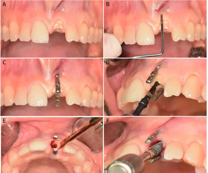
All implants were placed in the maxillary or man-dibular anterior area (canine to canine) with a flap-less approach (Fig. 1A). Following local anesthesia, the location for placing guiding pins was indicated on the muco-gingival junction (Fig. 1B). A pair of guiding pins was placed on the labial and lingual sides par-allel to the long axis of the adjacent tooth (Fig. 1C). Pilot and subsequent drilling were performed on the retained root. Based on the angulation and distance between the pins, the position and path of drilling were determined (Fig. 1D). After finishing the drilling
Fig. 1. (A) Preoperative view of maxillary left central incisal, fractured by secondary caries. (B) Indentation for the placement of guiding pin on the muco-gingival junction. (C) A pair of guiding pins were placed parallel to the long axis of adjacent tooth. (D) From the angula-tion and distance between pins, the posiangula-tion and path of drilling was determined. (E) After finishing the drilling procedure, the remaining root was extracted. (F) A cylindrical screw-type implant was placed in the extracted socket.
A
B
C
D
E
F
procedure, the remaining root was extracted (Fig. 1E). A cylindric screw-type implant was placed in the ex-tracted socket (Fig. 1F). Bone graft was not applied to all patients. A healing abutment was connected to the implant. Three months after the surgical intervention, a definitive ceramic implant crown was provided. The patients wore temporary denture until the delivery of the definite prosthesis. Customized abutments were provided so that the labial restorative margin was lo-cated 0.5mm below the gingiva. Computed tomogra-phy radiographs were taken before treatment, after surgery, and 3 months after surgery.
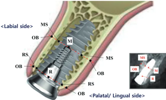
In order to describe the alteration in the alveolar bone, the landmarks were defined (Fig. 2). Changes in the alveolar bone were measured using CBCT
imag-es. The landmarks used to describe the alterations in the alveolar bone were as follows: the rim of the im-plant (R), the middle of the imim-plant (M), the surface of the implant at the rim of the implant (RS), the surface of the implant at the middle of the implant (MS), and the outer border of the bone crest (OB). The rim of the implant can be determined by finding the border between implant fixture and healing abutment. The middle of the implant can be constantly designat-ed by finding the tip of the abutment screw. The fol-lowing parameters were estimated: (1) RS to OB, the horizontal distance between the implant surface and the outer border of the alveolar bone at the implant rim level, and (2) MS to OB, the horizontal distance between the implant surface at the middle of the
im-Table 1. Characteristics of the study group N Number of patients 40 Number of implants 40 Male / female 15 / 25 Age (average) 49.3 Implant location Maxilla 20
Mandible 20 Reason for extraction Trauma 10 Caries 18
Periodontitis 12
Implant length (mm) 8.5 / 10 / 13 7 /32/ 1 Implant diameter (mm) 3.5 / 4.0 / 4.5 17 /16/ 7
* N = number
plant. The parameters were estimated for the labial and lingual side of the implant. The measurements were calibrated by two trained examiners. The first examiner measured the CBCT data repeating twice, and the second examiner double-checked the mea-surement from the capture image showing parame-ters on CBCT. The intra-examiner reliability was cal-culated by Pearson’s correlation coefficient (P < .001). The intraclass correlation coefficient was 0.87.
The results are expressed as mean ± standard de-viation (SD). Data were evaluated for homogeneity of variance based on Levene’s tests (α = .05). We used a paired t-test to compare the amount of alveolar bone alteration as the anatomic location of implants (labial side and palatal side, maxilla and mandible, and rim and middle). A P value of < .05 was considered sta-tistically significant. All statistical analyses were per-formed using SAS software version 9.4 (SAS Institute Inc., Cary, NC, USA).
RESULTS
This retrospective study included 40 patients, includ-ing 15 men and 25 women. Their ages ranged be-tween 24 and 68 years with an average age of 49.3.
Among the extracted teeth, 18, 12, and 10 were ex-tracted because of caries, periodontitis, and trauma, respectively. Implants replaced the maxillary and mandibular anterior teeth, including the central inci-sor, lateral inciinci-sor, and canine. The same number of implants was placed in the maxilla and mandible. The majority of the implants were 10 mm in length and 3.5 mm in diameter (Table 1).
Fig. 2. Landmarks used to describe the alterations in the alveolar bone. Rim of implant (R), middle of implant (M), surface of the implant corresponding to the rim of the implant (RS), surface of the implant corresponding to the the middle of the implant (MS), and outer bor-der of the bone crest (OB).
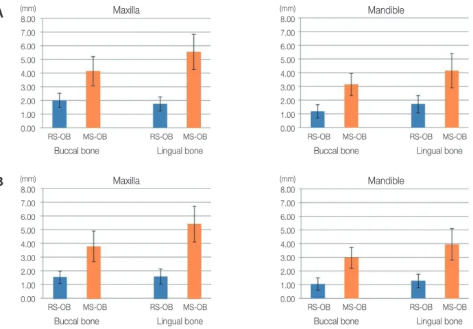
Figure 3 shows the parameters estimated immedi-ately after surgery and 3 months later. Table 2 shows the horizontal distance between the implant surface and outer border of the alveolar bone after implant placement, which indicates the labio-lingual position of the implants. In the maxilla, the differences be-tween the labial and lingual bones were 0.42 and 1.4 mm at the implant rim and implant middle, respec-tively. In the mandible, the differences between the labial and lingual bones were 0.3 and 1.01 mm at the implant rim level and implant middle, respectively.
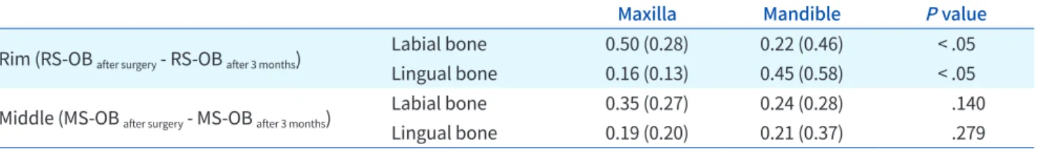
Table 3 shows the comparison of the alveolar bone alteration between the labial and lingual bone. In the maxilla, labial bone changes were significantly great-er than palatal bone changes, both at the rim of the implant (P < .05) and the middle of the implant (P < .05). In the mandible, there were no statistical differ-ences in the horizontal alteration between the labial and lingual bone, both at the rim of the implant and the middle of the implant (P > .05). Table 4 shows the comparison of the alveolar bone alteration between the maxilla and mandible. At the rim of the implant,
Table 2. The horizontal distance between implant surface and outer border of alveolar bone after implant placement
Labial bone Lingual bone Differences
Maxilla Rim (RS-OB) 2.18 (0.57) 1.76 (0.52) 0.42 Middle (MS-OB) 4.15 (1.08) 5.55 (1.27) 1.40 Mandible Rim (RS-OB) 1.20 (0.47) 1.50 (0.58) 0.30 Middle (MS-OB) 3.14 (0.80) 4.15 (1.24) 1.01
Fig. 3. The results of the study. (A) the horizontal distance between implant surface and the outer border of alveolar bone after surgery, (B) the horizontal distance between implant surface and the outer border of alveolar bone after 3 months.
8.00 7.00 6.00 5.00 4.00 3.00 2.00 1.00 0.00 Maxilla
RS-OB MS-OB RS-OB MS-OB
Buccal bone Lingual bone
(mm) 8.00 7.00 6.00 5.00 4.00 3.00 2.00 1.00 0.00 Maxilla
RS-OB MS-OB RS-OB MS-OB
Buccal bone Lingual bone
(mm) 8.00 7.00 6.00 5.00 4.00 3.00 2.00 1.00 0.00 Mandible
RS-OB MS-OB RS-OB MS-OB
Buccal bone Lingual bone
(mm) 8.00 7.00 6.00 5.00 4.00 3.00 2.00 1.00 0.00 Mandible
RS-OB MS-OB RS-OB MS-OB
Buccal bone Lingual bone
(mm)
A
Table 5. Comparison of the amount of alveolar bone alteration (RS-OB after surgery - RS-OB after 3 months or MS-OB after surgery -
MS-OB after 3 months) between the implant rim and the middle of implant Rim
(RS-OB after surgery - RS-OB after 3 months)
Middle
(MS-OB after surgery - MS-OB after 3 months) P value
Maxilla Labial bone 0.50 (0.28) 0.35 (0.27) .108 Lingual bone 0.16 (0.13) 0.19 (0.20) .984 Mandible Labial bone 0.22 (0.46) 0.24 (0.28) .255 Lingual bone 0.45 (0.58) 0.21 (0.37) .247
Table 4. Comparison of the amount of alveolar bone alteration (RS-OB after surgery - RS-OB after 3 months or MS-OB after surgery -
MS-OB after 3 months) between maxilla and mandibular bone
Maxilla Mandible P value
Rim (RS-OB after surgery - RS-OB after 3 months)
Labial bone 0.50 (0.28) 0.22 (0.46) < .05 Lingual bone 0.16 (0.13) 0.45 (0.58) < .05 Middle (MS-OB after surgery - MS-OB after 3 months) Labial bone 0.35 (0.27) 0.24 (0.28) .140
Lingual bone 0.19 (0.20) 0.21 (0.37) .279
Table 3. Comparison of the amount of alveolar bone alteration (RS-OB after surgery - RS-OB after 3 months or MS-OB after surgery -
MS-OB after 3 months) between labial and lingual bone
Labial bone Lingual bone P value
Maxilla Rim (RS-OB after surgery - RS-OB after 3 months) 0.50 (0.28) 0.16 (0.13) < .05 Middle (MS-OB after surgery - MS-OB after 3 months) 0.35 (0.27) 0.19 (0.20) < .05
Mandible Rim (RS-OB after surgery - RS-OB after 3 months) 0.22 (0.46) 0.45 (0.58) .104 Middle (MS-OB after surgery - MS-OB after 3 months) 0.24 (0.28) 0.21 (0.37) .734
more labial bone alteration was observed in the max-illa than in the mandible, and more lingual bone alter-ation was observed in the mandible than in the
max-illa with statistical significance (P < .05). At the middle
of the implant, there was no statistical difference in labial or lingual bone alteration between the maxilla and mandible (P > .05). Table 5 shows the comparison of alveolar bone alteration between the implant rim and the middle of the implant. There was no statis-tical difference in the alveolar bone alteration at the implant rim and at the implant middle (P > .05).
DISCUSSION
The greatest amount of alveolar bone alteration
oc-curs in horizontal dimension.15,16 To ensure sufficient
bone volume around the implant after immediate im-plant placement, the clinician should take into con-sideration the changes in alveolar bone around the
implant.8 In this study, the horizontal alteration of
al-veolar bone after implant placement was evaluated by placing implants in the center of the alveolar ridge using a pin-guide system. Implants were placed on the anterior alveolar ridges in the maxilla and
mandi-ble, and the alteration of bone volume was measured at the rim and middle of the implants. The results show that more resorption occurred towards the labi-al bone than the lingulabi-al bone in the maxilla, whereas a similar amount of resorption occurred at labial and lingual bone in the mandible.
Alveolar bone changes after tooth extraction vary with the associated local factors, including
alveo-lar bone and bone mass at skeletal sites.17 Bone
re-sorption following tooth loss also varies between the maxilla and mandible because anatomical features are different between the maxilla and the
mandi-ble.18 The outer cortex is thin in the maxilla and thick
in the mandible continuously till the basal bone,18
and the mandible shows higher alveolar bone density
than the maxilla.19 Similar to previous studies, in this
study, there was greater absorption of the labial bone than the lingual bone in the maxilla, but labial and lingual bones were similarly altered in the mandi-ble.18,20,21 Interestingly, these aspects were observed
both at the rim and middle of the implants. The re-sults highlight that clinicians should be cautious in maintaining sufficient distance between the implant and the outer border of the alveolar bone at the mid-dle level as well as the rim of the implant. A previous study that reported the dehiscence in the apical 1/3 part of the implant immediately placed after tooth
ex-traction supports the suggestion.22
Horizontal alteration of the alveolar ridge after tooth extraction should be considered for labial-lin-gual positioning of implants when they are immedi-ately placed. In the anterior maxilla, placing implants toward the lingual bone rather than the center of the extracted socket is recommended, as the resorption of the labial bone plate is more pronounced than that of the lingual bone plate both at the rim and middle of the implants. On the other hand, in the anterior mandible, placing implants at the center of the ex-tracted socket is recommended, as the bone resorp-tion at the rim and middle of the implants was similar between the labial and lingual bone plate. In addi-tion, the implant width should be determined con-sidering the total amount of horizontal resorption on the labial and lingual sides. For the maxilla, the alve-olar bone was horizontally altered by about 4 mm at the rim and 3 mm at the rim of the implant. In other
words, for the mandible, the horizontal alveolar bone resorption was approximately 3 mm at the rim and 7 mm at the rim of the implant.
The dimensional reduction of the alveolar ridge is related to the position of the implant in the socket, thickness of the socket wall, and patient-related
fac-tors.23 As the position of the implant placed into the
extraction socket can affect the alteration of the alve-olar ridge, for the exact evaluation of alvealve-olar ridge alteration, consistent constant positioning of im-plants is essential. In this study, a pin-guide system was used as the tool for adequate positioning of im-plants. A pair of guiding pins provided information on the distance between the labial and lingual outer bor-der and the angulation of the alveolar ridge. The hori-zontal distances between the implant surface and the outer border of the alveolar bone were similar at the labial and lingual bone, showing that the implants were positioned without the perforation of alveolar bone which causes adverse effect of alveolar bone al-teration. However, the other factors that affect to the alveolar resorption, including the distance between implant surface and alveolar bone, the vertical posi-tion of implant, and the thickness of alveolar bone, may be considered in the future studies.
As the physiological changes in the alveolar bone following tooth extraction occur within the first 3
months of socket healing,24 the alveolar ridge
chang-es were observed 3 months after tooth extraction. However, observation for longer periods may be nec-essary to evaluate the alteration in the alveolar bone around implants. For the repetitive measurement at the same point of the implant, distinguishable points were selected in the CBCT images. The rim of the im-plant can be distinguished by finding the border be-tween the implants and abutments. The middle of the implant was defined as the tip of the screw hole, which is also easily picked out as the radiolucent point of the implants. Although the point may not be exactly middle of implant, it indicates the point at a constant distance from the rim of implant.
CONCLUSION
Within the limits of this study, a greater amount of re-sorption was observed on the labial bone than at the
lingual bone in the maxilla. A similar amount of alter-ation in the labial and lingual bone was observed in the mandible. The results suggest labio-lingual posi-tioning of the implant towards the lingual bone in the maxilla and at the center of the socket in the mandi-ble, when placed immediately after tooth extraction.
REFERENCES
1. Chappuis V, Araújo MG, Buser D. Clinical relevance of dimensional bone and soft tissue alterations post-ex-traction in esthetic sites. Periodontol 2000 2017;73:73-83.
2. Araújo MG, Lindhe J. Dimensional ridge alterations following tooth extraction. An experimental study in the dog. J Clin Periodontol 2005;32:212-8.
3. Misawa M, Lindhe J, Araújo MG. The alveolar process following single-tooth extraction: a study of maxillary incisor and premolar sites in man. Clin Oral Implants Res 2016;27:884-9.
4. Tylman SD. Theory and practice of crown and bridge prosthodontics. 5th ed. St. Louis: Mosby; 1965. 5. Chen ST, Buser D. Clinical and esthetic outcomes of
implants placed in postextraction sites. Int J Oral Max-illofac Implants. 2009;24:186-217.
6. Schwartz-Arad D, Chaushu G. The ways and where-fores of immediate placement of implants into fresh extraction sites: a literature review. J Periodontol 1997;68:915-23.
7. Botticelli D, Berglundh T, Lindhe J. Hard-tissue alter-ations following immediate implant placement in ex-traction sites. J Clin Periodontol 2004;31:820-8. 8. Araújo MG, Silva CO, Souza AB, Sukekava F. Socket
healing with and without immediate implant place-ment. Periodontol 2000 2019;79:168-77.
9. Ferrus J, Cecchinato D, Pjetursson EB, Lang NP, Sanz M, Lindhe J. Factors influencing ridge alterations fol-lowing immediate implant placement into extraction sockets. Clin Oral Implants Res 2010;21:22-9.
10. Block MS, Emery RW, Cullum DR, Sheikh A. Implant placement is more accurate using dynamic naviga-tion. J Oral Maxillofac Surg 2017;75:1377-86.
11. Nisapakultorn K, Suphanantachat S, Silkosessak O, Rattanamongkolgul S. Factors affecting soft tissue level around anterior maxillary single-tooth implants. Clin Oral Implants Res 2010;21:662-70.
12. Belser UC, Buser D, Hess D, Schmid B, Bernard JP, Lang NP. Aesthetic implant restorations in partially edentulous patients-a critical appraisal. Periodontol 2000 1998;17:132-50.
13. Belser UC, Bernard JP, Buser D. Implant-supported restorations in the anterior region: prosthetic consid-erations. Pract Periodontics Aesthet Dent 1996;8:875-83; quiz 884.
14. Buser D, Martin W, Belser UC. Optimizing esthetics for implant restorations in the anterior maxilla: anatomic and surgical considerations. Int J Oral Maxillofac Im-plants 2004;19:43-61.
15. Van der Weijden F, Dell’Acqua F, Slot DE. Alveolar bone dimensional changes of post-extraction sock-ets in humans: a systematic review. J Clin Periodontol 2009;36:1048-58.
16. Lam RV. Contour changes of the alveolar processes following extractions. J Prosthet Dent 1960;10:25-32. 17. Bodic F, Hamel L, Lerouxel E, Baslé MF, Chappard D.
Bone loss and teeth. Joint Bone Spine 2005;72:215-21.
18. Malone WFP, Tylman SD, Koth DL. Tylman’s theory and practice of fixed prosthodontics. 8th ed. St. Louis: Ishiyaku Euro-America; 1989.
19. Devlin H, Horner K, Ledgerton D. A comparison of maxillary and mandibular bone mineral densities. J Prosthet Dent 1998;79:323-7.
20. Lekovic V, Kenney EB, Weinlaender M, Han T, Klok-kevold P, Nedic M, Orsini M. A bone regenerative ap-proach to alveolar ridge maintenance following tooth extraction. Report of 10 cases. J Periodontol 1997;68: 563-70.
21. Lekovic V, Camargo PM, Klokkevold PR, Weinlaender M, Kenney EB, Dimitrijevic B, Nedic M. Preservation of alveolar bone in extraction sockets using bioabsorb-able membranes. J Periodontol 1998;69:1044-9. 22. Lin CY, Pan WL, Wang HL. Facial fenestration and de-
hiscence defects associated with immediate implant placement without flap elevation in anterior max-il- lary ridge: a preliminary cone beam computed to- mography study. Int J Oral Maxillofac Implants 2018; 33:1112-8.
23. Tomasi C, Sanz M, Cecchinato D, Pjetursson B, Fer-rus J, Lang NP, Lindhe J. Bone dimensional variations at implants placed in fresh extraction sockets: a mul-tilevel multivariate analysis. Clin Oral Implants Res
2010;21:30-6.
24. Schropp L, Wenzel A, Kostopoulos L, Karring T. Bone healing and soft tissue contour changes following single-tooth extraction: a clinical and radiographic 12-month prospective study. Int J Periodont Restor Dent 2003;23:313-23.