이
이
이학
학
학 석
석
석사
사
사학
학
학위
위
위 논
논
논문
문
문
E
E
Ef
f
ff
f
fe
e
ec
c
ct
t
to
o
of
f
fC
C
Cy
y
ys
s
st
t
te
e
ei
i
i
n
n
ny
y
yl
l
lL
L
Le
e
eu
u
uk
k
ko
o
ot
t
tr
r
ri
i
i
e
e
en
n
ne
e
eD
D
D4
4
4
o
o
on
n
nC
C
Ch
h
he
e
em
m
mo
o
ok
k
ki
i
i
n
n
ne
e
eE
E
Ex
x
xp
p
pr
r
re
e
es
s
ss
s
si
i
i
o
o
on
n
n
v
v
vi
i
i
a
a
aC
C
Cy
y
ys
s
st
t
te
e
ei
i
i
n
n
ny
y
yl
l
lL
L
Le
e
eu
u
uk
k
ko
o
ot
t
tr
r
ri
i
i
e
e
en
n
ne
e
eR
R
Re
e
ec
c
ce
e
ep
p
pt
t
to
o
or
r
r1
1
1
i
i
i
n
n
nH
H
Hu
u
um
m
ma
a
an
nL
n
L
Lu
u
un
n
ng
g
gE
E
Ep
p
pi
i
i
t
t
th
h
he
e
el
l
l
i
i
i
a
a
al
lC
l
C
Ce
e
el
l
l
l
l
l
s
s
s,
,
,
A
A
A5
5
54
4
49
9
9
아
아
아 주
주
주 대
대
대 학
학 교
학
교
교 대
대
대 학
학
학 원
원
원
의
의
의 학
학
학 과
과
과
김
김
김 설
설
설 화
화
화
Effect
Effect
Effect
Effect of
of
of Cysteinyl
of
Cysteinyl
Cysteinyl
Cysteinyl Leukotriene
Leukotriene
Leukotriene D4
Leukotriene
D4
D4
D4
on
on
on
on Chemokine
Chemokine
Chemokine
Chemokine Expression
Expression
Expression
Expression
via
via
via
via Cysteinyl
Cysteinyl
Cysteinyl
Cysteinyl Leukotriene
Leukotriene
Leukotriene
Leukotriene Receptor
Receptor
Receptor
Receptor 1
1
1
1
in
in
in
in Human
Human
Human Lung
Human
Lung
Lung Epithelial
Lung
Epithelial
Epithelial Cells,
Epithelial
Cells,
Cells, A549
Cells,
A549
A549
A549
by
Seol-Hwa Kim
A Dissertation Submitted to The Graduate School of Ajou University
in Partial Fulfillment of the Requirements for the Degree of
MASTER OF SCIENCES
Supervised by
Hae-Sim Park. M.D., Ph.D.
Department
Department
Department
Department of
of
of
of Medical
Medical
Medical
Medical Sciences
Sciences
Sciences
Sciences
The
The
The
The Graduate
Graduate
Graduate
Graduate School,
School,
School, Ajou
School,
Ajou
Ajou
Ajou University
University
University
University
August,
August,
August,
김
김
김설
설
설화
화
화의
의
의 이
이
이학
학
학 석
석
석사
사
사학
학
학위
위
위 논
논
논문
문
문을
을
을 인
인
인준
준
준함
함
함.
.
.
심
심
심사
사
사위
위
위원
원
원장
장
장
박
박
박 해
해
해 심
심
심
인
인
인
심
심
심 사
사
사 위
위
위 원
원
원
박
박
박
선
선
선
인
인
인
심
심
심 사
사
사 위
위
위 원
원
원
김 승
김
김
승
승 현
현
현
인
인
인
아
아
아
아 주
주
주 대
주
대
대 학
대
학
학
학 교
교 대
교
교
대
대 학
대
학
학
학 원
원
원
원
2007
2007
2007
2007
년
년 6
년
년
6
6
6
월 22
월
월
월
22
22
22
일
일
일
일
-ABSTRACT-
Effect of Cysteinyl Leukotriene D4 on Chemokine Expression
via Cysteinyl Leukotriene Receptor 1
in Human Lung Epithelial Cells, A549
Background : Cysteinyl leukotrienes (CysLTs) such as LTC4, LTD4 and LTE4 are
proinflammtory lipid mediators synthesized by the 5-lipoxygenase pathway from arachidonic acid and play important roles in eosinophilic chemotactic activity to airway inflammation characterized by bronchoconstriction, mucus secretion and airway hyperresponsiveness through G-protein coupled receptor, cysteinyl leukotriene receptor 1(CysLTR1). CCL2 (MCP-1, Monocyte Chemoattractant protein-1) is one of the major chemokines responsible for attracting monocytes. CXCL10 (IP-10, IFN-γ-inducible protein 10kDa) is an IFN-γ-inducible chemokine that preferentially attracts activated Th1 lymphocytes.
Objective: The aim of this study is to investigate transcriptional regulation of MCP-1 and
IP-10 by LTD4 stimulation via CysLTR1 in human lung epithelial A549 cells.
Method: To investigate the effect of LTD4 and CysLTR1 on MCP-1 and IP-10,
Two
different kinds of epithelial cells, A549 cells and CysLTR1 over-expressed A549
cells, were prepared and treated with LTD4 100nM for 0.5, 1, 2, 4 and 8hours. After
LDT4 stimulation, mRNA levels of MCP-1 and IP-10 were examined by real-time
RT-PCR.
To make sure whether MCP-1 and IP-10 are involved with CysLTR1, prior to treatment of LTD4, cells were pre-treated with CysLTR1 antagonist, MK-571 100nM for 3hrs. To confirm the effect of LTD4 and/or CysLTR1 on releasing MCP-1 and IP-10, ELISA was performed with cell supernatant.Result: mRNA expression level of MCP-1 was augmented as soon as treated with LTD4.
mRNA expression level of IP-10 was increased 1hr after LTD4 100nM stimulation in A549cells. After transfection of CysLTR1, the mRNA expression of both MCP-1 and IP-10 were enhanced about more than 500-fold and 300-fold respectively. In case of released protein, IP-10 were increased about more than 100 times whereas MCP-1 was not affected. Although the tendency of the effect of LTD4 on IP-10 after transfection of CysLTR1 in mRNA level was similar with one before transfection, protein level of IP-10 was increased preferentially at 1hr after LTD4 treatment in CysLTR1-transfected A549 cells. MK571, CysLTR1 antagonist inhibited the expression of IP-10 in mRNA level while couldn’t block MCP-1 expression. IP-10 blocked by MK-571 was recovered immediately by LTD4 treatment.
Conclusion : IP-10 and MCP-1 are up-regulated by LTD4 stimulus both in transcriptional
and translational levels. Up-regulation of IP-10 by LTD4 was specifically inhibited by MK-571, cysLTR1 specific antagonist while the increased expression of MCP-1 was not. These data suggest that IP-10 expression may be involved in the pathogenic mechanism of asthma via CysLTR1.
Key words : asthma, cysteinyl leukotriene D4 (LTD4), cysteinyl leukotriene receptor 1
TABLE OF CONTENTS
ABSTRACT ··· i
TABLE OF CONTENTS ··· iv
LIST OF FIGURES ··· vi
LIST OF TABLES ··· viii
ABBREVIATION ··· viiii
Ⅰ. INTRODUCTION ··· 1
Ⅱ. MATERIALS AND METHODS··· 3
A. Materials ··· 3
B. Cell culture and transfection of CysLTR1 gene··· 3
C. Treatment of LTD4 and MK571 ··· 4
D. RT-PCR for analysis of CysLTR1 mRNA expression level ··· 5
E. Human inflammatory cytokines superarray assay ··· 5
F. Quantitative real-time RT-PCR for CysLTR1, MCP-1 and IP-10 ··· 6
G. ELISA ; Measurement of released cytokines··· 6
H. Flow cytometry ; Measurement of CysLTR1 receptor expression on the membrane··· 7
Ⅲ. RESULTS··· 8
A. Cytokines / chemokines induced by LTD4 in A549 cells ··· 8
B. MCP-1 and IP-10 induced by LTD4 in A549 cells ··· 13
C. mRNA and protein expression of transfected CysLTR1 ··· 13
D. Enhancement of MCP-1 and IP-10 by overexpressed CysLTR1 ··· 21
E. Inhibitory effect of MK-571 on MCP-1 and IP-10 ··· 26
IV. DISCUSSION··· 29
V. CONCLUSSION ··· 33
REFERENCES ··· 34
LIST OF FIGURES
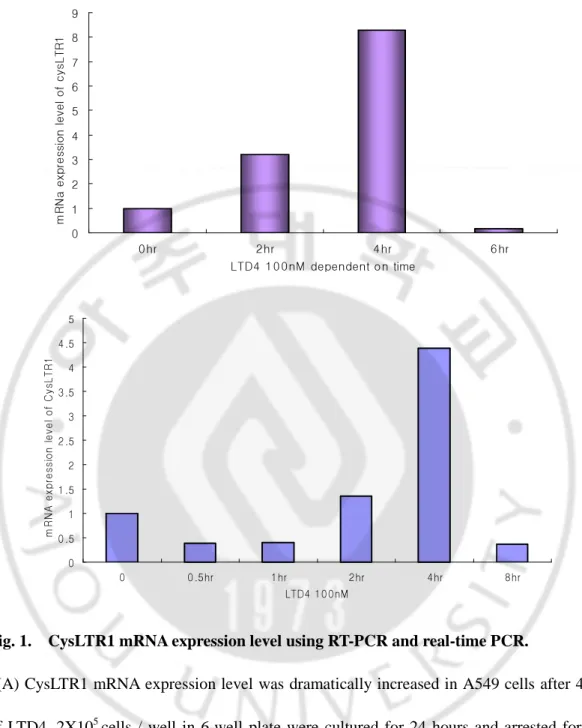
Fig. 1. CysLTR1 mRNA expression level using RT-PCR and real-time PCR ··· 9
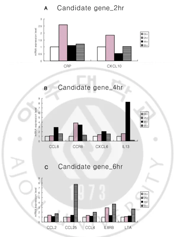
Fig. 2. The results of human inflammatory cytokines superarray using real-time PCR ·· 10
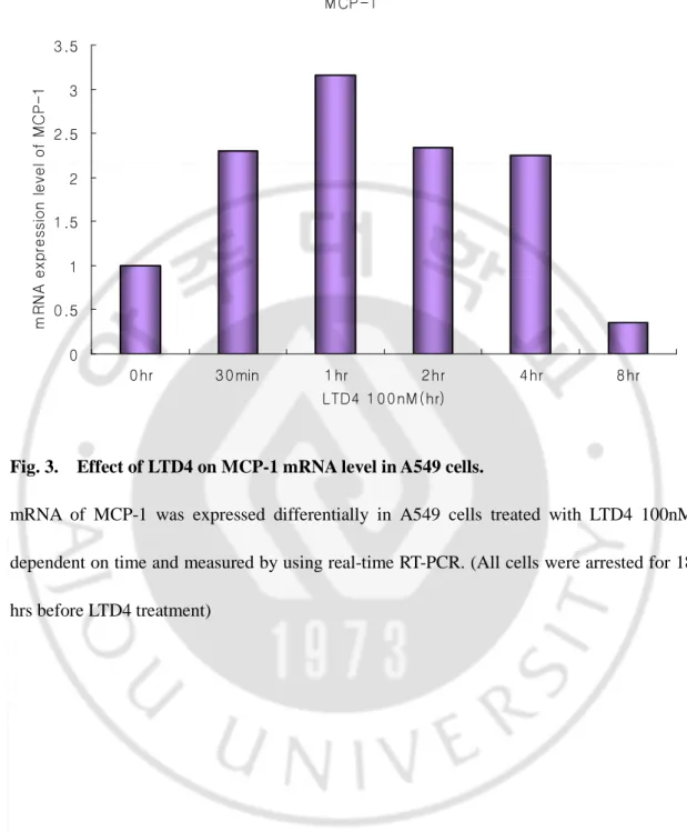
Fig. 3. Effect of LTD4 on the production of MCP-1 in A549 cells··· 14
Fig. 4. Effect of LTD4 on releasing MCP-1 in A549 cells··· 15
Fig. 5. Effect of LTD4 on the production of IP-10 in A549 cells··· 16
Fig. 6. mRNA expression of CysLTR1 after CysLTR1 transfection in A549 cells ··· 17
Fig. 7. Flow cytometric analysis of CysLTR1 surface expression ··· 18
Fig. 8. Effect of LTD4 on the production of MCP-1 in CysLTR1-overexpressed A549 cells ··· 19
Fig. 10. Effect of LTD4 on the production of IP-10 in CysLTR1-overexpressed A549
cells ··· 23
Fig. 11. Effect of LTD4 on releasing IP-10 in CysLTR1-overexpressed A549 cells··· 24
Fig. 12. Effect of MK571 to MCP-1 in CysLTR1-overexpressed A549 cells··· 25
Fig. 13. Effect of MK571 to IP-10 in CysLTR1-overexpressed A549 cells ··· 27
LIST OF TABLES
ABBREVIATION
LTD4 : cysteinyl leukotriene D4
CysLTR1 : Cysteinyl leukotriene receptor type I
MCP-1 : Monocyte Chemoattractant Protein – 1 (CCL2) IP-10 : Interferon-γ-inducible Protein 10kDa (CXCL10)
CCL4 : Macrophage inflammatory protein 1-beta, Lymphocyte activation gene 1 protein CCL8 : Monocyte chemoattractant protein 2
CCL 16 : small inducible cytokine subfamily A, member 16 CCL 23 : Macrophage inflammatory protein 3
CCL25 : Thymus expressed chemokine, Ck beta-15 CCR5 : chemokine (C-C motif) receptor 5
CCR8 : C-C chemokine receptor type 8 CRP : C-reactive protein precursor
CXCL6 : granulocyte chemotactic protein 2
CXCL11 : Interferon-inducible T-cell alpha chemoattractant CXCL12 : Pre-B cell growth-stimulating factor
CXCL13 : B lymphocyte chemoattractant
CXCL14 : Small inducible cytokine B14 precursor ICEBERG : Caspase-1 inhibitor Iceberg
IL1F6 : interleukin 1 family, member 6 (epsilon) IL13 : Interleukin-13 precursor
Ⅰ
Ⅰ
Ⅰ
Ⅰ. INTRODUCTION
Asthma is known as a chronic inflammatory disease caused by infiltration of inflammatory cells such as T cells, eosinophils, mast cells. In the pathogenesis of asthma, leukotrienes (LTs) play an important role by inducing bronchoconstriction, vascular hyperpermeability, mucus hypersecretion, airway hypersensitivity, and eosinophil recruitment (Kanaoka et al., 2004). They are metabolic products of arachidonic acid by cytosolic phospholipase A2 (cPLA2) from phospholipids in cellular membranes (Bandeira-Melo et al., 2003, Henderson, 1994, Wenzel, 2003). Especially, cysteinyl leukotrienes (CysLTs, known to consist of LTC4, and its conversion products, LTD4 and LTE4) are generated via 5-lipoxygenase pathway of arachidonic acid metabolism in a number of inflammatory cells including eosinophils, basophils, monocytes, and macrophages (Lewis et al., 1990, O'Byrne, 1997).
CysLTs affect the inflammatory response by inducing the production of cytokines required to stimulate T-cell responses or determine the homing of dendritic cells, and attract leukocytes to initiate inflammatory responses either directly or indirectly (Kanaoka et al., 2004). Many studies have reported that high level of cysLTs were detected in urine and/or exhaled air condensate of asthmatic patients (Antczak et al., 2002, Capra et al., 2006, Green et al., 2004, Montuschi et al., 2002, O'Byrne, 1997, Sampson et al., 1995). In addition, inhalation of LTD4 induced bronchoconstriction and sputum eosinophilia in patients with asthma, while inhaled methacholine, other bronchoconstrictor, didn’t affect eosinophil counts
(Diamant et al., 1997, Fregonese et al., 2002).
The bronchoconstrictive effect of cysLTs is exerted via two types of their G protein-coupled receptor, cysteinyl leukotriene receptor type 1 (CysLTR1) and type 2 (CysLTR2) (Brink et al., 2003, Capra et al., 2004). In particular, CysLTR1 is expressed in airway smooth muscle cells, eosinophils, B-lymphocytes, monocytes/macrophages and CD 34+ progenitor cells and has high affinity preferentially with LTD4 (Naik et al., 2005). The proinflammatory roles of cysLTs via CysLTR1 (Woszczek et al., 2005) have been certified by the observation that CysLT synthesis inhibitors or CysLTR1 antagonists not only reduced airway and/or blood eosinophilia but also downregulated bronchial constrictor responses to a variety of triggers. These improvements of baseline lung function in vivo and in vitro studies suggest that cysLTs contribute as potent factors to the development of asthma and some of the disease regulatory actions of cysLTs are mediated through CysLTR1 (Bjermer et al., 2002, Capra et al., 2006, Eum et al., 2003, Holgate et al., 2003, Nagata et al., 2003).
We hypothesized that LTD4, the potent mediator of asthma, might induce cytokines and/or chemokines that attract inflammatory cells directly or indirectly via CysLTR1. In this study, we investigated the effect of LTD4 via CysLTR1 on cytokine and chemokine expressions in human lung epithelial A549 cells, and also examined the inhibitory effect of CysLTR1 antagonist, MK-571 on the inflammatory role of LTD4.
Ⅱ
Ⅱ
Ⅱ
Ⅱ. MATERIALS AND METHODS
A. Materials
Phosphate buffered saline (PBS) was made by laboratory protocol, MK-571, selective CysLTR1 antagonist, was purchased from Biomol (Plymouth Meeting, PA), RPMI 1640, Fetal Bovine Serum (FBS), and anti-biotics were purchased from GIBCO (NY, USA), Lipofectamine reagent was purchased from Invitrogen (Seoul, Korea), LTD4 was purchased from SIGMA (MA, USA), easy-BLUE total RNA extract reagent was purchased from iNtRON (Daejeon, Korea), MMLV reverse transcriptase and RNasin were purchased from Promega (Madison, WI, USA), QIAGEN plasmid maxi and midi prep kit were purchased from QIAGEN (QIAGEN, Hilden, Germany), Human Quantikine for CCL2/MCP-1 and CXCL10/IP-10 were purchased from R&D Systems (TECHNE Corporation, MN, USA), Human cytokine superarray assay was purchased from Superarray (Bioscience corporation, MD, USA), rabbit anti-human CysLTR1 polyclonal antibody, rabbit anti-human IP-10 polyclonal antibody and goat anti-rabbit IgG conjugated to FITC were purchased from Santa Cruz (CA, USA), ABI 7500 real-time RT-PCR machine and Power SYBR Green PCR master mix were purchased from Applied Biosystems (CA, USA).
B. Cell culture and transfection of CysLTR1 gene
A549 cells were maintained in RPMI 1640 medium (GIBCO) supplemented with 10% fetal bovine serum, 100U/ml of penicillin G sodium, and 100ug/ml of streptomycin sulfate
(penicillin-streptomycin, GIBCO) and cultured at 37℃, and 5% CO2 in humidified incubator.
2 X 10⁵ Cells were seeded in each well of 6-well plate for 1 day prior to transfection so that they were attached with 60~70% confluency. To overexpress CysLTR1 in A549 cells, transfections were carried out in serum free medium using 5ul of Lipofectamine transfection reagent(Invitrogen), and 0.75ug of CysLTR1 construct and 0.75ug of 3xHA(Hemaglutanin) tagged CysLTR1 construct DNA per well according to the manufacturer’s protocol. 5hrs after transfection, cells were added FBS and cultured for 24hrs to express transfected CysLTR1 genes.
C. Treatment of LTD4 and MK571
After transfections, cells were starved with serum free media and arrested for 18 hrs before being treated with 100nM of MK571, selective CysLTR1 antagonist, and/or 100nM of LTD4. In case of treatment of MK571, it was pretreated for 3hrs and removed by being changed into new plain RPMI 1640 medium (treatment of MK571 for 3hrs was more effective to block CysLTR1 than for 18 hrs) . To harvest all cells of each well at the same time, cells were treated with 100nM of LTD4 for 8, 6, 4, 2, 1 and 0.5 hrs before harvest respectively. Cells and supernatant of each well were kept for real-time PCR and/or western blotting, and ELISA respectively. For real-time PCR, cells were added 1ml of easy-BLUE (Intron) directly after removing supernatants. For western blotting, cells were harvested with 0.25% Trypsin-EDTA solution (Sigma) , washed in phosphate-buffered saline and lysed in lysis buffer or frozen at -78℃
D. RT-PCR analysis for CysLTR1 mRNA expression level
Total RNA was extracted from cells by using Easy-Blue reagent (Intron Biotechnology, Korea) according to the manufacturer’s instructions. After denaturation of RNA and annealing with oligo dT 18mers (72℃, 5min), cDNA was synthesized from 2.5ug of RNA using 200U of MMLV-RTase (Promega) under optimized reaction conditions (RT buffer : 50mM pH 8.3Tris-HCl, 75mM KCl, 3mM MgCl2, 10mM DTT, 2mM dNTPs, 0.5U/ul RNase inhibitors (RNasin, Promega)) at 42℃ for 1hr and extended at 70℃ for 10min. To confirm the synthesis of cDNA or normalize the concentration of cDNA, β-actin RT-PCR was performed with primers TCCTTCTGCATCCTG TCGGC as forward primer and 5’-CAAGAGAT GGCCACGGCTGC as reverse primer using 0.03U of Taq polymerase (Solgent) under the following buffer and PCR condition (PCR-buffer : 20mM pH 8.4 Tris-HCl, 75mM KCl, 0.2mM dNTPs, 2mM MgCl2, 0.2uM forward and reverse primers, PCR condition : 22 cycles, denaturation : 95℃, 30sec ; annealing : 58℃, 30sec ; extension : 72℃, 30sec) using DNA Engine Thermal cycler (Bio-rad). Specific primers of target genes (CysLTR1, F : TGACCGCTGCCTTTTTAGTC, R : GAGAGGGTCAAAGCAACAA TTG ; MCP-1, F : TGCAGCTAACTTATTTTCCC, R : AGAACTGTGGTTCAAGAGGA ; CXCL10, F : CTAGAACCGTACGCTGTACCT, R : TCAGACATCTCT TCTCACCC ) were designed in their own coding regions
E. Human inflammatory cytokines superarray assay
cDNA from A549 and CysLTR1-tranfected A549 cells was prepared for template. Assay was performed using ABI 7500 real-time RT-PCR following the protocol provided by the
manufacturer (SuperArray Bioscience Corporation, USA). The assay plate contains 84 kinds of human cytokines primers and 5 different housekeeping genes primers for control. The results were analyzed by 2-ΔΔCt method using ABI 7500 prism software.
F. Quantitative Real-time RT PCR for CysLTR1, MCP-1 and IP-10
Quantitative real-time RT-PCR was performed using the SYBR Green PCR Master Mix (Applied Biosystems). Amplification conditions were 95℃ for 15min as the first step, 45 cycles of denaturation at 95℃ for 15sec, annealing at 58℃ for 30sec, and extension at 72℃ for 33sec. For relative quantitation, we used a method that compares the amount of target genes normalized to an endogenous reference gene such as β-actin. The formula was 2-∆∆Ct, representing the n-fold differential expression of a specific gene in a treated sample compared with the control, where Ct is the mean of threshold cycle, ∆Ct was the difference in the Ct values for the target gene and the reference gene, β-actin (in each sample), and
∆∆Ct represents the difference between the Ct from the control and each datum. Before
using this method, we performed a validation experiment comparing the standard curve of the reference and the target to show that efficiencies were equal. Because the annealing temperatures among reference gene and target genes, all real-time RT-PCR experiments in the same batch could be performed at the same time. Used primers were same with those for RT-PCR.
G. Measurement of released cytokines by ELISA
determined with ELISA specific kit (R&D Systems, Minneapolis, MN, USA). The detection limits for MCP-1 and IP-10 were 15.6pg/ml and 7.8pg/ml respectively. It was determined the optical density of each well within 30min, using a microplate reader set to 450nm.
H. Measurement of CysLT1 receptor expression on the membrane by flow cytometry
Cells were harvested using trypsin-EDTA , washed in PBS buffer and transferred to 5ml polystyrene round-bottom tube. For flow cytometry, cells were centrifuged at 1500rpm for 3min at 4℃ each time. And they were fixed with 4% paraformaldehyde-PBS for 30min at 4℃ and permeabilized with 0.3% Triton X-100-PBS supplemented with 5% FBS for 30min at 4℃. Cells were then labeled with a 1:100 dilution of an anti-CysLTR1 or anti-HA antibody in permeabilizing Triton X-100 solution for 40min at 4℃ and washed twice in PBS to remove unattached antibodies. And then cells were incubated with a 1:200 dilution of an anti-IgG conjugated to FITC in permeabilizing Triton X-100 solution for 40min at 4℃ and washed twice in PBS to remove unattached antibodies and get clear signal. For the last time, cells were resuspended in 200ul of PBS
Ⅲ
Ⅲ
Ⅲ
Ⅲ. RESULTS
A. Cytokines / chemokines induced by LTD4 in A549 cells
As a first step in assessing the effect of LTD4 on CysLTR1 expression, we analyzed mRNA expression level of CysLTR1 by real-time RT-PCR in various cell lines including U937 (Human leukemic monocyte lymphoma cell), differentiated U937 (macrophage-like cell), THP-1 (human monocytes) and A549 cells(human lung epithelial cells). Among these cell lines, CysLTR1 was prominently increased by stimulation with 100nM of leukotriene D4(LTD4) with time dependency in A549 cells and peaked at 4hrs after stimulation (Fig. 1A). This result was reproduced in the triplicate experiments (Fig. 1B).
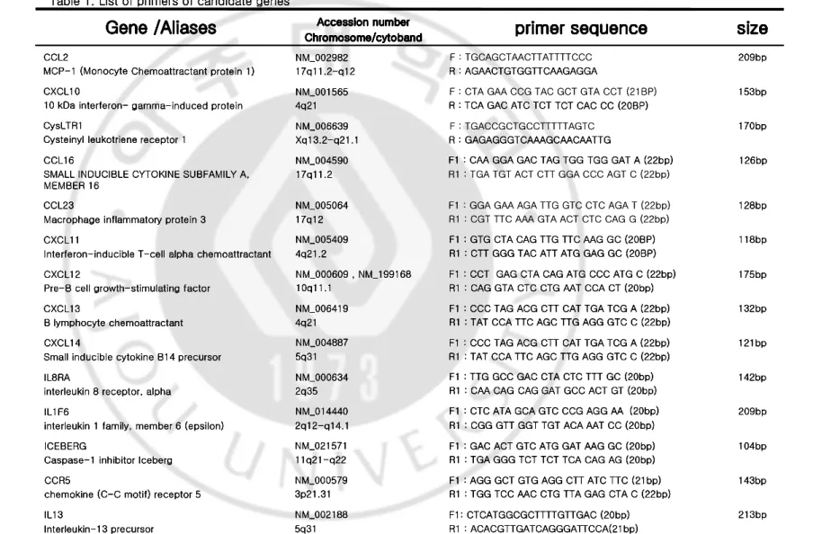
To search candidate inflammatory cytokines / chemokines which are sensitively responsible for LTD4, human inflammatory cytokines superarray assay containing 84 genes was performed. After stimulation of 100nM of LTD4 on A549, the cells were harvested in different time (0, 2, 4, and 6hrs) and then examined the expression levels of 84 inflammatory cytokines by real-time RT-PCR (Fig. 2.). CRP and CXCL10 were peaked at 2hr after LTD4 treatment (Fig. 2A.), CCL8, CCR8, CXCL6, and IL13 at 4hr (Fig. 2B.), and CCL2, CCL25, CCL4, IL8RB and LTA at 6hr (Fig. 2C.). Among them, MCP-1 and IP-10 were selected as target genes due to their biological roles in asthma. For MCP-1(CCL2) and IP-10(CXCL10) genes, gene-specific primers were designed respectively (Table 1.) and the mRNA expression levels were examined by real-time RT-PCR.
CysLT R1 ex pression in A549 by LT D4 CysLT R1 ex pression in A549 by LT D4CysLT R1 ex pression in A549 by LT D4 CysLT R1 ex pression in A549 by LT D4
0 1 2 3 4 5 6 7 8 9 0 hr 2 hr 4 hr 6 hr LTD4 1 0 0 nM dependent o n time m R N a e x p re s s io n l e v e l o f c y s L TR 1 0 0.5 1 1.5 2 2.5 3 3.5 4 4.5 5 0 0.5hr 1hr 2hr 4hr 8hr LTD4 10 0nM m R N A e x p re s s io n l e v e l o f C y s L T R 1
Fig. 1. CysLTR1 mRNA expression level using RT-PCR and real-time PCR.
(A) CysLTR1 mRNA expression level was dramatically increased in A549 cells after 4hrs of LTD4. 2X10⁵ cells / well in 6-well plate were cultured for 24 hours and arrested for 18 hours before treatment with LTD4 100nM for 0, 2, 4, and 8hours. (B) Same experiment as (A) was repeated to be confirmed and the result showed a similar tendency.
Candidate gene_2hr
0 0.5 1 1.5 2 2.5 3 CRP CX CL10 m R N A e x p re s s io n l e v e l 0h r 2h r 4h r 6h r A A A ACandidate gene_2hr
0 0.5 1 1.5 2 2.5 3 CRP CX CL10 m R N A e x p re s s io n l e v e l 0h r 2h r 4h r 6h r A A A ACandidate gene_4hr
0 1 2 3 4 5 6 7 8 9 CCL8 CCR8 CX CL6 IL13 m R N A e xp re s s io n l e ve l 0h r 2h r 4h r 6h r B B B BCandidate gene_4hr
0 1 2 3 4 5 6 7 8 9 CCL8 CCR8 CX CL6 IL13 m R N A e xp re s s io n l e ve l 0h r 2h r 4h r 6h r B B B BCandidate gene_6hr
0 1 2 3 4 5 6 7 8 9 CCL2 CCL25 CCL4 IL8RB LTA m R N A e x p re s s io n l e v e l 0hr 2hr 4hr 6hr C C C CCandidate gene_6hr
0 1 2 3 4 5 6 7 8 9 CCL2 CCL25 CCL4 IL8RB LTA m R N A e x p re s s io n l e v e l 0hr 2hr 4hr 6hr C C C CFig. 2. The results of human inflammatory cytokines superarray using real-time PCR.
100nM dependent upon time. 2(A), 4(B) and 6hr(C)-treated cells were normalized to untreated cells. (CRP, C-reactive protein precursor ; CXCL10 , IP-10 (Interferon-γ-inducible Protein 10kDa) ; CCL8, Monocyte chemoattractant protein 2 ; CCR8, C-C chemokine receptor type 8 ; CXCL6, granulocyte chemotactic protein 2 ; IL13, Interleukin-13 precursor ; CCL2, MCP-1 (Monocyte Chemoattractant Protein – 1) ; CCL25, Thymus expressed chemokine, Ck beta-15 ; CCL4, Macrophage inflammatory protein 1-beta, Lymphocyte activation gene 1 protein ; IL8RB, High affinity interleukin-8receptor B ; LTA, Lymphotoxin-alpha precursor, Tumor necrosis factor ligand superfamilymember 1)
1
2
-143bp F1 : AGG GCT GTG AGG CTT ATC TTC (21bp)
R1 : TGG TCC AAC CTG TTA GAG CTA C (22bp) NM_000579
3p21.31 CCR5
chemokine (C-C motif) receptor 5
104bp F1 : GAC ACT GTC ATG GAT AAG GC (20bp)
R1 : TGA GGG TCT TCT TCA CAG AG (20bp) NM_021571
11q21-q22 ICEBERG
Caspase-1 inhibitor Iceberg
209bp F1 : CTC ATA GCA GTC CCG AGG AA (20bp)
R1 : CGG GTT GGT TGT ACA AAT CC (20bp) NM_014440
2q12-q14.1 IL1F6
interleukin 1 family, member 6 (epsilon)
142bp F1 : TTG GCC GAC CTA CTC TTT GC (20bp)
R1 : CAA CAG CAG GAT GCC ACT GT (20bp) NM_000634
2q35 IL8RA
interleukin 8 receptor, alpha
121bp F1 : CCC TAG ACG CTT CAT TGA TCG A (22bp)
R1 : TAT CCA TTC AGC TTG AGG GTC C (22bp) NM_004887
5q31 CXCL14
Small inducible cytokine B14 precursor
132bp F1 : CCC TAG ACG CTT CAT TGA TCG A (22bp)
R1 : TAT CCA TTC AGC TTG AGG GTC C (22bp) NM_006419
4q21 CXCL13
B lymphocyte chemoattractant
175bp F1 : CCT GAG CTA CAG ATG CCC ATG C (22bp)
R1 : CAG GTA CTC CTG AAT CCA CT (20bp) NM_000609 , NM_199168
10q11.1 CXCL12
Pre-B cell growth-stimulating factor
118bp F1 : GTG CTA CAG TTG TTC AAG GC (20BP)
R1 : CTT GGG TAC ATT ATG GAG GC (20BP) NM_005409
4q21.2 CXCL11
Interferon-inducible T-cell alpha chemoattractant
128bp F1 : GGA GAA AGA TTG GTC CTC AGA T (22bp)
R1 : CGT TTC AAA GTA ACT CTC CAG G (22bp) NM_005064
17q12 CCL23
Macrophage inflammatory protein 3
126bp F1 : CAA GGA GAC TAG TGG TGG GAT A (22bp)
R1 : TGA TGT ACT CTT GGA CCC AGT C (22bp) NM_004590
17q11.2 CCL16
SMALL INDUCIBLE CYTOKINE SUBFAMILY A, MEMBER 16 170bp F : TGACCGCTGCCTTTTTAGTC R : GAGAGGGTCAAAGCAACAATTG NM_006639 Xq13.2-q21.1 CysLTR1
Cysteinyl leukotriene receptor 1
153bp F : CTA GAA CCG TAC GCT GTA CCT (21BP)
R : TCA GAC ATC TCT TCT CAC CC (20BP) NM_001565
4q21 CXCL10
10 kDa interferon- gamma-induced protein
209bp F : TGCAGCTAACTTATTTTCCC R : AGAACTGTGGTTCAAGAGGA NM_002982 17q11.2-q12 CCL2
MCP-1 (Monocyte Chemoattractant protein 1)
size
size
size
size
primer sequence
primer sequence
primer sequence
primer sequence
Accession number Accession number Accession number Accession number Chromosome/ Chromosome/ Chromosome/
Chromosome/cytobandcytobandcytobandcytoband
Gene /Aliases
Gene /Aliases
Gene /Aliases
Gene /Aliases
Table 1. List of primers of candidate genes
143bp F1 : AGG GCT GTG AGG CTT ATC TTC (21bp)
R1 : TGG TCC AAC CTG TTA GAG CTA C (22bp) NM_000579
3p21.31 CCR5
chemokine (C-C motif) receptor 5
104bp F1 : GAC ACT GTC ATG GAT AAG GC (20bp)
R1 : TGA GGG TCT TCT TCA CAG AG (20bp) NM_021571
11q21-q22 ICEBERG
Caspase-1 inhibitor Iceberg
209bp F1 : CTC ATA GCA GTC CCG AGG AA (20bp)
R1 : CGG GTT GGT TGT ACA AAT CC (20bp) NM_014440
2q12-q14.1 IL1F6
interleukin 1 family, member 6 (epsilon)
142bp F1 : TTG GCC GAC CTA CTC TTT GC (20bp)
R1 : CAA CAG CAG GAT GCC ACT GT (20bp) NM_000634
2q35 IL8RA
interleukin 8 receptor, alpha
121bp F1 : CCC TAG ACG CTT CAT TGA TCG A (22bp)
R1 : TAT CCA TTC AGC TTG AGG GTC C (22bp) NM_004887
5q31 CXCL14
Small inducible cytokine B14 precursor
132bp F1 : CCC TAG ACG CTT CAT TGA TCG A (22bp)
R1 : TAT CCA TTC AGC TTG AGG GTC C (22bp) NM_006419
4q21 CXCL13
B lymphocyte chemoattractant
175bp F1 : CCT GAG CTA CAG ATG CCC ATG C (22bp)
R1 : CAG GTA CTC CTG AAT CCA CT (20bp) NM_000609 , NM_199168
10q11.1 CXCL12
Pre-B cell growth-stimulating factor
118bp F1 : GTG CTA CAG TTG TTC AAG GC (20BP)
R1 : CTT GGG TAC ATT ATG GAG GC (20BP) NM_005409
4q21.2 CXCL11
Interferon-inducible T-cell alpha chemoattractant
128bp NM_005064
17q12 CCL23
Macrophage inflammatory protein 3
126bp F1 : CAA GGA GAC TAG TGG TGG GAT A (22bp)
NM_004590 17q11.2 CCL16
SMALL INDUCIBLE CYTOKINE SUBFAMILY A, MEMBER 16 170bp R : GAGAGGGTCAAAGCAACAATTG NM_006639 Xq13.2-q21.1 CysLTR1
Cysteinyl leukotriene receptor 1
153bp R : TCA GAC ATC TCT TCT CAC CC (20BP)
NM_001565 4q21 CXCL10
10 kDa interferon- gamma-induced protein
209bp R : AGAACTGTGGTTCAAGAGGA
NM_002982 17q11.2-q12 CCL2
MCP-1 (Monocyte Chemoattractant protein 1)
size
size
size
size
primer sequence
primer sequence
primer sequence
primer sequence
Accession number Accession number Accession number Accession number Chromosome/ Chromosome/ Chromosome/
Chromosome/cytobandcytobandcytobandcytoband
Gene /Aliases
Gene /Aliases
Gene /Aliases
Gene /Aliases
Table 1. List of primers of candidate genes
143bp F1 : AGG GCT GTG AGG CTT ATC TTC (21bp)
R1 : TGG TCC AAC CTG TTA GAG CTA C (22bp) NM_000579
3p21.31 CCR5
chemokine (C-C motif) receptor 5
104bp F1 : GAC ACT GTC ATG GAT AAG GC (20bp)
R1 : TGA GGG TCT TCT TCA CAG AG (20bp) NM_021571
11q21-q22 ICEBERG
Caspase-1 inhibitor Iceberg
209bp F1 : CTC ATA GCA GTC CCG AGG AA (20bp)
R1 : CGG GTT GGT TGT ACA AAT CC (20bp) NM_014440
2q12-q14.1 IL1F6
interleukin 1 family, member 6 (epsilon)
142bp F1 : TTG GCC GAC CTA CTC TTT GC (20bp)
R1 : CAA CAG CAG GAT GCC ACT GT (20bp) NM_000634
2q35 IL8RA
interleukin 8 receptor, alpha
121bp F1 : CCC TAG ACG CTT CAT TGA TCG A (22bp)
R1 : TAT CCA TTC AGC TTG AGG GTC C (22bp) NM_004887
5q31 CXCL14
Small inducible cytokine B14 precursor
132bp F1 : CCC TAG ACG CTT CAT TGA TCG A (22bp)
R1 : TAT CCA TTC AGC TTG AGG GTC C (22bp) NM_006419
4q21 CXCL13
B lymphocyte chemoattractant
175bp F1 : CCT GAG CTA CAG ATG CCC ATG C (22bp)
R1 : CAG GTA CTC CTG AAT CCA CT (20bp) NM_000609 , NM_199168
10q11.1 CXCL12
Pre-B cell growth-stimulating factor
118bp F1 : GTG CTA CAG TTG TTC AAG GC (20BP)
R1 : CTT GGG TAC ATT ATG GAG GC (20BP) NM_005409
4q21.2 CXCL11
Interferon-inducible T-cell alpha chemoattractant
128bp F1 : GGA GAA AGA TTG GTC CTC AGA T (22bp)
R1 : CGT TTC AAA GTA ACT CTC CAG G (22bp) NM_005064
17q12 CCL23
Macrophage inflammatory protein 3
126bp F1 : CAA GGA GAC TAG TGG TGG GAT A (22bp)
R1 : TGA TGT ACT CTT GGA CCC AGT C (22bp) NM_004590
17q11.2 CCL16
SMALL INDUCIBLE CYTOKINE SUBFAMILY A, MEMBER 16 170bp F : TGACCGCTGCCTTTTTAGTC R : GAGAGGGTCAAAGCAACAATTG NM_006639 Xq13.2-q21.1 CysLTR1
Cysteinyl leukotriene receptor 1
153bp F : CTA GAA CCG TAC GCT GTA CCT (21BP)
R : TCA GAC ATC TCT TCT CAC CC (20BP) NM_001565
4q21 CXCL10
10 kDa interferon- gamma-induced protein
209bp F : TGCAGCTAACTTATTTTCCC R : AGAACTGTGGTTCAAGAGGA NM_002982 17q11.2-q12 CCL2
MCP-1 (Monocyte Chemoattractant protein 1)
size
size
size
size
primer sequence
primer sequence
primer sequence
primer sequence
Accession number Accession number Accession number Accession number Chromosome/ Chromosome/ Chromosome/
Chromosome/cytobandcytobandcytobandcytoband
Gene /Aliases
Gene /Aliases
Gene /Aliases
Gene /Aliases
Table 1. List of primers of candidate genes
143bp F1 : AGG GCT GTG AGG CTT ATC TTC (21bp)
R1 : TGG TCC AAC CTG TTA GAG CTA C (22bp) NM_000579
3p21.31 CCR5
chemokine (C-C motif) receptor 5
104bp F1 : GAC ACT GTC ATG GAT AAG GC (20bp)
R1 : TGA GGG TCT TCT TCA CAG AG (20bp) NM_021571
11q21-q22 ICEBERG
Caspase-1 inhibitor Iceberg
209bp F1 : CTC ATA GCA GTC CCG AGG AA (20bp)
R1 : CGG GTT GGT TGT ACA AAT CC (20bp) NM_014440
2q12-q14.1 IL1F6
interleukin 1 family, member 6 (epsilon)
142bp F1 : TTG GCC GAC CTA CTC TTT GC (20bp)
R1 : CAA CAG CAG GAT GCC ACT GT (20bp) NM_000634
2q35 IL8RA
interleukin 8 receptor, alpha
121bp F1 : CCC TAG ACG CTT CAT TGA TCG A (22bp)
R1 : TAT CCA TTC AGC TTG AGG GTC C (22bp) NM_004887
5q31 CXCL14
Small inducible cytokine B14 precursor
132bp F1 : CCC TAG ACG CTT CAT TGA TCG A (22bp)
R1 : TAT CCA TTC AGC TTG AGG GTC C (22bp) NM_006419
4q21 CXCL13
B lymphocyte chemoattractant
175bp F1 : CCT GAG CTA CAG ATG CCC ATG C (22bp)
R1 : CAG GTA CTC CTG AAT CCA CT (20bp) NM_000609 , NM_199168
10q11.1 CXCL12
Pre-B cell growth-stimulating factor
118bp F1 : GTG CTA CAG TTG TTC AAG GC (20BP)
R1 : CTT GGG TAC ATT ATG GAG GC (20BP) NM_005409
4q21.2 CXCL11
Interferon-inducible T-cell alpha chemoattractant
128bp NM_005064
17q12 CCL23
Macrophage inflammatory protein 3
126bp F1 : CAA GGA GAC TAG TGG TGG GAT A (22bp)
NM_004590 17q11.2 CCL16
SMALL INDUCIBLE CYTOKINE SUBFAMILY A, MEMBER 16 170bp R : GAGAGGGTCAAAGCAACAATTG NM_006639 Xq13.2-q21.1 CysLTR1
Cysteinyl leukotriene receptor 1
153bp R : TCA GAC ATC TCT TCT CAC CC (20BP)
NM_001565 4q21 CXCL10
10 kDa interferon- gamma-induced protein
209bp R : AGAACTGTGGTTCAAGAGGA
NM_002982 17q11.2-q12 CCL2
MCP-1 (Monocyte Chemoattractant protein 1)
size
size
size
size
primer sequence
primer sequence
primer sequence
primer sequence
Accession number Accession number Accession number Accession number Chromosome/ Chromosome/ Chromosome/
Chromosome/cytobandcytobandcytobandcytoband
Gene /Aliases
Gene /Aliases
Gene /Aliases
Gene /Aliases
B. MCP-1 and IP-10 induced by LTD4 in A549 cells
To investigate the effect of LTD4 on MCP-1 and IP-10, additional experiments were performed. First of all, mRNA expression of MCP-1 after LTD4 stimulation was evaluated using real-time RT-PCR (Fig. 3.) MCP-1 mRNA level was 3 fold increased after 1hr treatment of LTD4 in A549 cells and then tailed out. Released MCP-1 level was gradually induced by LTD4 treatment (Fig. 4.) but the peaked point wasn’t observed exactly so it needs to extend the observation time. Same purposed experiments were performed for IP-10 (Fig. 5.) mRNA expression of IP-10 was induced after 1hr of LTD4 treatment but not as much as MCP-1(Fig. 5.). Released protein level of IP-10 was also analyzed in the supernatant but the concentration of IP-10 is less than detectable limit (data not shown).
C. mRNA and protein expression of transfected CysLTR1
In a further series of experiments, CysLTR1 overexpressed A549 cells were prepared by transient transfection with pCMV-CysLTR1 construct and then treated with 100nM of LTD4. Overexpressed mRNA level of CysLTR1 gene after transfection was confirmed by RT-PCR (Fig. 6.). The surface expression of CysLTR1 was also confirmed by flow cytometry (Fig. 7.). These results indicate that transfected CysLTR1 can be highly expressed in both transcriptional and translational levels and then efficiently localized as a functional receptor in plasma membrane.
M CP -1 0 0.5 1 1.5 2 2.5 3 3.5 0 hr 30 min 1 hr 2hr 4hr 8hr LTD4 1 00nM (hr) m R N A e x pr e s s io n l e v e l o f M C P -1
Fig. 3. Effect of LTD4 on MCP-1 mRNA level in A549 cells.
mRNA of MCP-1 was expressed differentially in A549 cells treated with LTD4 100nM dependent on time and measured by using real-time RT-PCR. (All cells were arrested for 18 hrs before LTD4 treatment)
1 3 0 0 1 3 5 0 1 4 0 0 1 4 5 0 1 5 0 0 1 5 5 0 1 6 0 0 0 hr 3 0 min 1 hr 2 hr 4 hr 8 hr LTD4 1 0 0 nM (hrs) M C P -1 c o n c e n tr a ti o n ( p g /1 0 ^ 6 c e lls )
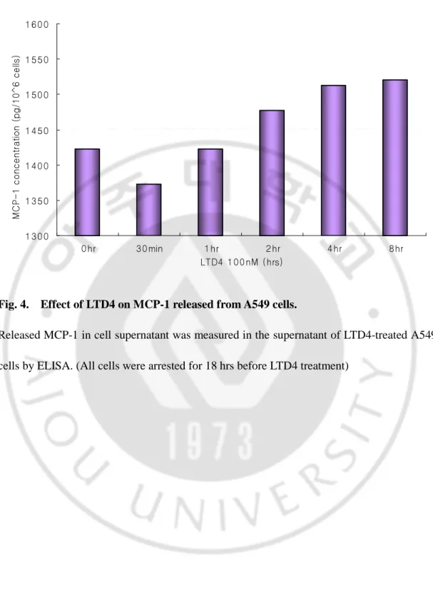
Fig. 4. Effect of LTD4 on MCP-1 released from A549 cells.
Released MCP-1 in cell supernatant was measured in the supernatant of LTD4-treated A549 cells by ELISA. (All cells were arrested for 18 hrs before LTD4 treatment)
0.00 0.20 0.40 0.60 0.80 1.00 1.20 1.40 1.60 0hr 30min 1hr 2hr 4hr 8hr LTD4 100nM m R N A e x p re s s io n l e v e l o f C X C L -1 0
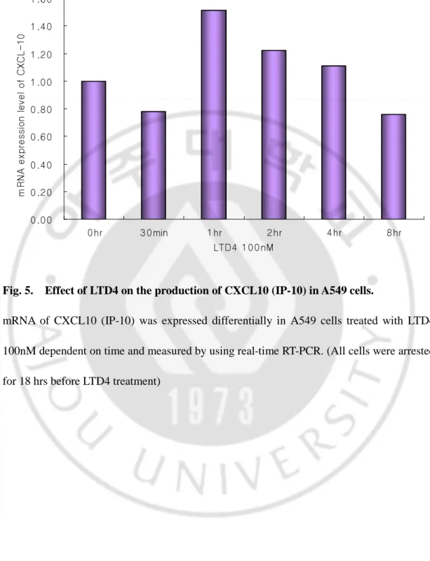
Fig. 5. Effect of LTD4 on the production of CXCL10 (IP-10) in A549 cells.
mRNA of CXCL10 (IP-10) was expressed differentially in A549 cells treated with LTD4 100nM dependent on time and measured by using real-time RT-PCR. (All cells were arrested for 18 hrs before LTD4 treatment)
M 1 2 3 4 5 6
M 1 2 3 4 5 6
M 1 2 3 4 5 6
M 1 2 3 4 5 6
M 1 2 3 4 5 6
M 1 2 3 4 5 6
M 1 2 3 4 5 6
M 1 2 3 4 5 6
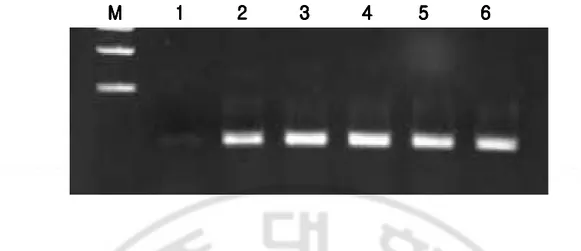
Fig. 6. mRNA expression of CysLTR1 after CysLTR1 transfection in A549 cells.
A549 cells were transfected with pCMV-CysLTR1 construct and then examined the mRNA level by RT-PCR. (M : 1Kb ladder , 1 : A : A549 , 2 : AC : CysLTR1 overexpressed A549 , 3 : ACL30min : CysLTR1 overexpressed A549 + LTD4 100nM 30min , 4 : ACL1hr : CysLTR1 overexpressed A549 + LTD4 100nM 1hr , 5 : ACL2hr : CysLTR1 overexpressed A549 + LTD4 100nM 2hr , 6 : ACL4hr : CysLTR1 overexpressed A549 + LTD4 100nM 4hr )
A549 cell only
7.2%
CysLTR1 transfected A549 cell
52%
A549 cell only
7.2%
A549 cell only
7.2%
CysLTR1 transfected A549 cell
52%
CysLTR1 transfected A549 cell
52%
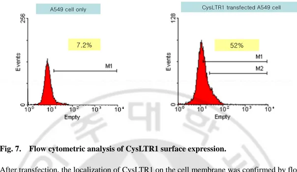
Fig. 7. Flow cytometric analysis of CysLTR1 surface expression.
After transfection, the localization of CysLTR1 on the cell membrane was confirmed by flow cytometry. Cells were labeled with anti-CysLTR1 Ab and stained with anti-IgG Ab conjugated to FITC.
M C P -1 T r a n s fe c t io n o f C y s L T R 1 A A C m R N A e x p re ss io n l ev el o f M 0 4 0 0 5 0 0 6 0 0 5 m R N A e x p re s s io n l e v e l o f M C P -1 M C P -1 T r a n s fe c t io n o f C y s L T R 1 A A C m R N A e x p re ss io n l ev el o f M 0 4 0 0 5 0 0 6 0 0 5 m R N A e x p re s s io n l e v e l o f M C P -1
A
A
A
A
M C P -1 T r a n s fe c t io n o f C y s L T R 1 A A C m R N A e x p re ss io n l ev el o f M 0 4 0 0 5 0 0 6 0 0 5 m R N A e x p re s s io n l e v e l o f M C P -1 M C P -1 T r a n s fe c t io n o f C y s L T R 1 A A C m R N A e x p re ss io n l ev el o f M 0 4 0 0 5 0 0 6 0 0 5 m R N A e x p re s s io n l e v e l o f M C P -1A
A
A
A
0 1 2 3 4 5 6 7AC ACL3 0 min ACL1 hr ACL2 hr ACL4 hr ACL8 hr C : transfectio n o f Cy sLTR 1 L : LTD4 1 0 0 nM m R N A e x p re s s io n l e v e l o f M C P -1
B
B
B
B
0 1 2 3 4 5 6 7AC ACL3 0 min ACL1 hr ACL2 hr ACL4 hr ACL8 hr C : transfectio n o f Cy sLTR 1 L : LTD4 1 0 0 nM m R N A e x p re s s io n l e v e l o f M C P -1
B
B
B
B
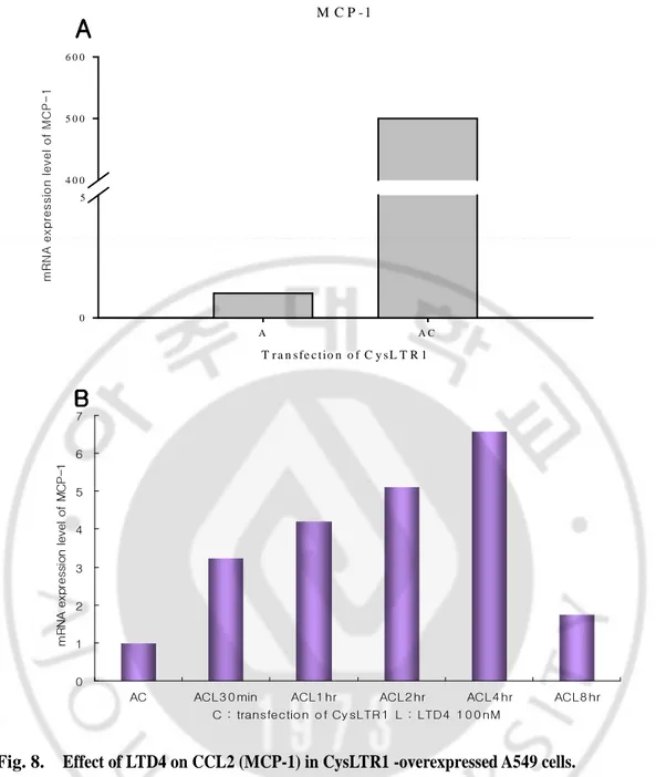
Fig. 8. Effect of LTD4 on CCL2 (MCP-1) in CysLTR1 -overexpressed A549 cells.
mRNA of MCP-1 was expressed differentially in A549 cells transfected with CysLTR1 and treated with LTD4 100nM dependent on time, and measured by real-time RT-PCR. After cultured for 24hrs, A549 cells were transfected with pCMV-CysLTR1 construct using lipofectamine for 5hours in serum free media and added FBS supplemented media for 24hrs.
And then cells were arrested for 15hrs before treatment of 100nM of LTD4. (A) After transfection of CysLTR1, mRNA expression of MCP-1 is significantly enhanced as much as 500-fold. (B) MCP-1 was upregulated gradually by LTD4 and maximally expressed at 4hrs and tailed out. (All cells were arrested for 18 hrs before LTD4 treatment)
D. Enhancement of MCP-1 and IP-10 by overexpressed CysLTR1
To exam whether LTD4 affects MCP-1 and/or IP-10 in CysLTR1-overexpressed A549 cells, mRNA and protein level of MCP-1 and IP-10 were analyzed. Interestingly, we found MCP-1 mRNA level was dramatically augmented as much as 400-fold over for transfected CysLTR1 alone (Fig. 8A). After LTD4 treatment on CysLTR1-overexpressed A549 cells, mRNA level of MCP-1 was gradually increased and peaked at 4hr, and tailed out at 8hr.
For released protein level of MCP-1, however, unexpected results were shown (Fig. 9.). While mRNA level of MCP-1 was augmented by overexpressed CysLTR1, released protein was rather decreased (data not shown). In addition, the effect of LTD4 on protein was also weak than that on mRNA. mRNA expression of IP-10 was also shown in similar tendency as that of MCP-1 in CysLTR1 overexpressed A549 (Fig. 10A.). It was enhanced by exaggerated CysLTR1, however, the mRNA level of 10 was slightly induced by LTD4 treatment. IP-10 protein was also augmented according to overexpressed CysLTR1. Even though the effect of LTD4 on IP-10 transcription level was weak (Fig. 10B.), LTD4 dramatically increased the released IP-10 protein level (Fig. 11.). When comparing to released MCP-1 protein, IP-10 is affected by overexpressed CysLTR1 even in translational level.
E. Inhibitory effect of MK-571 on MCP-1 and IP-10
0 500 1000 1500 2000 2500 3000 3500
AC ACL30min ACL1hr ACL2hr ACL4hr ACL8hr
C : CysLTR1 transfection L : LTD4 100nM c o n c e n tr a ti o n o f M C P -1 (p g /m l)
Fig. 9. Effect of LTD4 on releasing MCP-1 in CysLTR1-overexpressed A549 cells.
Released MCP-1 in cell supernatant was measured by ELISA. (All cells were arrested for 18 hrs before LTD4 treatment)
IP - 1 0 T r a n s f e c t i o n o f C y s L T R 1 A A C m R N A e x p re ss io n l ev el o f M 0 3 0 0 4 0 0 5 0 0 5 m R N A e x p re s s io n l e v e l o f C X C L 1 0 IP - 1 0 T r a n s f e c t i o n o f C y s L T R 1 A A C m R N A e x p re ss io n l ev el o f M 0 3 0 0 4 0 0 5 0 0 5 m R N A e x p re s s io n l e v e l o f C X C L 1 0
A
A
A
A
IP - 1 0 T r a n s f e c t i o n o f C y s L T R 1 A A C m R N A e x p re ss io n l ev el o f M 0 3 0 0 4 0 0 5 0 0 5 m R N A e x p re s s io n l e v e l o f C X C L 1 0 IP - 1 0 T r a n s f e c t i o n o f C y s L T R 1 A A C m R N A e x p re ss io n l ev el o f M 0 3 0 0 4 0 0 5 0 0 5 m R N A e x p re s s io n l e v e l o f C X C L 1 0A
A
A
A
0.00 0.20 0.40 0.60 0.80 1.00 1.20 1.40AC ACL30min ACL1hr ACL2hr ACL4hr ACL8hr C : transfection of CysLTR1 L : LTD4 100nM m R N A e x p re s s io n l e v e l o f C X C L 1 0
B
B
B
B
0.00 0.20 0.40 0.60 0.80 1.00 1.20 1.40AC ACL30min ACL1hr ACL2hr ACL4hr ACL8hr C : transfection of CysLTR1 L : LTD4 100nM m R N A e x p re s s io n l e v e l o f C X C L 1 0 0.00 0.20 0.40 0.60 0.80 1.00 1.20 1.40
AC ACL30min ACL1hr ACL2hr ACL4hr ACL8hr C : transfection of CysLTR1 L : LTD4 100nM m R N A e x p re s s io n l e v e l o f C X C L 1 0
B
B
B
B
Fig. 10. Effect of LTD4 on the production of IP-10 in CysLTR1-overexpressed A549 cells. mRNA of MCP-1 was expressed differentially in A549 cells transfected with CysLTR1
and treated with LTD4 100nM dependent on time, and measured by using real-time RT-PCR. (All cells were arrested for 18 hrs before LTD4 treatment)
0 100 200 300 400 500 600 700 0 0.5 1 2 4 8 LTD4 100nM (hr) IP -1 0 ( p g /4 X 1 0 6 c e lls ) 0 100 200 300 400 500 600 700 0 0.5 1 2 4 8 LTD4 100nM (hr) IP -1 0 ( p g /4 X 1 0 6 c e lls )
Fig. 11. Effect of LTD4 on releasing IP-10 in CysLTR1 overexpressed A549.
Released IP-10 in cell supernatant was measured by ELISA. (All cells were arrested for 18 hrs before LTD4 treatment)
- - 0.5 1 2 4 8 LTD4 100nM (hr) MK-571 10nM0 - + + + + + + 0.5 1 1.5 2 2.5 3 3.5 F o ld ( M C P -1 / b -a c ti n ) - - 0.5 1 2 4 8 LTD4 100nM (hr) MK-571 10nM0 - + + + + + + 0.5 1 1.5 2 2.5 3 3.5 F o ld ( M C P -1 / b -a c ti n )
Fig. 12. Effect of MK571 on mRNA expression of MCP-1 in CysLTR1 overexpressed A549. Cells were pretreated with MK571, CysLTR1 antagonist, for 3hrs and stimulated with
-ssion, it wasn’t certain that induction of MCP-1 and IP-10 by LTD4 are mediated through CysLTR1. To confirm whether LTD4 acts on the production of MCP-1 and IP-10 via CysLTR1, selective CysLTR1 antagonist, MK571 was treated in CysLTR1-overexpressed A549 cells and then mRNA expression levels of MCP-1 and IP-10 were analyzed. In case of MCP-1, as illustrated in Fig. 12. , MK571 didn’t inhibit the effect of LTD4 at all. In contrast, IP-10 expression was completely suppressed by pretreatment of the cells with MK571 despite being recovered by LTD4 100nM in 30min (Fig. 13.). Involvement of the CysLTR1 was confirmed by significant abrogation of IP-10 production in the presence of MK571 while nothing happened for MCP-1 (Fig.12., 14.). These observations suggest that the augmentation of IP-10 expression is mediated via CysLTR1 whereas MCP-1 isn’t.
0 0.5 1 1.5 2 2.5 3 - - 0.5 1 2 4 8 F o ld ( IP -1 0 / b -ac tin ) LTD4 100nM (hr) MK-571 10nM - + + + + + + 0 0.5 1 1.5 2 2.5 3 - - 0.5 1 2 4 8 F o ld ( IP -1 0 / b -ac tin ) LTD4 100nM (hr) MK-571 10nM - + + + + + +
Fig. 13. Effect of MK571 on mRNA expression of IP-10 in CysLTR1 overexpressed A549. Cells were pretreated with MK571, CysLTR1 antagonist, for 3hrs and stimulated with
CysLTR1 MCP-1 IP10 0 2000 4000 6000 8000 10000 12000 A AC ACM ACML 30min 25 50 CysLTR1 MCP-1 IP10 0 2000 4000 6000 8000 10000 12000 A AC ACM ACML 30min 25 50
Fig. 14. Dependency on CysLTR1.
A549 Cells were transfected with CysLTR1 construct, pre-treated with MK571 CysLTR1 antagonist, for 3hrs, and stimulated with LTD4 100nM for 30min.
Ⅳ
Ⅳ
Ⅳ
Ⅳ.
DISCUSSION
The cysLTs, particularly LTD4, is a potent lipid mediator that has been implicated in the pathogenesis of inflammatory processes, preferentially asthma characterized as bronchoconstriction, mucus secretion, and airway hyperresponsiveness. It has been shown that IL-4, the prototypical Th2 type cytokine up- regulates direct chemotactic response to cysLTs by increasing expression of CysLTR1 on human monocytes/macrophages (Thivierge et al., 2001). We hypothesized that the proinflammatory mediator, LTD4 might induce cytokines and/or chemokines which are attractive to inflammatory cells directly or indirectly. Thus, this present work was initiated to study the regulatory mechanisms of cytokines / chemokines, finally leading to persist chronic inflammation in asthmatic patients by LTD4 via CysLTR1. In the present study, we found two chemokines, MCP-1 and IP-10 are prominently up-regulated by LTD4 in human lung epithelial A549 cells.
A549 cells are a kind of alveolar epithelial cells type II that produce surfactant and act as progenitors to replace injured alveolar epithelial cells type I. Thus, they are located at the boundary between the alveolar airspace and the interstitium, and ideally situated to regulate the recruitment and activation of different types of leukocytes through the production of cytokines / chemokines in response to inflammatory stimulation from the alveolar space (Pechkovsky et al., 2005). Recently, it has been suggested that alveolar epithelial cells secrete a variety of mediators, including proinflammtory cytokines and chemokines important for the recruitment of monocytes / macrophages and T cells into the lung
interstitium and alveolar space (Barrett et al., 1998, Kay, 1983, Koyama et al., 1997). In this study, A549 cells rarely expressed CysLTR1 mRNA and they were transfected with CysLTR1 CDS and/or stimulated with LTD4 to represent a condition that cysLTs and CysLTR1 are accumulated in the lungs of patients with asthma. Moreover, CysLTR1 was enhanced by its agonist LTD4 dependent on autocrine signaling, which means A549 cells are stimulated with LTD4 through CysLTR1 and release LTD4 which binds its own receptor CysLTR1, causing self-stimulation. Thus, overexpressed CysLTR1 might elicit more effective signaling to downstream cytokines / chemokines.
Chemokines play an important role in the pathophysiology of asthma and allergy (Hung et al., 2006). A study has reported that airway hyperreactivity could be mediated by allergen-induced, as well as directly instilled-MCP-1 through CCR2, and significantly attenuated in CCR2-/- mice (Campbell et al., 1999). Besides, MCP-1 plays a key role in monocyte recruitment by integrin activation and by promoting migration to the vessel wall (Ashida et al., 2001) and it has been shown that its increased production is induced by cysLTs (LTD4, LTC4) in IL-4 primed THP-1 cells (Woszczek et al., 2005). So, MCP-1 and its receptor CCR2 are potentially important therapeutic targets for the treatment of hyperreactive airway disease and for repair of injured bronchial epithelial cells in asthma (Campbell et al., 1999). Nuclear factor (NF) – kappa (κ) B plays a major role in regulation of inflammatory genes including cytokines, chemokines, inflammatory enzymes, adhesion molecules, and others. Therefore NF-κB is considered as a target for novel anti-inflammtory therapies in diseases such as asthma and COPD (Newton et al., 2002) . It has been reported that NF-κB DNA binding activity was induced by LTD4 through activation of CysLTR1 (Thompson et al.,
2006) and the promoter region of MCP-1 gene contains binding site of NF-κB (Hong et al., 2007). A previous study showed that LTC4, -D4, and –E4 induced MCP-1 in THP-1 human monocytes and CD14+ monocytes/macrophage (Ichiyama et al., 2005). In that study, the inhibitory effect of CysLTR1 antagonist pranlukast on MCP-1 were examined and resulted in almost complete blockade of production of MCP-1 by cysLTs in THP-1 cells and partial inhibition in CD14+ cells. In present study, we examined the effect of LTD4 on MCP-1 in A549 cells and observed that mRNA of MCP-1 was induced by LTD4 in A549 cells as in the previous report in THP-1 cells, and excessive CysLTR1 augmented MCP-1. We also examined the inhibitory effect of CysLTR1 antagonist MK-571 on MCP-1 in CysLTR1-overexpressed A549 cells. In this study, however, CysLTR1 antagonist didn’t act as a inhibitor and didn’t make any difference of mRNA expression level between in presence and in absence of MK-571 suggesting MCP-1 wasn’t regulated via CysLTR1. And it was estimated that MCP-1 production is differently regulated according to cell type and is regulated by LTD4 partially, but might be mediated via CysLTR2, not CysLTR1.
IFN-induced protein of 10kDa (IP-10/CXCL10) is a member of CXCL class and target preferentially activated Th1 lymphocytes and natural killer cells through its receptor CXCR3 (Cole et al., 1998, Farber, 1997). IP-10 has been described in several cell types, including monocytes, keratocytes and neutrophils involved in the more severe form of asthma. Concentration of IP-10/CXCL10 was reported to be elevated in bronchoalveolar lavage fluid of atopic asthmatics following segmental allergen challenge (Bochner et al., 2003, Ying et al., 2005) and elevated numbers of cells expressing IP-10/CXCL10 mRNA and protein in bronchoalveolar lavage fluid and the bronchial mucosa have previously been detected in
asthmatics(Miotto et al., 2001, Ying et al., 2005). Similarly, It has been shown that in a murine model, IP-10 is up-regulated in allergic pulmonary inflammation and overexpressed IP-10 resulted in elevated eosinophil infiltration and IL-4 expression, and the airway hyperreactivity whereas IP-10/CXCL10 deficiency resulted in opposite effects (Medoff et al., 2002). And a study has reported MCP-1 and IP-10 are differently expressed and regulated in human alveolar epithelial cells type II ; MCP-1 is spontaneously expressed in alveolar epithelial cells type II whereas IP-10 is expressed in presence of IFN-γ (Pechkovsky et al., 2005). In present study, IP-10 was differentially expressed in response to LTD4 and elicited high expression by overexpressed CysLTR1 because LTD4 can affect IP-10 more strongly through excessive CysLTR1. The effect of LTD4 on IP-10 through CysLTR1 was evidenced by the inhibitory effect of MK-571 that blocked production of IP-10 in CysLTR1-overexpressed A549 cells, and production of IP-10 was recovered completely by LTD4 100nM in 30 minutes. They suggest IP-10 is regulated by LTD4 directly via CysLTR1. As noted above, NF-κB plays a key role as a regulator of inflammatory genes and the promoter region of IP-10 contains NF-κB and other transcription factor binding sites that are induced by LTD4 (Spurrell et al., 2005).
We demonstrated that IP-10 and MCP-1 are up-regulated by cysteinyl leukotriene D4 (LTD4) although it made a difference of effect respectively. Of interest, the expressions of IP-10 and MCP-1 were increased for overexpressed CysLTR1 alone. MK571, CysLTR1 antagonist inhibits the expression of IP-10 but not MCP-1 suggesting that IP-10 may be involved in CysLTR1 downstream directly, whereas other pathway may be involved in case of MCP-1.
Ⅴ
Ⅴ
Ⅴ
Ⅴ. CONCLUSION
This study suggests that pro-inflammatory chemokine MCP-1(CCL2) and IP-10 (CXCL-10) are up-regulated by cysteinyl leukotriene D4 (LTD4), and mRNA expression levels of IP-10 and MCP-1 are increased for overexpressed CysLTR1 alone. In case of IP-10, released protein production as well as mRNA is augmented by LTD4 and CysLTR1. MK571, CysLTR1 antagonist inhibits the expression of IP-10 but not MCP-1, suggesting that IP-10 may be involved in downstream pathway of CysLTR1 directly whereas other pathway has to be suggested for MCP.
REFERENCE
1. Antczak A, Montuschi P, Kharitonov S, Gorski P, Barnes PJ: Increased exhaled cysteinyl-leukotrienes and 8-isoprostane in aspirin-induced asthma. Am J Respir Crit
Care Med 166:301-306, 2002
2. Ashida N, Arai H, Yamasaki M, Kita T: Distinct signaling pathways for MCP-1-dependent integrin activation and chemotaxis. J Biol Chem 276:16555-16560, 2001
3. Bandeira-Melo C, Weller PF: Eosinophils and cysteinyl leukotrienes. Prostaglandins
Leukot Essent Fatty Acids 69:135-143, 2003
4. Barrett EG, Johnston C, Oberdorster G, Finkelstein JN: Silica-induced chemokine expression in alveolar type II cells is mediated by TNF-alpha. Am J Physiol 275:L1110-1119, 1998
5. Bjermer L, Diamant Z: The use of leukotriene receptor antagonists (LTRAs) as complementary therapy in asthma. Monaldi Arch Chest Dis 57:76-83, 2002
6. Bochner BS, Hudson SA, Xiao HQ, Liu MC: Release of both CCR4-active and CXCR3-active chemokines during human allergic pulmonary late-phase reactions. J
Allergy Clin Immunol 112:930-934, 2003
7. Brink C, Dahlen SE, Drazen J, Evans JF, Hay DW, Nicosia S, Serhan CN, Shimizu T,
Yokomizo T: International Union of Pharmacology XXXVII. Nomenclature for leukotriene and lipoxin receptors. Pharmacol Rev 55:195-227, 2003
8. Campbell EM, Charo IF, Kunkel SL, Strieter RM, Boring L, Gosling J, Lukacs NW:
Monocyte chemoattractant protein-1 mediates cockroach allergen-induced bronchial hyperreactivity in normal but not CCR2-/- mice: the role of mast cells. J Immunol 163:2160-2167, 1999
9. Capra V, Ravasi S, Accomazzo MR, Parenti M, Rovati GE: CysLT1 signal
transduction in differentiated U937 cells involves the activation of the small GTP-binding protein Ras. Biochem Pharmacol 67:1569-1577, 2004
10. Capra V, Thompson MD, Sala A, Cole DE, Folco G, Rovati GE: Cysteinyl-leukotrienes and their receptors in asthma and other inflammatory diseases: Critical update and emerging trends. Med Res Rev, 2006
11. Cole KE, Strick CA, Paradis TJ, Ogborne KT, Loetscher M, Gladue RP, Lin W, Boyd JG, Moser B, Wood DE, Sahagan BG, Neote K: Interferon-inducible T cell alpha chemoattractant (I-TAC): a novel non-ELR CXC chemokine with potent
activity on activated T cells through selective high affinity binding to CXCR3. J Exp
Med 187:2009-2021, 1998
12. Diamant Z, Hiltermann JT, van Rensen EL, Callenbach PM, Veselic-Charvat M, van
der Veen H, Sont JK, Sterk PJ: The effect of inhaled leukotriene D4 and methacholine on sputum cell differentials in asthma. Am J Respir Crit Care Med 155:1247-1253, 1997
13. Eum SY, Maghni K, Hamid Q, Campbell H, Eidelman DH, Martin JG: Involvement
of the cysteinyl-leukotrienes in allergen-induced airway eosinophilia and hyperresponsiveness in the mouse. Am J Respir Cell Mol Biol 28:25-32, 2003
14. Farber JM: Mig and IP-10: CXC chemokines that target lymphocytes. J Leukoc Biol 61:246-257, 1997
15. Fregonese L, Silvestri M, Sabatini F, Rossi GA: Cysteinyl leukotrienes induce human eosinophil locomotion and adhesion molecule expression via a CysLT1 receptor-mediated mechanism. Clin Exp Allergy 32:745-750, 2002
16. Green SA, Malice MP, Tanaka W, Tozzi CA, Reiss TF: Increase in urinary leukotriene LTE4 levels in acute asthma: correlation with airflow limitation. Thorax 59:100-104, 2004
17. Henderson WR, Jr.: The role of leukotrienes in inflammation. Ann Intern Med 121:684-697, 1994
18. Holgate ST, Peters-Golden M: Introduction: the anti-inflammatory role of cysteinyl leukotriene receptor antagonists in asthma. J Allergy Clin Immunol 111:S1-4, 2003
19. Hong MH, Kim MH, Chang HJ, Kim NH, Shin BA, Ahn BW, Jung YD:
(-)-Epigallocatechin-3-gallate inhibits monocyte chemotactic protein-1 expression in endothelial cells via blocking NF-kappaB signaling. Life Sci 80:1957-1965, 2007
20. Hung CH, Li CY, Hua YM, Chen CJ, Yang KD, Jong YJ: Effects of leukotriene receptor antagonists on monocyte chemotaxis, p38 and cytoplasmic calcium. Pediatr
Allergy Immunol 17:250-258, 2006
21. Ichiyama T, Hasegawa M, Ueno Y, Makata H, Matsubara T, Furukawa S: Cysteinyl
leukotrienes induce monocyte chemoattractant protein 1 in human
monocytes/macrophages. Clin Exp Allergy 35:1214-1219, 2005
22. Kanaoka Y, Boyce JA: Cysteinyl leukotrienes and their receptors: cellular distribution and function in immune and inflammatory responses. J Immunol 173:1503-1510, 2004
23. Kay AB: Mediators of hypersensitivity and inflammatory cells in the pathogenesis of bronchial asthma. Eur J Respir Dis Suppl 129:1-44, 1983
24. Koyama S, Sato E, Nomura H, Kubo K, Nagai S, Izumi T: Type II pneumocytes release chemoattractant activity for monocytes constitutively. Am J Physiol 272:L830-837, 1997
25. Lewis RA, Austen KF, Soberman RJ: Leukotrienes and other products of the 5-lipoxygenase pathway. Biochemistry and relation to pathobiology in human diseases.
N Engl J Med 323:645-655, 1990
26. Medoff BD, Sauty A, Tager AM, Maclean JA, Smith RN, Mathew A, Dufour JH, Luster AD: IFN-gamma-inducible protein 10 (CXCL10) contributes to airway hyperreactivity and airway inflammation in a mouse model of asthma. J Immunol 168:5278-5286, 2002
27. Miotto D, Christodoulopoulos P, Olivenstein R, Taha R, Cameron L, Tsicopoulos A, Tonnel AB, Fahy O, Lafitte JJ, Luster AD, Wallaert B, Mapp CE, Hamid Q: Expression of IFN-gamma-inducible protein; monocyte chemotactic proteins 1, 3, and 4; and eotaxin in TH1- and TH2-mediated lung diseases. J Allergy Clin Immunol 107:664-670, 2001
28. Montuschi P, Barnes PJ: Exhaled leukotrienes and prostaglandins in asthma. J
Allergy Clin Immunol 109:615-620, 2002
29. Nagata M, Saito K: The roles of cysteinyl leukotrienes in eosinophilic inflammation of asthmatic airways. Int Arch Allergy Immunol 131 Suppl 1:7-10, 2003
30. Naik S, Billington CK, Pascual RM, Deshpande DA, Stefano FP, Kohout TA, Eckman DM, Benovic JL, Penn RB: Regulation of cysteinyl leukotriene type 1 receptor internalization and signaling. J Biol Chem 280:8722-8732, 2005
31. Newton MF, O'Donnell DE, Forkert L: Response of lung volumes to inhaled salbutamol in a large population of patients with severe hyperinflation. Chest 121:1042-1050, 2002
32. O'Byrne PM: Leukotrienes in the pathogenesis of asthma. Chest 111:27S-34S, 1997
33. Pechkovsky DV, Goldmann T, Ludwig C, Prasse A, Vollmer E, Muller-Quernheim J,
Zissel G: CCR2 and CXCR3 agonistic chemokines are differently expressed and regulated in human alveolar epithelial cells type II. Respir Res 6:75, 2005
urinary leukotrienes after acute asthma. Arch Dis Child 73:221-225, 1995
35. Spurrell JC, Wiehler S, Zaheer RS, Sanders SP, Proud D: Human airway epithelial cells produce IP-10 (CXCL10) in vitro and in vivo upon rhinovirus infection. Am J
Physiol Lung Cell Mol Physiol 289:L85-95, 2005
36. Thivierge M, Stankova J, Rola-Pleszczynski M: IL-13 and IL-4 up-regulate cysteinyl leukotriene 1 receptor expression in human monocytes and macrophages. J Immunol 167:2855-2860, 2001
37. Thompson C, Cloutier A, Bosse Y, Thivierge M, Gouill CL, Larivee P, McDonald PP, Stankova J, Rola-Pleszczynski M: CysLT1 receptor engagement induces activator protein-1- and NF-kappaB-dependent IL-8 expression. Am J Respir Cell Mol Biol 35:697-704, 2006
38. Wenzel SE: The role of leukotrienes in asthma. Prostaglandins Leukot Essent Fatty
Acids 69:145-155, 2003
39. Woszczek G, Pawliczak R, Qi HY, Nagineni S, Alsaaty S, Logun C, Shelhamer JH:
Functional characterization of human cysteinyl leukotriene 1 receptor gene structure.
40. Ying S, O'Connor B, Ratoff J, Meng Q, Mallett K, Cousins D, Robinson D, Zhang G, Zhao J, Lee TH, Corrigan C: Thymic stromal lymphopoietin expression is increased in asthmatic airways and correlates with expression of Th2-attracting chemokines and disease severity. J Immunol 174:8183-8190, 2005