배양된 골수 간엽 간세포에서 골형성의 유도 및
그 표식 인자의 발현
경희대학교 의과대학 정형외과학교실, 연세대학교 의과대학 정형외과학교실*
석경수・김현우
・한수봉
Induction of Osteogenesis from Cultured Mesenchymal
Stem Cells and Their Marker Expression
Kyung-Soo Suk, M.D., Hyun-Woo Kim, M.D.*, Soo-Bong Hahn, M.D.*
Department of Orthopaedic Surgery, College of Medicine,
Kyung Hee University, Yonsei University*, Seoul, Korea
P u r p o s e: To induce osteogenesis from culuted mesenchymal stem cell isolated from human
bone marrow and to detect the marker of osteogenesis to develop cell therapy method
Materials & Methods: Mesenchymal stem cell (MSC) was isolated form human bone marrow
and cultured. In vitro and in vivo induction of osteogenesis was performed. And histological,
immunohistochemical and molecular biological study was performed to confirm osteogenesis.
R e s u l t s: Osteogenesis induced MSCs were strongly stained by ALP immunochemical stain.
Von Kossa stain showed black meneral deposition. Tetracyclin labeling revealed new bone
for-mation. RT-PCR for osteocalcin m-RNA showed increased osteocalcin m-RNA. H-E stain
showed penetration of cellular component into the pores of collagraft strip and showed formation
of osteoid.
Conclusion: Based on the results, osteogenesis could be induced from cultured mesenchymal
stem cell isolated from human bone marrow. Expression of markers for osteogenesis was
con-firmed by histologic, immunohistochemical, and molecular biologic method.
Key Words : Bone marrow, Osteogenesis, Mesenchymal stem cell
* Address for Correspondence : Soo-Bong Hahn, M.D.
Department of Orthopaedic Surgery, School of Medicine, Yonsei University, Seoul, Korea 134 Shinchon-dong, Seodaemun-gu, Seoul 120-752, Korea
Tel : 82-2-361-5640, Fax : 82-2-363-1139, E-mail : [email protected]
서
론
과거 골절치료의 발전은 기계적 안정성, 골 이식 술, 수술 수기의 개선 등으로 이루어졌다. 그러나 현 대에는 골절의 치유에 세포 분자 생물학 발전이 많은 도움을 주고 있다. 실험적으로 전기 자기장, 탈무기 질화한 골기질, 골형성 단백 및 자가 골수 세포를 이 용하여 골절의 지연 유합이나 불유합을 치료할 수 있 는 잠재력이 밝혀졌다.1 - 3 , 8 , 9 , 1 5 , 1 9 , 2 0 , 2 2 - 2 5 , 3 0 , 3 4 , 3 5 )골절의 불유합, 골 결손, 일리자로프 하지 연장술후 합병될 수 있는 골형성의 부족, 골 분절 이동술 후 골편 접 촉면의 불유합등 많은 분야에서 골 이식술이 필수적 이며 그 중 자가 장골 이식술이 가장 좋은 결과를 보 인다. 그러나 자가 장골 이식술은 마취 하에 정상적 인 장골의 일부를 채취하여 이식부에 이식하는 것으 로 그 양이 제한적이며 골 이식 공여부 동통 등의 단 점이 있다. 현재는 기존의 자가골 이식술과 위에서 기술된 골대체물의 단점을 극복하고자 조직공학 기법 을 이용한 자가 세포 배양 이식술의 적극적인 연구가 진행되고 있다. 자가 세포 배양 이식은 환자로부터 채취한 세포의 실험실 배양 과정을 통하여 세포의 증 식을 유도함으로써 이식에 필요한 충분한 수의 세포 를 확보하고 이를 원래의 조직과 같은 구조가 되도록 삼차원 배양하여 생체에 이식하는 술식이다. 이는 실 험실 배양 과정을 통해 충분한 수의 세포를 확보함으 로써 결손부위가 크더라도 무리 없이 사용할 수 있다 는 장점과 함께 자가 세포이식이므로 면역 거부반응 이 전혀 없다는 매우 유용한 장점을 가지고 있다. 최 근에는 자가세포 중 다양한 세포로 분화-증식할 수 있는 줄기 혹은 간세포(stem cells)에 대한 관심이 특히 높아지고 있다. 골수(bone marrow)는 지금 까지 혈액세포를 공급하는 줄기세포를 공급하는 것으 로 알려져 왔으나 다른 간엽 조직의 간세포도 존재하 는 것으로 확인되었다. 이 줄기세포는 골, 연골, 근 육, 지방세포 등으로 분화할 수 있는 다양한 능력을 가지고 있어 이를 골수 간엽 간세포(bone marrow mesenchymal stem cells)라 부르며 이를 임상에 적용하기 위한 다양한 시도가 현재 이루어지고 있 다.2 - 7 , 1 0 , 1 6 , 2 4 , 2 5 , 3 3 )1 9 9 9년 한과 석은 백서의 골수에서 분리 배양한 간엽 간세포의 시험관재 골형성 및 생체 내 골형성의 유도를 성공적으로 시행한 바가 있으며 12 본 연구에서는 이를 바탕으로 사람의 골수에서 분리 배양한 간엽 간세포를 이용하여 골형성을 유도 하여 자가골 이식술을 대체할 수 있는 방법을 개발하 기 위한 기초 자료를 제공하고자 한다.재료 및 방법
1. 간엽 간세포의 분리 및 배양 골이식술을 위하여 입원중인 환자에서 자가 장골을 채취하기 전 17 게이지 spinal needle을 이용하여 골수를 흡입, 채취한다. 채취한 골수를 포도당 1 g / L가 들어 있는 D u l b e c c o’s modified Eagle’s medium(DMEM-LG, Gibco BRL, Grand Island, NY, U.S.A.)에 부유 시킨다. 골수 세포의 부유물을 원심 분리기에 1500 rpm으로 1 0분간 두 어 상층액을 버린후 Phosphate buffered saline(PBS: 137 mM NaCl, 2.7 mM KCl, 4.3 mM Na2HPO4-7H2O, KH2PO4, pH 7.3)으로 3회 세척한 후 10% 우태혈청(fetal bovine serum), penicillin(100U/ml, Gibco BRL)과 steptomycin(100 μg/ml, Gibco BRL)이 포함된 DMEM-LG 배양액이 들어있는 75 cm2 조직 배양 용기에 5×1 07 개의 세포수의 농도로 넣어 3 7℃, 5% CO2 배양기에서 배양을 시작하였다. 4일 후 부 유하는 세포는 배양액을 교환하며 버리고 이후 3일 간격으로 2주간 배양액을 교환하여 간엽 간세포의 분리 및 일차 배양을 시도하였다. 이후 역시 3일 간 격으로 배양액을 교환하여 배양 용기 바닥이 가득차 면 0.25% trypsin-EDTA(Gibco BRL)를 3 7℃에 서 5분간 처리하여 세포를 분리하여 계대 배양하였 다. 상기한 방법으로 충분한 양의 세포가 얻어지면 일부 세포는 - 7 0℃에서 보관하였다. 2. 배양된 간엽 간세포에서 시험관내 골형성의 유도 간엽 간세포를 10% 우태혈청, penicillin과s t e p t o m y c i n이 포함된 DMEM-LG 배양액이 들어 있는 6-well(10 cm2
) tissue culture plate에 3 ×1 03c e l l s / c m2의 농도로 2 4시간 배양한후 골형성 을 유도할 간엽 간세포가 들어있는 조직 배양 용기에 는 100 nM dexamethasone(Sigma Chemical Co., St. Louis, MO, U.S.A.), 10 mM β- g l y c-erophosphate(Sigma), 0.05 mM L-ascorbic acid-2-phosphate(Gibco BRL)로 구성된 골형성 유도 용액을 추가하였고 대조군으로 골형성을 유도하 지 않을 간엽 간세포가 들어있는 조직 배양 용기에는 골형성 유도 용액을 추가하지 않고 10% 우태혈청-D M E M만으로 배양하였다. 3일 간격으로 배양액을 교환하며 골형성 유도군에는 골형성 유도 용액도 추 가하며 2주간 배양하였다. 3. 시험관내 골형성의 평가 가.알칼리 인산 분해 효소의 활성도의 정량적 측정 각 배지의 배양액을 버리고 P B S로 세척한 후 세포 에 발색 반응을 나타낼 수 있는 알칼리 인산 분해 효 소의 기질(2.5 mM p-nitrophenyl phosphate) 이 포함된 알칼리 인산 분해 효소 기질 완충액( 5 0 mM glycine, 1 mM magnesium chloride, pH10.5) 1 ml를 가하여 3 7℃에서 1 5분간 반응시 켰다. 15분 후 배지에서 완충액을 모아 완충액에 1N NaOH 1 ml를 가하여 반응을 중단시켰다. 완충 액을 ELISA plate reader(Bio-Rad, Melville, NY, USA)를 이용하여 405 nm에서 흡광도를 측정 하였다. 나. 알칼리 인산 분해 효소 조직 화학 염색 Sigma kit #85를 이용하여 2 5℃에서 1시간 동 안 시행하였다. 반응하는 동안 배지의 세포가 건조해 지거나 빛에 노출되지 않도록 하였으며 세척시에는 비이온수(deionized water)를 사용하였다. 다. 무기질 침착의 염색(von Kossa 염색) 세포를 10% formaldehyde로 1시간 고정한 후 암실에서 2% silver nitrate 용액( S i g m a )으로 1 0 분간 반응시켰다. 비이온수로 세척하고 밝은 광선에 1 5분간 노출하였다.
라. Osteocalcin mRNA 분석(Northern blot hybridization 및 R T - P C R )
1×1 06개의 간엽 간세포를 100 cm2 petri dish 에 분주한 후 상기한 시험관내 골형성 유도 방법에 따라 골 형성 유도군과 대조군으로 나누어 2주간 배 양하였다. 실험군과 대조군에서 0.25% trypsin-E D T A로 처리하여 세포를 분리한 후 RNeasy Mini Kit(Qiagene, Hilden, Germany)를 사용하여 total RNA를 분리하였다. 분리된 total RNA를 s p e c t r o p h o t o m e t e r로 260 nm에서 정량을 실시 한 후 동량(10 μg )의 total RNA를 1% formalde-hyde-agarose gel에서 전기 영동을 실시한 후 nylon membrane(Amersham, U.S.A.)으로 t r a n s f e r하였다. Transfer된 nylon membrane 은 [α-3 2P]dCTP(Amersham, U.S.A.)로 l a b e l-i n g한 cDNA probe를 사용하여 h y b r l-i d l-i z a t l-i o n을 시행하였다. hybridization된 m e m b r a n e을 X -ray film에 2 4시간 동안 감광시킨 후 o s t e o c a l c i n m R N A를 정량하여 실험군과 대조군간의 차이를 조 사하였다. Osteocalcin의 mRNA 염기서열은 Genebank accession No. X04141의 염기서열 을 기초로 하여 500 bp 크기의 cDNA probe를 역 전사-중합효소 연쇄반응을 이용하여 제조하였으며 제 작된 cDNA probe는 s e q u e n c i n g을 통하여 염기서 열을 확인하였다. 제작된 c D N A는 r e d i - P r i m e labeling kit(Amershan, U.S.A.)를 이용하여 [α-3 2P]dCTP(Amersham, U.S.A.)로 l a b e l i n g하 여 h y b r i d i z a t i o n에 사용하였다.
4. 생체내 간엽 간세포의 이식
실험 동물로는 생후 4주된 면역 결핍 수컷 백서 (species: rats, strain: Cr:NIH-RNU)를 사용하 였다. Ketamine(40 mg/kg, Ketara , 유한양 행)을 복강내 주사하여 마취하였고 배부를 삭모하고 피부절개를 가하여 피하조직에 공간을 만들었다. 5 ×5×3 mm 크기의 Type I collagen이 함유된 다 공성의 calcium phosphate ceramic인 c o l l
a-graft strip(Zimmer Orthopaedic, Warsaw, IN, U.S.A.)을 간엽 간세포의 부유물에 2 4시간 두 어 간엽 간세포를 collagraft strip에 부착시킨후 준비된 피하 조직의 공간에 이식하였다. 대조군으로 는 간엽 간세포를 이식하지 않은 c o l l a g r a f t만을 이 식하였다. 5. 생체내 골형성의 평가 Collagraft strip을 이식후 3, 6, 9, 12주에 실 험 동물을 희생하여 채취하였다. 채취한 c o l l a g r a f t s t r i p은 f o r m a l i n용액에 고정한 후 1일간 탈석회화 시키고 paraffin 속에 묻고 5 μm 두께로 잘라 조직 슬라이드를 만들었다. 조직학적 검사 방법으로 H e m a t o x y l i n과 Eosin(H-E) 염색을 시행하여 염 색된 표본을 광학 현미경으로 관찰하여 c o l l a g r a f t s t r i p의 다공속으로 세포의 침투 및 골 및 연골의 형 성 정도를 평가하였다. 새로이 형성되는 유골 (newly-formed osteoid) 형성을 보기 위하여 t e t r a c y c l i n으로 l a b e l i n g을 시행한 후 형광 현미 경으로 관찰하였다.
결
과
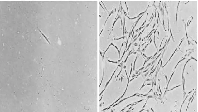
1. 간엽 간세포의 분리 및 배양 간엽 간세포는 배지에 부착되어 섬유아세포와 유사 한 형태를 나타내었다.(Fig. 1) 2. 시험관내 골형성 가. 알칼리 인산 분해 효소의 활성도 골형성을 유도한 실험군의 알칼리 인산 분해 효소 의 활성도는 평균 11 p-nitrophenol nmol/ min/105 cells이었고 대조군의 활성도는 평균 5 p-nitrophenol nmol/min/105 cells이었다.나. 알칼리 인산 분해 효소 조직 화학 염색
Fig. 1. Microscopic finding of bone marrow cell just after irrigated from bone marrow (left). Microscopic finding of
mesenchymal stem cells cultured in control medium for two weeks. The cells appeared fibroblastoid. (right,
골형성을 유도한 실험군에서는 보라색으로 짙게 염 색되는 양성의 반응을 보였고 세포들이 결절을 형성 하며 모여있는 소견을 보였으나 대조군에서는 음성의 반응을 보였다.(Fig. 2) 다. 무기질 침착 무기질 침착을 보기위한 von Kossa 염색상 골형 성을 유도한 실험군에서는 검은 색으로 염색되는 무 기질의 침착이 있었으며 그 주위로 세포들이 모여있 는 소견을 볼 수 있었으나 대조군에서는 검게 염색되 는 무기질의 침착을 볼 수 없었다.(Fig. 3) 라. Osteocalcin mRNA의 분석 Osteocalcin mRNA의 표현을 보기 위하여 Northern blot analysis를 시행하였는데 골형성을 유도한 실험군에서는 o s t e o c a l c i n에 대한 m R N A의 발현이 대조군에 비하여 증가하였다. 실험군의 r e l a-tive density ratio는 12.1, 대조군이 r e l a t i v e density ratio는 6 . 1으로 실험군에서 o s t e o c a l c i n m R N A의 발현이 약 2배 많았다. RT-PCR 소견상 골형성 유도군에서는 m R N A가 발현되었다.(Fig. 4) 3. 생체내 골 형성 H-E 염색 소견상 이식 3주에 채취한 실험군의 표 본에서 collagraft strip내로 세포의 침투가 관찰되 었으며 6주 이후의 표본에서는 세포가 c o l l a g r a f t s t r i p의 다공을 모두 채우고 있었으며 미성숙 골로 생각되는 분홍색으로 염색되는 기질의 형성을 볼 수 있었다. 대조군에서는 다공내에 세포가 드물게 있었 으며 골형성은 관찰할 수 없었다.(Fig. 5) 새로이 형성되는 유골형성을 보기 위하여 시행한 t e t r a c y c l i n으로 l a b e l i n g을 시행한 후 형광 현미 경으로 검사한 관찰한 결과 실험군에서 노란 형광을 띄는 유골( o s t e o i d )를 관찰할 수 있었다.(Fig. 6)
고
찰
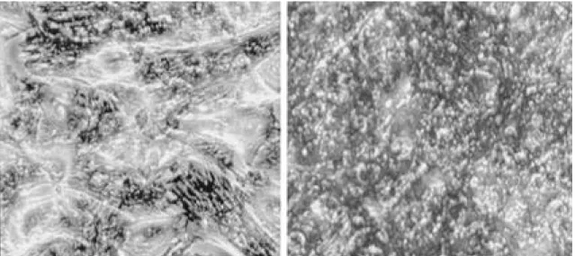
골수는 조혈계, 내피계, 기질계의 세포로 구성된 복잡한 조직이다. 골수는 조혈 미세 환경을 구성할 수 있는 능력 외에도 기질계에서 유래한 세포들이 골 형성 세포(osteogenic cell), 연골형성 세포( c h o n-drogenic cell), 지방 세포(adipocytic cell), 섬유Fig. 2. Microscopic findings of alkaline phosphatase staining of mesenchymal stem cell cultured in control medium
for two weeks. The cells were not stained with violet color (left, ×100). Microscopic findings of alkaline phosphatase staining of mesenchymal stem cell cultured in osteogenic medium for two weeks. The cells were stained with deep violet color (right, ×100).
간엽 간세포에서 골형성 세포로의 분화는 많은 저자 들에 의하여 연구되었으며 이러한 분화를 위한 생활 성화 인자(bioactive factor)로 o s t e o g e n i n3 0 ), 골 형성 단백( B M P - 2 ) ,2 , 3 , 8 , 2 0 , 2 7 , 3 1 , 3 5 )골형성 성장 펩타이 드(osteogenic growth peptide),2 8 )
덱사메사존 (synthetic glucocorticoid dexametha-s o n e )1 4 , 1 7 , 2 7 , 3 1 )
등이 보고되었다. 최근에는 골형성 단 백을 생산하는 유전자를 이용한 유전자 치료가 관심 을 끌고 있다.1 1 , 1 8 )또한 간엽 간세포의 성장 및 분화 를 촉진하는 인자로 T G F -β(transforming growth f a c t o r -β), PDGF-BB(platelet derived growth factor-BB), FGF(fibroblast growth factor)등 의 연구가 활발하다.7 , 3 2 ) 본 연구에서는 100 nM dexamethasone, 10 mM β- g l y c e r o p h o s p h a t e , 0.05 mM L-ascorbic acid-2-phosphate로 구성 된 골형성 유도 용액을 사용하였다.
1 9 9 6년 Cassiede 등7 )은 간엽 간세포와 hydroxyapatite 60%, tricalcium phosphate
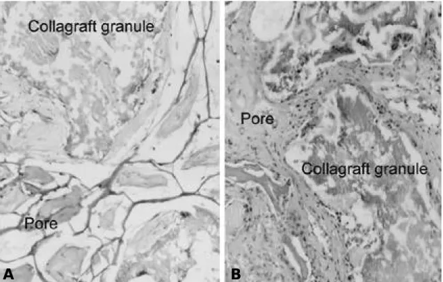
Fig. 5. A. Microscopic findings of H-E stained specimen (collagraft only), which was grafted on the back of rat 6
weeks before. Pores of collagraft were filled with scanty cells and some fibrous tissue . B. Microscopic find -ings of H-E stained specimen (collagraft +mesenchymal stem cells), which was grafted on the back of rat 6 weeks before. Pores of collagraft were filled with abundant cells and osteoid.
Fig. 6. Fluorescent microscopic findings of specimen,
which was grafted on the back of rat 6 weeks before. Yellow fluorescence indicate newly formed osteoid.
아 세포(fibroblastic cell), 망상 세포( r e t i c u l a r cell) 등을 형성할 수 있는 다양한 능력이 있는 간세 포(stem cell)를 함유하고 있는 것으로 생각된다.2 -7 , 1 0 , 1 6 , 2 4 , 2 5 , 3 3 )골수 세포를 배지에 배양하면 대부분의 조혈세포는 부유 상태에 있게 되고 기질 세포는 배양 용기에 부착하게 된다. 본 연구에서는 이러한 골수 세포의 성질을 이용하여 조혈 세포와 간엽 간세포를 분리할 수 있었다. 즉 골수 세포를 배지에 넣고 3일 간 배양한 후 배양액을 버리면 부유중인 조혈세포는 배양액과 함께 제거되고 배양 용기에 부착한 간엽 간 세포만 남게 된다. 이와 같은 방법을 2주간 반복하여 골수 세포에서 간엽 간세포를 분리하였다. 분리한 간 엽 간세포는 골형성 실험에서 osteocalcin m-RNA 에 대한 Nothern blot analysis에서 골형성을 유 도하지 않았을 때는 표현이 되지 않았으나 골형성을 유도한 후에는 표현이 강하게 나타나는 것으로 보아 간엽 간세포임을 알 수 있었다. 본 연구에서는 간엽 간세포를 상기한 방법으로 간접적으로 확인할 수 있 었으나 최근 세포 표면 항원을 이용하여 간엽 간세포 를 확인하는 연구가 활발히 진행되고 있다. 조혈세포 의 표면 항원으로는 CD45, CD14, CD34 등이 알 려져 있으며 간엽 간세포의 표면 항원으로는 S H 2 , SH3, SH4 등이 알려져 있으며 이는 조혈세포에서 는 표현되지 않는다.1 3 , 2 6 ) 병아리,1 7 ) 마우스,7 , 2 9 ) 백서,2 1 ) 가토,1 4 , 2 8 ) 돼지3 4 ) 의
Fig. 3. Gross and microscopic findings of von-Kossa staining of mesenchymal stem cell cultured in osteogenic
medi-um for three weeks. There were dark black staining mineral depositions (×40).
Fig. 4. Results of RT-PCR for osteocalcin m-RNA.
There was thick white band in osteogenesis induced group compared to the control group. (con: control group, osteo: osteogenesis induced group).
4 0 %로 구성된 세라믹을 마우스에 이식하여 간엽 간 세포에 의한 생체내 골형성을 보고한 바 있다. 이들 은 이소 부위(heterotopic site)인 마우스의 배부 피하조직에 이식하였으며 공통 유전형의 마우스 (syngeneic mouse)를 사용하였다. 본 연구에서는 간엽 간세포의 공여자로 사람을 사용하였으며 이식을 받는 백서는 공통 유전형이 아닌 면역 결핍 백서 (species: rats, strain: Cr:NIH-RNU)를 사용하 였으며 담체로는 hydroxyapatite 60%, tricalci-um 40%, 제 1형 콜리젠이 함유된 골과 유사한 세 라믹을 사용하였다. 물론 동종의 동물의 간엽 간세포 를 사용하여 공통 유전형( s y n g e n e i c )의 동물에 이 식한다면 면역 반응이 없어 자가 간엽 간세포 이식 (autogenous mesenchymal stem cell graft) 과 같은 결과를 보일 것이다. 그러나 본 연구에서 이 종간인 사람의 간엽 간세포를 면역 결핍 백서에 이식 한 이유는 향후 시행할 사람의 간엽 간세포 이식을 위한 기초적 연구이기 때문이었다. 즉, 사람의 간엽 간세포 이식을 인체에 바로 시행하는 것은 윤리적으 로 허용될 수 없기에 결국 사람의 간엽 간세포를 동 물에 이식하기 위하여는 면역 결핍 동물의 사용이 필 수적이며 그 기초적 자료를 얻기 위하여 면역 결핍 백서를 사용하였다. 이제까지 많은 연구자들이 생체내 골형성을 유도하 는 연구에서 불유합 모델을 만들어 사용하였다. 즉, 골 결손을 만든후 간엽 간세포를 이식하거나 골형성 단 백 을 이 식 하 는 실 험 등 을 시 행 하 였 다.8 , 9 , 1 5 , 2 0 , 2 4 , 3 0 , 3 1 , 3 3 ) 그러나 골 결손 모델에서의 골형성 유도는 새로이 형성된 골질이 이식한 간엽 간세포에 서 만들어졌는지 골 결손 부위에서 유래한 간엽 간세 포에 의하여 골질이 형성되었는지는 구별하기가 어려 울 수 있다. 따라서 본 연구에서는 골 형성의 이소성 부위(heterotopic site)인 배부의 피하조직에 간엽 간세포를 이식함으로써 이식한 간엽 간세포에서 골형 성이 이루어 졌음을 알 수 있었다. 1 9 9 7년 Bruder 등5 )은 골수에서 분리한 간엽 간 세포가 냉동 보관( c r y o p r e s e v a t i o n )후 3 0회 이상 계대 배양하여도 골 및 연골형성 능력이 있음을 보고 한 바 있다. 본 연구에서도 영하 7 0도에 냉동 보관 한 간엽 간세포를 계대 배양 및 골형성을 유도하였을 때 골형성이 됨을 확인할 수 있었다. 이와 같이 간엽 간세포는 보관 및 계대 배양을 통한 증식이 용이하 며, 보관 및 증식 후에도 골형성 능력이 있으므로 자 가골 이식을 대치할 수 있는 간엽 간세포 이식술의 임상적 적용이 가능할 것으로 생각되었다.
결
론
사람의 골수에서 분리 배양한 간엽 간세포에서 시 험관내 및 생체내 골형성을 유도하여 간엽 간세포를 이용한 골형성을 위한 세포 치료의 기초적 자료를 얻 기 위하여 본 연구를 시행하였으며 골형성 유도 후 조직학적, 면역조직화학적, 분자생물학적 방법으로 골형성을 확인할 수 있었다. 특히 골형성의 특징적인 지표인 osteocalcin mRNA의 발현을 확인하였다. 특히 사람의 간엽 간세포를 c o l l a g r a f t에 이식하여 생체내에서 골형성을 유도한 것은 임상적으로 골결손 의 치료에 적용할 수 있는 의의가 있다. 본 연구를 통하여 골결손의 치료를 위한 간엽 간세 포를 이용한 세포 치료의 방법을 개발할 수 있을 것 으로 사료된다.REFERENCES
01) Aaron RK, Cuombor DM. Acceleration of experi-mental endochondral ossification by biophysical stimulation of the progenitor cell pool. J Orthop
Res, 14:582-9, 1996.
02) An J, Rosen V, Cox K, Beauchemin N, Sulivan AK. Recombinant human bone morphogenetic protein-2 induces a hematopoietic microenviron-ment in the rat that supports the growth of stem cells. Exp Hematol, 24:768-76, 1996.
03) Balk ML, Bray J, Day C, Epperly M, Greenberger J, Evans CH, et al. Effect of rhBMP-2 on the osteogenic potential of bone marrow stromal cells from an osteogenesis imperfecta mouse. B o n e, 21:7-15, 1997.
04) Bergman RJ, Gazit D, Kahn AJ, Gruber H, Mcdougall S, Hahn TJ. Age-related change in osteogenic stem cells in mice. J Bone Miner Res,
11:568-77, 1996.
05) Bruder SP, Jaiswal N, Haynesworth SE. Growth kinetics, self-renewal and the osteogenic potential of purified human mesenchymal stem cells during extensive subcultivation and following cryopreser-vation. J Cell Biochem, 64:278-94, 1997.
06) Caplan AI, Elyaderani M, Mochizuki Y, Wakitani S, Goldberg VM. Principle of cartilage repair and regeneration. Clin Orthop Rel Res, 342:254-69, 1997.
07) Cassiede P, D e n n i s J E , M a F , C a p l a n AI. Osteochondrogenic potential of marrow mes-enchymal progenitor cells exposed to TGF-β1 or PDGF-BB as assayed in vivo and in vitro. J Bone
Miner Res, 11:1264-73, 1996.
08) Cook SD, Wolfe MW, Salkeld SL, Rueger DC. Effect of recombinant human osteogenic protein-1 on healing of segmental defects in non-human pri-mates. J Bone Joint Surg [Am], 77:734-50, 1995.
09) Einhorn TA, Lane JM, Burstein AH, Kopman CR, Vigorita VJ. The healing of segmental bone defects induced by demineralized bone matrix: a radiographic and biomechanical study. J Bone
Joint Surg [Am], 66:274-9, 1984.
10) Erben RG, Scutt AM, Miao D, Kollenkirchen U, Haberey M. Short-term treatment of rats with high dose 1,25-dihydroxyvitamin D3 stimulates bone formation and increases the number of osteoblast precursor cells in bone marrow. E n d o c r i n o l o g y, 138:4629-35, 1997.
11) Fang J, Jhu YY, Smiley E, Bonadio J, Rouleau JP, Goldstein SA, McKauley LK, Davidson BL, Roessler BJ. Stimulation of new bone formation by direct transfer of osteogenic plasmid genes.
Proc Natl Acad Sci USA, 93:5753-8, 1996.
12) Hahn SB and Suk KS. In vitro and in vivo induc-tion of osteogenesis in cultured mesenchymal stem cells isolated from rat bone marrow. J of Korean
Orthopaedic Research Society , 2(2):102-110,
1999.
13) Haynesworth S, Baber M, Caplan A. Cell surface antigens on human marrow-derived mesenchymal cells are detected by monoclonal antibodies. Bone,
13:69-80, 1992.
14) Howlett CR, Cavo J, Williamson M, Framer J, Ali SY, Bab I, et al. Mineralization in in vitro cultures of rabbit bone marrow stromal cells. Clin Orthop
Rel Res, 213:251-63, 1986.
15) Hunt TR, Schwappach JR, Anderson HC. Healing of a segmenta defect in the rat femur with use of an extract from a cultured human osteosarcoma cell-line(Saos-2). J Bone Joint Surg [Am], 78:41-8, 1996.
16) Jaiswal N, Haynesworth SE, Caplan AI, Bruder SP. Osteogenic differentiation of purified, culture-expanded human mesenchymal stem cells in vitro.
J Cell Biochem, 64:295-312, 1997.
17) Kamalia N, McCulloch CAG, Tennenbaum HC, Limeback H. Dexamethasone recruitment of self-renewing osteoprogenitor cells in chick bone mar-row stromal cell cultures. Blood, 79:320-26, 1992. 18) Kazhdan I, Rickard D, Leboy PS. HLH
transcrip-tion factor activity in osteogenic cells. J Cell
Biochem, 65:1-10, 1997.
19) Klokkevold PR, Vandermark L, Kenney EB, Bernard GW. Osteogenesis enhanced by chitosan (poly-N-acetyl glucosaminoglycan) in vitro. J
Periodontol, 67:1170-5, 1996.
20) Lee SC, Shea M, Battle MA, Kozitza K, Ron E, Turek T, et al. Healing of large segmental defects in rat femurs is aided by rhBMP-2 in PLGA matrix. J Biomed Mater Res, 28:1149-56, 1994. 21) Maniatopolous C, Sodek J, Melcher AH. Bone
for-mation in vitro by stromal cells obtained from bone marrow of young adult rats. Cell Tissue Res, 254:317-30, 1988.
22) Murray SS, Grisanti MS, Bentley GV, Kahn AJ, Urist MR, Murray EJB. The calpain-calpastatin system and cellular proliferation and differentia-tion in rodent osteoblastic cells. Exp Cell Res, 233:297-309, 1997.
23) Nulend JK, Roelofsen J, Semeins CM, Bronckers AL, Burger EH. Mechanical stimulation of osteo-pontin mRNA expression and synthesis in bone cell cultures. J Cell Physiol, 170:1744-81, 1997. 24) Ohgushi H, Goldberg VM, Caplan AI. Repair of
bone defects with marrow cells and porous ceram-ic: experiments in rats. Acta Orthop Scand , 60:334-9, 1989.
25) O t t o T E , N u l e n d J K , P a t k a P , B u r g e r E H , Haarman HJTM. Effect of (poly)-L-lactic acid on the proliferation and differentiation of primary bone cells in vitro. J Biomed Mater Res , 32:513-8, 1996.
26) Pittinger MF, Mackay A, Beck SC. Multilineage potential of adult human mesenchymal stem cells.
Science, 284:143-7, 1999.
27) Richard DJ, Sulivan TA, Shenker BJ, LeBoy PS, Kazhdan I. Induction of rapid osteoblast differenti-ation in rat bone marrow stromal cell cultures by dexamethasone and BMP-2. Dev Biol, 161:218-28, 1994.
28) Robinson D, Bab I, Nervo, Z. Osteogenic growth peptide regulates proliferation and osteogenic mat-uration of human and rabbit bone marrow stromal cells. J Bone Miner Res, 10:690-6, 1995.
29) Schoeters GER, de Saint-Georges L, Van Den Heuvel R, Vanderborght O. Mineralization of adult mouse bone marrow in vitro. Cell Tissue Knet, 21:363-74, 1988.
30) Stevenson S, Cunningham N, Toth J, Davy D, Reddi AH. The effect of osteogenin (a bone mor-phogenetic protein) on the formation of bone in
orthotopic segmental defects in rats. J Bone Joint
Surg [Am], 76:1676-87, 1994.
31) Takaki K, Urist MR. The role of bone morpho-genetic protein-induced repair of femoral massive diaphyseal defects. Clin Orhop, 171:224-31, 1982. 32) Thomson BM, Bennet J, Dean V, Triffit J, Meikle
MC, Loveridge N. Preliminary characterization of porcine bone marrow stromal cells: Skeletogenic potential, colony-forming activity, and response to dexamethasone, transforming growth factor beta, and basic fibroblast growth factor. J Bone Miner
Res, 8:1173-83, 1993.
33) Werntz JR, Lane JM, Burstein AH, Justin R, Klein R, Tomin E. Qualitative and quantitative analysis of orthotopic bone regeneration by marrow. J
Orthop Res, 14:85-93, 1996.
34) Wolff D, Goldberg VM, Stevenson S. Histomorphometric analysis of the repair of a seg-mental diaphyseal defect with ceramic and titani-um fibermetal implants: effects of bone marrow. J
Orthop Res, 12:439-46, 1994.
35) Yasuko AW, Lane JM, Fellinger EJ, Rosen V, Wozney JM, Wang EA. The healing of segmental bone defects, induced by human bone morpho-genetic protein (rh BMP-2). J Bone Joint Surg