Plasma Klotho concentration is associated
with the presence, burden and progression of
cerebral small vessel disease in patients with
acute ischaemic stroke
Ho Geol WooID1‡, Yoonkyung Chang2‡, Dong-Ryeol Ryu3, Tae-Jin SongID2*
1 Department of Neurology, Ewha Womans University Seoul Hospital, Ewha Womans University College of
Medicine, Seoul, Korea, 2 Department of Neurology, Ewha Womans University Mokdong Hospital, Ewha Womans University College of Medicine, Seoul, Korea, 3 Department of Nephrology, Ewha Womans University College of Medicine, Seoul, Korea
‡ Both authors equally contributed as first authors *knstar@ewha.ac.kr
Abstract
Klotho is a soluble or membrane-bound anti-aging protein, whose protective actions are important for a prudential function of many organs. Because Klotho and cerebral small vessel disease (SVD) are associated with ageing process and endothelial dysfunction, it is possible that Klotho has an association with cerebral SVD. We aimed to investigate the association of plasma Klotho concentration with the presence, burden and progression of cerebral SVD. We prospectively enrolled 262 patients with first-ever acute cerebral infarction, performed brain MRI and collected their blood samples within 24 hours of admission. An enzyme-linked immunosorbent assay was used for evaluating plasma Klotho concentration. We estimated the total SVD score of each patient after determining the presence and burden of high-grade white matter hyperintensities (HWMHs), cerebral microbleeds (CMBs), high-grade perivas-cular spaces (HPVSs) and asymptomatic lacunar infarctions (ALIs). Univariate and multivari-ate analyses were conducted to investigmultivari-ate association of Klotho with cerebral SVD and the total SVD score. Of the 262 patients, 152 (58.0%) were men. The mean age of these patients was 64.7 years. The mean±standard deviation of plasma Klotho concentration was 329.8±
194.1 pg/mL. In multivariate analysis, plasma Klotho concentration was negatively associ-ated with the presence of HWMHs [Odds ratio (OR): 0.13, p = 0.047], HPVSs (OR: 0.22, p = 0.024) and ALIs (OR: 0.53, p = 0.021) but not associated with the presence of CMBs (OR: 0.39, p = 0.404). Plasma Klotho concentration was also negatively related to the total SVD score (unstandardized coefficients beta:−0.895, standard error = 0.317, p = 0.005, R2= 0.239). Furthermore, plasma Klotho concentration was negatively related to the presence (OR: 0.75, 95% CI: 0.59–0.96, p = 0.025) and severity of cerebral SVD progression (OR: 0.72, 95% CI: 0.56–0.92, p = 0.009). In conclusion, plasma Klotho concentration was nega-tively associated with the presence, burden and progression of cerebral SVD.
a1111111111 a1111111111 a1111111111 a1111111111 a1111111111 OPEN ACCESS
Citation: Woo HG, Chang Y, Ryu D-R, Song T-J
(2019) Plasma Klotho concentration is associated with the presence, burden and progression of cerebral small vessel disease in patients with acute ischaemic stroke. PLoS ONE 14(8): e0220796. https://doi.org/10.1371/journal.pone.0220796 Editor: Oliver Chen, Tufts University, UNITED
STATES
Received: January 6, 2019 Accepted: July 23, 2019 Published: August 9, 2019
Copyright:© 2019 Woo et al. This is an open access article distributed under the terms of the Creative Commons Attribution License, which permits unrestricted use, distribution, and reproduction in any medium, provided the original author and source are credited.
Data Availability Statement: All relevant data are
within the paper and its Supporting Information files.
Funding: This project was supported by grant from
the Basic Science Research Program through the National Research Foundation of Korea funded by the Ministry of Education
(2018R1D1A1B07040959 to T-JS). This work was supported by the Ewha Womans University Research Grant of 2018 to T-JS. The funders had no role in study design, data collection and
Introduction
Klotho was a single-pass transmembrane protein encoded by Klotho gene that is predomi-nantly present in the distal tubular cells of the kidney, choroid plexus of the brain and parathy-roid glands [1]. The importance of this Klotho has been highlighted in the study involving the Klotho knock-out mouse model, which revealed that Klotho deficiency was related to reduced life expectancy [2] and that Klotho overexpression was associated with life span prolongation [3]. Human Klotho exists in the form of membrane Klotho and circulatory Klotho. In the kid-ney, membrane Klotho acts as a co-receptor for fibroblast growth factor and is involved in phosphate secretion [4], whereas circulatory Klotho plays a role in the regulation of nitric oxide in vascular endothelial cells and in calcium homeostasis [5]. Circulatory Klotho is recog-nized as having the enzyme activity of autocrine, endocrine and paracrine hormone. Previous clinical study demonstrated that potential link between klotho deficiency and enhanced oxida-tive stress in patients with kidney disease [6].
Cerebral small vessel disease (SVD) is characterized by pathologic changes in a small artery or arteriole and manifests as white matter hyperintensities (WMHs), perivascular spaces (PVSs), cerebral microbleeds (CMBs) and asymptomatic lacunar infarctions (ALIs) on specific brain MR image sequences [7]. These cerebral SVD forms share common risk factors or mech-anism of development despite each forms demonstrating a different clinical impact or associa-tion; for example, i.e. WMHs, PVSs and ALIs are mainly related to cerebral ischaemia, whereas CMBs are closely associated with future cerebral haemorrhagic events [8,9]. Because these cerebral SVD forms are closely linked with future stroke or cognitive decline events [7,
10,11], studies to determine the associated risk factors for various cerebral SVD forms are warranted.
Previous studies using animal model were demonstrated that klotho protein protects the endothelial dysfunction through nitric oxide production by humoral pathways [12,13]. In addition, high Klotho concentration was associated with a reduced risk of developing stroke and macroangiopathies in clinical study [14]. Furthermore, cerebral SVD is also associated with ageing process and endothelial dysfunction in human studies [15,16]. Therefore, Klotho may have a possibility of association with cerebral SVD. In our study, we hypothesized that plasma Klotho concentration would be negatively associated with the presence, burden and progression of cerebral SVD.
Materials and methods
Subjects
Between June 2014 and May 2016, we prospectively enrolled 262 patients with first cerebral infarction who were admitted to our hospital within 7 days of symptom onset and whose stroke subtypes were classified as large artery atherosclerosis, cardioembolism or lacunar infarction. All patients were evaluated according to the standard protocol of our hospital, which includes 12-lead electrocardiography, chest X-ray, routine blood tests [white blood cell count and creatinine levels at admission and levels of vitamin D (25(OH)D), fasting glucose, HbA1c, triglyceride, total cholesterol, low-density lipoprotein, haemoglobin, total calcium, phosphate, albumin, alkaline phosphatase, uric acid and C-reactive protein after 12 hours fast-ing period] and imagfast-ing studies (CT and/or MRI, CT angiography, MR angiography or digital subtraction angiography) [17,18]. Patients were not enrolled in the study if they did not pro-vide consent for participation as well as when they did not propro-vide consent for blood collection to determine plasma Klotho concentration. We excluded patients had a previous history of cancer or autoimmune disease, because impaired immune system caused by predisposing to analysis, decision to publish, or preparation of the
manuscript.
Competing interests: The authors have declared
autoimmune disease or cancer impedes an adequate control of systemic inflammation and sys-temic inflammation is the main downregulator of levels of Klotho [19,20]. Furthermore, because Klotho is involved in calcium homeostasis in the kidney, we excluded patients with bone fractures in the last 2 months [21].
The definition of risk factors was established in the Supplementary methods section in a previous study (S1 Appendix) [22]. Cerebral atherosclerosis was defined as the presence of one or more vessels with over 50% stenosis or occlusion in extracranial or intracranial cerebral arteries [23]. The stroke subtype was classified according to the Trial of Org 10172 in Acute Stroke Treatment (TOAST) classification system [24]. Briefly, the diagnosis was large artery atherosclerosis if the patient had significant stenosis (�50%) or occlusion of the relevant large artery of the lesion. Cardioembolism was diagnosed when the patient had a possible cardioem-bolic source. Lacunar infarction was diagnosed when the patient with acute lacunar syndrome had small (<15 mm) and deep lesion but no significant stenosis (<50%) of the relevant artery of the lesion and no possible cardioembolic source [25]. This study was approved by our Insti-tutional Review Board (Ewha Clinical Trial Center 2014-04-023), and we received informed consents from all participants and their closest relatives.
Brain MR protocol and definition of cerebral small vessel diseases
The detailed brain MR image sequence and definition of cerebral SVD were established in pre-vious studies [26–28]. All subjects were examined using a 3.0 T MRI scanner (Achieva 3.0 T, Philips Medical Systems, Best, Netherlands; or MAGNETOM Trio 3.0 T, Siemens, Germany). MRI protocol, acquired along the axis of the orbitomeatal line, included T2-weighted images [time repetition (TR), 9000 ms; time echo (TE), 100 ms; field of view (FOV), 230×230 mm; slice thickness, 5 mm; pixel spacing 0.240×0.240 mm], fluid attenuated inversion recovery (FLAIR) (TR, 9000 ms; TE, 120 ms; FOV, 230×230 mm; slice thickness, 5 mm; pixel spacing, 0.449×0.449 mm), gradient echo (GRE) (TR, 600 ms; TE, 16 ms; FOV, 250×250 mm; slice thickness, 5 mm; pixel spacing, 0.449×0.449 mm) and diffusion weighted image (DWI) sequence along six different directions (x, y, z, xy, yz, zx) (TR, 2600–6500 ms; TE, 42–70 ms; FOV, 230×230 mm; slice thickness, 5 mm; intersection gap, 2 mm; b-values, 0 and 600 mm2/s) [28,29].
According to Fazekas’ scoring system, the extent of WMHs was referred to as a periven-tricular white matter or deep white matter on FLAIR images [30]. High-grade white matter hyperintensities (HWMHs) were regarded as a Fazekas score of �2 in periventricular white matter and/or �2 in deep white matter. The PVSs were observed as punctate and/or linear hyperintense lesions located in the centrum semi-ovale or basal ganglia with a diameter of
<3 mm on T2-weighted images [31]. High-grade perivascular spaces (HPVSs) were regarded as grade 2–4 PVSs based on a previous report [9]. The CMBs were regarded to be round hypointense lesions with a diameter of <10 mm on GRE images [29]. The ALIs were referred to as round or ovoid, subcortical, fluid-filled cavities (signal similar to cerebrospi-nal fluid) measuring 3–15 mm in diameter that manifested as hyperintense lesions on T2-weighted images and as hypointense lesions on T1-weighted images, with no history of relevant symptoms or signs. The presence of HWMHs, HPVSs, CMBs and ALIs was defined outside the acute infarcted area (based on DWI), and these lesions were indepen-dently evaluated by two neurologists (T.J.S. and Y.C.) who were blinded to the clinical information.
The total SVD score was obtained on an ordinal scale ranging from 0 to 4 by assigning a rating (0 or 1) to the presence of each of the four SVD features, namely HWMHs, HPVSs, CMBs and ALIs [32]. Inter-observer agreements for the presence of HWMHs, HPVSs,
CMBs and ALIs were 0.956, 0.938, 0.912 and 0.888, respectively (allp<0.05) [26]. Any disagreement between the two raters regarding the presence of SVD was solved by consensus.
For evaluating the association of Klotho with cerebral SVD progression, we retrospectively included the 223 patients (85.1%: 223/262) who performed brain MRI at 2 years (± 6 months) after index stroke. Follow up brain MRI (FLAIR images, T2-weighted images and GRE images at 2 years after index stroke) is routinely recommended in our clinic for evaluating progression of cerebral SVD. The cerebral SVD progression is defined as increased total SVD score more than 1 score compared to the baseline total SVD score.
Measurement of plasma Klotho concentration
For estimating plasma Klotho concentration, venous samples in EDTA tubes were immedi-ately collected at admission. Plasma was separated by centrifugation (1900 g, 15 min) at 4˚C [25]. The acquired plasma was stored at−80˚C until analysis. An enzyme-linked immunosor-bent assay (Immuno-Biological Laboratories, Gunma, Japan) was used for the evaluation of plasma Klotho concentration [33]. The detection range of this assay was 31.25–4000 pg/mL. Plasma Klotho concentration was measured in duplicate and averaged by researchers who were blinded to the clinical information (Y.C. and D.Y.R). Intra-assay and inter-assay coeffi-cients of variability were 3.1% and 6.9%, respectively.
Statistical analysis
Statistical analysis was performed using SPSS software package (version 21.0, Chicago, IL, USA). Continuous variables were analysed using independent t-test or Mann–Whitney test, and categorical variables were analysed using Chi-square or Fisher’s exact test. Univariate and multivariate analyses were conducted to investigate the association of Klotho with cerebral SVD. To compare Klotho concentration among stroke subtypes, Kruskal–Wallis test was per-formed. The variables with a p value of < 0.1 in univariate analysis, sex and age were entered into the multivariate analyses.
To investigate the relationship of Klotho with the total SVD score, univariate and multivari-ate linear regression analyses were performed. In model 1, sex, age, body mass index, risk fac-tors and clinical variables (hypertension, diabetes mellitus, hypercholesterolemia, coronary artery disease, smoking, alcohol intake, pre-stroke anti-thrombotics, pre-stroke statins, cere-bral atherosclerosis and stroke subtype) were adjusted. In model 2, sex, age, body mass index and blood laboratory findings (levels of fibroblast growth factor-23, vitamin D 25(OH)D, fast-ing glucose, HbA1c, triglyceride, total cholesterol, low-density lipoprotein, haemoglobin, cre-atinine, total calcium, phosphate, albumin, alkaline phosphatase, uric acid and C-reactive protein as well as white blood cell count) were adjusted. Moreover, in model 3, sex, age, body mass index and variables with a p value of < 0.1 in univariate analysis (hypertension, coronary artery disease, smoking, stroke subtype and levels of fibroblast growth factor-23, vitamin D 25 (OH)D, low-density lipoprotein, creatinine and albumin) were adjusted. For convenience, intracranial and extracranial cerebral atherosclerosis were dichotomized as cerebral athero-sclerosis owing to over-fitting in logistic regression and linear regression. To evaluating the association of Klotho with cerebral SVD progression, binary logistic regression for presence of cerebral SVD progression and ordinal logistic regression for severity of cerebral SVD progres-sion were performed. The sex, age, body mass index and variables with a p value of < 0.1 in univariate analysis were entered into the multivariate analyses. A p value of < 0.05 was consid-ered to be statistically significant.
Results
Demographic data and comparative analysis according to the presence of
cerebral SVD
Demographic data of patients are shown inTable 1. Of the 262 patients, 152 (58.0%) were men. The mean age of the study patients was 64.7 years. The mean± standard deviation of plasma Klotho concentration was 329.8± 194.1 pg/mL. Regarding stroke subtype, large artery atherosclerosis (41.2%, 108/262) was most commonly noted, followed by small vessel occlusion (40.8%) and cardioembolism (17.9%). The HWMHs were prevalent in 24.4% of subjects (64/ 262); HPVSs, 9.2% (24/262); CMBs, 19.5% (51/262) and ALIs, 19.5% (51/262). One hundred forty-nine (56.9%) patients had a total SVD score of 0. The total SVD score of 1 was most com-monly noted (25.6%, 67/262) (Table 1). Plasma Klotho concentration did not differ according to stroke subtype (p = 0.974).
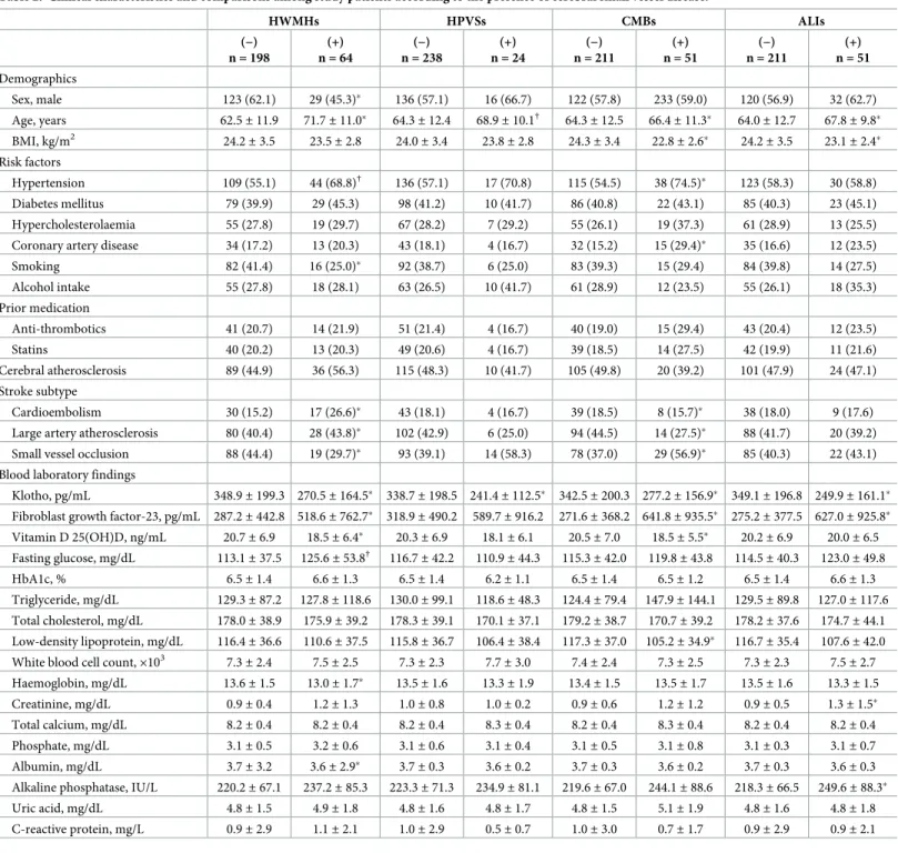
As opposed to patients without HWMHs, patients with HWMHs had significantly lower concentration of Klotho (270.5± 164.5 pg/mL vs. 348.9 ± 199.3 pg/mL), levels of vitamin D 25 (OH)D (18.5± 6.4 ng/mL vs. 20.7 ± 6.9 ng/mL) and albumin (3.6 ± 2.9 mg/dL vs. 3.7 ± 3.2 mg/dL) and a higher levels of fibroblast growth factor-23 (518.6± 762.7 pg/mL vs.
287.2± 442.8 pg/mL) and haemoglobin (13.6 ± 1.5 mg/dL vs. 13.0 ± 1.7 mg/dL). The patients with HPVSs had a lower plasma Klotho concentration than those without HPVSs
(241.4± 112.5 pg/mL vs. 338.7 ± 198.5 pg/mL). As opposed to patients without CMBs, those with CMBs had a higher prevalence of hypertension (74.5% vs. 54.5%), coronary artery disease (29.4% vs. 15.2%) and small vessel occlusion stroke subtype (56.9% vs. 37.0%). The plasma Klotho (277.2± 156.9 pg/mL vs. 342.5 ± 200.3 pg/mL), levels of vitamin D 25(OH)D (18.5± 5.5 ng/mL vs. 20.5 ± 7.0 ng/mL) and low-density lipoprotein (105.2 ± 34.9 mg/dL vs. 117.3± 37.0 mg/dL) were lower in patients with CMBs than in those without CMBs. The patients with ALIs had a lower plasma Klotho concentration than those without ALIs
(249.9± 161.1 pg/mL vs. 349.1 ± 196.8 pg/mL). In contrast, patients with ALIs had higher lev-els of fibroblast growth factor-23 (627.0± 925.8 pg/mL vs. 275.2 ± 377.5 pg/mL), creatinine (1.3± 1.5 mg/dL vs. 0.9 ± 0.5 mg/dL) and alkaline phosphatase (249.6 ± 88.3 IU/L vs. 218.3± 66.5 IU/L) than those without ALIs (Table 2).
Association between plasma Klotho concentration and cerebral small vessel
disease
Considering the plasma Klotho concentration, the study population was categorized into quar-tiles according to the plasma Klotho concentration (136.2± 49.7 pg/mL, 248.1 ± 25.1 pg/mL, 347.4± 28.8 pg/mL and 588.7 ± 190.8 pg/mL). Cerebral atherosclerosis, presence of HWMHs and ALIs, total SVD score and fibroblast growth factor-23 were significantly different accord-ing to quartiles of the plasma Klotho concentration (S1 TableandFig 1). In multivariate analy-sis, plasma Klotho concentration was negatively associated with the presence of HWMHs [Odds ratio (OR): 0.13, 95% confidence interval (CI): 0.01–0.97, p = 0.047)], HPVSs (OR: 0.22, 95% CI: 0.10–0.60, p = 0.024), and ALIs (OR: 0.53, 95% CI: 0.40–0.64, p = 0.021) but not with the presence of CMBs (OR: 0.39, 95% CI: 0.04–3.46, p = 0.404) (Table 3). No statistically signif-icant interaction of Klotho with demographics, risk factors and blood laboratory findings were noted with regard to the presence of HWMHs, HPVSs, CMBs and ALIs.
Considering the total SVD score, the study population was categorized into total SVD score. Age, plasma Klotho concentration, fibroblast growth factor-23, fasting glucose and cre-atinine were significantly different according to total SVD score (S2 Table). In multivariate analysis, plasma Klotho concentration was negatively related to the total SVD score in model 1
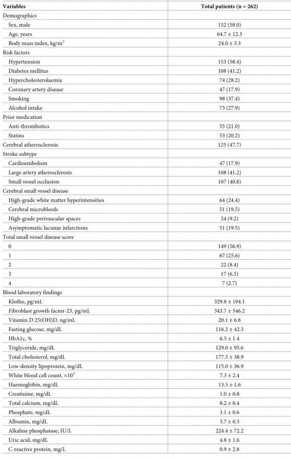
Table 1. Clinical characteristics of enrolled patients.
Variables Total patients (n = 262)
Demographics
Sex, male 152 (58.0)
Age, years 64.7± 12.3
Body mass index, kg/m2 24.0± 3.3
Risk factors
Hypertension 153 (58.4)
Diabetes mellitus 108 (41.2)
Hypercholesterolaemia 74 (28.2)
Coronary artery disease 47 (17.9)
Smoking 98 (37.4) Alcohol intake 73 (27.9) Prior medication Anti-thrombotics 55 (21.0) Statins 53 (20.2) Cerebral atherosclerosis 125 (47.7) Stroke subtype Cardioembolism 47 (17.9)
Large artery atherosclerosis 108 (41.2)
Small vessel occlusion 107 (40.8)
Cerebral small vessel disease
High-grade white matter hyperintensities 64 (24.4)
Cerebral microbleeds 51 (19.5)
High-grade perivascular spaces 24 (9.2) Asymptomatic lacunar infarctions 51 (19.5) Total small vessel disease score
0 149 (56.9)
1 67 (25.6)
2 22 (8.4)
3 17 (6.5)
4 7 (2.7)
Blood laboratory findings
Klotho, pg/mL 329.8± 194.1
Fibroblast growth factor-23, pg/mL 343.7± 546.2
Vitamin D 25(OH)D, ng/mL 20.1± 6.8 Fasting glucose, mg/dL 116.2± 42.3 HbA1c, % 6.5± 1.4 Triglyceride, mg/dL 129.0± 95.6 Total cholesterol, mg/dL 177.5± 38.9 Low-density lipoprotein, mg/dL 115.0± 36.9 White blood cell count,×103 7.3± 2.4
Haemoglobin, mg/dL 13.5± 1.6
Creatinine, mg/dL 1.0± 0.8
Total calcium, mg/dL 8.2± 0.4
Phosphate, mg/dL 3.1± 0.6
Albumin, mg/dL 3.7± 0.3
Alkaline phosphatase, IU/L 224.4± 72.2
Uric acid, mg/dL 4.8± 1.6
C-reactive protein, mg/L 0.9± 2.8
Data are shown as n (%) or mean± standard deviation
Table 2. Clinical characteristics and comparisons among study patients according to the presence of cerebral small vessel disease. HWMHs HPVSs CMBs ALIs (−) n = 198 (+) n = 64 (−) n = 238 (+) n = 24 (−) n = 211 (+) n = 51 (−) n = 211 (+) n = 51 Demographics Sex, male 123 (62.1) 29 (45.3)� 136 (57.1) 16 (66.7) 122 (57.8) 233 (59.0) 120 (56.9) 32 (62.7) Age, years 62.5± 11.9 71.7± 11.0� 64.3± 12.4 68.9± 10.1† 64.3± 12.5 66.4± 11.3� 64.0± 12.7 67.8± 9.8� BMI, kg/m2 24.2± 3.5 23.5± 2.8 24.0± 3.4 23.8± 2.8 24.3± 3.4 22.8± 2.6� 24.2± 3.5 23.1± 2.4� Risk factors Hypertension 109 (55.1) 44 (68.8)† 136 (57.1) 17 (70.8) 115 (54.5) 38 (74.5)� 123 (58.3) 30 (58.8) Diabetes mellitus 79 (39.9) 29 (45.3) 98 (41.2) 10 (41.7) 86 (40.8) 22 (43.1) 85 (40.3) 23 (45.1) Hypercholesterolaemia 55 (27.8) 19 (29.7) 67 (28.2) 7 (29.2) 55 (26.1) 19 (37.3) 61 (28.9) 13 (25.5) Coronary artery disease 34 (17.2) 13 (20.3) 43 (18.1) 4 (16.7) 32 (15.2) 15 (29.4)� 35 (16.6) 12 (23.5)
Smoking 82 (41.4) 16 (25.0)� 92 (38.7) 6 (25.0) 83 (39.3) 15 (29.4) 84 (39.8) 14 (27.5) Alcohol intake 55 (27.8) 18 (28.1) 63 (26.5) 10 (41.7) 61 (28.9) 12 (23.5) 55 (26.1) 18 (35.3) Prior medication Anti-thrombotics 41 (20.7) 14 (21.9) 51 (21.4) 4 (16.7) 40 (19.0) 15 (29.4) 43 (20.4) 12 (23.5) Statins 40 (20.2) 13 (20.3) 49 (20.6) 4 (16.7) 39 (18.5) 14 (27.5) 42 (19.9) 11 (21.6) Cerebral atherosclerosis 89 (44.9) 36 (56.3) 115 (48.3) 10 (41.7) 105 (49.8) 20 (39.2) 101 (47.9) 24 (47.1) Stroke subtype Cardioembolism 30 (15.2) 17 (26.6)� 43 (18.1) 4 (16.7) 39 (18.5) 8 (15.7)� 38 (18.0) 9 (17.6)
Large artery atherosclerosis 80 (40.4) 28 (43.8)� 102 (42.9) 6 (25.0) 94 (44.5) 14 (27.5)� 88 (41.7) 20 (39.2)
Small vessel occlusion 88 (44.4) 19 (29.7)� 93 (39.1) 14 (58.3) 78 (37.0) 29 (56.9)� 85 (40.3) 22 (43.1)
Blood laboratory findings
Klotho, pg/mL 348.9± 199.3 270.5 ± 164.5� 338.7± 198.5 241.4 ± 112.5� 342.5± 200.3 277.2 ± 156.9� 349.1± 196.8 249.9 ± 161.1�
Fibroblast growth factor-23, pg/mL 287.2± 442.8 518.6 ± 762.7� 318.9± 490.2 589.7± 916.2 271.6± 368.2 641.8 ± 935.5� 275.2± 377.5 627.0 ± 925.8�
Vitamin D 25(OH)D, ng/mL 20.7± 6.9 18.5± 6.4� 20.3± 6.9 18.1± 6.1 20.5± 7.0 18.5± 5.5� 20.2± 6.9 20.0± 6.5 Fasting glucose, mg/dL 113.1± 37.5 125.6± 53.8† 116.7± 42.2 110.9± 44.3 115.3± 42.0 119.8± 43.8 114.5± 40.3 123.0± 49.8 HbA1c, % 6.5± 1.4 6.6± 1.3 6.5± 1.4 6.2± 1.1 6.5± 1.4 6.5± 1.2 6.5± 1.4 6.6± 1.3 Triglyceride, mg/dL 129.3± 87.2 127.8± 118.6 130.0± 99.1 118.6± 48.3 124.4± 79.4 147.9± 144.1 129.5± 89.8 127.0± 117.6 Total cholesterol, mg/dL 178.0± 38.9 175.9± 39.2 178.3± 39.1 170.1± 37.1 179.2± 38.7 170.7± 39.2 178.2± 37.6 174.7± 44.1 Low-density lipoprotein, mg/dL 116.4± 36.6 110.6± 37.5 115.8± 36.7 106.4± 38.4 117.3± 37.0 105.2± 34.9� 116.7± 35.4 107.6± 42.0
White blood cell count,×103 7.3
± 2.4 7.5± 2.5 7.3± 2.3 7.7± 3.0 7.4± 2.4 7.3± 2.5 7.3± 2.3 7.5± 2.7 Haemoglobin, mg/dL 13.6± 1.5 13.0± 1.7� 13.5± 1.6 13.3± 1.9 13.4± 1.5 13.5± 1.7 13.5± 1.6 13.3± 1.5 Creatinine, mg/dL 0.9± 0.4 1.2± 1.3 1.0± 0.8 1.0± 0.2 0.9± 0.6 1.2± 1.2 0.9± 0.5 1.3± 1.5� Total calcium, mg/dL 8.2± 0.4 8.2± 0.4 8.2± 0.4 8.3± 0.4 8.2± 0.4 8.3± 0.4 8.2± 0.4 8.2± 0.4 Phosphate, mg/dL 3.1± 0.5 3.2± 0.6 3.1± 0.6 3.1± 0.4 3.1± 0.5 3.1± 0.8 3.1± 0.3 3.1± 0.7 Albumin, mg/dL 3.7± 3.2 3.6± 2.9� 3.7± 0.3 3.6± 0.2 3.7± 0.3 3.6± 0.2 3.7± 0.3 3.6± 0.3
Alkaline phosphatase, IU/L 220.2± 67.1 237.2± 85.3 223.3± 71.3 234.9± 81.1 219.6± 67.0 244.1± 88.6 218.3± 66.5 249.6± 88.3�
Uric acid, mg/dL 4.8± 1.5 4.9± 1.8 4.8± 1.6 4.8± 1.7 4.8± 1.5 5.1± 1.9 4.8± 1.6 4.8± 1.8 C-reactive protein, mg/L 0.9± 2.9 1.1± 2.1 1.0± 2.9 0.5± 0.7 1.0± 3.0 0.7± 1.7 0.9± 2.9 0.9± 2.1
Data are shown as n (%) or mean± standard deviation.
�:p<0.05, †:
p<0.1.
HWMHs: high-grade white matter hyperintensities, HPVSs: high-grade perivascular spaces, CMBs: cerebral microbleeds, ALIs: asymptomatic lacunar infarctions, BMI: body mass index.
(unstandardized coefficients beta:−1.314, standard error = 0.321, p = 0.001, R2
= 0.199), model 2 (unstandardized coefficients beta:−0.888, standard error = 0.329, p = 0.007, R2= 0.260) and model 3 (unstandardized coefficients beta:−0.895, standard error = 0.317, p = 0.005, R2= 0.239) (Table 4).
Number of total SVD score was lowest in patients with 4th quartile of the plasma Klotho concentration, followed by those with 3rd quartile of the plasma Klotho concentration, those with 2nd quartile of the plasma Klotho concentration, compared with those with 1st quartile of the plasma Klotho concentration (p = 0.013).
Association between plasma Klotho concentration and cerebral small vessel
disease progression
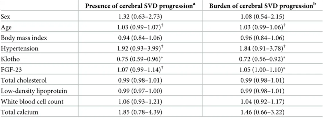
Among 223 patients who performed follow up brain MRI, 58 (26.0%) patients showed increased total SVD score [36 (16.1%) patients were increased with one point, 11 (4.9%) patients were increased with two points, 7 (3.1%) patients were increased with three points and 4 (1.8%) patients were increased with four points]. The patients with cerebral SVD pro-gression had significantly old age (68.4± 8.8 years vs. 62.5 ± 12.8 years), higher prevalence of hypertension (72.4% vs. 53.3%) and lower level of plasma Klotho (251.8± 146.3 pg/mL vs. 354.3± 202.7 pg/mL) than those without cerebral SVD progression (S3 Table). Considering the relationship of Klotho with cerebral SVD progression, plasma Klotho concentration was negatively related with cerebral SVD progression (presence of progression; OR: 0.75, 95% CI: 0.59–0.96, p = 0.025, severity of progression; OR: 0.72, 95% CI: 0.56–0.92, p = 0.009) (Table 5).
Discussion
To date, the association of Klotho with cerebral SVD has been rarely reported. In previous studies involving patients with cognitive dysfunction, reduced plasma Klotho level was Fig 1. Number of total SVD score stratified by quartiles of the plasma Klotho concentration.
independently correlated with the degree of cerebral WMHs and was accompanied by cogni-tive decline [34], especially vascular dementia [35]. In a previous study which dealt with Klotho and vascular surrogate markers or cardiovascular outcome, low level of Klotho was found to be associated with the presence and severity of coronary artery disease [36]. Moreover, in patients with renal dysfunction, decreased level of circulating Klotho was correlated with bra-chial-ankle pulse wave velocity which represented arterial stiffness [37]. In patients who received dialysis therapy, decreased serum Klotho level was associated with cardiovascular morbidity and mortality [38]. In line with results of these previous studies, the key finding of our study is that 1) plasma Klotho concentration was negatively associated with the presence of cerebral SVD, such as HWMHs, HPVSs, CMBs (even though this was noted in univariate analysis only) and ALIs, 2) plasma Klotho concentration was negatively correlated an increased burden of cerebral SVD and 3) plasma Klotho concentration was also negatively related with progression of cerebral SVD in stroke patients.
Although our study did not suggest the exact mechanism of underlying the relationship between plasma Klotho and cerebral SVD, several hypotheses can explain this association. Table 3. Multivariate analysis for the presence of cerebral small vessel disease.
Presence of cerebral small vessel disease
HWMHsa HPVSsb CMBsc ALIsd
Sex 0.98 (0.44–2.17) 2.37 (0.88–6.34)† 1.21 (0.57–2.56) 2.13 (1.00–4.57)†
Age 1.07 (1.03–1.11)� 1.04 (1.00–1.08)� 1.00 (0.96–1.04) 1.02 (0.99–1.05)
Body mass index 0.83 (0.73–0.94)� 0.89 (0.80–1.00)†
Hypertension 1.27 (0.63–2.54) 2.45 (1.11–5.44)�
Coronary artery disease 2.01 (0.87–4.66)
Smoking 0.78 (0.34–1.76)
Stroke subtype
SVO 1.01 (0.47–2.18) 3.79 (1.62–8.86)�
LAA 1.18 (0.49–2.80) 1.34 (0.44–4.04)
CE Reference Reference Reference Reference
Klotho 0.13 (0.01–0.97)� 0.22 (0.10–0.60)� 0.39 (0.04–3.46) 0.53 (0.40–0.64)�
Fibroblast growth factor-23 1.22 (0.70–2.11) 2.85 (1.35–5.99)� 1.86 (0.99–3.52)†
Vitamin D 25(OH)D 0.96 (0.91–1.01) 0.97 (0.92–1.03) Fasting glucose 1.01 (1.00–1.02)� Low-density lipoprotein 0.98 (0.97–1.00)† Haemoglobin 1.03 (0.80–1.32) Creatinine 1.22 (0.84–1.79) Albumin 0.50 (0.16–1.57) Alkaline phosphatase 1.00 (1.00–1.01)�
Data are shown as odds ratio (95% confidence interval)
�:p<0.05, †
:p<0.1.
HWMHs: high-grade white matter hyperintensities, HPVSs: high-grade perivascular spaces, CMBs: cerebral microbleeds, ALIs: asymptomatic lacunar infarctions, SVO: Small vessel occlusion, LAA: Large artery atherosclerosis, CE: Cardioembolism
a
adjusted for sex, age, hypertension, smoking, stroke subtype and levels of fibroblast growth factor-23, vitamin D 25(OH)D, fasting glucose, haemoglobin, and albumin.
b
adjusted for sex and age.
c
adjusted for sex, age, BMI, hypertension, coronary artery disease, stroke subtype and levels of fibroblast growth factor-23, vitamin D 25(OH)D, and low-density lipoprotein.
d
adjusted for sex, age, BMI and levels of fibroblast growth factor-23, creatinine, and alkaline phosphatase.
First, endothelial dysfunction may explain correlation of Klotho and cerebral SVD [15], because Klotho may have vasoprotective effects on the endothelium and involve in vasorelaxa-tion that reduces endothelial dysfuncvasorelaxa-tion via nitric oxide pathway [13,39] and endothelial dys-function is associated with cerebral SVD [40]. Second, reactive oxygen species may explain correlation of Klotho and cerebral SVD, because Klotho is involved in vascular protection by inhibiting the production of reactive oxygen species and a previous study has suggested that reactive oxygen species including oxidative stress are closely related to cerebral SVD [41,42]. Third, Klotho inhibited expression of adhesion molecules, pro-inflammatory, and pro-throm-botic factors which are concerned with cell apoptosis and inflammation [43]. Because those factors are associated with oxidative stress, relationship between plasma Klotho and cerebral SVD may be explained.
Table 4. Association of plasma Klotho with total SVD score.
Unstandardized coefficients Standardized coefficients beta T p value R2
B SE Univariate −1.288 0.322 −0.240 −3.994 0.001 0.058 Multivariate Model 1 −1.314 0.321 −0.245 −4.094 0.001 0.199 Model 2 −0.888 0.329 −0.166 −2.703 0.007 0.260 Model 3 −0.895 0.317 −0.167 −2.828 0.005 0.239
SVD: small vessel disease, SE: Standard error.
Model 1: adjusted for sex, age, body mass index, risk factors and clinical variables (hypertension, diabetes mellitus, hypercholesterolemia, coronary artery disease, smoking, alcohol intake, pre-stroke anti-thrombotics, pre-stroke statins, cerebral atherosclerosis, and stroke subtype)
Model 2: adjusted for sex, age, body mass index and blood laboratory findings (levels of fibroblast growth factor-23, vitamin D 25(OH)D, fasting glucose, HbA1c, triglyceride, total cholesterol, low-density lipoprotein, haemoglobin, creatinine, total calcium, phosphate, albumin, alkaline phosphatase, uric acid, and C-reactive protein as well as white blood cell count)
Model 3: adjusted for sex, age, body mass index and variables with a p value of <0.1 in univariate analysis (hypertension, coronary artery disease, smoking, stroke subtype and levels of fibroblast growth factor-23, vitamin D 25(OH)D, low-density lipoprotein, creatinine, and albumin)
https://doi.org/10.1371/journal.pone.0220796.t004
Table 5. Multivariate analysis for the progression of cerebral small vessel disease.
Presence of cerebral SVD progressiona Burden of cerebral SVD progressionb
Sex 1.32 (0.63–2.73) 1.08 (0.54–2.15)
Age 1.03 (0.99–1.07)† 1.03 (0.99–1.06)†
Body mass index 0.94 (0.84–1.06) 0.96 (0.84–1.06) Hypertension 1.92 (0.93–3.99)† 1.84 (0.91–3.78)†
Klotho 0.75 (0.59–0.96)� 0.72 (0.56–0.92)�
FGF-23 1.07 (0.99–1.14)† 1.05 (1.00–1.10)�
Total cholesterol 0.99 (0.98–1.01) 0.99 (0.98–1.01) Low-density lipoprotein 0.99 (0.97–1.00) 0.99 (0.98–1.01) White blood cell count 1.06 (0.93–1.21) 1.04 (0.92–1.17) Total calcium 1.85 (0.78–4.39) 1.46 (0.66–3.22)
Data are shown as odds ratio (95% confidence interval)
�:p<0.05, †
:p<0.1. FGF-23: fibroblast growth factor-23.
a
: presence of~: any increase in total SVD score.
b
: burden of ~: increased point in total SVD score.
In our study, the presence of CMBs was not independently associated with plasma Klotho concentration despite showing an association in univariate analysis. The reason for this result may be that the sample size of our study may be small to indicate statistical significance and that other factors, such as hypertension, are more strongly associated with CMBs than Klotho. Additionally, because cerebral SVD such as HWMHs, HPVs and ALIs is more closely linked with cerebral ischaemia mechanisms than CMBs, which are more closely related to the cere-bral bleeding mechanism, Klotho may be more closely related to ischaemia or infarction-related mechanisms than brain haemorrhage. Further research is warranted in this context.
This study had limitations. We did not investigate blood samples from the general popula-tion. However, the main goals of our study were to demonstrate the association of the pres-ence, burden, and progression of cerebral SVD. Second, all of our blood samples and brain image findings were acquired from patients with acute stroke at the time of admission. There-fore, we were unable to investigate serial changes in Klotho concentration and progression in cerebral SVD findings of brain MRI. Third, we did not perform lumbar puncture for CSF study, although Klotho which is present in choroid plexus might be good to test for association Klotho with cerebral SVD. The reason was because lumbar puncture for CSF study was risky procedure in acute ischemic stroke patients because patients was taking medication including anti-thrombotic agent.
Conclusions
Our study demonstrated that plasma Klotho concentration was negatively associated with the presence, burden and progression of cerebral SVD in patients with first-ever ischaemic stroke. We attribute these associations to the pleiotropic roles of Klotho in cerebral SVD.
Supporting information
S1 Dataset. Open data extraction.(XLS)
S1 Appendix. Supplementary methods.
(DOCX)
S2 Appendix. STROBE statement. Checklist of items that should be included in reports of
observational studies. (DOC)
S1 Table. Characteristics of the study subjects according to plasma Klotho concentration quartile.
(DOCX)
S2 Table. Characteristics of the study subjects according to total small vessel disease score.
(DOCX)
S3 Table. Characteristics of the study subjects according to progression of cerebral small vessel disease.
(DOCX)
Author Contributions
Conceptualization: Ho Geol Woo, Tae-Jin Song.
Formal analysis: Ho Geol Woo, Yoonkyung Chang, Dong-Ryeol Ryu, Tae-Jin Song. Funding acquisition: Tae-Jin Song.
Investigation: Ho Geol Woo, Yoonkyung Chang, Dong-Ryeol Ryu, Tae-Jin Song. Methodology: Ho Geol Woo, Yoonkyung Chang, Dong-Ryeol Ryu, Tae-Jin Song. Project administration: Ho Geol Woo, Tae-Jin Song.
Resources: Ho Geol Woo, Yoonkyung Chang, Dong-Ryeol Ryu, Tae-Jin Song. Software: Ho Geol Woo, Tae-Jin Song.
Supervision: Yoonkyung Chang, Dong-Ryeol Ryu. Validation: Yoonkyung Chang, Dong-Ryeol Ryu. Visualization: Ho Geol Woo, Tae-Jin Song.
Writing – original draft: Ho Geol Woo, Tae-Jin Song. Writing – review & editing: Ho Geol Woo, Tae-Jin Song.
References
1. Drueke TB, Massy ZA. Circulating Klotho levels: clinical relevance and relationship with tissue Klotho expression. Kidney Int. 2013; 83(1):13–5.https://doi.org/10.1038/ki.2012.370PMID:23271484.
2. Kurosu H, Yamamoto M, Clark JD, Pastor JV, Nandi A, Gurnani P, et al. Suppression of aging in mice by the hormone Klotho. Science. 2005; 309(5742):1829–33.https://doi.org/10.1126/science.1112766
PMID:16123266
3. Semba RD, Cappola AR, Sun K, Bandinelli S, Dalal M, Crasto C, et al. Plasma klotho and cardiovascu-lar disease in adults. J Am Geriatr Soc. 2011; 59(9):1596–601.https://doi.org/10.1111/j.1532-5415. 2011.03558.xPMID:21883107
4. Urakawa I, Yamazaki Y, Shimada T, Iijima K, Hasegawa H, Okawa K, et al. Klotho converts canonical FGF receptor into a specific receptor for FGF23. Nature. 2006; 444(7120):770–4.https://doi.org/10. 1038/nature05315PMID:17086194.
5. Chang Q, Hoefs S, van der Kemp AW, Topala CN, Bindels RJ, Hoenderop JG. The beta-glucuronidase klotho hydrolyzes and activates the TRPV5 channel. Science. 2005; 310(5747):490–3.https://doi.org/ 10.1126/science.1114245PMID:16239475.
6. Oh HJ, Nam BY, Lee MJ, Kim CH, Koo HM, Doh FM, et al. Decreased circulating klotho levels in patients undergoing dialysis and relationship to oxidative stress and inflammation. Perit Dial Int. 2015; 35(1):43–51.https://doi.org/10.3747/pdi.2013.00150PMID:24497597
7. Pantoni L. Cerebral small vessel disease: from pathogenesis and clinical characteristics to therapeutic challenges. Lancet Neurol. 2010; 9(7):689–701.https://doi.org/10.1016/S1474-4422(10)70104-6
PMID:20610345.
8. Greenberg SM, Vernooij MW, Cordonnier C, Viswanathan A, Al-Shahi Salman R, Warach S, et al. Cerebral microbleeds: a guide to detection and interpretation. Lancet Neurol. 2009; 8(2):165–74.
https://doi.org/10.1016/S1474-4422(09)70013-4PMID:19161908
9. Doubal FN, MacLullich AM, Ferguson KJ, Dennis MS, Wardlaw JM. Enlarged perivascular spaces on MRI are a feature of cerebral small vessel disease. Stroke. 2010; 41(3):450–4.https://doi.org/10.1161/ STROKEAHA.109.564914PMID:20056930.
10. Kim BJ, Lee SH. Cerebral microbleeds: their associated factors, radiologic findings, and clinical implica-tions. J Stroke. 2013; 15(3):153–63.https://doi.org/10.5853/jos.2013.15.3.153PMID:24396809 11. Kalaria RN, Erkinjuntti T. Small vessel disease and subcortical vascular dementia. J Clin Neurol. 2006;
2(1):1–11.https://doi.org/10.3988/jcn.2006.2.1.1PMID:20396480
12. Nagai R, Saito Y, Ohyama Y, Aizawa H, Suga T, Nakamura T, et al. Endothelial dysfunction in the klotho mouse and downregulation of klotho gene expression in various animal models of vascular and metabolic diseases. Cell Mol Life Sci. 2000; 57(5):738–46. PMID:10892340.
13. Saito Y, Yamagishi T, Nakamura T, Ohyama Y, Aizawa H, Suga T, et al. Klotho protein protects against endothelial dysfunction. Biochem Biophys Res Commun. 1998; 248(2):324–9. PMID:9675134.
14. Pan HC, Chou KM, Lee CC, Yang NI, Sun CY. Circulating Klotho levels can predict long-term macro-vascular outcomes in type 2 diabetic patients. Atherosclerosis. 2018; 276:83–90.https://doi.org/10. 1016/j.atherosclerosis.2018.07.006PMID:30048945.
15. Nezu T, Hosomi N, Aoki S, Kubo S, Araki M, Mukai T, et al. Endothelial dysfunction is associated with the severity of cerebral small vessel disease. Hypertens Res. 2015; 38(4):291–7.https://doi.org/10. 1038/hr.2015.4PMID:25672660.
16. Cuadrado-Godia E, Dwivedi P, Sharma S, Ois Santiago A, Roquer Gonzalez J, Balcells M, et al. Cere-bral Small Vessel Disease: A Review Focusing on Pathophysiology, Biomarkers, and Machine Learning Strategies. J Stroke. 2018; 20(3):302–20.https://doi.org/10.5853/jos.2017.02922PMID:30309226 17. Song TJ, Kim YD, Yoo J, Kim J, Chang HJ, Hong GR, et al. Association between Aortic Atheroma and
Cerebral Small Vessel Disease in Patients with Ischemic Stroke. J Stroke. 2016; 18(3):312–20.https:// doi.org/10.5853/jos.2016.00171PMID:27488980
18. Chang Y, Kim J, Kim MH, Kim YJ, Song TJ. Interarm Blood Pressure Difference is Associated with Early Neurological Deterioration, Poor Short-Term Functional Outcome, and Mortality in Noncardioem-bolic Stroke Patients. J Clin Neurol. 2018; 14(4):555–65.https://doi.org/10.3988/jcn.2018.14.4.555
PMID:30284767
19. Witkowski JM, Soroczynska-Cybula M, Bryl E, Smolenska Z, Jozwik A. Klotho—a common link in physi-ological and rheumatoid arthritis-related aging of human CD4+ lymphocytes. J Immunol. 2007; 178 (2):771–7.https://doi.org/10.4049/jimmunol.178.2.771PMID:17202338.
20. Chen CD, Li H, Liang J, Hixson K, Zeldich E, Abraham CR. The anti-aging and tumor suppressor protein Klotho enhances differentiation of a human oligodendrocytic hybrid cell line. J Mol Neurosci. 2015; 55 (1):76–90.https://doi.org/10.1007/s12031-014-0336-1PMID:24907942
21. Wei K, Yin Z, Xie Y. Roles of the kidney in the formation, remodeling and repair of bone. J Nephrol. 2016; 29(3):349–57.https://doi.org/10.1007/s40620-016-0284-7PMID:26943181
22. Song TJ, Chang Y, Chun MY, Lee CY, Kim AR, Kim Y, et al. High Dietary Glycemic Load is Associated with Poor Functional Outcome in Patients with Acute Cerebral Infarction. J Clin Neurol. 2018; 14 (2):165–73.https://doi.org/10.3988/jcn.2018.14.2.165PMID:29629525
23. Chang Y, Choi GS, Lim SM, Kim YJ, Song TJ. Interarm Systolic and Diastolic Blood Pressure Differ-ence Is Diversely Associated With Cerebral Atherosclerosis in Noncardioembolic Stroke Patients. Am J Hypertens. 2017; 31(1):35–42.https://doi.org/10.1093/ajh/hpx126PMID:28985258.
24. Adams HP Jr., Bendixen BH, Kappelle LJ, Biller J, Love BB, Gordon DL, et al. Classification of subtype of acute ischemic stroke. Definitions for use in a multicenter clinical trial. TOAST. Trial of Org 10172 in Acute Stroke Treatment. Stroke. 1993; 24(1):35–41.https://doi.org/10.1161/01.str.24.1.35PMID:
7678184.
25. Song TJ, Kim J, Yang SH, Park JH, Lee HS, Nam CM, et al. Association of plasma osteoprotegerin lev-els with stroke severity and functional outcome in acute ischaemic stroke patients. Biomarkers. 2012; 17(8):738–44.https://doi.org/10.3109/1354750X.2012.727027PMID:23030274.
26. Song TJ, Chang Y, Kim AR, Kim Y, Kim YJ. High dietary glycemic load was associated with the pres-ence and burden of cerebral small vessel diseases in acute ischemic stroke patients. Nutr Res. 2018; 51:93–101.https://doi.org/10.1016/j.nutres.2017.12.009PMID:29459114.
27. Song TJ, Kim J, Song D, Yoo J, Lee HS, Kim YJ, et al. Total Cerebral Small-Vessel Disease Score is Associated with Mortality during Follow-Up after Acute Ischemic Stroke. J Clin Neurol. 2017; 13 (2):187–95.https://doi.org/10.3988/jcn.2017.13.2.187PMID:28406586
28. Song TJ, Kim J, Song D, Nam HS, Kim YD, Lee HS, et al. Association of cerebral microbleeds with mor-tality in stroke patients having atrial fibrillation. Neurology. 2014; 83(15):1308–15.https://doi.org/10. 1212/WNL.0000000000000862PMID:25186853.
29. Song TJ, Kim J, Lee HS, Nam CM, Nam HS, Kim EH, et al. Differential impact of unrecognised brain infarction on stroke outcome in non-valvular atrial fibrillation. Thromb Haemost. 2014; 112(6):1312–8.
https://doi.org/10.1160/TH14-02-0176PMID:25231184.
30. Song TJ, Kim J, Kim YD, Nam HS, Lee HS, Nam CM, et al. The distribution of cerebral microbleeds determines their association with arterial stiffness in non-cardioembolic acute stroke patients. Eur J Neurol. 2014; 21(3):463–9.https://doi.org/10.1111/ene.12332PMID:24330330.
31. Wardlaw JM, Smith EE, Biessels GJ, Cordonnier C, Fazekas F, Frayne R, et al. Neuroimaging stan-dards for research into small vessel disease and its contribution to ageing and neurodegeneration. Lan-cet Neurol. 2013; 12(8):822–38.https://doi.org/10.1016/S1474-4422(13)70124-8PMID:23867200 32. Staals J, Makin SD, Doubal FN, Dennis MS, Wardlaw JM. Stroke subtype, vascular risk factors, and
total MRI brain small-vessel disease burden. Neurology. 2014; 83(14):1228–34.https://doi.org/10. 1212/WNL.0000000000000837PMID:25165388
33. Yamazaki Y, Imura A, Urakawa I, Shimada T, Murakami J, Aono Y, et al. Establishment of sandwich ELISA for soluble alpha-Klotho measurement: Age-dependent change of soluble alpha-Klotho levels in healthy subjects. Biochem Biophys Res Commun. 2010; 398(3):513–8.https://doi.org/10.1016/j.bbrc. 2010.06.110PMID:20599764
34. Kuriyama N, Ozaki E, Mizuno T, Ihara M, Mizuno S, Koyama T, et al. Association between alpha-Klotho and Deep White Matter Lesions in the Brain: A Pilot Case Control Study Using Brain MRI. J Alzheimers Dis. 2018; 61(1):145–55.https://doi.org/10.3233/JAD-170466PMID:29154273.
35. Brombo G, Bonetti F, Ortolani B, Morieri ML, Bosi C, Passaro A, et al. Lower Plasma Klotho Concentra-tions Are Associated with Vascular Dementia but Not Late-Onset Alzheimer’s Disease. Gerontology. 2018; 64(5):414–21. Epub 2018/05/17.https://doi.org/10.1159/000488318PMID:29768278.
36. Navarro-Gonzalez JF, Donate-Correa J, Muros de Fuentes M, Perez-Hernandez H, Martinez-Sanz R, Mora-Fernandez C. Reduced Klotho is associated with the presence and severity of coronary artery dis-ease. Heart. 2014; 100(1):34–40.https://doi.org/10.1136/heartjnl-2013-304746PMID:24165855.
37. Kitagawa M, Sugiyama H, Morinaga H, Inoue T, Takiue K, Ogawa A, et al. A decreased level of serum soluble Klotho is an independent biomarker associated with arterial stiffness in patients with chronic kid-ney disease. PLoS One. 2013; 8(2):e56695.https://doi.org/10.1371/journal.pone.0056695PMID:
23431388
38. Marcais C, Maucort-Boulch D, Drai J, Dantony E, Carlier MC, Blond E, et al. Circulating Klotho Associ-ates With Cardiovascular Morbidity and Mortality During Hemodialysis. J Clin Endocrinol Metab. 2017; 102(9):3154–61.https://doi.org/10.1210/jc.2017-00104PMID:28402487.
39. Saito Y, Nakamura T, Ohyama Y, Suzuki T, Iida A, Shiraki-Iida T, et al. In vivo klotho gene delivery pro-tects against endothelial dysfunction in multiple risk factor syndrome. Biochem Biophys Res Commun. 2000; 276(2):767–72.https://doi.org/10.1006/bbrc.2000.3470PMID:11027545.
40. Poggesi A, Pasi M, Pescini F, Pantoni L, Inzitari D. Circulating biologic markers of endothelial dysfunc-tion in cerebral small vessel disease: A review. J Cereb Blood Flow Metab. 2016; 36(1):72–94.https:// doi.org/10.1038/jcbfm.2015.116PMID:26058695
41. Rakugi H, Matsukawa N, Ishikawa K, Yang J, Imai M, Ikushima M, et al. Anti-oxidative effect of Klotho on endothelial cells through cAMP activation. Endocrine. 2007; 31(1):82–7. PMID:17709902.
42. Grochowski C, Litak J, Kamieniak P, Maciejewski R. Oxidative stress in cerebral small vessel disease. Role of reactive species. Free Radic Res. 2018; 52(1):1–13.https://doi.org/10.1080/10715762.2017. 1402304PMID:29166803.
43. Maekawa Y, Ishikawa K, Yasuda O, Oguro R, Hanasaki H, Kida I, et al. Klotho suppresses TNF-alpha-induced expression of adhesion molecules in the endothelium and attenuates NF-kappaB activation. Endocrine. 2009; 35(3):341–6.https://doi.org/10.1007/s12020-009-9181-3PMID:19367378.