저작자표시-비영리-변경금지 2.0 대한민국 이용자는 아래의 조건을 따르는 경우에 한하여 자유롭게 l 이 저작물을 복제, 배포, 전송, 전시, 공연 및 방송할 수 있습니다. 다음과 같은 조건을 따라야 합니다: l 귀하는, 이 저작물의 재이용이나 배포의 경우, 이 저작물에 적용된 이용허락조건 을 명확하게 나타내어야 합니다. l 저작권자로부터 별도의 허가를 받으면 이러한 조건들은 적용되지 않습니다. 저작권법에 따른 이용자의 권리는 위의 내용에 의하여 영향을 받지 않습니다. 이것은 이용허락규약(Legal Code)을 이해하기 쉽게 요약한 것입니다. Disclaimer 저작자표시. 귀하는 원저작자를 표시하여야 합니다. 비영리. 귀하는 이 저작물을 영리 목적으로 이용할 수 없습니다. 변경금지. 귀하는 이 저작물을 개작, 변형 또는 가공할 수 없습니다.
Minimum 5-year follow-up
after arthroscopic repair of large to
massive rotator cuff tears:
complete repair with aggressive release
versus partial repair alone
Jeong Jeung Yeol
Department of Medicine
The Graduate School, Yonsei University
[UCI]I804:11046-000000522365
[UCI]I804:11046-000000522365
Minimum 5-year follow-up
after arthroscopic repair of large to
massive rotator cuff tears:
complete repair with aggressive release
versus partial repair alone
Jeong Jeung Yeol
Department of Medicine
Minimum 5-year follow-up
after arthroscopic repair of large to
massive rotator cuff tears:
complete repair with aggressive release
versus partial repair alone
Directed by Professor Chun Yong-Min
The Master's Thesis
submitted to the Department of Medicine
the Graduate School of Yonsei University
in partial fulfillment of the requirements for the degree
of Master of Medical Science
Jeong Jeung Yeol
This certifies that the Master's Thesis
of Jeong Jeung Yeol is approved.
---
Thesis Supervisor : Chun Yong-Min
---
Thesis Committee Member#1 : Jung Min
---
Thesis Committee Member#2 : Lee Young Han
The Graduate School
Yonsei University
ACKNOWLEDGEMENTS
Now, at 12th year after my first step in Orthopedics, this
experiment including pre-experiment planning, performing of
experiment, analysis of results, and completion of manuscript
gave me big pleasure of study and research.
I sincerely thank Pf. Chun Yong-Min for giving me the
opportunity to do great experiment with lots of encouragement
and supports, and admire his achievements devising the basis of
the research which is the starting point of these serial
experiments.
I would like to express my sincere gratitude to committee
members, Pf. Jung Min, Pf. Lee Young Han for your valuable
advice and help in evaluating and correcting this research.
<TABLE OF CONTENTS>
ABSTRACT··· 1
I. INTRODUCTION ··· 3
II. MATERIALS AND METHODS ··· 5
1. Study Population ··· 5
2. Functional and Radiological Assessments ··· 6
3. Postoperative Rehabilitation ··· 8
4. Statistical analysis ··· 8
III. RESULTS ··· 8
1. Patient Demographics ··· 8
2. Arthroscopic Findings and Concomitant Procedures ··· 10
3. Functional and Radiological Outcomes ··· 10
IV. DISCUSSION ··· 16
V. CONCLUSION ··· 20
REFERENCES ··· 21
LIST OF FIGURES
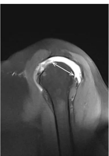
Figure 1. MRA sagittal oblique image showing Tear size ···· 7
LIST OF TABLES
Table 1. Patients’ demographics ··· 9
Table 2. Functional scores for both groups ··· 11
Table 3. Active range of motions of both groups ··· 12
Table 4. Preoperative tear size and follow-up re-tear size/ residual
defect on T1-weighted fat suppression sagittal oblique image in
MRA/CTA ··· 14
Table 5. The preoperative and postoperative stage of fatty
infiltration and muscle atrophy in the supraspinatus and
infraspinatus ··· 15
Table 6. Preoperative and final follow-up acromiohumeral distance
1
ABSTRACT
Minimum 5-year follow-up
after arthroscopic repair of large to massive rotator cuff tears:
complete repair with aggressive release versus partial repair alone
Jeong Jeung Yeol
Department of Medicine
The Graduate School, Yonsei University
(Directed by Professor Chun Yong-Min)
Background: The purpose of this retrospective study was to compare the clinical and radiological outcomes of a minimum 5-year follow-up period for repair of a large to massive rotator cuff tear performed using arthroscopic complete repair with a posterior interval slide or partial repair without the posterior interval slide. Methods: This study included fifty-eight patients with large to massive rotator cuff tears for whom arthroscopic complete repair was not feasible using anterior interval slide and margin convergence alone. Each patient underwent either arthroscopic complete repair with an additional posterior interval slide and a following side-to-side repair of the interval slide edge (Group C) or arthroscopic partial repair with margin convergence alone, without the additional posterior interval slide (Group P). Patient assignment to treatment group was not randomized. Functional assessments included the visual analog scale pain score, subjective shoulder value, the American Shoulder and Elbow Surgeons score, the University of California at Los Angeles
2
shoulder score, and active range of motion. The preoperative and 6-month follow-up magnetic resonance arthrography (MRA) images were compared within- and between-groups.
Results: At the final follow-up evaluation, there were no significant differences between groups in functional values despite significant improvement in both groups, compared with the preoperative values (p < 0.001). A re-tear was identified in 88% (22/25) of the patients in Group C; they had larger re-tear sizes (p = 0.001) and shorter acromiohumeral distances (p = 0.007), compared with Group P patients. Conclusion: At the early postoperative follow-up MRA, the complete repair group with a posterior interval slide (Group C) had 88% re-tear rate and larger re-tear sizes than the partial repair group without a posterior interval slide (Group P). This early inferior radiological outcome led to significantly shorter acromiohumeral distances in the complete repair group, although there were no significant differences between groups in clinical outcomes during the minimum 5-year follow-up period.
Key Words : large to massive rotator cuff tear, complete repair, partial repair, interval slide, integrity, long-term follow up
3
Minimum 5-year follow-up
after arthroscopic repair of large to massive rotator cuff tears:
complete repair with aggressive release versus partial repair alone
Jeong Jeung Yeol
Department of Medicine
The Graduate School, Yonsei University
(Directed by Professor Chun Yong-Min)
I. INTRODUCTION
In many cases, large to massive rotator cuff tears have associated retraction of a torn tendon and fatty infiltration of the affected cuff muscle.1 Due to improvements in, and wide adoption of arthroscopic techniques (e.g., margin convergence and interval slide), arthroscopic repair of large to massive rotator cuff tears is currently feasible. However, it is challenging to achieve complete repair without tension overload, and satisfactory cuff healing and integrity.2
Tauro et al.3 introduced the interval slide technique to address large to massive rotator cuff tears. This technique releases the coracohumeral ligament in the rotator interval from the supraspinatus tendon to improve mobility of the retracted tendon. Lo and Burkhart subsequently extended this concept by adding the posterior interval slide, and it is used when an anterior interval slide alone does not acheive acceptable mobility.4 The posterior interval slide is a release of the retracted rotator
4
cuff tendon at the interval between the supraspinatus and infraspinatus tendons along the scapula spine, thereby further improving mobility of the tethered torn tendon and reducing tension at the repair site.4,5
Although these techniques can convert incomplete repair to complete repair by improving mobility,5 this aggressive technique has a potential risk of devascularization of the repaired tendon.5,6 Kim et al.5 found a re-tear rate up to 91% in patients treated using complete repair group with aggressive release. Also, the re-tear size was significantly larger than the residual defects in the partial repair group without aggressive release. We are well aware that satisfactory functional improvement can be achieved despite re-tear. However, there is a paucity of literature that examines this issue for arthroscopic repair of large to massive rotator cuff tears with longer follow-up periods.
The purpose of this retrospective study was to compare the clinical and radiological outcomes of a minimum 5-year follow-up period for repair of a large to massive rotator cuff tear performed using arthroscopic complete repair with a posterior interval slide or partial repair without the posterior interval slide. We hypothesized that the patients in the complete repair group with the posterior interval slide would experience inferior clinical and radiological outcomes, compared with the patients in the partial repair group without the posterior interval slide.
5
II. MATERIALS AND METHODS 1. Study Population
The study population included 107 patients from March 2008 to July 2012; each had a large to massive rotator cuff tear for which arthroscopic complete repair was not feasible with anterior interval slide and margin convergence. They underwent either arthroscopic complete repair with an additional posterior interval slide and a following side-to-side repair of the interval slide edge (Group C) or arthroscopic partial repair with margin convergence alone, without the additional posterior interval slide (Group P). Patient assignment to treatment group was not randomized.
The inclusion criteria were: (1) the presence of a large to massive rotator cuff tear with an anteroposterior diameter of ≥3 cm or involvement of two or more tendons identified during a preoperative MRA and arthroscopic evaluation, (2) a tear for which complete repair was not feasible using anterior interval slide and margin convergence alone, (3) follow-up MRA images were available at 6 months after surgery for assessment of structural integrity, and (4) ≤grade 2 shoulder arthritis according to the Hamada classification system.7 The exclusion criteria were (1) follow-up data not available for a minimum of 5 years after surgery (38 patients), (2) partial repair despite use of the posterior interval slide and a following side-to-side repair of the interval slide edge (10 patients), (3) a concomitant irreparable subscapularis tear or stage III or higher fatty infiltration of the subscapularis muscle (8 patients), (4) a history of surgery on the affected shoulder (12 patients), and (5)
6
reoperation (3 patients). Fifty-eight patients (25 patients in Group C, complete repair; 33 patients in Group P, partial repair) who met the inclusion and exclusion criteria were included in the study. The data, including radiological images, were reviewed retrospectively. Our institutional review board approved the study protocol and waived the requirement for patient informed consent.
2. Functional and Radiological Assessments
Functional assessments included the visual analog scale (VAS) pain score (range, 0 to 10), the subjective shoulder value (SSV; patient’s self-estimation of the affected shoulder as a percentage of the normal shoulder value), the American Shoulder and Elbow Surgeons (ASES) score, the University of California at Los Angeles (UCLA) shoulder score, and an active range of motion (ROM) assessment. The shoulder ROM included three movements. The forward flexion angle was measured in the scapular plane, external rotation with the arm at the side, and internal rotation. Internal rotation was determined by measuring the highest spinal segment reached by the patient’s thumb. For ease of statistical analysis, the spinal segments were converted into numbers. The levels T1 to T12 were represented using 1 through 12; the levels L1 to L5 were represented using 13 through 17; the sacrum was represented using 18.5,8-10 An independent examiner who was blinded to the group assignments performed the preoperative and follow-up functional assessments, including the shoulder scores and active ROMs.
7
The preoperative radiological evaluation included true shoulder anteroposterior and axillary radiography and MRA. For the follow-up images, a postoperative MRA was acquired to evaluate the structural integrity of the repaired tendons at 6 months after surgery. The preoperative tear size and the postoperative retear size or the residual defect after partial repair were defined as the maximum anteroposterior width on a fat-suppressed T1-weighted sagittal oblique image in an MRA (Figure 1). The pre- and postoperative degree of fatty infiltration was determined on the most lateral T1-weighted sagittal oblique image where contact of the scapular spine with the scapula was observed. The acromiohumeral distance was also evaluated on both the preoperative and follow-up true shoulder anteroposterior radiographic images. Two independent orthopaedic surgeons who were blinded to the group assignments performed the assessments of the radiological values.
Figure 1. The tear size is defined as the maximum anteroposterior width on a fat-suppressed T1-weighted sagittal oblique image.
8
3. Postoperative Rehabilitation
After surgery, the affected arm was kept in abduction for 6 weeks. During the first 6 weeks, pendulum and gentle self-assisted circumduction exercise were begun. Six weeks after surgery, self-assisted passive ROM exercises (e.g., the table sliding stretch and forward flexion of the shoulder in the supine position) were initiated. At 8 weeks after surgery, each patient was encouraged to perform active assisted ROM exercises during the subsequent period. Three months after surgery, isotonic strengthening exercises using an elastic band were begun. Six months after surgery, the patients were allowed to gradually return to their premorbid levels of sports activity.
4. Statistical Analysis
The SPSS statistics software (version 25; IBM, Armonk, New York) was used to analyze the data. The Mann-Whitney test was used to analyze continuous or continuous ranked data between the two groups. The Wilcoxon signed-ranked test was used to compare preoperative and postoperative values. The Chi-square test was used to compare categorical data, such as fatty infiltration, between the two groups. Statistical significance was set at p < 0.05.
III. RESULTS
1. Patient Demographics
9
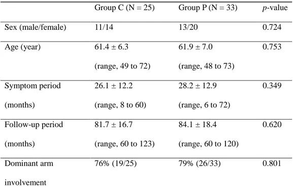
women. The mean ages of the patients at the time of surgery were 61.4 years (range, 49–72 years) in Group C and 61.9 years (range, 48–73 years) in Group P. The mean periods of symptoms before surgery were 26.1 months in Group C and 28.2 months in Group P. The mean follow-up periods for Groups C and P were 81.7 months (range, 60 to 123 months) and 84.1 months (range, 60 to 120 months), respectively. Dominant arm involvement was 76% (19/25) in Group C and 79% (26/33) in Group P. There were no statistically significant differences in patient demographics values (Table 1)
Table 1. Patients’ demographics
Group C (N = 25) Group P (N = 33) p-value
Sex (male/female) 11/14 13/20 0.724 Age (year) 61.4 ± 6.3 (range, 49 to 72) 61.9 ± 7.0 (range, 48 to 73) 0.753 Symptom period (months) 26.1 ± 12.2 (range, 8 to 60) 28.2 ± 12.9 (range, 6 to 72) 0.349 Follow-up period (months) 81.7 ± 16.7 (range, 60 to 123) 84.1 ± 18.4 (range, 60 to 120) 0.620 Dominant arm involvement 76% (19/25) 79% (26/33) 0.801
10
Group C: achieved a complete repair with posterior interval slide and following margin convergence; Group P: achieved a partial repair with side to side sutures margin convergence alone, without posterior interval slide. The values are given as the mean and standard deviation.
2. Arthroscopic Findings and Concomitant Procedures
A biceps lesion was identified in 13 patients (52%) in Group C and 16 patients (49%) in Group P. These patients underwent biceps tenotomy or tenodesis. A concomitant subscapularis partial or full-thickness tear requiring repair was identified in 9 patients (36%) in Group C and 11 patients (33%) in Group P. The supraspinatus and infraspinatus tears were repaired using either a single-row (19 patients in Group C and 25 patients in Group P) or a double-row suture-bridge type row (6 patients in Group C and 8 patients in Group P). After repair, the mean residual defect in Group P was 12.2 ± 4.4 mm (range, 5 to 25 mm).
3. Functional and Radiological Outcomes
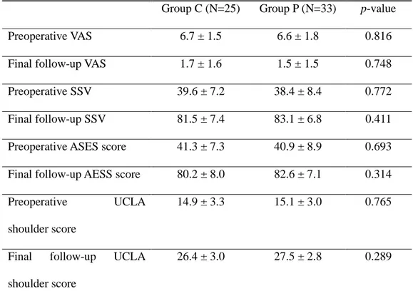
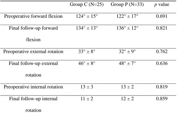
At the final follow-up, the mean VAS pain score, SSV, the ASES score, and the UCLA shoulder score improved significantly in both groups, compared to preoperative values (p < 0.001). However, in all shoulder function values, no postoperative differences between groups were significant (Table 2). Active forward flexion, external rotation with the arm at side, and internal rotation also, improved significantly in both groups, compared to preoperative values (p < 0.001). however,
11
in all ROM measurements, there were no significant differences between groups (Table 3). Three patients (two from Group C and one from Group P) underwent reoperation (reverse total shoulder arthroplasty; 7 years (two) and 8 years (one) after surgery) due to pain and disability associated with progressing arthropathy.
Table 2. Visual analogue scale (VAS), subjective shoulder value (SSV), and American Shoulder and Elbow Surgeon (ASES) score, University California of Los Angeles (UCLA) shoulder score for both groups
Group C (N=25) Group P (N=33) p-value
Preoperative VAS 6.7 ± 1.5 6.6 ± 1.8 0.816
Final follow-up VAS 1.7 ± 1.6 1.5 ± 1.5 0.748 Preoperative SSV 39.6 ± 7.2 38.4 ± 8.4 0.772 Final follow-up SSV 81.5 ± 7.4 83.1 ± 6.8 0.411 Preoperative ASES score 41.3 ± 7.3 40.9 ± 8.9 0.693 Final follow-up AESS score 80.2 ± 8.0 82.6 ± 7.1 0.314 Preoperative UCLA
shoulder score
14.9 ± 3.3 15.1 ± 3.0 0.765
Final follow-up UCLA shoulder score
26.4 ± 3.0 27.5 ± 2.8 0.289
12
margin convergence; Group P: achieved a partial repair with side to side sutures margin convergence alone, without posterior interval slide. The values are given as the mean and standard deviation.
Table 3. Active range of motions of both groups
Group C (N=25) Group P (N=33) p value
Preoperative forward flexion 124° ± 15° 122° ± 17° 0.691 Final follow-up forward
flexion
134° ± 13° 136° ± 12° 0.821
Preoperative external rotation 33° ± 8° 32° ± 9° 0.762 Final follow-up external
rotation
46° ± 8° 48° ± 7° 0.636
Preoperative internal rotation 13 ± 3 13 ± 2 0.819 Final follow-up internal
rotation
11 ± 2 12 ± 2 0.859
Group C: achieved a complete repair with posterior interval slide and following margin convergence; Group P: achieved a partial repair with side to side sutures margin convergence alone, without posterior interval slide. The values are given as the mean and standard deviation. For the internal rotation, the spinal segments as reference were converted into continuous number: T1-12 to 1-12, L1-5 to 13-17, and sacrum to 18.
13
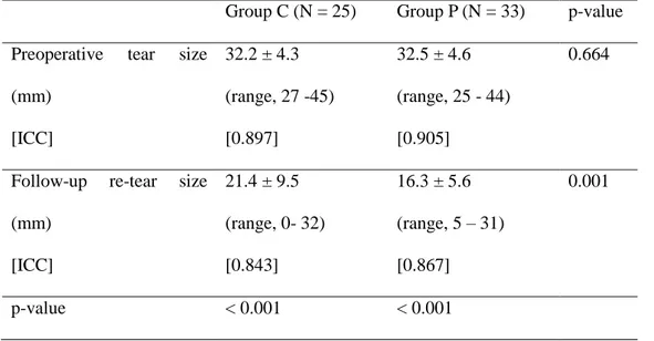
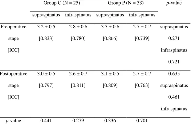
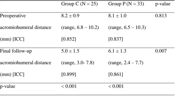
The preoperative mean tear size was 32.2 mm in Group C and 32.5 cm in Group P. On the follow-up MRA, a re-tear was identified in 88% (22/25) of the patients in Group C; this result was associated with a mean re-tear size of 21.4 mm. In Group P, the mean residual defect on follow-up MRA after partial repair was reduced to 16.3 mm. The difference in re-tear size and residual defect was significant between groups (p = 0.001) (Table 4). Regarding the mean preoperative and postoperative fatty infiltration stage in the supraspinatus and infraspinatus muscles, there were no significant differences between groups. (Table 5). At the final follow-up, the mean acromiohumeral distance was significantly decreased, from 8.2 to 5.0 mm in Group C (p < 0.001) and 8.1 to 6.1 mm in Group P (p < 0.001). There was also a significant difference between the groups final acromiohumeral distance (Table 6), (p = 0.007).
14
Table 4. Preoperative tear size and follow-up re-tear size/ residual defect on T1-weighted fat suppression sagittal oblique image in MRA/CTA
Group C (N = 25) Group P (N = 33) p-value Preoperative tear size
(mm) [ICC] 32.2 ± 4.3 (range, 27 -45) [0.897] 32.5 ± 4.6 (range, 25 - 44) [0.905] 0.664
Follow-up re-tear size (mm) [ICC] 21.4 ± 9.5 (range, 0- 32) [0.843] 16.3 ± 5.6 (range, 5 – 31) [0.867] 0.001 p-value < 0.001 < 0.001
Group C: achieved a complete repair with posterior interval slide and following margin convergence; Group P: achieved a partial repair with side to side sutures margin convergence alone, without posterior interval slide. The values are given as the mean and standard deviation. ICC: Intra-class correlation coefficient for inter-observer reliability.
15
Table 5. The preoperative and postoperative stage of fatty infiltration and muscle atrophy in the supraspinatus and infraspinatus
Group C (N = 25) Group P (N = 33) p-value
supraspinatus infraspinatus supraspinatus infraspinatus Preoperative stage [ICC] 3.2 ± 0.5 [0.833] 2.8 ± 0.6 [0.780] 3.3 ± 0.6 [0.866] 2.7 ± 0.7 [0.739] supraspinatus 0.271 infraspinatus 0.721 Postoperative stage [ICC] 3.0 ± 0.5 [0.797] 2.6 ± 0.7 [0.811] 3.1 ± 0.5 [0.809] 2.7 ± 0.7 [0.763] 0.635 supraspinatus 0.461 infraspinatus p-value 0.441 0.279 0.336 0.701
Group C: achieved a complete repair with posterior interval slide and following margin convergence; Group P: achieved a partial repair with side to side sutures margin convergence alone, without posterior interval slide. The values are given as the mean and standard deviation. ICC: Intra-class correlation coefficient for inter-observer reliability.
16
Table 6. Preoperative and final follow-up acromiohumeral distance on true anteroposterior radiograph of the shoulder
Group C (N = 25) Group P (N = 33) p-value Preoperative acromiohumeral distance (mm) [ICC] 8.2 ± 0.9 (range, 6.8 – 10.2) [0.852] 8.1 ± 1.0 (range, 6.5 – 10.3) [0.837] 0.813 Final follow-up acromiohumeral distance (mm) [ICC] 5.0 ± 1.5 (range, 3.0- 7.8) [0.899] 6.1 ± 1.3 (range, 2.4 – 7.7) [0.861] 0.007 p-value < 0.001 < 0.001
Group C: achieved a complete repair with posterior interval slide and following margin convergence; Group P: achieved a partial repair with side to side sutures margin convergence alone, without posterior interval slide. The values are given as the mean and standard deviation. ICC: Intra-class correlation coefficient for inter-observer reliability.
IV. DISCUSSION
This study was designed to compare the clinical and radiological outcomes of a minimum 5-year follow-up period for repair of large to massive rotator cuff tear
17
performed via either arthroscopic complete repair with posterior interval slide or partial repair without the posterior interval slide. The follow-up MRAs after repair revealed tears in 88% of the patients in the complete repair group. The mean re-tear size in the complete repair group was also significantly larger than the mean residual defect in the partial repair group. Also, the acromiohumeral distance at final follow-up was significantly shorter in the complete repair group. These results might be associated with the aggressive release and a following impaired tendon-to-bone healing. Nonetheless, despite inferior radiological outcomes, there were no significant differences in clinical outcomes at the final follow-up between the two groups. This result was not consistent with our hypothesis.
In repair of large to massive contracted rotator cuff tears, surgical techniques such as margin convergence or interval side have been introduced to minimize tension at repair site and maximize tendon mobility.4,11 Margin convergence (side-to-side repair) is useful for repair of longitudinal-type rotator cuff tears for which direct reduction of the tendon (in particular, the apex of the torn tendon) may not be possible. In this circumstance, even if direct reduction is possible, the repair is likely to be achieved under significant tension. However, through the use of margin convergence, tension at the repair site at the corner of each leaf can be decreased.5 Several investigators have reported satisfactory clinical outcomes using this approach.12,13
18
this concept to the use of an additional posterior interval slide, indicating that these anterior and posterior (double) interval slides could achieve significant improvements in lateral excursion.4 These early investigators reported satisfactory outcomes using this interval slide technique, but they did not include results on structural integrity on the follow-up imaging studies.
Kim et al. also used this double interval slide for arthroscopic rotator cuff repair of massive contracted tears for which complete repair could not be achieved without a double interval slide.5 However, they found a high re-tear rate of up to 91% for the complete repair, despite improvements in lateral excursion and attempts to reduce the repair tension. The re-tear sizes in the complete repair group were larger than the residual defects in the partial repair group without the posterior interval slide. Berduso et al.15 also used interval slide techniques to treat massive contracted immobile rotator cuff tears in their series. Although they did not indicate the status of preoperative fatty infiltration of the affected cuff muscle in detail, massive re-tearing to the original size was identified in 55% of the patients. Nonetheless, they reported that the overall shoulder function score was satisfactory, and they suggested that use of interval slide techniques can result in satisfactory clinical and radiological outcomes. However, as Kim and Iagulli et al.5,6 reported, tissue devascularization and subsequent impairment of healing potential through use of this aggressive release may occur. Therefore, Kim et al. indicated that the use of aggressive release did not increase benefit, although it may facilitate the complete
19
repair.5 Despite a high re-tear rate and larger re-tear size in the complete repair with aggressive release group, clinical outcomes were not significantly different between the two groups at a 2-year follow-up.5 At early postoperative period, the current study found that the re-tear rate was similar to those of Kim et al.5. We supposed that with time, this inferior radiological outcome in the complete repair group with the aggressive release would result in significant deterioration of clinical outcomes. However, despite significantly shorter acromiohumeral distances in the complete repair group, there were no significant differences between the groups in clinical outcomes for at least the 5-year follow-up period. We did not determine why larger re-tear sizes at the early postoperative period and shorter acromiohumeral distances at the final follow-up were not associated with inferior clinical outcomes. We assume that this clinical outcome may be attributed to maintained transverse force combined with a robust subscapularis and teres minor tendon and muscle in both groups. Unfortunately, at the same final clinical follow-up, we could not confirm the structural integrity of these tendons and muscles using the MR images.
Recently, Collin et al.16 reported 10-year follow-up clinical outcomes after repair of massive posterosuperior rotator cuff tears with of a 10-year assessment of repair integrity. Although they did not describe the release techniques used, complete repair was accomplished in all cases. At the 10-year follow-up, the postoperative MRIs showed promising outcomes; the re-tear rate was just 34%. This result might be attributed to the fact that most of the patients had stage 2 or less fatty infiltration
20
of the affected cuff muscle, which was different from the patients in our current study and in previous studies that examined massive rotator cuff tears in the context of stage 3 or 4 fatty infiltration.
This study had some limitations. First, this was a retrospective study with non-randomized patient assignment. The complete repairs with the posterior interval slide were performed earlier in the study period and the partial repairs without it were performed later. Second, inclusion of a limited number of patients may be associated with type II error with low statistical power. Third, we did not have final MRA follow-up images that matched changes in fatty infiltration with the final clinical assessments assessing the integrity of the supraspinatus/infraspinatus and the subscapularis/teres minor muscles.
V. CONCLUSION
On follow-up MRA during the early postoperative period, the complete repair group with posterior interval slide had 88% of the re-tear rate and larger re-tear sizes than the partial repair group without a posterior interval slide for repair of large to massive rotator cuff tears. Although there were no statistically significant differences in clinical outcomes between the two groupa, this early inferior radiological outcome resulted in significantly shorter acromiohumeral distances in the patients in the complete repair group at the final follow-up.
21
REFERENCES
1. Cordasco FA, Bigliani LU. The rotator cuff. Large and massive tears. Technique of open repair. Orthop Clin North Am. 1997 Apr;28(2):179-93.
2. Mori D, Funakoshi N, Yamashita F. Arthroscopic surgery of irreparable large or massive rotator cuff tears with low-grade fatty degeneration of the infraspinatus: patch autograft procedure versus partial repair procedure. Arthroscopy. 2013 Dec;29(12):1911-21.
3. Tauro JC. Arthroscopic repair of large rotator cuff tears using the interval slide technique. Arthroscopy. 2004 Jan;20(1):13-21.
4. Lo IK, Burkhart SS. Arthroscopic repair of massive, contracted, immobile rotator cuff tears using single and double interval slides: technique and preliminary results. Arthroscopy. 2004 Jan;20(1):22-33.
5. Kim SJ, Kim SH, Lee SK, Seo JW, Chun YM. Arthroscopic repair of massive contracted rotator cuff tears: aggressive release with anterior and posterior interval slides do not improve cuff healing and integrity. J Bone Joint Surg Am. 2013 Aug 21;95(16):1482-8.
6. Iagulli ND, Field LD, Hobgood ER, Ramsey JR, Savoie FH, 3rd. Comparison of partial versus complete arthroscopic repair of massive rotator cuff tears. Am J Sports Med. 2012 May;40(5):1022-6.
22
7. Hamada K, Fukuda H, Mikasa M, Kobayashi Y. Roentgenographic findings in massive rotator cuff tears. A long-term observation. Clin Orthop Relat Res. 1990 May(254):92-6.
8. Oh JH, Kim SH, Shin SH, Chung SW, Kim JY, Kim SH, Kim SJ. Outcome of rotator cuff repair in large-to-massive tear with pseudoparalysis: a comparative study with propensity score matching. Am J Sports Med. 2011 Jul;39(7):1413-20.
9. Oh JH, Oh CH, Kim SH, Kim JH, Yoon JP, Jung JH. Clinical features of partial anterior bursal-sided supraspinatus tendon (PABST) lesions. J Shoulder Elbow Surg. 2012 Mar;21(3):295-303.
10. Kim SJ, Lee IS, Kim SH, Woo CM, Chun YM. Arthroscopic repair of
concomitant type II SLAP lesions in large to massive rotator cuff tears: comparison with biceps tenotomy. Am J Sports Med. 2012 Dec;40(12):2786-93.
11. Burkhart SS. Reconciling the paradox of rotator cuff repair versus debridement: a unified biomechanical rationale for the treatment of rotator cuff tears. Arthroscopy. 1994 Feb;10(1):4-19.
12. Kim SJ, Lee IS, Kim SH, Lee WY, Chun YM. Arthroscopic partial repair of irreparable large to massive rotator cuff tears. Arthroscopy. 2012 Jun;28(6):761-8. 13. Calvert PT, Packer NP, Stoker DJ, Bayley JI, Kessel L. Arthrography of the
shoulder after operative repair of the torn rotator cuff. J Bone Joint Surg Br. 1986 Jan;68(1):147-50.
23
14. Tauro JC. Arthroscopic "interval slide" in the repair of large rotator cuff tears. Arthroscopy. 1999 Jul-Aug;15(5):527-30.
15. Berdusco R, Trantalis JN, Nelson AA, Sohmer S, More KD, Wong B, Boorman RS, Lo IK. Arthroscopic repair of massive, contracted, immobile tears using interval slides: clinical and MRI structural follow-up. Knee Surg Sports Traumatol Arthrosc. 2015 Feb;23(2):502-7.
16. Collin P, Colmar M, Thomazeau H, Mansat P, Boileau P, Valenti P, Saffarini M, Nover L, Kempf JF. Clinical and MRI Outcomes 10 Years After Repair of Massive Posterosuperior Rotator Cuff Tears. J Bone Joint Surg Am. 2018 Nov
24
ABSTRACT(IN KOREAN)
크기가 큰 회전근 개 파열의 관절경 봉합 후 최소 5 년 추적 관찰: 적극적인 이완술을 동반한 완전 봉합과 부분 봉합만의 비교<지도교수 천 용 민>
연세대학교 대학원 의학과
정 증 열
배경: 이 후향적 연구의 목적은 후방 간격 슬라이드를 통해서 완전봉합 또는 후부 간격슬라이드 없이 부분적인 봉합을 사용하여 수행된 대형 회전근 개 파열의 수복을 위한 최소 5 년 추적 관찰 기간동안 임상 및 방사선 학적 결과를 비교하는 것입니다. 대상 및 방법: 이 연구에는 전방 간격 슬라이드와 마진 수렴만으로는 관절경 완전 봉합이 불가능한 대형 회전근 개 파열을 가진 58 명의 환자가 포함되었습니다. 각각 환자는 추가적인 후방 간격 슬라이드와 후속 측면 수선을 이용한 관절경 완전 봉합(그룹 C)과 추가적인 후방 간격 슬라이드 없이 마진 수렴만으로 관절경 부분 봉합(그룹 P)치료중에 하나를 받았습니다. 치료 군에 대한 환자 할당은 무작위화되지 않았습니다. 기능 평가에는 시각 아날로그 척도 통증 점수, 주관적인 어깨 값, 미국 어깨 및 팔꿈치 외과 의사 점수, 로스 앤젤레스 캘리포니아 대학 어깨 점수 및 능동 어깨 활동 범위가 포함되었습니다. 수술 전 및 6 개월의 후속 자기 공명 관절 조영술 (MRA) 이미지를 그룹25 내 및 그룹 간 비교하였습니다. 결과: 수술 전 값과 비교하여 두 그룹 모두에서 상당한 개선에도 불구하고 최종 추시 평가에서, 기능적 값의 그룹간에 유의 한 차이는 없었습니다 (p <0.001). 재 파열은 C 군 환자의 88 % (22/25)에서 확인되었으며; 그들은 P군 환자에 비해 더 큰 재 파열 크기 (p = 0.001)와 더 짧은 견봉상완골두 거리 (p = 0.007)를 가졌습니다. 결론: 수술 후 초기 추종 MRA에서 후방 간격 슬라이드 (그룹 C)가있는 완전 봉합 그룹은 후방 간격 슬라이드 (그룹 P)가없는 부분 봉합 그룹보다 88 %의 재 파열비율과 더 큰 재 파열 크기를 가졌습니다. 최소 5 년의 추적 관찰 기간 동안 임상 결과들에서 두 그룹간에 유의한 차이는 없었지만 이 초기 열등한 방사선 학적 결과는 완전 봉합 그룹에서 견봉상완골두간의 거리를 유의하게 단축시켰습니다.