pISSN: 0378-6471 eISSN: 2092-9374 http://dx.doi.org/10.3341/jkos.2013.54.9.1365 = 증례보고 =
두 종류의 스펙트럼영역 빛간섭단층촬영기를 사용한
정상 한국인의 맥락막 두께 측정의 재현성
이광현⋅이성철⋅이승규 연세대학교 의과대학 안과학교실목적: 두 종류(Cirrus, Spectalis)의 스펙트럼영역 빛간섭단층촬영기(Optical coherence tomography, OCT)를 사용하여 건강한 한국 성인의 맥락막 두께를 측정하고, 두 기계간의 재현성 및 측정자간의 재현성을 비교하였다.
대상과 방법: 안과적 이상이 없는 정상 성인 30명의 60안을 대상으로, Cirrus OCT의 1-line raster 및 Spectialis OCT의 Enhanced depth imaging을 사용하여 맥락막을 검사하였고, 맥락막 두께는 독립된 두 측정자가 중심와 아래, 1.5 mm 비측, 1.5 mm 이측에서 측정하였다.
결과: 총 검사 대상자는 30명이었으며 남성 15명, 여성 15명으로 평균 연령은 50.73 ± 15.09세(24-75세)였다. 두 기계간에서 측정한 맥락막 두께는 통계적으로 차이를 보이지 않았으며 강한 상관관계를 보였다. Bland-Altman plot에서 bias는 관찰되지 않았다. 두 측정 자간의 재현성도 우수하였다.
결론: 정상 한국인의 맥락막 두께 측정에 대한 Cirrus 및 Spectralis OCT의 재현성은 우수하였다. <대한안과학회지 2013;54(9):1365-1370>
■Received: 2013. 2. 2. ■ Revised: 2013. 5. 3. ■Accepted: 2013. 7. 23.
■Address reprint requests to Christopher Seungkyu Lee, MD Department of Ophthalmology, Severance Hospital, #50 Yonsei-ro, Seodaemun-gu, Seoul 120-749, Korea
Tel: 82-2-2228-3570, Fax: 82-2-312-0541 E-mail: [email protected]
* The Association for Research in Vision and Ophthalmology Annual Meeting, May, 6-10,2012, Fort Lauderdale, Florida.
맥락막은 망막색소상피 아래에 위치하며 망막외층에 혈 액을 공급하는 조직으로, 맥락막 종양,1 황반변성,2중심장 액맥락망막병증3 등의 안과적 질환과 관련되어 있는 것으 로 보고되었다. 이러한 중요성에도 불구하고 맥락막은 검안 경, 안저사진, 형광안저혈관조영술을 통해서는 검사가 어려 웠고, 망막색소상피로 인해 기존의 OCT (Optical coher-ence tomography, OCT)로도 관찰이 어려웠다. 그러나 최 근 Spaide et al4에 의해 고안된 Enhanced depth imaging (EDI) 방법을 통해 맥락막 두께의 측정이 가능하게 되었다. 이 러한 EDI 방법은 Spectralis OCT (Heidelberg Engineering, Heidelberg, Germany)에서 최초로 제공되었으며 이후 Cirrus OCT (Carl Zeiss Meditec Inc., Dublin, CA)에서도 맥락막 두께의 측정이 가능하게 되었다.5-8
현재 임상적으로 사용되고 있는 OCT에는 Topcon사의 OCT-1000, Canon 사의 SPOCT, Spectralis HRA+OCT (Vista, Heidelberg, Germany), Spectral OCT/SLO (Ophthalmic
Technologies Inc, Toronto, Ontario, Canada), RTVue-100 (Optovue Inc, Fremont, Califonia, USA), 3D SD-OCT (Bioptigen, Research Triangle Park, North Carolina, USA), Cirrus OCT (Carl zeiss Meditec Inc, Dublin, CA) 등이 있으며 본 연구에서는 한국에서 널리 사용되고 있는 2 종류의 OCT (Heidelberg Spectralis-OCT, Cirrus-OCT) 를 사용하여 건강한 성인 한국인에서 맥락막 두께를 측정 하고, 두 기계 사이의 재현성(reproducibility) 및 측정자 간 재현성을 알아보았다.
대상과 방법
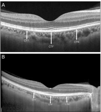
2011년 2월부터 4월까지 본원 안과에 내원한 정상인을 대상으로 하였으며, 세극등, 골드만 안압측정, 최대교정시 력, 안저검사를 시행하였다. 굴절이상은 자동굴절검사기 (Canon, Inc., Tokyo, Japan)를 사용하여 측정하였다. 대 상자 중 안과질환의 과거력이 있거나 소견이 보이는 환자, 최대교정시력이 0.8 미만인 경우, 이전에 안과적 수술의 과거력이 있거나 맥락막과 망막의 질환이 발견된 경우, 녹 내장을 진단 받았거나 안압이 21 mmHg 이상인 경우, 그 리고 당뇨와 고혈압과 같은 전신적인 질환이 있는 환자는 대상에서 제외하였다. 굴절력이 -6 디옵터 미만인 고도 근시 및 +6 디옵터 이상인 원시의 경우도 제외하였다.Figure 1. Optical coherence tomography scans showing
cho-roidal thicknesses of the same subject on Cirrus and Spectralis. (A) An OCT image of the subject on Cirrus (Carl Zeiss Meditec Inc., Dublin, CA). Image averaging is used for cho-roidal visualization. chocho-roidal thickness measurements was taken perpendicularly from the outer edge of the hyper-re-flective retinal pigment epithelium to the inner sclera at the fo-vea, 1500 μm temporal to the fofo-vea, and 1500 μm nasal to the fovea. (B) An OCT image of subject on Spectralis. Image averaging with the aid of the eye tracking and EDI are used for choroidal visualization. Choroidal thickness measurements was taken at the fovea, 1500 μm temporal to the fovea, and 1500 μm nasal to the fovea (CTN: choroidal thickness meas-ured at 1.5 mm nasal to the fovea, CTF subfoveal choroidal thickness, CTT choroidal thickness measured at 1.5 mm tem-poral to the fovea).
400 350 300 250 200 150 100 50 A ve ra ge th ic kn es s ( m ) µ
Nasal Subfoveal Temporal
Spectralis Cirrus
Figure 2. Graph showing mean choroidal thickness
measure-ment in normal eyes at the fovea, 1.5 mm nasal to the fovea, and 1.5 mm temporal to the fovea.
모든 대상자는 Cirrus OCT와 Spectiralis OCT로 검사를 시행 받았으며, 총 검사시간은 맥락막 두께의 일중변화를 고려해 30분이 넘지 않도록 하였다. Cirrus OCT를 사용한 촬영시에는 software version 6.0에서 제공되는 EDI 방법 을 사용하지 않고, 기존의 1-line raster 모드로 검사를 시 행하였다(Fig. 1A).7 Cirrus OCT의 1-line raster는 유리 체망막 간격(vitreoretinal interface)을 최대한 0으로 하여 맥락막을 촬영하였다. Cirrus OCT의 경우 상이 뒤집힐 경 우 영상의 질이 떨어질 수가 있어서 뒤집힌 상을 얻어 맥락 막의 두께를 측정하는 방법은 사용하지 않았다. Spectralis OCT는 EDI 모드를 사용하여 맥락막을 검사하였다(Fig. 1B).4두 독립된 측정자가 서로의 결과를 알지 못하는 상황 에서 맥락막 두께를 각각 중심와 아래, 1.5 mm 비측, 1.5 mm 이측에서 측정하였다.
통계학적 분석은 PASW Statistics 18.0 version (SPSS inc., Chicago, IL)을 이용하였으며 One-way analysis of variance (ANOVA)를 이용하여 중심와 아래, 비측, 이측에 서 측정된 맥락막 두께를 비교 하였고, 측정자간의 재현성은 Pearson 상관분석을 사용하여 분석하였다. Bland-Altman plot을 사용하여 두 기계간의 측정치간의 차이를 분석하였 다. p<0.05인 경우를 통계학적으로 유의한 것으로 하였다.
결
과
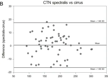
대상군은 총 30명으로 이 중 남자는 15명 30안, 여자는 15명 30안이었고, 평균 연령은 50.73 ± 15.09세(24-75 세)였다. 평균 굴절력은 -0.74 ±1.98 디옵터였다. 모든 안에서 정상적인 망막 소견을 보였고, 맥락막 경계를 측정 할 수 있었다. 중심와 아래의 맥락막 두께는 Cirrus에서 238.33 ± 55.00 μm, Spectralis에서 238.90 ±53.34 μm였으며, 비 측은 Cirrus 178.93 ± 49.72 μm, Spectralis 179.67 ± 49.37 μm, 이측은 Cirrus 218.65 ±46.84 μm, Spectralis 219.94 ±48.82 μm였다. 맥락막 두께는 중심와 아래가 가 장 두꺼웠으며 그 다음으로 이측이 두꺼웠고, 비측이 가장 얇았다(Fig. 2). 두 기계로 측정한 맥락막 두께 사이에 통계적으로 유의 한 차이는 없었으며, 강한 상관관계를 보였다. Pearson 상 관계수는 0.984 (p<0.001)였다. Bland-Altman plot에서 두 기계간에 유의한 bias는 관찰되지 않았다(Fig. 3). Pearson 상관분석을 사용하여 두 평가자간의 상관관계도 분석하였으며, 모든 측정치에서 높은 상관관계를 관찰할 수 있었다(Table 1).A
B
30 20 10 0 -10 -20 -30 100 150 200 250 300 350 400 D iff er en ce ( sp ec tra lis -c irr us )
Average of spectralis and cirrus
Mean 1.96 ± SD Mean Mean 1.96 ± SD CTF spectralisvs cirrus 30 20 10 0 -10 -20 50 100 150 200 250 300 350 D iff er en ce ( sp ec tra lis -c irr us )
Average of spectralis and cirrus
Mean 1.96 ± SD
Mean 1.96 ± SD Mean
CTN spectralis vs cirrus
Average of spectralis and cirrus
100 150 200 250 300 350 40 30 20 10 0 -10 -20 -30 D iff er en ce ( -c irr us ) sp ec tra lis Mean 1.96 ± SD Mean 1.96 ± SD Mean CTT spectralis vs cirrus
Figure 3. Bland-Altman plots for Cirrus vs. Spectralis. (A)
Bland-Altman plot for subfoveal choroidal thickness. Mean dif-ference is 2.425 (95% CI -0.174 to 5.024). The 95% limits of agreement for choroidal thickness measurements are -17.694 to 22.544. (B) Bland-Altman plot for choroidal thickness of the na-sal fovea. Mean difference is 0.733 (95% CI -1.489 to 2.956). The 95% limits of agreement are -16.476 to 17.942. (C) Bland- Altman plot forchoroidal thickness of the temporal fovea. Mean difference is 1.292 (95% CI -1.659 to 4.242). The 95% limits of agreement for choroidal thickness measurements between Cirrus and Spectralis are -21.549 to 24.133.
Table 1. Inter-grader comparison of choroidal thickness measurements for cirrus and spectralis
Location Spectralis Cirrus
Correlation coefficient p-value Correlation coefficient p-value
CTN* 0.945 <0.001§ 0.919 <0.001§
CTF† 0.977 <0.001§ 0.966 <0.001§
CTT‡ 0.953 <0.001§ 0.890 <0.001§
*Choroidal thickness measured at 1.5 mm nasal to the fovea; †Subfoveal choroidal thickness; ‡Choroidal thickness measured at 1.5 mm
temporal to the fovea; §p-value is significant at the 0.05 level (two-tailed).
고
찰
최근 수년동안 빛간섭 단층 촬영기를 사용한 정상인 및 질병이 있는 안구의 맥락막 두께 및 구조에 대한 연구들이 보고되었다.1,4-7,9-14 본 연구 결과에 따르면 Spectralis OCT와 Cirrus OCT 사이의 맥락막 두께 측정에 대한 재현 성은 매우 뛰어났으며 평가자 간 재현성도 우수한 것으로 나타났다. 이러한 결과는 Branchini et al5및 Yamashita et al8의 결과와 일치하는 것으로, Branchini et al5는 Spectralis, Cirrus, RTvue 간에 뛰어난 재현성이 있음을 밝혔고, Yamashita et al8
은 Spectralis, Cirrus, Topcon에서 높은
재현성을 발표하였다. 본 연구에서는 기존 연구에 비해 더 욱 넓은 범위에서 맥락막 두께를 측정(1.5 mm versus 0.75 mm)하였고, 두 기계간의 높은 재현성이 더 넓은 영역 에서도 나타남을 확인하였다. 본 연구에서 주목할 점은 Cirrus OCT에서 EDI 방법을 사용하지 않은 측정임에도 맥 락막의 관찰이 가능했다는 점이다. 고도근시, 황반 변성의 경우 맥락막의 두께가 건강인보다 얇으므로15,16임상에서는 EDI 방법을 사용하지 않아도 맥락막 두께의 측정이 많은 환자군에서 가능할 것으로 생각한다. 그러나 각막 혼탁, 백 내장 등이 동반된 안구의 경우 일반적인 방법으로는 맥락 막 두께의 측정이 어려울 수 있을 것이라 생각되며, 이에
A
B
C
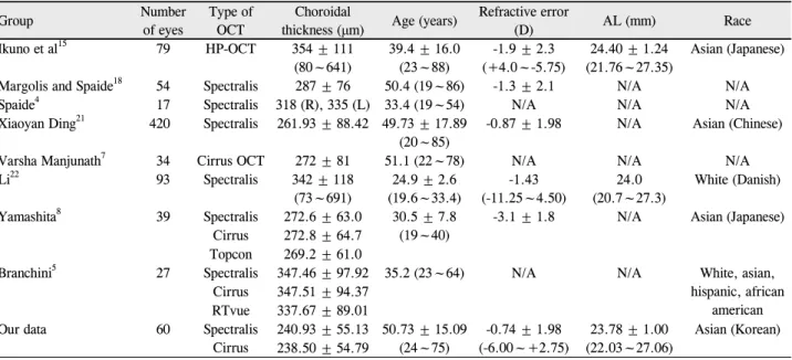
Table 2. Summary of previous studies on demographics and choroidal thickness measurements using various optical coherence
to-mographies in normal subjects
Group Number of eyes Type of OCT thickness (μm)Choroidal Age (years) Refractive error (D) AL (mm) Race Ikuno et al15 79 HP-OCT 354 ± 111 (80~641) 39.4 ± 16.0 (23~88) -1.9 ± 2.3 (+4.0~-5.75) 24.40 ± 1.24 (21.76~27.35) Asian (Japanese) Margolis and Spaide18 54 Spectralis 287 ± 76 50.4 (19~86) -1.3 ± 2.1 N/A N/A
Spaide4 17 Spectralis 318 (R), 335 (L) 33.4 (19~54) N/A N/A N/A
Xiaoyan Ding21 420 Spectralis 261.93 ± 88.42 49.73 ± 17.89
(20~85)
-0.87 ± 1.98 N/A Asian (Chinese)
Varsha Manjunath7 34 Cirrus OCT 272 ± 81 51.1 (22~78) N/A N/A N/A
Li22 93 Spectralis 342 ± 118 (73~691) 24.9 ± 2.6 (19.6~33.4) -1.43 (-11.25~4.50) 24.0 (20.7~27.3) White (Danish) Yamashita8 39 Spectralis 272.6 ± 63.0 30.5 ± 7.8 (19~40)
-3.1 ± 1.8 N/A Asian (Japanese) Cirrus 272.8 ± 64.7
Topcon 269.2 ± 61.0
Branchini5 27 Spectralis 347.46 ± 97.92 35.2 (23~64) N/A N/A White, asian,
hispanic, african american Cirrus 347.51 ± 94.37
RTvue 337.67 ± 89.01
Our data 60 Spectralis
Cirrus 240.93 ± 55.13 50.73 ± 15.09 (24~75) -0.74 ± 1.98 (-6.00~+2.75) 23.78 ± 1.00 (22.03~27.06) Asian (Korean) 238.50 ± 54.79
Values are presented as mean ± SD.
AL = axial length; N/A = not applicable; HP-OCT = high-penetration optical coherence tomography.
대한 확인을 위해서는 다양한 질환군을 대상으로 한 추가 적인 연구가 필요할 것이다. 본 연구에서 망막 중심 오목의 평균 맥락막 두께는 Cirrus에서 232.38 ±55.48 μm, Spectralis에서는 242.20 ±56.01 μm였으며 이 수치는 이전의 연구들에 비해 얇은 것으로, Hirata et al17이 시행한 연구 만이 202.6 ±83.5 μm로 본 연구 결과보다 얇은 두께를 보고하였다. Margolis and Spaide18는 OCT의 EDI방법으로 정상시력을 가지는 대 상(평균나이 50.4세)에서 맥락막 두께를 측정하여 평균 287 μm로 보고하였으며, Ikuno et al15은 평균 37.6세의 대 상자에서 고해상도 OCT (high-penetration OCT)를 이용 하여 평균 292.7 μm, 같은 대상자에서 EDI기법으로 평균 283 μm로 보고하였다. 한국인을 대상으로 한 연구에서는 Kim et al19이 건강한 대조군(평균 59.75세)의 중심와아래 맥락막 두께는 평균 247 μm로 보고하였다. 이러한 연구간 의 차이는 연구 대상자들의 연령 및 굴절력의 차이 때문으 로 생각한다(Table 2).5,7,12,18,20 정상인의 맥락막 두께는 중심와에서 가장 두꺼운 것으로 보고되었으며,18 이러한 결과는 본 연구와도 일치한다. 본 연구에서는 중심와 아래에서 측정한 맥락막이 가장 두꺼웠 으며, 다음으로 이측, 비측 순으로 얇았다. 흥미로운 점은 총 60안 중 19안에서 이측 맥락막이 중심와 아래 맥락막보 다 두꺼웠다. Fujiwara et al11이 최근 보고한 결과에 따르 면 고도근시안에서는 이측 맥락막이 가장 두껍고 그 다음 으로 중심와 아래에서 측정한 맥락막의 두께가 두꺼웠으며 비측 맥락막이 가장 얇았다. 이전 연구에서는 이러한 정상 안과 고도근시안 사이의 맥락막 분포의 차이가 후포도종 (posterior staphyloma) 때문일 것으로 추측하였다. 그러나 이측 맥락막이 더 두꺼운 19안에서 후포도종은 발견할 수 없었다. 따라서 맥락막 두께의 분포와 연령, 안축장 길이, 굴절력 등이 상관관계가 있는지에 대한 추가적인 연구가 필요할 것으로 생각한다.
결론적으로 Cirrus와 Spectralis OCT는 정상 한국인의 맥락막 두께 측정에서 높은 재현성을 보였으며, 각각 측정 자간의 재현성도 우수하였다. 앞으로 더 나아가, 다양한 종 류의 OCT를 이용하여 여러 질환 별 측정치의 비교와 반복 성 및 재현성에 관한 추가적인 연구가 필요할 것으로 생각 한다.
REFERENCES
1) Torres VL, Brugnoni N, Kaiser PK, Singh AD. Optical coherence tomography enhanced depth imaging of choroidal tumors. Am J Ophthalmol 2011;151:586-93.e2.
2) Spaide RF. Age-related choroidal atrophy. Am J Ophthalmol 2009;147:801-10.
3) Gemenetzi M, De Salvo G, Lotery AJ. Central serous chorioretin-opathy: an update on pathogenesis and treatment. Eye (Lond) 2010;24:1743-56.
4) Spaide RF, Koizumi H, Pozzoni MC. Enhanced depth imaging spectral-domain optical coherence tomography. Am J Ophthalmol 2008;146:496-500.
of choroidal thickness measurements across three spectral domain optical coherence tomography systems. Ophthalmology 2012;119: 119-23.
6) Manjunath V, Fujimoto JG, Duker JS. Cirrus HD-OCT high defi-nition imaging is another tool available for visualization of the cho-roid and provides agreement with the finding that the chocho-roidal thickness is increased in central serous chorioretinopathy in com-parison to normal eyes. Retina 2010;30:1320-1; author reply 1321-2.
7) Manjunath V, Taha M, Fujimoto JG, Duker JS. Choroidal thickness in normal eyes measured using Cirrus HD optical coherence tomography. Am J Ophthalmol 2010;150:325-9.
8) Yamashita T, Yamashita T, Shirasawa M, et al. Repeatability and reproducibility of subfoveal choroidal thickness in normal eyes of Japanese using different SD-OCT devices. Invest Ophthalmol Vis Sci 2012;53:1102-7.
9) Benavente-Pérez A, Hosking SL, Logan NS, Bansal D. Reproducibility- repeatability of choroidal thickness calculation using optical co-herence tomography. Optom Vis Sci 2010;87:867-72.
10) Carrasco S, Torres JP, Torner L, et al. Enhancing the axial reso-lution of quantum optical coherence tomography by chirped qua-si-phase matching. Opt Lett 2004;29:2429-31.
11) Fujiwara T, Imamura Y, Margolis R, et al. Enhanced depth imaging optical coherence tomography of the choroid in highly myopic eyes. Am J Ophthalmol 2009;148:445-50.
12) Ikuno Y, Maruko I, Yasuno Y, et al. Reproducibility of retinal and choroidal thickness measurements in enhanced depth imaging and high-penetration optical coherence tomography. Invest Ophthalmol Vis Sci 2011;52:5536-40.
13) Imamura Y, Fujiwara T, Margolis R, Spaide RF. Enhanced depth imaging optical coherence tomography of the choroid in central se-rous chorioretinopathy. Retina 2009;29:1469-73.
14) Imamura Y, Iida T, Maruko I, et al. Enhanced depth imaging optical coherence tomography of the sclera in dome-shaped macula. Am J Ophthalmol 2011;151:297-302.
15) Ikuno Y, Kawaguchi K, Nouchi T, Yasuno Y. Choroidal thickness in healthy Japanese subjects. Invest Ophthalmol Vis Sci 2010;51: 2173-6.
16) Manjunath V, Goren J, Fujimoto JG, Duker JS. Analysis of choroi-dal thickness in age-related macular degeneration using spec-tral-domain optical coherence tomography. Am J Ophthalmol 2011;152:663-8.
17) Hirata M, Tsujikawa A, Matsumoto A, et al. Macular choroidal thickness and volume in normal subjects measured by swept- source optical coherence tomography. Invest Ophthalmol Vis Sci 2011;52:4971-8.
18) Margolis R, Spaide RF. A pilot study of enhanced depth imaging optical coherence tomography of the choroid in normal eyes. Am J Ophthalmol 2009;147:811-5.
19) Kim SW, Oh J, Kwon SS, et al. Comparison of choroidal thickness among patients with healthy eyes, early age-related maculopathy, neovascular age-related macular degeneration, central serous cho-rioretinopathy, and polypoidal choroidal vasculopathy. Retina 2011;31:1904-11.
20) Esmaeelpour M, Povazay B, Hermann B, et al. Three-dimensional 1060-nm OCT: choroidal thickness maps in normal subjects and improved posterior segment visualization in cataract patients. Invest Ophthalmol Vis Sci 2010;51:5260-6.
21) Ding X, Li J, Zeng J, et al. Choroidal thickness in healthy Chinese subjects. Invest Ophthalmol Vis Sci 2011;52:9555-60.
22) Li XQ, Larsen M, Munch IC. Subfoveal choroidal thickness in re-lation to sex and axial length in 93 Danish university students. Invest Ophthalmol Vis Sci 2011;52:8438-41.
=ABSTRACT=
Reproducibility of Choroidal Thickness in Normal Korean Eyes Using
Two Spectral Domain Optical Coherence Tomography
Kwang Hyun Lee, MD, Sung Chul Lee, MD, Christopher Seungkyu Lee, MD
Department of Ophthalmology, Yonsei University College of Medicine, Seoul, Korea
Purpose: To investigate the reproducibility of choroidal thickness measurements in healthy Koreans using two spectral
do-main optical coherence tomography (SD-OCT) instruments: Zeiss Cirrus HD-OCT (Carl Zeiss Meditec Inc., Dublin, CA, USA) and Heidelberg Spectralis (Heidelberg Engineering, Heidelberg, Germany).
Methods: Images were obtained in 60 eyes of 30 healthy undilated volunteers without ocular pathology in a clinical setting.
The choroid was imaged in all subjects using Cirrus HD 1-line raster and Spectralis enhanced depth imaging (EDI). The choroid was measured subfoveally, 1500 µm temporal, and 1500 µm nasal to the fovea. All measurements were per-formed by two independent observers. One-way analysis of variance (ANOVA), Pearson correlation, and Bland–Altman analysis were used to compare measurements.
Results: The study group consisted of 15 males and 15 females. The mean age was 50.73 ± 15.09 years (range, 24-75
years). There was no significant difference in the mean choroidal thickness (p > 0.05) between systems for any location. The choroidal thickness measurements using two instruments (Cirrus vs. Spectralis) were also strongly correlated (p < 0.001).
Conclusions: In the present study of healthy Korean adults, good reproducibility was observed between choroidal
thick-ness measurements of images obtained from Cirrus and Spectralis. J Korean Ophthalmol Soc 2013;54(9):1365-1370
Key Words: Choroidal thickness, Optical coherence tomography, Reproducibility of results
Address reprint requests to Christopher Seungkyu Lee, MD Department of Ophthalmology, Severance Hospital #50 Yonsei-ro, Seodaemun-gu, Seoul 120-749, Korea