Muscle Functional MRI of
Exercise-Induced Rotator Cuff Muscles
INTRODUCTION
Strengthening of the rotator cuff muscles is one of the most integral parts of a rehabilitation program for athletes with shoulder injuries who must perform throwing motions during sports activities. Additionally, exercise-induced muscle activity is essential in sports medicine and rehabilitation medicine. Therefore, previous studies have been conducted to identify the most effective exercise for strengthening rotator
This is an Open Access article distributed under the terms of the Creative Commons Attribution Non-Commercial License (http://creativecommons.org/licenses/ by-nc/4.0/) which permits unrestricted non-commercial use, distribution, and reproduction in any medium, provided the original work is properly cited. Received: July 3, 2020 Revised: September 26, 2020 Accepted: November 2, 2020 Correspondence to: Noriyuki Tawara, Ph.D. Department of Radiological Sciences, Faculty of Health Sciences, Japan Health Care College, Sapporo 004-0819, Japan. Tel. +81-11-885-7711 Fax. +81-11-885-5757 E-mail: n_tawara@nihoniryo-c. ac.jp
Copyright © 2021 Korean Society of Magnetic Resonance in Medicine (KSMRM)
Original Article
The aim of this study was to provide a new assessment of rotator cuff muscle activity. Eight male subjects (24.7 ± 3.2 years old,171.2 ± 9.8 cm tall, and weighing 63.8 ± 11.9 kg) performed the study exercises. The subjects performed 10 sets of the exercise while fixing the elbow at 90 degrees flexure and lying supine on a bed. One exercise set consisted of the subject performing external shoulder rotation 50 times using training equipment. Two imaging protocols were employed: (a) true fast imaging with steady precession (TrueFISP) at an acquisition time of 12 seconds and (b) multi-shot spin-echo echo-planar imaging (MSSE-EPI) at an acquisition time of 30 seconds for one echo. The main method of assessing rotator cuff muscle activity was functional T2 mapping using ultrafast imaging (fast-acquired muscle functional MRI [fast-mfMRI]). Fast-mfMRI enabled real-time imaging for the identification and evaluation of the degree of muscle activity induced by the exercise. Regions of interest were set at several places in the musculus subscapularis (sub), musculus supraspinatus (sup), musculus teres minor (ter), and deltoid muscle (del). We used the MR signal of the images and transverse relaxation time (T2) for comparison. Most of the TrueFISP signal was not changed by exercise and there was no significant difference from the resting values. Only the T2 in the musculus teres minor was increased after one set and the change were seen on the T2 images. Additionally, except for those after one and two sets, the changes in T2 were significant compared to those at rest (P < 0.01). We also demonstrated identify and visualize the extent to which muscles involved in muscle activity by exercise. In addition, we showed that muscle activity in a region such as a shoulder, which is susceptible to B0 inhomogeneity, could be easily detected using this technique.
Keywords: Muscle functional magnetic resonance imaging (mfMRI); Multi-shot spin-echo spin-echo-planar imaging (MSSE-EPI); TrueFISP; Transverse relaxation time (T2); Rotator cuff
Noriyuki Tawara1, 2, Atsushi Nishiyama1
1Department of Radiological Sciences, Faculty of Health Sciences, Japan Health Care College, Sapporo, Japan
mfMRI of Exercise-Induced Rotator Cuff Muscles | Noriyuki Tawara, et al.
cuff muscles (1-3). However, the most effective exercise has varied between studies. Several factors may have contributed to the divergent results. One of them was that electromyography (EMG) was commonly used as a technique to assess muscle activity induced by exercise in these reports. Although a higher level of EMG reproducibility was reported by Kelly et al. (3), shortfalls with the use of EMG still exist. The intramuscular dual fine wire electrodes used in EMG detect the activity of only a small number of muscle fibers and sometimes migrate during exercise. This may help explain the relatively poor reproducibility of EMG (4-6) and may be another possible reason for the divergent data.
1H-magnetic resonance imaging (MRI) can evaluate not
only superficial muscles but also deep muscles. In addition, increases in muscle proton transverse relaxation time (T2) were reported to positively correlate with the intensity of concentric and eccentric muscle contractions (7-10). Furthermore, the relationship between T2 and EMG activity from contraction amplitude is linear (11). Therefore, this technique is also useful in evaluating muscle use after short periods of resistance training. Those findings indicate that MRI can be used to study skeletal muscle function, with T2 serving as a quantitative index of activity. In the field of exercise physiology and rehabilitation medicine, this technique is called muscle functional magnetic resonance imaging (mfMRI) (12-15). Researchers in several studies have tabulated the changes in muscle T2 to monitor the pattern of muscle recruitment during various exercises in human subjects (7, 8, 10, 16-20). However, as reported by Takeda et al. (21), it is shared as an experienced knowledge that the detectability of T2 changes is limited if the exercise is not performed until fatigue. Therefore, it is difficult to use this evaluation approach efficiently during rehabilitation.
We considered that the problem of detectability seems to involve the temporal resolution of image acquisition for calculating the T2. To reduce the acquisition time and thus, increase temporal resolution, we theorized and then verified that the T2 values calculated from spin-echo echo-planar-imaging (SE-EPI) images could highly detect slight muscle activity induced by acute exercise because the high temporal resolution of SE-EPI, data can be collected while T2 is elevated, although the elevated T2 returns to its resting value due to exercise (22). And muscle functional MRI using the T2 calculated from SE-EPI images was able to detect the slight impact of muscle activity induced by exercise. Ultrafast imaging has been shown to allow the visualization of exercise-induced muscle activity (23).
SE-EPI has been used to refer to a sequence in which the data from all of the k-space for an entire 2D plane is collected following a single RF-excitation pulse. More recently, the term has been expanded to include any rapid spin-echo sequence in which the k-space is traversed in one or a small number of excitations (24). In the modern lexicon, these are termed single-shot spin-echo echo-planar imaging (SSSE-EPI) and multi-shot spin-echo echo-planar imaging (MSSE-EPI), respectively.
The advantage of SSSE-EPI is that the image acquisition time can be significantly shortened, so the influence of motion artifacts can be reduced. However, this technique places more stress on the gradient and phase errors more time to build up. In particular, the variation in phase error is more susceptible to it as the number of k-space trajectory readings during a single TR increase. SSSE-EPI is susceptible because it has the highest number of k-space trajectory to be read during one TR. Thus, image distortion and shimming malfunction are likely to occur due to the influence of susceptibility effects. In contrast, MSSE-EPI takes longer to perform than does SSSE-EPI but can reduce image distortion and the influence of susceptibility effects. Therefore, although it is necessary to verify T2 calculations using SE-EPI in the rotator cuff region where susceptibility effects have a large influence, no previous studies have been reported.
The purpose of this study was to evaluate mfMRI of the rotator cuff muscles by T2 calculations using SE-EPI, and whether it could be used to visualize muscle activity in rotator cuff muscles.
MATERIALS AND METHODS
Subjects and exercise protocol
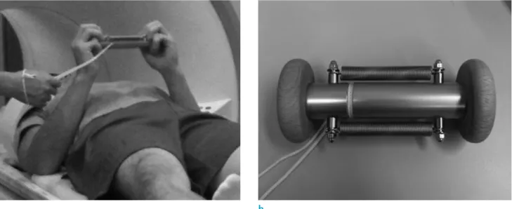
Eight male subjects (24.7 ± 3.2 years old, 171.2 ± 9.8 cm tall, and weighing 63.8 ± 11.9 kg) performed the exercise. The subjects performed 10 sets of an exercise while fixing the elbow at 90 degrees flexure and lying supine on a bed (Fig. 1). One exercise set consisted of the subject performing external shoulder rotation 50 times using training equipment (Arm Twista; Sanriki Corporation, Tokyo, Japan). All subjects provided written informed consent according to as approved by the Ethics Committee of the Japan Institute of Sports Sciences.
MR imaging and data analysis
performed on a 1.5-Tesla whole-body scanner (Symphony; Siemens AG, Erlangen, Germany) with a small shoulder array coil. Two protocols were employed: (a) true fast imaging with steady precession (TrueFISP) with a repetition time (TR) of 4.72 ms, an echo time (TE) of 2.36 ms, a matrix size of 256 × 256, a flip angle (FA) of 50, a bandwidth (BW) 501 Hz/Px, an acquisition time of 12 seconds; and (b) MSSE-EPI with a TR of 2000 ms, a TE of 20, 30, 40, 50 ms (4 echoes), a matrix size of 128 × 128 interpolated into 256 × 256, an FA of 90, a BW of 1392 Hz/Px, and an acquisition time of 30 seconds (for one echo). A slice thickness of 5 mm, a FOV of 240 × 240 mm, and a NEX of 1 were common factors. The imaging conditions for SSSE-EPI could not be adjusted, so it could not be used in the experiment.
Among the fast imaging, TrueFISP can acquire high spatial resolution image data and therefore easy to obtain anatomical information. EPI, on the other hand, is extremely low in spatial resolution, so it is difficult to obtain anatomical information from the image data. In rest, TrueFISP images as morphological images and MSSE-EPI images as T2-weighted images for calculating T2 were acquired in order. The subjects exercised on the bed of the MR scanner and after each set of exercises, only MSSE-EPI images were acquired immediately. By not resetting the positioning information of the device, the time required from the end of each exercise to the start of scanning was shortened to less than one minute. After the experiment was completed, the image data were transferred to a computer for analysis, T2 images were created using the
T2-weighted images acquired by MSSE-EPI, and image analysis was performed.
T2 images were calculated using the least-squares fitting mono-exponential of the MSSE-EPI images. In extracting the MR signal from the TrueFISP images (TrueFISP signal), and muscle T2 from the T2 images, regions of interest (ROI) were set at several places in the musculus subscapularis (sub), musculus supraspinatus (sup), musculus teres minor (ter), and deltoid muscle (del). The average and standard deviation of the T2 for each muscle set as the ROI was calculated. The T2 was calculated using Interactive Data Language (IDL: L3Harris Geospatial, Boulder, CO, USA) with least-squares fitting mono-exponential regression of the MSSE-EPI images. To visualize the area of the activated muscle, we used the fast-mfMRI technique (23, 25).
Statistical analysis
A previous study reported that there were approximately 10% changes in the T2 in the same muscle (26). Based on this finding, the obtained data were subjected to one-way repeated-measures ANOVA with Scheffe’s post-hoc test. Differences with P-values of < 0.05 were considered significant.
RESULTS
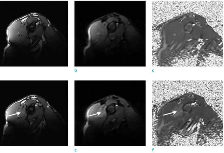
Figure 2 shows representative MR images of the right shoulder at rest and after 10 exercise sets. In the images
Fig. 1. The exercise scheme of external rotation exercises of the shoulders was repeated 50 times starting from a neutral position. (a) The practical landscape of the exercises and (b) training equipment.
mfMRI of Exercise-Induced Rotator Cuff Muscles | Noriyuki Tawara, et al.
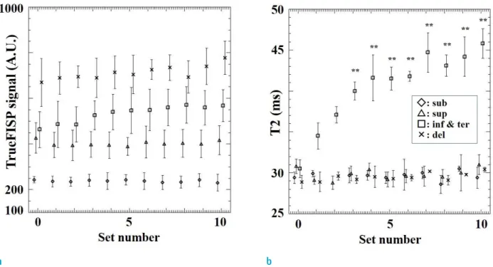
obtained after 10 sets, especially on the MSSE-EPI and T2 images, the areas of the activated musculus teres minor were well-enhanced and the details were preserved. Figure 3 shows changes in the TrueFISP signals after each set and changes in the T2. All subjects showed the same findings. Most of MR signal detected from TrueFISP images were not changed by exercise and there was no significant difference from the resting values. Only T2 in the ter was increased after one set, and the change was seen on T2 images. Additionally, except for those after one and two sets, the changes in T2 were significant compared to those at rest (P < 0.01).
Figure 4 shows representative MR images of the right shoulder after 10 sets of exercise. The inf and ter showed slight changes in signal intensity (SI) after exercise, However, it was difficult to quantify the SI difference (Fig. 4a). Additionally, although the TrueFISP images were superior in spatial resolution, the identification of the
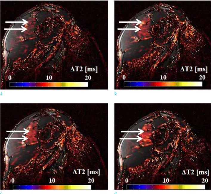
activated muscle using changes in SI in the subtracted images was difficult (Fig. 4c). The T2 images improved the image contrast in muscle activity induced by exercise (Fig. 4b) and the subtracted images (Fig. 4d) could identify muscle activity induced by exercise in the inf and ter. Figure 5 shows the fusion images generated using the fast-mfMRI technique. This technique can facilitate the visualization of muscle activity induced by exercise. As shown in the figure, this technique is applicable even in the rotator cuff region, which is susceptible to B0 inhomogeneity. Additionally, muscle activity due to slight exercise could be detected using MSSE-EPI to obtain excellent T2 measurements in relation to temporal resolution.
DISCUSSION
First, we tried to adjust the imaging conditions of
SSSE-Fig. 2. Representative MR images. (a) TrueFISP image at rest, (b) MSSE-EPI image at rest, (c) T2 image at rest, (d) TrueFISP image after 10 sets, (e) MSSE-EPI image after 10 sets, and (f) T2 image after 10 sets. In (b) and (e), the echo was 50 ms. The arrows denote the activated musculus teres minor.
a b c
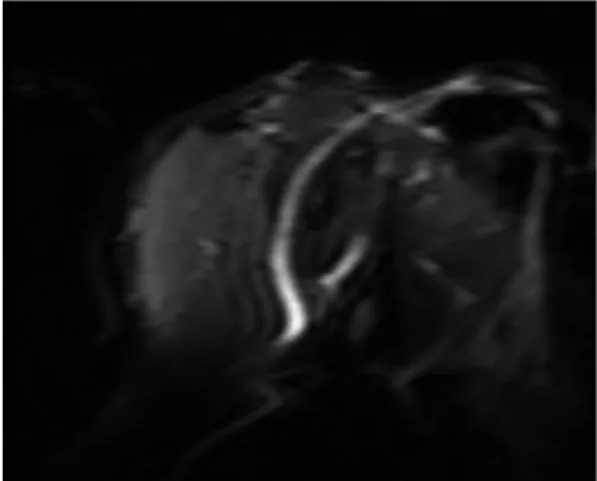
EPI for obtaining rotator cuff MR images, but we could not use SSSE-EPI because the MRI unit used in this study could not eliminate the influence of fat artifacts, as shown in Figure 6.
In this study, the SE-EPI we used was multi-shot SE-EPI (MSSE-EPI) instead of single-shot SE-EPI (SSSE-EPI). An advantage of MSSE-EPI is that phase errors have less time to accumulate compared to SSSE-EPI. Thus, MSSE-EPI can reduce susceptibility artifacts. This advantage is thought to act on sites such as the shoulder where susceptibility artifacts occur due to B0 inhomogeneity.
Muscle activity induced by exercise was only detected in the T2 images. In T2 using the ultrafast imaging method, it was possible to detect small impacts of slight muscle activity induced by acute exercise. Figure 3a shows that the shoulder is a region that is susceptible to B0 inhomogeneity. Conversely, it is an advantage that the spin-echo sequences could correct B0 inhomogeneity to some extent. Additionally, we showed that muscle activity due to slight exercise could be detected if we used MSSE-EPI to obtain excellent T2 measurements in relation to temporal resolution. Therefore, it was suggested that when a site is susceptible to B0 inhomogeneity, T2 calculation using MSSE-EPI could resolve the problem of image distortion.
Since MSSE-EPI and SSSE-EPI are pulse sequences having
the characteristics of high temporal resolution but low spatial resolution, it is difficult to obtain morphological information about the boundaries of each muscle. In addition, although the SE sequence can easily obtain morphological information, the scanning time of SE is generally long. Thus, TrueFISP images were selected as morphological images that satisfied the two requirements of shortened imaging acquisition time and high spatial resolution. In the same way, we also tried to visualize muscle activity induced by exercise using our mfMRI method. Although the MR signal alone could not identify the muscles involved by exercised, only T2 could be used to extract the area of the muscle in which activity was induced by exercise. The usual MR signals obtained with sequences such as steady-state free precession (SSFP) using TrueFISP and gradient-echo (GE and/or GrE) cannot compensate for the effects of B0 inhomogeneity, which is common in images of the shoulder region, and the effect of B0 inhomogeneity on image quality is significant. However, T2 images calculated from images obtained from SE sequences using refocusing pulses are less susceptible to B0 inhomogeneity (22), and we confirmed that T2-weighted images were an excellent method for identifying activated muscles in the shoulder, including the rotator cuff, as shown in Figure 4d.
Fig. 3. TrueFISP signal and T2 at rest (set number = 0) and after exercise in representative subjects. (a) TrueFISP signal. (b) T2. Significantly different from the value at rest, * : P<0.05, ** : P<0.01. sub (◊), sup (△), inf & ter (□), and del (×).
mfMRI of Exercise-Induced Rotator Cuff Muscles | Noriyuki Tawara, et al.
The simplest examination for the increases in T2 after exercise is that the increase in osmolarity (phosphate, lactate, sodium) in the cytoplasm during activity results in the influx of fluid, diluting the effect of the myofibrillar proteins on water relaxation. Therefore, T2 is actively used to evaluate exercise-induced muscle activity (12).
The recovery of muscle activity may affect T2 calculations because it took a few minutes to collect the imaging data
for T2 calculations. However, in the exercise protocol conducted in this paper, an increase in T2 could be detected, as shown in Figure 3. Therefore, it was suggested that the exercise protocol conducted in this paper had no effect on the recovery of muscle activity.
The usefulness of changes in muscle T2 has been reported by a number of previous studies. In this study, the image distortion in SE-EPI was improved by using
MSSE-Fig. 4. Representative right shoulder MR images after 10 sets of exercise. (a) TrueFISP image, (b) T2 image, (c) TrueFISP images subtracted from 10 sets at rest, and (d) T2 images subtracted from 10 sets at rest. The arrows denote activated infraspinatus & teres minor (inf & ter) muscles.
a b
EPI and showed muscle activity in a region such as a shoulder, which is susceptible to B0 inhomogeneity. Also, muscle activity induced by slight exercise was successfully visualized. It is thought that changes in muscle T2 induced by exercise were strongly influenced by temporal resolution.
In addition, SE-EPI involves pulse sequences with the application of fat saturation until the RF pulse. Therefore, the fat MR signal was suppressed in the SE-EPI images. In skeletal muscle, there are intramyocellular triglycerides (IMCL) and extramyocellular triglycerides (EMCL). Therefore, it was feared that IMCL and EMCL affected the muscle T2. However, in the 1.5-T MRI scanner, we demonstrated that
there was no significant difference in the T2 whether or not the MSE images included fat suppression (26). These results suggest that MR images acquired by 1.5-T imager cannot detect fat within muscle, and therefore also suggest that it cannot detect about IMCL and EMCL too. This suggests that the results from the 1.5-T imager used to calculate muscle T2 were unaffected by IMCL and EMCL.
Since SE-EPI is required to achieve high homogeneity in the static magnetic field, even a small magnetic field difference causes a large spatial distortion. In addition, at the boundaries where the difference in susceptibility effects is large, shimming to eliminate fat artifacts due to
Fig. 5. Fused image using fast-mfMRI obtained after 2 sets (a), 4 sets (b), 6 sets (c), and 8 sets (d) of exercise. The color bar shows the differences in the T2 (delta T2). The arrows denote activated infraspinatus & teres minor (inf & ter) muscles.
a b
mfMRI of Exercise-Induced Rotator Cuff Muscles | Noriyuki Tawara, et al.
bandwidth becomes defective and the artifacts may not be removed. This suggests that the image data obtained by SSSE-EPI, which has high temporal resolution but is greatly affected by susceptibility effects, was poor.
In contrast, in MSSE-EPI, the k-space is divided into multiple segments, each of which is acquired by a separate EPI train. The advantage is that susceptibility effects can be reduced compared to SSSE-EPI. Therefore, MSSE-EPI showed that image data was acquired even in the boundaries where the difference in susceptibility effects was large like in the shoulders.
At present, the limitations of this technique are as follows. Originally, fast-mfMRI (25) was a method of using SSSE-EPI as the pulse sequence for T2 acquisition to significantly reduce the imaging acquisition time for T2 measurements. SSSE-EPI, which is the pulse sequence with a high temporal resolution, is greatly affected by image distortion due to inhomogeneity of the magnetic field, so SSSE-EPI cannot be used in regions susceptible to B0 inhomogeneity. In contrast, although MSSE-EPI is useful for suppressing image distortion due to inhomogeneity of the magnetic field, the image acquisition time is extended 15 times in the scanning conditions used in this paper. Temporal resolution and image distortion are trade-off relationships for SE-EPI, and condition adjustments related to these relationships cannot exceed the performance of the MRI units themselves. Although the application of fast-mfMRI to the whole-body region is possible, the problematic trade-offs in SE-EPI require further research.
CONCLUSION
In this study, we demonstrated the detectability of rotator cuff activity using the T2 calculated from MSSE-EPI images and showed the high detectability of muscle activity in a region, such as a shoulder, which is susceptible to B0 inhomogeneity. Detectability reached a plateau after exercise. The T2 calculated from SE-EPI images indicated the high detectability of slight muscle activity induced by acute exercise. This study also demonstrated the visualization of the activity of individual muscles of the rotator cuff. In addition, we showed that muscle activity in a region such as a shoulder, which is susceptible to B0 inhomogeneity, could easily be detected using the MSSE-EPI technique. Acknowledgments
This work was supported by a Grant-in-Aid for Scientific Research (B) from the Ministry of Education, Culture, Sports, Science, and Technology (25282170). We thank the Japan Institute of Sports Sciences (JISS) for only assistance with data collection.
REFERENCES
1. Blackburn TA WB, McLeod WD, Wofford L. EMG analysis of posterior rotator cuff EMG analysis of posterior rotator cuff exercises. Athletic Training 1990;25:40-45
2. Jobe FW, Moynes DR. Delineation of diagnostic criteria and a rehabilitation program for rotator cuff injuries. Am J Sports Med 1982;10:336-339
3. Ke l l y BT, Ka d r m a s W R , S p e e r K P. T h e m a n u a l muscle examination for rotator cuff strength. An electromyographic investigation. Am J Sports Med 1996;24:581-588
4. Giroux B, Lamontagne M. Comparisons between surface electrodes and intramuscular wire electrodes in isometric and dynamic conditions. Electromyogr Clin Neurophysiol 1990;30:397-405
5. Komi PV, Buskirk ER. Reproducibility of electromyographic measurements with inserted wire electrodes and surface electrodes. Electromyography 1970;10:357-367
6. Morris AD, Kemp GJ, Lees A, Frostick SP. A study of the reproducibility of three different normalisation methods in intramuscular dual fine wire electromyography of the shoulder. J Electromyogr Kinesiol 1998;8:317-322
7. Adams GR, Duvoisin MR, Dudley GA. Magnetic resonance imaging and electromyography as indexes of muscle function. J Appl Physiol (1985) 1992;73:1578-1583
Fig. 6. T2-weighted MR image of a rotator cuff by
SSSE-EPI. Fat artifacts due to subcutaneous fat cannot be removed and remain.
8. Fisher MJ, Meyer RA, Adams GR, Foley JM, Potchen EJ. Direct relationship between proton T2 and exercise intensity in skeletal muscle MR images. Invest Radiol 1990;25:480-185
9. Fleckenstein JL, Watumull D, McIntire DD, Bertocci LA, Chason DP, Peshock RM. Muscle proton T2 relaxation times and work during repetitive maximal voluntary exercise. J Appl Physiol (1985) 1993;74:2855-2859
10. Shellock FG, Fukunaga T, Mink JH, Edgerton VR. Acute effects of exercise on MR imaging of skeletal muscle: concentric vs eccentric actions. AJR Am J Roentgenol 1991;156:765-768
11. Adams GR, Harris RT, Woodard D, Dudley GA. Mapping of electrical muscle stimulation using MRI. J Appl Physiol (1985) 1993;74:532-537
12. Meyer RA, Prior BM. Functional magnetic resonance imaging of muscle. Exerc Sport Sci Rev 2000;28:89-92 13. Ploutz-Snyder LL, Yackel-Giamis EL, Rosenbaum AE,
Formikell M. Use of muscle functional magnetic resonance imaging with older individuals. J Generontol A Biol Sci Med Sci 2000;55:B504-511
14. Akima H, Kinugasa R, Kuno S. Recruitment of the thigh muscles during sprint cycling by muscle functional magnetic resonance imaging. Int J Sports Med 2005;26:245-252
15. Dickx N, Cagnie B, Achten E, Vandemaele P, Parlevliet T, Dannels L. Changes in lumber muscle activity because of induced muscle pain evaluated by muscle functional magnetic resonance imaging. Spine (Phila Pa 1976) 2008;33: E983-989
16. Disler DG, Cohen MS, Krebs DE, Roy SH, Rosenthal DI. Dynamic evaluation of exercising leg muscle in healthy subjects with echo planar MR imaging: work rate and total work determine rate of T2 change. J Magn Reson Imag 1995;5:588-593
17. Fleckenstein JL, Canby RC, Parkey RW, Peshock RM. Acute
effects of exercise on MR imaging of skeletal muscle in normal volunteers. AJR Am J Roentgenol 1988;151:231-237
18. Horrigan JM, Shellock FG, Mink JH, Deutsch AL. Magnetic resonance imaging evaluation of muscle usage associated with three exercises for rotator cuff rehabilitation. Med Sci Sports Exerc 1999;31:1361-1366
19. Price TB, McCauley TR, Duleba AJ, Wilkens KL, Gore JC. Changes in magnetic resonance transverse relaxation times of two muscles following standardized exercise. Med Sci Sports Exerc 1995;27:1421-1429
20. Yue G, Alexander AL, Laidlaw DH, Gmitro AF, Unger EC, Enoka RM. Sensitivity of muscle proton spin-spin relaxation time as an index of muscle activation. J Appl Physiol 1994;77:84-92
21. Takeda Y, Kashiwaguchi S, Endo K, Matsuura T, Sasa T. The most effective exercise for strengthening the supraspinatus muscle: evaluation by magnetic resonance imaging. Am J Sports Med 2002;30:374-381
22. Tawara N, Nitta O, Kuruma H, et al. Exercise-induced muscle activities of the trunk: detectability of the slight impact using muscle functional MRI. In Proceedings of Joint Annual Meeting of ISMRM and ESMRMB, Stockholm, Sweden, 2010:865
23. Tawara N, Nitta O, Kuruma H, et al. Functional T(2) mapping of the trunkal muscle. Magn Reson Med Sci 2009;8:81-83
24. Hashemi RH, Lisanti CJ, Bradley WG. MRI Basics. 4th ed. Philadelphia: Wolters Kluwer, 2018:269-279
25. Tawara N, Nitta O, Kuruma H, Niitsu M, Itoh A. T2 mapping of muscle activity using ultrafast imaging. Magn Reson Med Sci 2011;10:85-91
26. Tawara N, Nitta O, Itoh A. Comparison of pulse sequences for T2 measurement of human skeletal muscle. Japan J Magn Reson 2008;28:25-34