https://doi.org/10.5624/isd.20200132
Introduction
Ameloblastoma is the most common odontogenic neo plasm, primarily originating from the oral epithelium. It usually involves the jaw bone and accounts for 11% to
13% of all odontogenic tumors. Ameloblastoma, formerly known as solid/multicystic ameloblastoma,1 is considered the most frequent type of ameloblastoma in the mandible, especially in the posterior region. Ameloblastoma presents characteristics of local invasion and progressive infiltration of the medullary bone, as well as expansion of the cortical bone.2 It can cause displacement of the adjacent teeth, root resorption, and extensive bone expansion, thereby leading to facial asymmetry.
The intimate proximity of ameloblastoma to the mandi bular canal can result in clinical symptoms of paresthesia,
Directions of mandibular canal displacement in ameloblastoma: A computed tomography
mirrored-method analysis
Karine Evangelista
1,*, Lincoln Cardoso
2, Ítalo Toledo
2, Giovanni Gasperini
2,
José ValladaresNeto
1, Lucia Helena Soares Cevidanes
3, Antonio Carlos de Oliveira Ruellas
3,
Maria Alves Garcia Silva
41Department of Orthodontics, School of Dentistry, Federal University of Goiás, Goiânia, Brazil 2University Clinical Hospital, Federal University of Goiás, Goiânia, Brazil
3Department of Orthodontics and Pediatric Dentistry, School of Dentistry, University of Michigan, Ann Arbor, MI, USA 4Department of Stomatological Sciences, School of Dentistry, Federal University of Goiás, Goiânia, Brazil
ABSTRACT
Purpose: This study was performed to investigate mandibular canal displacement in patients with ameloblastoma
using a 3dimensional mirroredmodel analysis.
Materials and Methods: The sample consisted of computed tomographic scans of patients with ameloblastoma
(n=10) and healthy controls(n=20). The amount of mandibular canal asymmetry was recorded as a continuous variable, while the buccolingual(yaw) and superoinferior(pitch) directions of displacement were classified as categorical variables. The ttest for independent samples and the Fisher exact test were used to compare groups in terms of differences between sides and the presence of asymmetric inclinations, respectively(P<0.05).
Results: The length of the mandibular canal was similar on both sides in both groups. The ameloblastoma group
presented more lateral(2.40±4.16mm) and inferior(-1.97±1.92mm) positions of the mental foramen, and a more buccal(1.09±2.75mm) position of the middle canal point on the lesion side. Displacement of the mandibular canal tended to be found in the anterior region in patients with ameloblastoma, occurring toward the buccal and inferior directions in 60% and 70% of ameloblastoma patients, respectively.
Conclusion: Mandibular canal displacement due to ameloblastoma could be detected by this superimposed mirrored
method, and displacement was more prevalent toward the inferior and buccal directions. This displacement affected the mental foramen position, but did not lead to a change in the length of the mandibular canal. The control group presented no mandibular canal displacement.(Imaging Sci Dent 2021; 51: 17-25)
KEY WORDS: Ameloblastoma; Mandibular Nerve; Facial Asymmetry; Multidetector Computed Tomography; Cone
Beam Computed Tomography
Copyright ⓒ 2021 by Korean Academy of Oral and Maxillofacial Radiology
This is an Open Access article distributed under the terms of the Creative Commons Attribution NonCommercial License(http://creativecommons.org/licenses/bync/3.0) which permits unrestricted noncommercial use, distribution, and reproduction in any medium, provided the original work is properly cited.
Imaging Science in Dentistry·pISSN 22337822 eISSN 22337830
This study was partially funded by the Coordination for the Improvement of Higher Education PersonnelBrazil(CAPES)Finance Code 001 and NIH grant NIDCR R01 DE024450.
Received May 27, 2020; Revised August 15, 2020; Accepted September 9, 2020 *Correspondence to : Prof. Karine Evangelista
School of Dentistry, Federal University of Goiás, Avenida Universitária esquina com 1a Avenida, S/N. Zip Code: 74605220, Goiânia, GoiásBrazil
especially in advancedstage lesions.3 The outcomes of a thorough clinical evaluation in conjunction with different imaging modalities and histopathology are crucial for the successful management of ameloblastoma.4 Both cone beam computed tomography(CBCT) and multislice com puted tomography(MSCT) are efficient imaging modalities for assessing ameloblastomas, as they enable a 3dimen sional(3D) evaluation of the lesion and the anatomical structures adjacent to the tumor, especially the relationship with the mandibular canal.
In cases of ameloblastoma, mandibular canal displace ment is a common radiological finding, especially in lesions closer to the neurovascular bundle. Ameloblastomas cause buccal displacement of the inferior alveolar nerve(IAN) in 84.6% of cases and lingual displacement in 15.4%.5 When treating patients with ameloblastoma, it is essential to know the pathway of the mandibular canal when determining the direction of biopsy access and the safety limits of surgical excision in order to prevent neurovascular injuries.5,6 Precise localization of the IAN may be particularly crucial for post erior mandibular lesions, especially if preservation of the neurovascular bundle is included in the surgical planning.7
Depending on how early a patient presents for evalua tion, mandibular canal displacement may not be evident on imaging. The mandibular canal has an Sshaped pattern in the bone, forming a pathway to the mental foramen.8 Threedimensionalreconstructed volumes show the path way, rather than sectional views of the mandibular canal. Until now, mandibular canal displacement has been studied through qualitative analyses, without using 3D models.5,6 Over the last decade, the diagnostic use of CBCT exams has been enhanced by the emergence of specific tools for 3D symmetry analyses of both craniofacial sides.9,10 Dis placement of the IAN associated with jaw lesions could be an essential radiographic finding. Recognizing its direction could contribute to a better understanding of the biological behavior and management of the tumor. This study set out to investigate ameloblastomarelated mandibular canal dis placement, using a 3D mirroredmodel analysis.
Materials and Methods
This observational retrospective study was approved by the Institutional Review Board of the University of Michi gan(ID: HUM00161968) and the Federal University of Goiás(02681118.5.0000.5078).
Sample
This sample consisted of a secondary data analysis of
available MSCT tomographic scans(n =5) and CBCT scans(n=25), exported as Digital Imaging and Communi cations in Medicine(DICOM) files, divided into 2 groups of patients: the ameloblastoma group, which consisted of 10 patients(7 men and 3 women) with an ameloblastoma lesion who were treated between 2013 and 2018 at the Oral Surgery Department of the Clinical Hospital of Federal University of Goiás, Brazil, and the control group, which included 20 healthy patients(10 men and 10 women) sel ected from deidentified preorthodontic files of the Dental and Craniofacial Bionetwork for Image Analysis of the University of Michigan, USA. The control group patients were selected by pairing their ages with those in the amelo blastoma group at a 2 : 1 ratio. The sample size calculation considered the presence of inclination asymmetry between both sides of the mandibular canal with a power of 80% and an alpha of 0.05 for a 2tailed test. The sample size required for a ratio of 2 : 1 for control and ameloblastoma patients was 16 : 8.
The following inclusion criteria were applied: 1) MSCT and CBCT scans exported as DICOM files; 2) tomographic images presenting no distortion or movement artifacts and a field of view(FOV) covering the full extent of the mandi bular canal; 3) ameloblastoma with histopathological con firmation and the lesion located in the mandibular body or ramus; and 4) healthy patients without severe mandibular asymmetry(<4mm)11 or anatomical variations in the man
dibular canal(bifid mandibular canal). The exclusion crite ria were: 1) images suggestive of facial trauma; 2) images of patients with syndromes or congenital craniofacial anomal ies, such as cleft lip and palate; and 3) images suggestive of previous mandibular surgical procedures.
Image acquisition and 3D assessment
The MSCT scans, which had been taken for 5 ameloblas toma patients before treatment as a complementary exam for diagnosis or surgical planning, were performed on a Syngo CT device(Siemens Healthcare GmbH, Nuremberg, Germany) at the University Clinical Hospital, with an expo sure time of 6.44s, a thickness of 0.8mm, a peak kilovol tage of 130kVp, and a tube current of 25mA. The CBCT scans of 5 ameloblastoma patients and the entire control sample were acquired using an iCat unit(Imaging Sciences International, Hatfield, PA, USA), with a scan time of 40s, an FOV of 23cm×17cm, and a voxel size of 0.4mm3.
The 3D model analysis was performed by a single exami ner, who had previously undergone training in this method with an expert in 3D model analysis, and by an oral radiolo gist. Calibration was achieved by performing all the steps
in 5 different scans before the study.
The 3D analysis was carried out in the following steps, beginning with the conversion of DICOM files to GIPL files using ITKSNAP, an opensource software(version 2.4.0; www.itksnap.org). The original scan was convert ed from a 0.4mm3 voxel size to 0.5mm3 voxels using 3D Slicer(ver. 4.0; www.slicer.org) to reduce the compu tational power and time required for the image analysis. Automatic segmentation of the mandible was executed, followed by the creation of a volumetric label map and a virtual 3D model using 3D Slicer. For the 3D model of the mandible, position orientation was carried out using the 3D Slicer tool(a fixedcoordinate system) in order to stan dardize the mandibular canal position in the image analysis view. The base of the mandibular body was matched with the horizontal reference plane on both sides and the sagittal reference plane was perpendicular to the horizontal position and centered in the mandibular symphysis. The coronal plane of reference was matched to the gonial angle on both sides to correct the mandibular rotation. When cortical ex pansion was present, only the healthy side was considered in the orientation. The prelabeling step consisted of cre ating a segmented label map of the mandibular canal with landmarks using ITKSNAP.12 The landmarks denoted 3 regions in the mandibular canal: anterior region(mental fora men), posterior region(mandibular foramen), and the middle
region(orthogonal projection of the center point located on the connecting line between the mental foramen and man dibular foramen). The landmarks are shown and described in Figure 1.
The mirroring steps started with mirroring the prelabeled mandible, the mandibular canal volumetric labels with landmarks, and the corresponding scans using 3D Slicer.9,10 The reference for the mirroring was the superimposition of the healthy side onto the lesion side in the ameloblastoma group, and the superimposition of the left side onto the right side in the control group. Then, the mirrored scan was man ually approximated over the original oriented scan, using the healthy bilateral regions of the mandibular symphysis as a bestfit reference. Mirrored and oriented files(scans and segmentation) were then registered, after which the mirrored and original segmentation files were converted to surface models with prelabeled landmarks using 3D Slicer. The prelabeled anatomical landmarks were detected in the original and mirrored surface models using the Q3DC tool in 3D Slicer, and then the Q3DC tool was used to calculate the quantitative linear distances of mandibular canal length, and the number of directional changes of the mandibular canal in the mediolateral(ML), anteroposterior(AP), and superoinferior(SI) axes. The variables were measured on both sides, as described in Table 1. The qualitative yaw and pitch variables of the anterior and posterior regions were
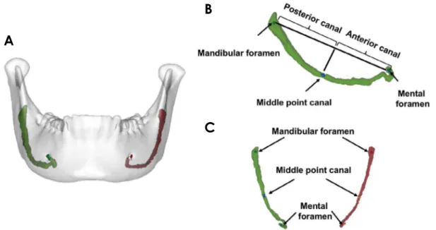
Fig. 1. A. Threedimensional(3D) model of the mandible, including the right and left mandibular canals from a healthy sample subject.
B. Lateral 3D view of the mandibular canal and landmarks; mental foramen: center of the mental foramen, mandibular foramen: center of the mandibular canal foramen, middle point of the canal: point projected perpendicularly from the center of the line between the mental foramen and the mandibular foramen. C. Superior 3D view of the mandibular canal and landmarks of the mental foramen, mandibular fo ramen, and middle point of the canal.
A
B
determined through a visual evaluation of the 3D superim posed models. An example of linear and angular(yaw and pitch) measurements is shown in Figure 2.
Statistical analysis
The statistical analysis was performed with SPSS version 23.0(IBM Corp., Armonk, NY, USA). A post hoc power analysis was undertaken using G*Power software, ver sion 3.2.9.2. The random error was determined using the Dahlberg formula, while systematic error was detected by intraclass correlation coefficients with a confidence level
of 95% to verify reproducibility after repeating placements of all the prelabeled landmarks and measurements of all patients at a 15day interval. The distributions of all vari ables were tested using the KolmogorovSmirnov test, and all variables showed a normal distribution. The ttest for independent samples was used to compare groups in terms of differences between both sides, while the Fisher exact test was used to compare groups in terms of the presence of asymmetric inclinations of the anterior and posterior canal. The level of significance was set at 0.05 for all tests.
Table 1. Mandibular canal measurements
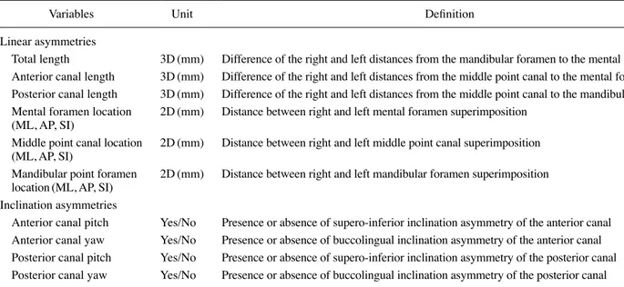
Variables Unit Definition
Linear asymmetries
Total length 3D(mm) Difference of the right and left distances from the mandibular foramen to the mental foramen Anterior canal length 3D(mm) Difference of the right and left distances from the middle point canal to the mental foramen Posterior canal length 3D(mm) Difference of the right and left distances from the middle point canal to the mandibular foramen Mental foramen location
(ML, AP, SI) 2D(mm) Distance between right and left mental foramen superimposition Middle point canal location
(ML, AP, SI) 2D(mm) Distance between right and left middle point canal superimposition Mandibular point foramen
location(ML, AP, SI) 2D(mm) Distance between right and left mandibular foramen superimposition Inclination asymmetries
Anterior canal pitch Yes/No Presence or absence of superoinferior inclination asymmetry of the anterior canal Anterior canal yaw Yes/No Presence or absence of buccolingual inclination asymmetry of the anterior canal Posterior canal pitch Yes/No Presence or absence of superoinferior inclination asymmetry of the posterior canal Posterior canal yaw Yes/No Presence or absence of buccolingual inclination asymmetry of the posterior canal
ML: mediolateral, AP: anteroposterior, SI: superoinferior, 3D: 3dimensional, 2D: 2dimensional
Fig. 2. A. Threedimensional(3D) models of the mandible(white), ameloblastoma lesion(blue), and right(green) and left(red) mandibular
canals from an ameloblastoma patient. B. Lateral view of the mandibular canal of the lesion side(red) mirrored on mandibular canal of the healthy side(green) showing the respective landmarks, correspondence of the posterior canal direction, and inferior displacement of the lesion side in the anterior region. C. Superior view of the mandibular canal of the lesionaffected side(red) mirrored onto the mandibular canal of the healthy side(green), showing the respective landmarks, equivalence of the posterior canal direction, and buccal displacement of the lesionaffected side in the anterior region.
Results
Reliability and sample characteristics
All 3D measurements showed a high level of intra examiner agreement. The lowest ICC value was that of the AP middle point(0.881), while the highest was that of the
SI mandibular foramen position(0.894). The operator error measurements varied between 0.06 and 0.51mm.
The sample power analysis of our study(20 : 10) resulted in a power of 92.96%. Table 2 presents the sample distribu tion(sex, age, side of the lesion, lesion location, and cortical expansion). The ameloblastoma and control groups showed a similar age distribution. Ameloblastoma lesions were seen more frequently in men, on the left side, spread over both the anterior and posterior regions, and with cortical expansion.
Intergroup comparison
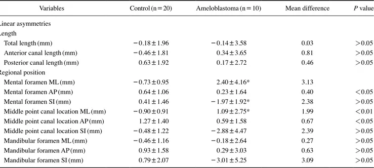
The intergroup comparison of the differences between both sides of the mandibular canal is shown in Table 3. All length measurements of the mandibular canal presented similar differences between both sides in the control and ameloblastoma groups. The quantitative measurements showed that the ameloblastoma group had a more buccal (2.40±4.16mm) and inferior position(-1.97±1.92mm) of the mental foramen, and a more buccal(1.09±2.75mm) position of the middle canal point. No similar differences between both sides in the ameloblastoma and control groups were shown for the AP position of the mental foramen, the AP and SI positions of the middle canal point, and all positions of the mandibular foramen.
In the qualitative analysis of buccolingual(yaw) and SI (pitch) displacement of the mandibular canal, the amelo
Table 2. Sample characteristics of control and ameloblastoma groups
(number and percentage)
Control(n=20) Ameloblastoma(n=10) Sex
Female 10(50.0) 3(30.0)
Male 10(50.0) 7(70.0)
Mean age(years) 27.0 26.3
Lesion side Right NA 2(20.0) Left NA 7(70.0) Both NA 1(10.0) Lesion location* Anterior NA 2(20.0) Posterior NA 1(10.0) Both NA 7(70.0) Cortical expansion Yes NA 6(60.0) No NA 4(40.0)
*: related to mandibular canal, NA: not applicable
Table 3. Linear asymmetry between both sides of the mandibular canal in the control and ameloblastoma groups
Variables Control(n=20) Ameloblastoma(n=10) Mean difference P value
Linear asymmetries Length
Total length(mm) -0.18±1.96 -0.14±3.58 0.03 >0.05
Anterior canal length(mm) -0.46±1.81 0.34±3.65 0.81 >0.05
Posterior canal length(mm) 0.63±1.92 0.17±2.72 0.46 >0.05
Regional position
Mental foramen ML(mm) -0.73±0.95 2.40±4.16* 3.13
Mental foramen AP(mm) 0.64±1.06 0.23±1.64 0.40 <0.05
Mental foramen SI(mm) 0.41±1.46 -1.97±1.92* 2.38 >0.05
Middle point canal location ML(mm) -0.90±0.91 1.09±2.75* 1.99 <0.01
Middle point canal location AP(mm) 1.27±1.40 0.59±1.58 0.67 <0.05
Middle point canal location SI(mm) -0.48±1.22 -2.88±4.47 2.39 >0.05
Mandibular foramen ML(mm) -0.46±1.16 -0.18±2.64 0.27 >0.05
Mandibular foramen AP(mm) 0.93±1.58 0.29±3.03 0.63 >0.05
Mandibular foramen SI(mm) 0.79±2.07 -3.01±5.25 3.09 >0.05
Length measurements have positive values for greater values on the left side or lesion side, and negative values for greater values on the right or healthy sides. Positive values of regional position represent buccal, superior, or anterior displacement of the left or lesion landmarks. Negative values of regional position represent lingual, inferior, or posterior displacement of the left or lesion landmarks. *: P<0.05 compared with the control group. ML: mediolateral, AP: anteroposterior, SI: superoinferior
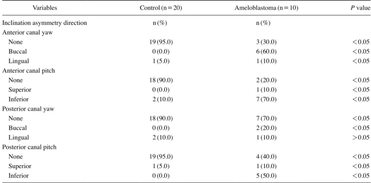
blastoma group presented statistically significant differ ences in anterior canal yaw, anterior canal pitch, posterior canal yaw, and posterior canal pitch when compared to the control group(P<0.05). The lingual inclination of the
posterior canal did not show a statistically significant dif ference(P>0.05). Displacement of the mandibular canal
in ameloblastoma patients tended to occur in the anterior region, toward the buccal and inferior directions in 60% and 70% of patients, respectively, and a high prevalence of the corresponding posterior canal yaw was also found(70%). The control group presented a marked predominance of the corresponding positions of anterior and posterior canal inclinations in the right and left sides of the mandible. Fig ures 3 and 4 show mandibular canal superimposition in the ameloblastoma and control groups, respectively.
Discussion
This study presented a 3D evaluation of mandibular canal displacement using 3D mirrored models in a sample diag nosed with ameloblastoma compared with a symmetric con trol group. This methodology was capable of 3dimension
ally detecting the behavior of the mandibular canal position in the presence of a benign but aggressive lesion, and also of identifying the correspondence of the mandibular canal pathway in the symmetric group of healthy patients. The results showed that the most significant changes in patients with ameloblastoma were related to asymmetric inclina tions in the anterior regions of the mandibular canal and that few changes were expressed in the posterior region. The mandibular canal length showed stable measurements, both in the anterior and posterior regions.
Ameloblastoma is usually found in patients in their third decade of life,13 has no sex predilection,14,15 and is mainly located in the posterior part of the mandible, such as the ramus, mandibular angle, and mandibular body.16 Cortical bone expansion and mandibular canal displacement are common signs present in ameloblastoma lesions. These signs are related to the length of time involved in the devel opment of the lesion, as well as to its size,17 and produce facial deformities and erosion of cortical bone. These les ions also produce paresthesia when close to the mandibular canal. In the present study, the ameloblastoma lesions were also found in young adult patients, and were more prevalent
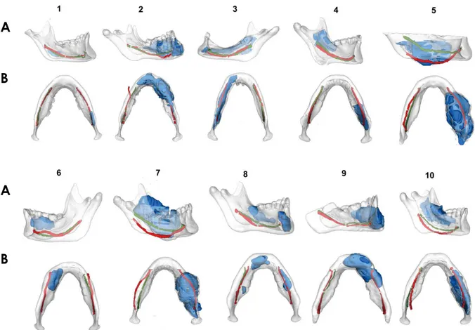
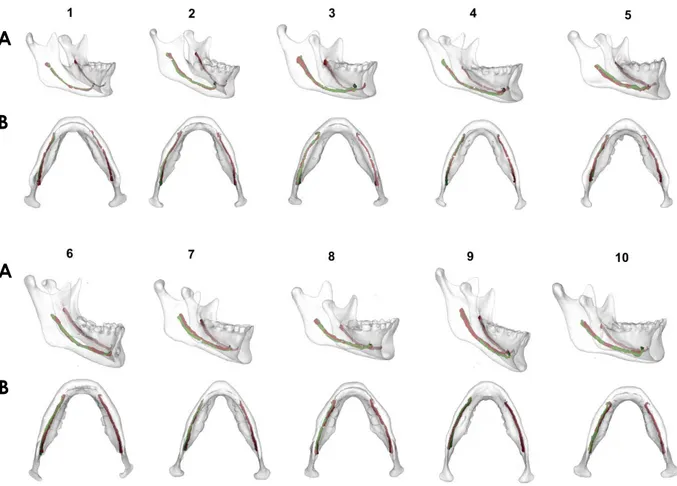
Fig. 3. Lateral(A) and inferior(B) views of all cases of ameloblastoma superimposed onto 3dimensional models of the mandible(white),
ameloblastoma lesion(blue), and healthy(green) and lesionaffected(red) sides of the mandibular canal.
A
B
A
in men, as has generally been reported in South American populations.18
Earlier studies on the relationship between ameloblastoma and mandibular canal displacement used tomographic images of 3 different regions of the mandible and showed the mandibular displacement in a buccolingual direction.5,6 These studies also used the external cortical bone(buccal or lingual) in the lesion side as the anatomical reference to carry out qualitative measurements. However, as these stud ies used no control groups, their results showing buccolin gual directions of mandibular canal displacement are con troversial. Such methods are not capable of detecting the extent of the displacement through quantitative data. Our study presents a methodology based on 3D model super impositions of the healthy and lesionaffected sides of the mandibular canal. Moreover, the control group of symmetric mandibles added information about mandibular canal cor respondence between both sides to validate the method, as seen in Figure 4.
Our results showed significant mandibular canal displace ment in ameloblastoma patients when compared to healthy
patients, especially in the anterior region. However, the qualitative analysis did not agree with the results of earlier studies,5,6 which found a higher prevalence of buccal dis placement in the posterior region. One possible explanation for the conflicting results is the use of different reference parameters. While our study was based on the pathway of the healthy mirrored mandibular canal, the other studies used the lesion itself or the cortical bone on the lesion side to identify the displacement of the mandibular canal. Such an approach can bias the results because the relationship between the mandibular canal position and the ameloblas toma lesion or cortical bone only indicates the location, and not the direction, of displacement. Furthermore, our imaging study traced the trajectory of displacement, which is different for every tumor. This statement is supported by cases that showed inexpressive buccolingual displacement in the posterior region(30%), and no statistical significance when lingual displacement was present(P>0.05), as seen
in Figure 3 and Table 4. This individual variability reinfor ces the fact that each case must be evaluated on an indivi dual basis, mainly using the parameters of the patients them
Fig. 4. A. Lateral(A) and inferior(B) views of 10 cases of the control group with superimposed 3dimensional models of the mandible
(white) and the right(green) and left(red) sides of the mandibular canal.
A
B
A
selves, such as the healthy side as compared to the lesion side.
Other new information from our study includes quan titative data on mandibular canal length and its regions of asymmetries. As seen in Table 3, the mandibular canal length of the anterior and posterior regions of the ameloblas toma group was similar on the healthy and lesionaffected sides. In addition, the comparison between the ameloblas toma and control groups showed similar lengths for both sides. The similarity in canal lengths on both sides suggests that ameloblastoma is incapable of significantly changing the canal length. The mandibular canal was most often dislocated by the ameloblastoma in the lateromedial and superior directions. The variability in quantitative data in all regional positions also reinforces that there is no specific pattern of mandibular canal displacement, especially in the posterior region.
The clinical application of the results of this study is di rectly related to surgical treatment planning, and to the early diagnosis of mandibular canal displacement in ameloblas toma, a phenomenon that occurs before cortical expansion. In our study sample, 60% of patients showed evident cor tical expansion, while 80% had mandibular canal displace ment(Fig. 3). This information suggests that mandibular canal displacement could happen before cortical expansion occurs, and could be an essential detail in the diagnosis. Imaging analysis of the mandibular canal using tomographic
exams is usually based on different sectional views.5,6 3D reconstructed models provide a better comparison of the man di bular canal pathway than tomographic sectional views. The 3D superimposition of the original and mirrored models has been studied for mandibular asymmetries and constitutes a valuable and accurate approach to analyzing craniofacial and mandibular asymmetries.9,10 The results of a thorough clinical evaluation in conjunction with different imaging modalities and histopathology are crucial for the successful management of ameloblastoma, irrespective of its histological subtype.4
Both primary and recurrent ameloblastoma are usually treated surgically. For every type of surgical approach, knowledge of the relationship between the tumor and the IAN is of fundamental importance for avoiding postopera tive complications. The impact of surgery on facial growth and development in young patients should also be consi dered during treatment planning. The use of surgical treat ment for pediatric ameloblastoma patients remains contro versial. In general, conservative surgery prioritizes the main tenance of a good posttreatment quality of life over the possibility of recurrence in pediatric ameloblastoma pati ents.19
Mandibular canal displacement can be present in patients diagnosed with ameloblastoma, although this imaging fea ture is not considered pathognomonic. Canal displacement is probably related to the nature of the lesion, such as the
Table 4. Frequency of inclination asymmetry between both sides of the mandibular canal in the control and ameloblastoma groups
Variables Control(n=20) Ameloblastoma(n=10) P value
Inclination asymmetry direction n(%) n(%)
Anterior canal yaw
None 19(95.0) 3(30.0) <0.05
Buccal 0(0.0) 6(60.0) <0.05
Lingual 1(5.0) 1(10.0) <0.05
Anterior canal pitch
None 18(90.0) 2(20.0) <0.05
Superior 0(0.0) 1(10.0) <0.05
Inferior 2(10.0) 7(70.0) <0.05
Posterior canal yaw
None 18(90.0) 7(70.0) <0.05
Buccal 0(0.0) 2(20.0) <0.05
Lingual 2(10.0) 1(10.0) >0.05
Posterior canal pitch
None 19(95.0) 4(40.0) <0.05
Superior 1(5.0) 1(10.0) <0.05
Inferior 0(0.0) 5(50.0) <0.05
direction of spread, slow growth rate, and its lower aggres siveness when compared to other lesions. Knowing the prevalence of the direction of mandibular canal displace ment is invaluable information, especially when conserva tive treatment of ameloblastoma is indicated.
The limitations of this study include the use of retrospec tive data and a relatively small sample size with different ameloblastoma volumes and directions of spread. Further studies, including groups with lesions with different vol umes and biological behaviors, could be useful for identi fying the association of mandibular canal displacement and clinical symptoms, such as paresthesia.
This study concludes that the displacement of the mandi bular canal due to an ameloblastoma lesion can be detected by this superimposed mirrored method. Displacement is more prevalent toward the inferior and buccal directions and affects the mental foramen position, but does not lead to a change in the length of the mandibular canal. The results also suggest that the direction of mandibular canal displace ment should be taken into account as a tomographic sign to support decisionmaking concerning diagnostic and thera peutic management.
Conflicts of Interest: None
References
1. ElNaggar AK, Chan JK, Grandis JR, Takata T, Slootweg PJ. WHO classification of head and neck tumours. Lyon: Interna tional Agency for Research on Cancer; 2017. p. 2157. 2. Becelli R, Carboni A, Cerulli G, Perugini M, Iannetti G. Man
dibular ameloblastoma: analysis of surgical treatment carried out in 60 patients between 1977 and 1998. J Craniofac Surg 2002; 13: 395400.
3. Nakamura N, Mitsuyasu T, Higuchi Y, Sandra F, Ohishi M. Growth characteristics of ameloblastoma involving the inferior alveolar nerve: a clinical and histopathologic study. Oral Surg Oral Med Oral Pathol Oral Radiol Endod 2001; 91: 55762. 4. Effiom OA, Ogundana OM, Akinshipo AO, Akintoye SO. Ame
loblastoma: current etiopathological concepts and management. Oral Dis 2018; 24: 30716.
5. Abdi I, Taheri Talesh K, Yazdani J, Keshavarz Meshkin Fam S, Ghavimi MA, Arta AS. The effect of ameloblastoma and keratocystic odontogenic tumor on the displacement pattern of inferior alveolar canal in CBCT examinations. J Dent Res Dent Clin Dent Prospects 2016; 10: 15561.
6. Kolokythas A, AlGhamia H, Miloro M. Does a difference exist in inferior alveolar canal displacement caused by commonly encountered pathologic entities? An observational study. J Oral Maxillofac Surg 2011; 69: 194451.
7. Mortazavi H, Baharvand M, Safi Y, Dalaie K, Behnaz M, Safari F. Common conditions associated with mandibular canal widen ing: a literature review. Imaging Sci Dent 2019; 49: 8795. 8. Iwanaga J, Mikushi S, Tohara H. Oral cavity and pharynx. In:
Watanabe K, Shoja MM, Loukas M, Tubbs RS. Anatomy for plastic surgery of the face, head, and neck. New York: Thieme; 2016. p. 183200.
9. AlHadidi A, Cevidanes LH, Mol A, Ludlow J, Styner M. Com parison of two methods for quantitative assessment of mandi bular asymmetry using cone beam computed tomography image volumes. Dentomaxillofac Radiol 2011; 40: 3517.
10. Cevidanes LH, Alhadidi A, Paniagua B, Styner M, Ludlow J, Mol A, et al. Threedimensional quantification of mandibular asymmetry through conebeam computerized tomography. Oral Surg Oral Med Oral Pathol Oral Radiol Endod 2011; 111: 757 70.
11. Thiesen G, Griebel BF, Freitas MP, Oliver DR, Kim KB. Man dibular asymmetries and associated factors in orthodontic and orthognathic surgery patients. Angle Orthod 2018; 88: 54551. 12. Ruellas AC, Huanca Ghislanzoni LT, Gomes MR, Danesi C,
Lione R, Nguyen T, et al. Comparison and reproducibility of 2 regions of reference for maxillary regional registration with conebeam computed tomography. Am J Orthod Dentofacial Orth op 2016; 149: 53342.
13. Reichart PA, Philipsen HP, Sonner S. Ameloblastoma: biologi cal profile of 3677 cases. Eur J Cancer B Oral Oncol 1995; 31B: 8699.
14. Sehdev MK, Huvos AG, Strong EW, Gerold FP, Willis GW. Proceedings: ameloblastoma of maxilla and mandible. Cancer 1974; 33: 32433.
15. Ghandhi D, Ayoub AF, Pogrel MA, MacDonald G, Brocklebank LM, Moos KF. Ameloblastoma: a surgeon’s Dilemma. J Oral Maxillofac Surg 2006; 64: 10104.
16. Sham E, Leong J, Maher R, Schenberg M, Leung M, Mansour AK. Mandibular ameloblastoma: clinical experience and litera ture review. ANZ J Surg 2009; 79: 73944.
17. Black CC, Addante RR, Mohila CA. Intraosseous ameloblasto ma. Oral Surg Oral Med Oral Pathol Oral Radiol Endod 2010; 110: 58592.
18. Hendra FN, Van Cann EM, Helder MN, Ruslin M, de Visscher JG, Forouzanfar T, et al. Global incidence and profile of ame loblastoma: A systematic review and metaanalysis. Oral Dis 2020; 26: 1221.
19. Huang IY, Lai ST, Chen CH, Chen CM, Wu CW, Shen YH. Surgical management of ameloblastoma in children. Oral Surg Oral Med Oral Pathol Oral Radiol Endod 2007; 104: 47885.