석 사 학 위 논 문
RAW 264.7 cell 에서 당유자
(
C. grandis
Osbeck) 잎 추출물의
염증 억제 효과
제주대학교 대학원
의 학 과
양 은 진
2008년 12월
RAW 264.7 cell 에서 당유자 (C. grandis Osbeck)
잎 추출물의 염증 억제 효과
지도교수 유 은 숙
양 은 진
이 논문을 의학 석사학위 논문으로 제출함
2008년 12월
양은진의 의학 석사학위 논문을 인준함
제주대학교 대학원
2008년 12월
Anti-inflammatory effect of
Citrus grandis Osbeck leaves
in RAW 264.7 cells
Eun-Jin Yang
(Supervised by Professor Eun-Sook Yoo)
A thesis submitted in partial fulfillment of the
requirement for the degree of Master of Science in
Medicine.
2008. 12
This thesis has been examined and approved.
DEPARTMENT OF MEDICINE
GRADUATE SCHOOL
I
ABSTRACT
Citrus grandis Osbeck is a native plant growing only on Jeju Island in Korea, it is consisted of several component such as limonene, obacunone, nomiline, naringin. In this study, we investigated the anti-inflammatory effect of C. grandis Osbeck leaves in the murine macrophage cell line. RAW 264.7 murine macrophage cells were stimulated with LPS (1 ㎍/㎖) to induce pro-inflammatory markers (IL-6 and iNOS). The crude extract (80% MeOH Ex.) and solvent fractions (hexane, CHCl3, EtOAc, BuOH and H2O Ex.) were
obtained from C. grandis leaves. Among them, the CHCl3 fraction inhibited
the nitric oxide (NO) and interleukin-6 (IL-6) production in a dose-dependent manner. Also, the CHCl3 fraction inhibited mRNA expression and
protein level of iNOS in a dose-dependent manner. To investigate their action mechanism, we examined the effects of the CHCl3 fraction on
LPS-induced NF-κB activation and phosphorylation of MAP kinase (ERK, JNK and
p38). As a result, western blot revealed that the CHCl3 fraction inhibited
phosphorylation of MAP kinase family and activation of NF-κB in cytosol. Moreover, luciferase assay revealed that CHCl3 fraction suppressed the
transcriptional activity of NF-κB in nucleus. These results suggest that the C. grandis leaves may inhibit LPS-induced production of inflammatory markers (IL-6 and iNOS) by blocking NF-κB activation and MAPKs phosphorylation in RAW 264.7 cells.
Key word: Citrus grandis Osbeck, inflammation, inflammatory markers (IL-6
II 목 차 A B S T R A C T … … … Ⅰ 목 차 … … … Ⅱ LIST OF SCHEME ……… Ⅳ LIST OF TABLES ……… Ⅴ LIST OF FIGURES ……… Ⅵ Ⅰ. 서 론 ………1 Ⅱ. 재료 및 방법 ………5 1. 당유자 잎 시료의 추출 2. 세포배양 3. 시약 4. 세포 독성 평가
5. Nitric oxide (NO) 측정
6. Pro-inflammatory cytokines (IL-6)의 정량 7. Western blot analysis
8. RNA 분리
9. cDNA 합성 및 PCR
10. Transient transfection과 luciferase assay 11. 통계처리
Ⅲ. 결과 ………13 1. LPS로 자극한 대식세포에서 당유자 잎 추출물 및 분획물의 독성 평가 및
III 1) 당유자 잎 추출물 및 분획물이 RAW 264.7 세포에 미치는 영향 2) 당유자 잎 추출뮬 및 분획물이 NO와 iNOS 생성에 미치는 영향 (1) Nitric Oxide 생성 억제 효과 (2) iNOS 발현 억제 효과 3) 당유자 잎 추출물 및 분획물이 IL-6 생성에 미치는 영향 (1) IL-6 생성 억제 효과 (2) IL-6 발현 억제 효과 2. LPS로 자극한 대식세포에서 당유자 잎 분획물의 항염증 작용기전 1) 당유자 잎 CHCl3 분획물에 의한 IκB-α의 degradation 억제효과 2) 당유자 잎 CHCl3 분획물에 의한 NF-κB 활성억제 효과 3) 당유자 잎 CHCl3 분획물에 의한 MAPKs (ERK, JNK, p38) 활성 억제효과 Ⅳ. 고찰 ………36 Ⅴ. 참고문헌 ………39 Ⅵ. 적요 ………46
IV
LIST OF SCHEME
Scheme 1. Systemic purification using solvent partitioning from Citrus
V
LIST OF TABLE
Table 1. Primer sequences and the expected product size in RT-PCR analysis … … … 1 1
VI
LIST OF FIGURES
Figure 1. Effect of crude extract and solvent fractions from C. grandis
Osbeck leaves on the nitric oxide (NO) production in RAW 264.7 cells ………14
Figure 2. Effect of CHCl3 fraction from C. grandis Osbeck leaves on the
nitric oxide (NO) production in RAW 264.7 cells ………15
Figure 3. Effect of crude extract and solvent fractions from C. grandis
Osbeck leaves on the mRNA expression of iNOS in LPS-stimulated RAW 264.7 cells ………17
Figure 4. Effect of CHCl3 fraction from C. grandis Osbeck leaves on the
protein level of iNOS in LPS-stimulated RAW 264.7 cells …………18
Figure 5. Effect of CHCl3 fraction from C. grandis Osbeck leaves on the
production level of IL-6 in LPS-stimulated RAW 264.7 cells ……20
Figure 6. Effect of crude extract and solvent fractions from C. grandis
Osbeck leaves on the mRNA expression of IL-6 in LPS-stimulated RAW 264.7 cells ………21 Figure 7. Overview of the pathway leading to NF-κB and MAPKs activation ………23
Figure 8. Effect of CHCl3 fraction from C. grandis Osbeck leaves on the
degradation of IκB-α in LPS-stimulated RAW 264.7 cells ………26
Figure 9. Effect of CHCl3 fraction from C. grandis Osbeck leaves on the
phosphorylation of NF-κB in LPS-stimulated RAW 264.7 cells ……27
VII
NF-κB activation in LPS stimulated RAW 264.7 cells ………28
Figure 11. Effect of CHCl3 fraction from C. grandis Osbeck leaves on the
activation of ERK 1/2 in LPS stimulated RAW 264.7 cells …………30
Figure 12. Effect of CHCl3 fraction from C. grandis Osbeck leaves on the
activation of JNK 1/2 in LPS stimulated RAW 264.7 cells …………31
Figure 13. Effect of CHCl3 fraction from C. grandis Osbeck leaves on the
activation of p38 in LPS stimulated RAW 264.7 cells ………32 Figure 14. Structure and HPLC chromatogram of nobiletin ………34
1
I. 서 론
오늘날 사회가 고령화되고 삶의 수준이 향상됨에 따라 자신의 건강에 대한 관 심이 한층 높아지고 있으며, 고령화 사회에 따른 만성염증질환이 증가하면서 약 에 의존하는 계층이 더욱 늘어나게 되었다 (Han, 2003). 현재까지는 비스테로이 드성 항염증 약물 (nonsteroidal anti-inflammatory drugs, NSAIDs)의 사용으로 인해 염증의 완화가 상당하게 이루어졌으나, 위장관 미란과 신장 손상 등의 심 각한 부작용을 가지고 있어 제한적으로 사용되고 있다 (Seibert et al., 1994). 이러한 부작용을 극복하기 위해 최근에는 민간에서 사용되고 있는 천연물에서 그 활성 성분을 찾으려는 연구가 활발히 진행되고 있다. 인류는 옛날부터 산과 들에서 식물의 잎·줄기·뿌리 등을 채취해 맛을 보고 독이 있거나 없음을 알아내어 식용 또는 약용 등으로 이용하였다. 약용식물과 관계하여 중국에서는 쑥에서 말라리아 치료제인 알테미시닌을 개발하고, 간장질 환 치료제인 시잔드린을 오미자로부터 분리, 개발하였다. 이처럼 천연식물 중에 서의 신약개발은 아주 보편적인 것으로, 대표적인 것으로는 양귀비에서 모르핀, 키나피에서 키니네, 버드나무에서 살리실산, 은행잎에서 징크플라보노이드 등이 있다. 천연 약용식물에서 유용한 신약이 개발되는 것은 수천 년에 걸쳐 인간의 경험적 사용을 통하여 효능과 안정성이 입증되었다 (Chae et al., 2007). 염증반응은 생체나 조직이 병원체의 자극으로 인해 손상이 일어나면, 손상부 위를 수복 재생하려는 기전으로, 일단 자극이 가해지면 국소적으로 prostaglandins, hydroxyleicosa-tetraenoic acid (HETE), leukotriene과 같은 혈관 활성 물질이 유리되어 혈관 투과성이 증대되면서 염증을 유발한다. 그러나 지속적인 염증반응은 오히려 점막손상을 촉진하고 (Willoughby, 1975), 결과적 으로 만성 염증질환인 류마티스 관절염, 심장 자극, 알츠하이머 (Balkwill, 2004), 암 등을 초래한다 (Li, 2007).
2
Lipopolysaccharide (LPS)는 그람음성 세균의 세포 외막에 존재하는 내독소 (endotoxin)로, 대식세포로부터 다양한 염증발병 요인으로 알려진 tumor necrosis factor-α (TNF-α), interleukin-6 (IL-6)와 같은 pro-inflammatory
cytokines을 증가시키는 것으로 알려져 있다 (Shin et al., 2004). TNF-α는 류
마티스 관절염, 패혈성 쇼크와 기타 만성 염증 질환시 증가하는 것으로 알려져 있으며, 최근 IL-6는 골다공증에 중요하게 관여하는 것으로 보고되고 있다 (Dai
et al., 2000; Kim et al., 1996).
체내 염증 과정에서는 과량의 nitric oxide (NO)와 prostaglandin E2 (PGE2)가
생성된다 (Willoughby et al., 2000). Nitric oxide (NO)는 짧은 반감기를 가진
free radical로서 신경전달, 혈관 항상성, 숙주방어, 면역조절, 혈압조절, 혈소 판 응집억제 등의 다양한 생물학적 기능의 매개체 역할을 하는 것으로 알려져 있다 (Kim et al., 2006; Rho et al., 1999 ).
Nitric oxide (NO)는 nitric oxide synthase (NOS)에 의해 생성이 조절되는데
(Moeslinger et al., 2006), 포유류에서 NOS는 물리화학적 성상에 따라
inducible NOS (iNOS), neuronal NOS (nNOS), endothelial NOS (eNOS) (Son et
al., 2006) 그리고 최근 밝혀진 mitochondrial NOS (mNOS)등 4가지의 동종 효소
로 분류된다 (Ying et al., 2007). 이중 iNOS에 의한 NO의 생성이 절대적으로
중요한데, 대식세포가 lipopolysacchride (LPS)나 interferon-γ (IFN-γ)에 의 해서 자극을 받으면 iNOS가 발현되어 과량의 NO를 생성하고, 과량 생성된 NO는 조직의 파괴, 자가면역 질환, 패혈성 쇼크, 류마티스 관절염, 궤양성 대장염 등
을 유발한다 (Jun et al., 1998; Lin et al, 2007; Blonska et al., 2002).
최근, Nuclear factor (NF)-κB와 mitogen-activated protein (MAP) kinases
는 iNOS 전사조절에 관련 하는 것으로 보고되어 있다 (Klemer et al., 2003;
Kim et al., 2006). NF-κB는 염증과 면역에 중요한 전사인자로 여러 가지 종류 의 단백질이 밝혀졌는데 가장 대표적인 p65와 p50의 단백질은 동질 이형체를 이
3
루어 존재한다 (Nomura et al., 2001). NF-κB는 활성화 되지 않은 세포의 세포
질 내에서 억제 단백질인 IκB와 결합하여 존재하나 세포가 자극을 받아 IκB가
활성화 되면 분리되어 핵 내로 들어가 전사 인자로 작용하게 된다 (Rossi et
al., 2000). LPS로 자극된 murine macrophage 세포에 NF-κB 억제제인 PDTC
(pyrrolidine dithicarbamate)를 처리하면 iNOS의 발현이 차단되는데, 이것은
NF-κB 활성의 억제를 통한 것이라고 알려져 있다 (Surh et al., 2001). MAPK는
extracellular signal-regulated kinase (ERK), p38 그리고 stress-activated
protein kinase/c-Jun NH3-terminal kinase (SAPK/JNK)로 구분되는데 (Kim et
al., 2006), ERK는 주로 세포의 증식, 분화 및 생존 등의 반응을 촉발시키는 성
장인자에 의해 활성화 된다고 알려져 있고, ERK의 지속적인 활성은 종양형성에 관련된 세포내 신호 전달계 에서 중요한 역할을 하는 것으로 보고되고 있다
(Kimura et al., 1999). p38과 JNK는 여러 가지 스트레스에 의해 활성화되어 세
포 자연사에 관여하며 다양한 병리적 과정에 관여하는 것으로 알려져 이들을
stress-activated protein kinase (SAPKs)라고 한다 (Xia et al., 1995).
우리나라는 기상학적, 지리학적으로 감귤재배지 중 최북단에 위치하고 있어 내
한성이 강한 만다린계의 온주밀감이 감귤의 주종을 이루고 있다 (Rhyu et al.,
2002). 감귤류는 운향과 (Rutacae) 감귤아과 (Auranti-oideae)에 속하는 식물로, 감귤로 취급하는 것에는 감귤속 (Citrus), 금귤속 (Fortunella), 탱자나무속 (Ponicirus) 과 클리메니아 (Clymennia)의 4속이 있다 (고정삼, 2001). 감귤류 에는 neohesperidin, naringin, flavonoids, nobiletin등의 많은 유효 성분이
함유되어 있는데 flavonoids는 항염·항암 효과 (Manthey et al., 2001),
hesperidin은 지질 과산화물 형성 억제를 통한 항산화, 항염증 효과가 보고 되
었고 (Choi et al., 2006), 최근 Nobiletin도 항염증 효과가 있음이 보고되어
감귤류의 중요한 물질로 대두되고 있다. (Li et al., 2007).
4 는 제주도 에서만 자라는 종으로 제주에서는 왕귤 또는 뎅유지로 불리우고 (Lim et al., 2002), 동의보감에는 등자피 (橙子皮)로 기재되어 있다. 동의보감에 따 르면“ 당유자는 성질이 따스하고 맛이 쓰고 매우며 독이 없으니 먹은 것을 소 화시키고 장과 위 속의 악기와 부풍을 흩어 버린다 ” 고 전하고 있다 (허준, 1995). 최근에는 당유자의 효능이 검증되고 있는데, 당유자 미숙과는 항염증 (Lee et al., 2006), 항산화 효과를 지니는 것이 보고 되었으며 (Lim et al., 2006), 제주 재래종 감귤류 미숙과 중 당유자 미숙과에서 neohesperidin과 naringin의 함량이 높은 것으로 보고되었고 성숙해 질수록 이들의 함량이 점점 감소된다고 보고되었다 (Rhyu et al., 2002). 현재 당유자 과실의 효능에 관한 보고는 계속되고 있으나 잎은 어떠한 활성과 기전을 가지고 있는지 보고된 바가 없다. 이에 본 연구에서는 제주도에서 재배되는 당유자 잎 추출물이 LPS로 활성화된 murine macrophage RAW 264.7 세포에서 염증성 인자로 알려진 iNOS, IL-6의 억
제 효과를 조사하였으며, 이들의 작용 메커니즘을 알아보기 위하여 당유자 잎 추출물이 NF-κB 활성화 및 MAPKs (ERK, JNK, p38) 활성화에 미치는 영향에 대 하여 조사하였다. 또한, 당유자 잎 추출물로부터 활성물질을 찾기 위하여 감귤
5 II. 재료 및 방법 1. 당유자 잎 시료의 추출 제주도에서 재배되는 당유자 잎을 2005년 8월에 채집하여 동결건조 후, 분쇄 기로 갈아 미세말로 하였다. 미세말 시료 (50.0 g)를 80% methanol (MeOH)로 2 회 교반 추출 후 여과하고 감압 농축하였다. 여기에서 얻은 MeOH 추출물 (10.81 g)을 계통적 추출방법에 의하여 hexane 분획 (0.71 g), chloroform (CHCl3)분획
(0.25 g), ethylacetate (EtOAc) 분획 (1.27 g), buthanol (n-BuOH) 분획 (2.17
6
Scheme 1. Systemic purification using solvent partitioning from
Citrus grandis Osbeck leaves.
7
2. 세포배양
Murine macrophage cell line인 RAW 264.7 세포를 KCLB (Korean Cell Line Bank)로부터 분양 받아 100 units/ml penicillin-streptomycin과 10% fetal
bovine serum (FBS)이 함유된 DMEM배지를 사용하여 37 ℃, 5% CO2 항온기
(incubator)에서 배양하였으며, 3일에 한번씩 계대 배양을 실시하였다.
3. 시약
세포 계대 배양에 사용된 DMEM (Dulbecco’s Modified Eagle’s Medium) 배지 와 fetal bovine serum (FBS)은 Gibco (Grand Island, NY, USA)에서 구입하였고, lipopolysaccharide (LPS, E. coli serotype 0111:B4)는 sigma (St. Louis, MO) 로부터 구입하였다. IL-6 정량을 위한 enzyme-linked immunosorbent assay (ELISA) kit는 R&D System (Minneapolis, MN, USA)로부터 구입하였다. Western blot을 위한 Antibody (IκB-α, phospho-NF-κB (p65), p38, ERK 1/2, phospho-ERK 1/2, JNK 1/2 와 phospho-JNK 1/2)는 모두 Cell Signaling Technology (Beverly, MA, USA) 로부터 구입하였고, phospho-p38 은 BD Biosciences (San Diego, USA)로부터 구입하였다. 모든 시약은 sigma등급의 시 약을 사용하였다.
4. 세포 독성 평가
당유자 시료의 독성은 MTT assay를 이용하여 실험하였다. 생존하면서 대사가 왕성한 세포는, 세포내 mitochondria의 탈 수소 효소작용에 의하여 수용성의 노 란색 MTT [3-(4,5-dimethylthiaxol-2-yl)-2,5-diphenyl tetrazolium bromide]를
환원시켜 자주색을 띠는 비 수용성의 formazan을 형성한다 (Gerlier et al.,
1986; Liu, 1999).
8
24시간 배양한 후, MTT (Sigma, MO, USA) 용액 200 ㎕ (2 ㎖/㎎)를 첨가하여 4 시간 동안 반응 시킨다. 상층액을 완전히 제거하고 dimethylsulfoxide (DMSO : Sigma, MO, USA)를 500 ㎕를 가하여 침전물을 완전히 용해시킨 후, microplate reader (Amersham Pharmacia Biotech, NY, USA)를 사용하여 540 nm에서 흡광도 를 측정하였다. 각 시료군에 대한 평균 흡광도 값을 구하였으며, 대조군의 흡광 도 값과 비교하여 독성 정도를 평가하였다.
5. Nitric oxide (NO) 측정
RAW 264.7 세포 (1.5×10⁵ cells/㎖)를 18시간 전 배양 후 시료와 LPS (1 ㎍/
㎖)를 동시 처리하여 24시간 배양하였고, 생성된 NO는 Griess 시약을 이용하여
세포 배양액 중에 존재하는 NO2- 의 형태로 측정하였다. 세포배양 상층액 100 ㎕
와 Griess시약 [1% (w/v) sulfanilamide, 0.1% (w/v) naphtylethylenediamine in 2.5% (v/v) phosphoric acid]을 동량 혼합하여 10분간 실온암소에서 반응시 킨 후 ELISA reader를 이용하여 540 nm에서 흡광도를 측정하였다. 표준농도 곡
선은 sodium nitrite (NaNO2)를 serial dilution하여 얻었다 (10-100 µM).
6. Pro-inflammatory cytokines (IL-6)의 정량
Murine macrophage cell line인 RAW 264.7 세포 (4.0×10⁵ cells/㎖)를 18시간
전 배양하고 시료와 LPS (1 ㎍/㎖)를 동시처리 하여 24시간 배양하였다. 24시간 후 배양 배지를 원심분리 (12,000 rpm, 3 min) 하여 얻어진 상층액의 IL-6 함량 을 측정하였다. 정량은 murine enzyme-linked immunosorbent assay (ELISA) kit (R&D system, Inc, USA)를 이용하여 정량하였으며 standard에 대한 표준곡선의
r2 값은 0.99 이상이었다 (Cho et al
., 2000).
9
RAW 264.7세포 (5.0×10⁵ cells/㎖)를 18시간 전 배양한 후, 시료와 LPS (1
㎍/㎖)를 동시 처리하여 24시간 배양하였다. 세포를 PBS (phosphate buffered saline)로 2회 세척한 후 200 ㎕의 lysis buffer [50 mM Tris-HCl (pH7.5), 150
mM NaCl, 1% Nonidet P-40, 2 mM EDTA, 1 mM EGTA, 1 mM NaVO3, 10 mM NaF, 1
mM dithiothreitol, 1 mM phenylmethylsulofonyl fluoride, 25 ㎍/㎖ aprotinin, 25 ㎍/㎖ leupeptin]를 첨가하여 4 ℃에서 30분~1시간 동안 lysis시킨 후 15,000 rpm에서 15분간 원심하여 세포막 성분 등을 제거하였다. 단백질 농도는 BSA (bovine serum albumin)를 표준화하여 Bio-Rad Protein Assay Kit를 사용하 여 정량하였다. 20~30 ㎍의 lysate를 10~12% mini gel SDS-PAGE (Poly Acrylamide Gel Electrophoresis)로 변성 분리하여, 이를 PVDF membrane (BIO-RAD, HC, USA)에 200 V로 2시간 동안 transfer하였다. 그리고 membrane의 blocking 처리는 5% skim milk가 함유된 TTBS (TBS+ 0.1% Tween 20) 용액에서 overnight 하였다. iNOS의 발현 양을 확인하기 위한 항체로는 anti-mouse iNOS (1:1000) (Santa-Cruz)을 TTBS용액에 희석하여 상온에서 2시간 반응시킨 후 TTBS로 4회 세정하였다. 2차 항체로는 HRP (Horse Radish Peroxidase)가 결합된 anti-mouse IgG (Vector Laboratiories, Burlingame, USA)를 1:5000으로 희석하 여 상온에서 30분 간 반응시킨 후, TTBS로 4회 세정한다. IκB-α, phospho-NF-κB (p65), p38, ERK 1/2, JNK 1/2의 발현 양을 확인하기 위한 항체로는 anti-rabbit (1:1000)을 TTBS용액에 희석하여 4 ℃에서 overnight한다. 2차 항체로는 HRP가 결합된 anti-rabbit IgG를 1:5000으로 희석하여 상온에서 30분 간 반응시 킨 후, TTBS로 4회 세정한다. 반응이 완료된 membrane을 ECL 기질 (Intron Biotechnology, Inc, Korea)과 1분 간 반응 후 X-ray 필름에 감광하였다.
8. RNA분리
10
를 동시 처리하여 24시간 배양하였다. Total RNA 추출은 TRI-reagent (MRC, Cincinnati, OH, USA)를 이용하였다. 세포에 TRI-reagent를 첨가하여 균질화 한 후, 클로로포름을 첨가하여 원심 분리하였다. 상층액을 분리한 후 동량의 이소 프로판올을 첨가하여 원심 분리하여 RNA를 침전시키고 75%의 DEPC 처리된 에탄 올로 세척한 후, 건조시켜 DEPC 처리된 증류수에 녹였다. 260 nm에서 흡광도를 측정하여 RNA를 정량 하였고, A260/A280 nm의 비율이 1.6~1.8 범위 내의 값을 갖는 RNA를 실험에 사용하였다. 모든 실험은 RNase-free 한 조건에서 실시하였 다. 9. cDNA 합성 및 RT-PCR
4 ㎍의 total RNA를 oligo (dT)15 primer와 mix후 70 ℃ 에서 5min 반응시킨 후
dNTP (0.5 µM), 1 unit RNase inhibitor, M-MuLV reverse transcriptase (2U), 5× reaction buffer를 넣고 25 ℃ 5min, 37 ℃ 60min, 70 ℃ 15min 반응 시킨
후 중지하였다. cDNA 합성은 Improm-IITM cDNA kit (Promega, WI, USA)를 이용하
였다.
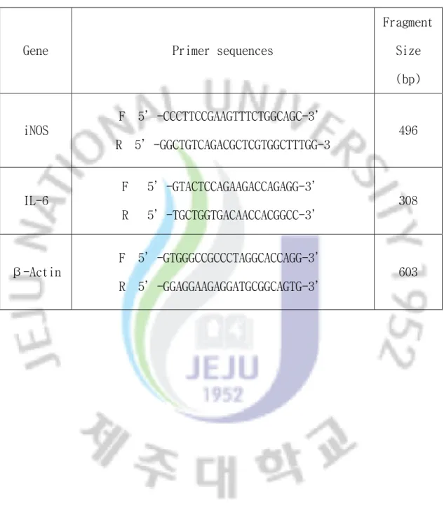
Polymerase chain reaction (PCR)은 합성된 cDNA로부터 IL-6, iNOS, β-actin 을 증폭시키기 위하여 3 ㎕ cDNA, 4 µM의 5’ 과 3’ primer, 10× PCR buffer [100 mM Tris-HCl (pH 8.3), 500 mM KCl, 20 mM MgCl2 , Enhancer solution], 10
mM dNTPs (2.5 mM each), 25 mM MgCl2, i-TaqTM DNA polymerase (5 U/㎕) (Intron
Biotechnology, Inc, Korea)를 넣고 distilled water로 최종 볼륨을 25 ㎕로 맞 추고 Perkin-Elmer Thermal Cycler (Bio-rad, HC, USA)를 이용하여 PCR을 실시 하였다. 이때 PCR 조건은 94 ℃/45초, 55~60 ℃/45초, 72 ℃/60초, 30회이며, PCR에 의해 생성된 산물은 1.0% agarose gel을 이용하여 전기영동을 실시하고, ethidium bromide (EtBr)로 염색하여 특정 band를 확인하였다 (Table 1).
11
Table 1. Primer sequences and the expected product size in RT-PCR analysis.
Gene
Primer sequences
Fragment
Size
(bp)
iNOS
F 5’-CCCTTCCGAAGTTTCTGGCAGC-3’
R 5’-GGCTGTCAGACGCTCGTGGCTTTGG-3
496
IL-6
F 5’-GTACTCCAGAAGACCAGAGG-3’
R 5’-TGCTGGTGACAACCACGGCC-3’
308
β-Actin
F 5’-GTGGGCCGCCCTAGGCACCAGG-3’
R 5’-GGAGGAAGAGGATGCGGCAGTG-3’
603
12
10. Transient transfection과 luciferase assay
세포에 유전자 도입을 위해 transfection을 수행하였다. 우선 RAW 264.7 세포
를 96 well plate에 1.5×10⁵ cells/㎖로 200 ㎕씩 접종시켜 18시간 배양 후, 혈
청이 없는 배지에 50 ng의 NF-κB promoter-based luciferase reporter plasmid (Panomics, Redwood City, CA, USA)와 10 ng의 Renilla luciferase reporter plasmid (Promega, Madison, WI, USA)를 TransFastTM transfection reagent
(Promega Madison, WI, USA)를 이용하여 일시적으로 transfection하였다. 1시간 방치 후 혈청함유 배지로 채워주고 24~30 시간 가량 반응시킨다. 그 후, LPS (1
㎍/㎖)와 당유자 잎의 CHCl3 fraction을 농도별로 처리한 후 염증 반응을 유발
하였다. 염증 반응 유발 24시간 후 각 lysate를 얻었고 Dual-Luciferase Reporter Assay Kit (Promega Corp., Madison, WI, USA)를 이용하여 측정하였다. Luciferase activity는 luminometer를 이용하여 LPS 비처리 군과 비교하여 나타 내었다.
11. 통계처리
실험결과는 3번 이상의 독립적인 실험의 데이터를 mean ± standard error (SE) 값으로 나타내었다. 실험군 사이의 통계적 유의성 검정은 student ’ s t-test 분석방법을 사용하였다.
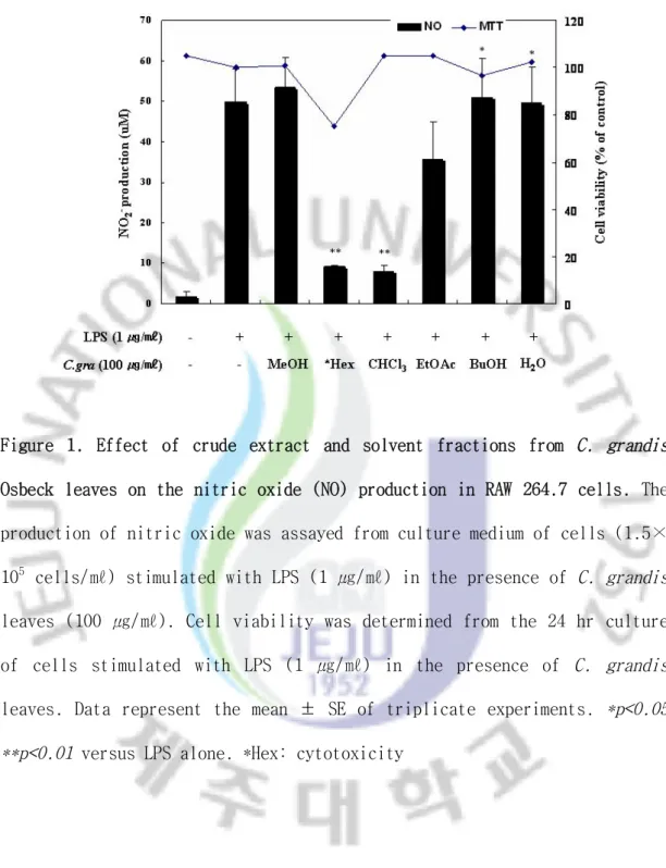
13 III. 결 과 1. LPS로 자극한 대식세포에서 당유자 잎 추출물 및 분획물의 독성 평가 및 항 염증 효과 1) 당유자 잎 추출물 및 분획물이 RAW 264.7 세포에 미치는 영향 RAW 264.7 세포 (1.5×10⁵ cells/㎖)에 당유자 잎 각 분획별 시료 (100 ㎍/㎖) 와 LPS (1 ㎍/㎖)를 동시 처리하여 24시간 배양한 후, MTT assay 방법을 이용하 여 세포 생존율을 확인하였다. 당유자 잎 분획물 중 hexane 분획에서 대조군에 비하여 세포 성장이 약 25% 감소하여 강한 세포 독성이 나타났으나 나머지 분획 에서는 독성이 나타나지 않았다 (Figure 1). 2) 당유자 잎 추출물 및 분획물의 NO와 iNOS 생성에 미치는 영향 (1) Nitric Oxide 생성 억제 효과 최근 체내 염증 과정에서 중요한 역할을 하는 것으로 알려진 nitric oxide (NO) 생성에 대하여 당유자 잎 추출물 및 분획물이 미치는 영향을 조사하였다. RAW 264.7 세포 (1.5×10⁵ cells/㎖)에 LPS (1 ㎍/㎖)와 당유자 분획물 (100 ㎍ /㎖)을 동시 처리 하여 24시간 배양하였다. 생성된 NO의 양은 Griess 시약을 이 용하여 세포 배양액에 존재하는 NO2- 의 형태로 측정하였다. 그 결과 LPS 단독
처리군에서 49.9 uM로 NO가 과량 생성되었으며, hexane 분획물과 CHCl3 분획물에
서 각각 9 uM과 7.9 uM로 LPS에 의해 유도된 NO 생성량이 감소되었음을 확인할 수 있었다. 그러나 hexane 분획물의 NO 생성의 억제는 세포독성에 의한 결과로
사료된다. 또한, NO생성에 강한 억제 효과를 나타낸 CHCl3 분획물을 농도별
(12.5, 25, 50, 100 ㎍/㎖)로 처리하여 확인한 결과, 농도의존적으로 억제효과 를 보였으며 세포독성도 관찰되지 않았다 (Figure 2).
14
Figure 1. Effect of crude extract and solvent fractions from C. grandis
Osbeck leaves on the nitric oxide (NO) production in RAW 264.7 cells. The production of nitric oxide was assayed from culture medium of cells (1.5×
105 cells/㎖) stimulated with LPS (1 ㎍/㎖) in the presence of C. grandis
leaves (100 ㎍/㎖). Cell viability was determined from the 24 hr culture of cells stimulated with LPS (1 ㎍/㎖) in the presence of C. grandis
leaves. Data represent the mean ± SE of triplicate experiments. *p<0.05 **p<0.01 versus LPS alone. *Hex: cytotoxicity
15
Figure 2. Effect of CHCl3 fraction from C. grandis Osbeck leaves on the
nitric oxide (NO) production in RAW 264.7 cells. The production of nitric oxide was assayed culture medium of cells (1.5×10⁵ cells/㎖) stimulated
with LPS (1 ㎍/㎖) in the presence of C. grandis leaves CHCl3 fraction
(12.5, 25, 50, 100 ㎍/㎖). Cell viability was determined from the 24 hr culture of cells stimulated with LPS (1 ㎍/㎖) in the presence of C. grandis leaves. Data represent the mean ± SE of triplicate experiments.
16
(2) iNOS mRNA 발현 및 protein level 억제효과
RAW 264.7 세포 (1.5×10⁵ cells/㎖)에 당유자 시료와 LPS (1 ㎍/㎖)를 동시
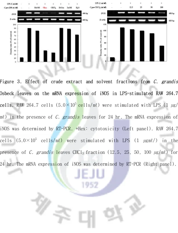
처리하고 24시간 배양하여, 당유자 추출물 및 분획물에 의한 iNOS의 발현 억제 정도를 RT-PCR과 western blot을 통해 확인해 보았다. LPS에 의해 iNOS의 발현
은 현저히 증가하였고, 당유자 분획물을 처리한 결과 CHCl3 분획물이 iNOS의
mRNA 발현을 강하게 억제하였다 (Figure 3). 또한, CHCl3 분획물을 농도별 (12.5,
25, 50, 100 ㎍/㎖)로 처리하여 확인한 결과 모두 농도 의존적으로 억제되었다 (Figure 3, 4).
17
Figure 3. Effect of crude extract and solvent fractions from C. grandis
Osbeck leaves on the mRNA expression of iNOS in LPS-stimulated RAW 264.7
cells. RAW 264.7 cells (5.0×10⁵ cells/㎖) were stimulated with LPS (1 ㎍/
㎖) in the presence of C. grandis leaves for 24 hr. The mRNA expression of
iNOS was determined by RT-PCR. *Hex: cytotoxicity (Left panel). RAW 264.7
cells (5.0×10⁵ cells/㎖) were stimulated with LPS (1 ㎍㎖/) in the
presence of C. grandis leaves CHCl3 fraction (12.5, 25, 50, 100 ㎍/㎖) for
18
Figure 4. Effect of CHCl3 fraction from C. grandis Osbeck leaves on the
protein level of iNOS in LPS-stimulated RAW 264.7 cells. RAW 264.7 cells
(5.0×10⁵ cells/㎖) were stimulated with LPS (1 ㎍/㎖) in the presence of
C. grandis leaves CHCl3 fraction (12.5, 25, 50, 100 ㎍/㎖) for 24 hr.
Whole-cell lysates (25 ㎍) were prepared and the protein level was subjected to 10% SDS-PAGE, and expression of iNOS and β-actin were determined by western blotting. The β-actin was a loading control.
19
3) 당유자 잎 추출물 및 분획물의 IL-6 생성에 미치는 영향 (1) IL-6 생성 억제 효과
LPS로 자극한 RAW 264.7 세포에서 생성되는 IL-6에 대한 당유자 잎 CHCl3 분획
의 억제 활성을 ELISA (enzyme-linked immunosorbent assay) 방법으로 조사하였
다. IL-6의 생성과 억제 정도를 확인하기 위해 RAW 264.7 세포 (1.5×10⁵
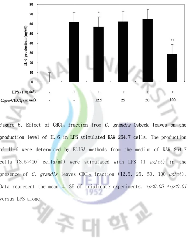
cells/㎖)에 LPS (1 ㎍/㎖)와 당유자 잎 CHCl3 분획물을 농도별 (12.5, 25, 50,
100 ㎍/㎖)로 처리하여 확인한 결과, IL-6의 생성을 100 ㎍/㎖의 농도에서 약 53.4% 억제하였다 (Figure 5).
(2) IL-6 mRNA 발현 억제 효과
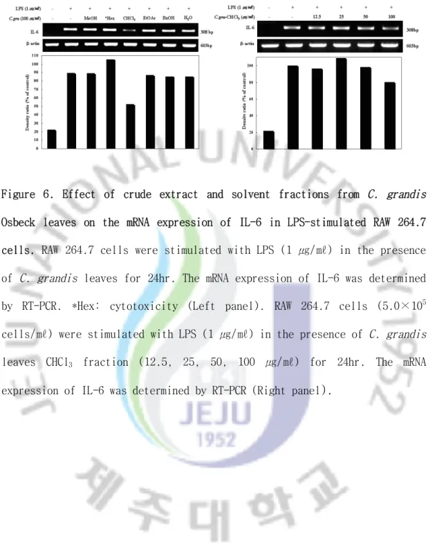
ELISA 결과와 같이 cytokines의 mRNA 발현도 억제하는지를 조사하기 위하여 RT-PCR로 억제 효과를 확인하였다. 또한, β-actin을 같이 나타내어 당유자 자 체가 세포에 영향을 주지 않는 것을 확인하였다. 당유자 각 분획물을 처리한 결
과 CHCl3 분획물이 IL-6의 발현을 억제한다는 것을 확인 하였고, 당유자 잎
CHCl3 분획물을 농도별로 처리한 결과 ELISA와 동등한 결과를 나타냈다 (Figure
20
Figure 5. Effect of CHCl3 fraction from C. grandis Osbeck leaves on the
production level of IL-6 in LPS-stimulated RAW 264.7 cells. The production of IL-6 were determined by ELISA methods from the medium of RAW 264.7
cells (3.5×10⁵ cells/㎖) were stimulated with LPS (1 ㎍/㎖) in the
presence of C. grandis leaves CHCl3 fraction (12.5, 25, 50, 100 ㎍/㎖).
Data represent the mean ± SE of triplicate experiments. *p<0.05 **p<0.01
21
Figure 6. Effect of crude extract and solvent fractions from C. grandis
Osbeck leaves on the mRNA expression of IL-6 in LPS-stimulated RAW 264.7 cells. RAW 264.7 cells were stimulated with LPS (1 ㎍/㎖) in the presence of C. grandis leaves for 24hr. The mRNA expression of IL-6 was determined by RT-PCR. *Hex: cytotoxicity (Left panel). RAW 264.7 cells (5.0×10⁵
cells/㎖) were stimulated with LPS (1 ㎍/㎖) in the presence of C. grandis
leaves CHCl3 fraction (12.5, 25, 50, 100 ㎍/㎖) for 24hr. The mRNA
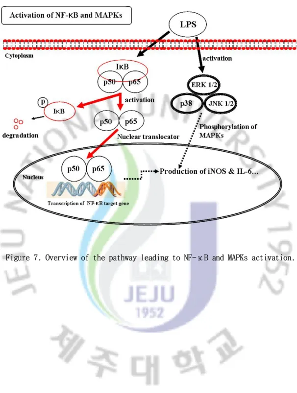
22 2. LPS로 자극한 대식세포에서 당유자 잎 분획물의 항염증 작용기전 외부의 자극이 없을 때 NF-κB (Nuclear factorκB)는 다른 전사인자와는 다르 게 IκB (inhibitor κB)라 불리는 분자에 의해 불활성 상태로 세포질에 존재한 다. NF-κB는 p50, p52, p65, c-Rel등이 결합한 형태로 존재하는데 주로 p65-p50 heterodimer의 NF-κB가 주로 관찰된다. 비 활성 상태에서는 IκB와 결합한 상태로 세포질 내에 존재하며 LPS나 UV등과 같은 다양한 외부자극에 의해서 Mitogen-activated protein kinases (MAPKs)가 활성화 되고, IKK (Inhibitor κ B kinase) pathway를 통해 IκB가 분해되면서 NF-κB가 핵 속으로 이동하여
promoter 부위에 결합하여 전사를 촉진시킨다. (Murakami et al., 2005;
Hambleton et al., 1995; Grossman et al., 2002; Ahn et al., 2005; Prabhu et al., 2002; Pan et al., 1999; Chen et al., 1997).
MAPKs 신호전달체계는 세포가 외부의 환경변화에 따른 자극들을 인지하여 그 정보를 세포질 및 세포핵 내부로 전달하는 과정에 관여하는 신호전달체계이다. 척추 동물에 존재하는 MAPKs는 extracellular signal-related kinase (ERK), stress-activated protein kinase (SAPK/JNK), p38 MAPkinase 등이 있다. ERK는 다양한 성장인자, cytokines에 의해서 활성화되고, 많은 세포에서 세포증식과 분화를 유도하는 기능을 나타낸다. 반면에, JNK와 p38은 염증 유도 cytokines, 자외선 노출, 스트레스에 의해 활성이 증가하는 것으로 알려져 있고, 특히 JNK 는 pro-inflammatory cytokines의 발현에 중요한 작용을 한다. 따라서, NF-κB 와 MAPKs의 활성을 억제하는 것은 염증을 억제하는 중요한 작용이 된다 (Figure 7).
23
24 1) 당유자 잎 CHCl3 분획물에 의한 IκB-α의 degradation 억제효과 세포를 LPS로 자극하였을 때 세포질 내의 IκB-α는 10분부터 감소하기 시작하 여 20분에 최대로 감소하였고, 30분부터 다시 증가하였다 (Figure 8A). 당유자 잎 CHCl3 분획물이 IκB-α degradation 억제에 영향을 주는지 확인하기 위하여 LPS로 자극한 RAW 264.7 세포 (degradation이 최대로 일어나는 20분)에 NF-κB 표준 억제제인 PDTC (25 uM)와 CHCl3 분획물을 농도별로 처리하여 확인한 결과, PDTC 처리군은 LPS 단독 처리군에 비해 IκB-α의 degradation을 억제하였으나, CHCl3 분획물은 LPS 단독 처리군에 비해 IκB-α의 degradation을 억제하지 않은 것으로 나타났다 (Figure 8B). 2) 당유자 잎 CHCl3 분획물에 의한 NF-κB활성 억제효과 NF-κB의 활성을 억제하는 것은 염증을 억제하는 중요한 작용이 된다. 세포를 LPS로 자극하였을 때 10분 후에 NF-κB의 인산화가 가장 많이 증가 하였고, 점 점 감소하다가 약 1시간 후 부터 다시 증가하는 추세를 보였다 (Figure 9A). 당 유자 잎 CHCl3 분획물이 NF-κB의 인산화 억제에 영향을 주는지 확인하기 위하 여 LPS로 자극한 RAW 264.7 세포 (인산화가 최대로 일어나는 10분)에 NF-κB 억 제제인 PDTC (25 uM)와 CHCl3 분획물을 농도별로 처리하여 확인한 결과, CHCl3 분획물이 NF-κB 인산화를 농도 의존적으로 억제하였다 (Figure 9B). 다음으로는 NF-kB의 프로모터 활성에 대해 알아보기 위해 luciferase assay를 수행하였다. NF-kB의 프로모터가 있는 유전자를 RAW 264.7 세포에 일시적으로 transfection한 후 LPS만 처리하였을 때 15 시간 후에 NF-kB의 활성이 급격히 증가하였으며, 그 이후에 감소하는 것을 확인하였다 (Figure 10A). 당유자 잎 CHCl3 분획물이 NF-κB의 프로모터 활성에 영향을 주는지 확인하기 위하여 RAW 264.7 세포를 NF-κB의 프로모터가 있는 유전자로 일시적으로 transfection한 후 LPS와 당유자 잎 CHCl3 분획물을 농도별로 처리하여 NF-κB의 프로모터 활성
25
억제 정도를 확인하였다. 그 결과, LPS만 단독 처리한 세포에서 NF-κB의 활성
이 크게 증가 하였고, CHCl3 분획물을 처리한 결과 농도 의존적으로 NF-κB의
26
Figure 8. Effect of CHCl3 fraction from C. grandis Osbeck leaves on the
phosphorylation and degradation of IκB-α in LPS-stimulated RAW 264.7
cells. (A) RAW 264.7 cells (1.0×10⁶ cells/㎖) were stimulated with LPS (1
㎍/㎖) for indicate times. Whole-cell lysate (25 ㎍) were prepared and the protein level was subjected to 12% SDS-PAGE, and expression of IκB-α was determined by western blotting. (B) RAW 264.7 cells were stimulated with LPS (1 ㎍/㎖) in the presence of C. grandis leaves CHCl3 fraction (12.5,
25, 50, 100 ㎍/㎖) or PDTC (25 uM). Whole cell lysates (25 ㎍) were prepared and the protein level was subjected to 10% SDS-PAGE, and expression of IkB-a (20 min) and β-actin were determined by western blotting. PDTC: NF-κB inhibitor
27
Figure 9. Effect of CHCl3 fraction from C. grandis Osbeck leaves on the
phosphorylation of NF-κB in LPS-stimulated RAW 264.7 cells. (A) RAW 264.7
cells (1.0×10⁶ cells/㎖) were stimulated with LPS (1 ㎍/㎖) for indicate
times. Whole-cell lysates (25 ㎍) were prepared and the protein level was subjected to 10% SDS-PAGE, and expression of NF-κB was determined by western blotting. (B) RAW 264.7 cells were stimulated with LPS (1 ㎍/㎖) in the presence of C. grandis leaves CHCl3 fraction (12.5, 25, 50, 100 ㎍/
㎖) or PDTC (25 uM). Whole cell lysates (25 ㎍) were prepared and the protein level was subjected to 10% SDS-PAGE, and expression of NF-kB (10 min) and β-actin were determined by western blotting. PDTC: NF-κB inhibitor
28
Figure 10. Effect of CHCl3 fraction from C. grandis leaves on the activation
of NF-κB in LPS stimulated RAW 264.7 cells. RAW 264.7 cells (1.5×10⁵ cells/㎖) were transiently cotransfected with NF-kB promoter-based luciferase reporter plasmid (pNF-kB-Luc) and Renilla luciferase reporter plasmid (pRL-null) as internal control for 24 hr. Then the cells were stimulated with LPS (1 ㎍/㎖) for time course (Left panel). RAW 264.7
cells (1.5×10⁵ cells/㎖) were transiently cotransfected with NF-kB
promoter-based luciferase reporter plasmid (pNF-kB-Luc) and Renilla luciferase reporter plasmid (pRL-null) as internal control for 24 hr. Then the cells were stimulated with LPS (1 ㎍/㎖) in the presence of CHCl3 fraction of C. grandis leaves for 15 hr. The luciferase activity was measured and data were normalized to Renilla luciferase expression (Right panel). Data represent the mean ± SE of triplicate experiments.
29
3) 당유자 잎 CHCl3 분획물에 의한 MAPKs (ERK, JNK, p38)활성 억제 효과
Mitogen-activated protein kinase (MAPK)에 속하는 ERK 1/2, JNK 1/2, p38은
LPS로 자극된 대식세포의 초기 신호 경로 중 하나이다 (Ronald et al., 2006).
따라서, 당유자 잎 CHCl3 분획물이 MAPKs를 억제하여 염증인자들의 생성을 억제
하는지 western blot을 통해 확인 하였다. RAW 264.7 세포 (1.0×10⁶ cells/㎖)
를 LPS (1 ㎍/㎖)로 자극하였을 때 ERK는 15~20분, JNK는 20~30분 그리고 p38은
15분에 인산화가 최대로 증가하였다 (Figure 11A, 12A, 13A). 당유자 잎 CHCl3
분획물이 MAPKs의 인산화 억제에 영향을 주는지 확인하기 위하여 LPS로 자극한 RAW 264.7 세포 (인산화가 최대로 일어나는 각각의 시간)에 ERK 억제제인 PD98059 (50 uM), JNK 억제제인 SP600125 (50 uM), p38 억제제인 SB203580 (25 uM)과 CHCl3 분획물을 농도별 (12.5, 25, 50, 100 ㎍/㎖)로 처리하여 결과를 확 인하였다. 당유자 잎 CHCl3 분획물을 농도별로 처리한 결과 ERK, JNK, p38의 인 산화가 모두 농도 의존적으로 억제 되었다 (Figure 11B, 12B, 13B).
30
Figure 11. Effect of CHCl3 fraction from C. grandis Osbeck leaves on the
activation of ERK 1/2 in LPS stimulated RAW 264.7 cells. (A) RAW 264.7
cells (1.0×10⁶ cells/㎖) were stimulated with LPS (1 ㎍/㎖) for indicate
times. (B) RAW 264.7 cells (1.0×10⁶ cells/㎖) were stimulated with LPS (1
㎍/㎖) in the presence of C. grandis leaves (12.5, 25, 50, 100 ㎍/㎖) or PD98059 (50 uM) for 15 minutes. Whole-cell lysate (25 ㎍) were prepared and the protein level was subjected to 10% SDS-PAGE, and expression of phospho-ERK and ERK were determined by western blotting. PD98059: ERK inhibitor
31
Figure 12. Effect of CHCl3 fraction from C. grandis Osbeck leaves on the
activation of JNK 1/2 in LPS stimulated RAW 264.7 cells. (A) RAW 264.7
cells (1.0×10⁶ cells/㎖) were stimulated with LPS (1 ㎍/㎖) for indicate
times. (B) RAW 264.7 cells (1.0×10⁶ cells/㎖) were stimulated with LPS (1
㎍/㎖) in the presence of C. grandis leaves (12.5, 25, 50, 100 ㎍/㎖) or
SP600125 (50 uM) for 30 minutes. Whole-cell lysate (25 ㎍) were prepared and the protein level was subjected to 10% SDS-PAGE, and expression of phospho-JNK and JNK were determined by western blotting. SP600125: JNK inhibitor
32
Figure 13. Effect of CHCl3 fraction from C. grandis Osbeck leaves on the
activation of p38 in LPS stimulated RAW 264.7 cells. (A) RAW 264.7 cells
(1.0×10⁶ cells/㎖) were stimulated with LPS (1 ㎍/㎖) for indicate times.
(B) RAW 264.7 cells (1.0×10⁶ cells/㎖) were stimulated with LPS (1 ㎍/㎖)
in the presence of C. grandis leaves (12.5, 25, 50, 100 ㎍/㎖) or SB203580
(25 uM) for 15 minutes. Whole-cell lysate (25 ㎍) were prepared and the p r o te in le v el w a s s ub je c te d t o 1 0% SDS - P AG E, an d e xp r es s io n o f phospho-p38 and p38 were determined by western blotting. SB203580: p38 inhibitor
33
3. 당유자의 nobiletin 함유 측정
Nobiletin은 6개의 methyl기를 가진 flavonoid로 주로 감귤류에서 검출되는
성분이며, 항암 (Ishiwa et al., 2000), 항염증, 항산화 기능을 가진다고 보고
되었다 (Murakami et al., 2005). 이러한 결과를 토대로, 당유자 잎의 함염증
활성이 nobiletin과 관련이 있는지 확인하기 위하여 nobiletin을 표준물질로 하
여 당유자 잎 추출물 (MeOH)과 분획물 (CHCl3, EtOAc, BuOH, H2O)의 nobiletin
함유를 HPLC를 이용해 확인해 보았다. 그 결과, 당유자 잎 추출물과 분획물 중
뛰어나게 염증 인자를 억제했던 당유자 잎 CHCl3 분획물이 nobiletin을 함유한
34
35
Figure 15. HPLC chromatogram of C. grandis leaves. (A) The HPLC
chromatogram of C. grandis leaves. (B) The merge of HPLC chromatogram of nobiletin and C. grandis leaves CHCl3 fraction.
36
Ⅳ. 고 찰
제주도 재래 감귤의 하나인 당유자 (Citrus grandis Osbeck)는 우리나라에서
는 제주도 에서만 자생하는 감귤류로 (Lim et al., 2002) 미숙과는
neohesperidin과 naringin 등을 함유하고 있으며 (Rhyu et al., 2002) 최근, 당
유자 미숙과의 항산화, 항염증 효과에 관한 보고는 계속되고 있으나 (Lee et al., 2006, Lim et al., 2006) 잎은 어떠한 활성과 기전을 가지고 있는지 보고된 바가 없다. 이에 본 연구에서는 제주도에서 재배되는 당유자 잎 추출물로부터 대식세 포에서 염증반응의 중요한 인자로 알려진 NO와 IL-6의 억제효과를 조사하였으며, NF-κB와 MAPKs의 활성을 측정하여 당유자 잎의 염증억제 작용기전을 밝혔다. 대식세포를 LPS와 같은 TLR 리간드로 자극시키면 세포내 downstream signal인 NF-κB와 MAPKs가 활성화되어 NO또는 IL-6같은 염증성 인자들을 분비하고, 이러
한 작용으로 선천 면역반응이 일어나게 된다 (Ronald et al., 2006). 따라서
RAW 264.7 세포를 LPS (1 ㎍/㎖)로 자극하였을 때, 당유자 잎이 NO와 IL-6같은 염증성 인자들을 억제하는지 확인해 보았다. 당유자 잎 추출물과 분획물 중
CHCl3 분획물이 세포독성 없이 NO의 생성을 강하게 억제하였으며, 농도별로 처
리하였을 때 농도의존적으로 억제하는 것을 확인할 수 있었다. 또한, iNOS의 mRNA와 단백질 발현 억제를 RT-PCR과 western blot방법을 통해 확인한 결과
CHCl3 분획물이 iNOS의 발현을 농도 의존적으로 억제한다는 것을 확인 할 수 있
었다. NO와 함께 중요한 염증성 인자의 하나로 알려진 IL-6의 억제효과를
RT-PCR과 ELISA assay를 통해 확인한 결과, CHCl3 분획물이 IL-6 mRNA의 발현을 억
제하였고, 이것은 ELISA 결과와 유사한 양상을 보였다. 이러한 결과를 요약하면,
당유자 잎 CHCl3 분획물이 대식세포에서 LPS에 의해 생성되는 NO와 IL-6를 억제
하여 항염증 효과를 갖는 것으로 사료된다.
37
인자를 생성한다 (Hambleton et al., 1995 ). NF-κB (Nuclear factorκB)는 I
κB (inhibitor κB)라 불리는 분자에 의해 불활성 상태로 세포질에 존재하며, 주로 p65-p50 heterodimer의 NF-κB가 주로 관찰된다. 비 활성 상태에서는 IκB 와 결합된 상태로 세포질 내에 존재하며 LPS나 UV등과 같은 다양한 외부자극에 의해서 IKK (Inhibitor κB kinase) pathway를 통해 IκB가 분해되면서 NF-κB 가 핵 속으로 이동하여 promoter 부위에 결합하여 염증 인자들의 전사를 촉진시
킨다 (Ahn et al., 2005). 이와 관련해서, LPS로 자극한 RAW 264.7 세포에서 생
성되는 NO와 IL-6에 대한 당유자 잎 CHCl3 분획물의 억제효과가 NF-κB의 활성
억제에 대한 결과인지 western blot과 luciferase assay를 통해 확인하여 보았
다. 그 결과, 당유자 잎 CHCl3 분획물이 세포질내 NF-kB의 인산화와 핵 내에서 전사인자로서의 작용을 억제한다는 것을 확인하였다. 하지만 IkBa의 degradation에는 영향을 주지 않는 것으로 확인되었다. 이러한 결과를 요약하면 당유자 잎 CHCl3 분획물이 염증 인자의 생성을 억제하는 것은 NF-kB의 인산화 억제와 핵 내 promoter로서의 작용을 억제한 결과이며, 이것은 IκB-α의 경로 와 관계없이 활성화된 후의 NF-kB를 억제한 결과로 사료된다.
이외에도 murine macrophage RAW 264.7 세포와 같은 대식세포에서 pro-inflammatory mediator의 생성은 MAPKs pathway를 통해서도 이루어 진다고 알려
져 있다 (Lee et al., 2008). JNK와 p38 억제제를 처리하였을 때 LPS에 의해 유
도되는 NO, TNF-α와 IL-1β의 생성이 억제되었고(Kwak et al., 2005), ERK가
특이적으로 IL-6생성의 억제에 관여한다고 보고되었다 (Ghazizadeh et al.,
2007). 이러한 결과와 관련하여, 당유자 잎 CHCl3 분획물이 MAPKs 인산화에 관
여하여 NO와 IL-6의 생성을 억제하는지 확인하기 위하여, LPS로 자극한 RAW
264.7 세포에 당유자 잎 CHCl3 분획물을 처리하여 확인한 결과, ERK1/2, JNK1/2
와 p38 모두를 효과적으로 억제하였다. 따라서, CHCl3 분획물이 ERK의 인산화를
38 히 억제한다는 것을 확인하였다. 또한, 감귤류에서 분리되는 주요 flavonoid성분 중 하나인 nobiletin은 기관 지 평활근의 이완에 효과를 미쳐 기관지 천식에 중요한 효과를 나타낼 것으로 기대되며 (Baek et al., 2002), 항암 (Ishiwa et al., 2000), 항염증, 항산화 기능을 가진다 (Murakami et al., 2005). 이러한 내용에 착안하여 당유자 잎의 항염증 효과가 nobiletin의 영향을 받은 것인지 조사하기 위하여 nobiletin의 함유를 확인하였을 때, 당유자 잎 CHCl3 분획에서만 nobiletin의 존재가 확인되 었다. 따라서, 당유자 잎 CHCl3 분획물의 염증억제효과에는 nobiletin이 주요 한 작용을 하고 있다고 사료되며, 더 나아가 약리학적 활성을 가진 물질을 분리 해 내는 것이 중요하다고 사료된다. 본 실험 결과를 요약하면, 당유자 잎 CHCl3 분획물이 대식세포에서 염증성 인자인 NO와 IL-6의 생성억제 효과를 나타내었으며 , 이것은 이들의 생성과 관련된 중요한 신호전달 기전인 NF-κB의 활성과 MAPKs (ERK, JNK, p38)의 인산화 억제에 의한 결과였다. 또한, CHCl3 분획에서만 nobiletin이 특이적으로 검출됨에 따라, nobiletin이 당유자 잎 활성성분의 하 나임을 확인할 수 있었다. 이러한 결과들은 제주 감귤류가 염증의 예방과 치료를 위한 주요 천연식물이며,특히, 감귤류의 잎이 식품으로 개발될 수 있는 과실에 비해 약리학적으로 이용 가치가 크고 주요한 성분을 가지고 있 다고 사료되어 그 활성성분의 분리와 작용기전 연구에 중요한 기초자료 가 될 것으로 사료된다.
39
Ⅴ. 참고문헌
Ahn, K.S., and Aqqarwal, B.B., Transcription factor NF-kappaB: a sensor for smoke and stress signals. Ann N Y Acad Sci, 1056, 218-233 (2005) Back, S.O., Han, Y.H., Chun, H.J., Han, D.S., Han, S.S., and Kim, I.K.,
Tracheal muscle smoothing effect of nobiletin isolated from Korean Chung-pi. Journal of the Korean chemical society, 46 (2002).
Balkwill, F., and Coussens, L.M., Cancer: an inflammatory link. Nature, 431, 405-406. (2004)
Blonska, M., Czuba, Z.P., and Krol, W., Effect of flavone derivatives on interleukin-1β (IL-1β) mRNA expression and IL-1β protein synthesis in stimulated RAW 264.7 macrophages. Scand J Immunol, 57, 162-166. (2003)
Cao, Y.Z., Weaver, J.A., Reddy, C.C., and Sordillo, L.M., Selenium deficiency alters the formation of eicosanoids and signal transduction in rat lymphocytes. Prostaglandis Other Lipid Mediat, 70, 131-143 Chen, F., Lu, Y., Kuhn, D.C., Maki, M., Shi, X., Sun, S.C., and Demers,
L.M., Calpain contributes to silica-induced I kappa B-alpha
degradation and nuclear factor-kappa B activation. Arch Biochem
Biophys, 352, 383-388
Cho, J.Y., Baik, K.U., Jung, J.H., and Park, M.H., In vitro anti-inflammatory effects of cynaropicrin, a sesquiterpene lactone, from Saussurea lappa. Eur J Pharmacol, 398, 399-407 (2000)
Choi, I.Y., Kim, S.J., Jeong, H. J., Park, S.H., Song, Y.S., Lee, J.H., Kang, T.H., Park, J.H., Hwang, G.S., Lee, E.J., Hong, S.H., Kim, H.M.,
40
and Um, J.Y., Hesperidin inhibits expression of hypoxia inducible factor-1 alpha and inflammatory cytokine production from mast cells.
Mol Cell Biochem, 305, 153-161 (2007)
Choi, S.Y., Hwang, J.H., Ko, H.C., Paek, J.G., and Kim, S.J., Nobiletin from citrus fruit peel inhibits the DNA-binding activity of NF-κB and ROS production in LPS-activated RAW 264.7 cells. J Enthnopharmacol, 113, 149-155 (2007)
Dai, J., Lin, D., Zhang, J., Habib, P., Smith, P., Murtha, J., Fu, Z., Yao, Z., Qi, Y., and Keller, E. T., Chronic alcohol ingestion induces osteoclastogenesis and bone loss through IL-6 in mice. J Clin Invest, 106, 887-895 (2000)
Gerlier, D., and Thomasser, N., Use of MTT colorimetricassay to measure cell activation. J Immunol Methods, 94, 57-63 (1986)
Guha, M., and Mackman, N., LPS induction of gene expression in human monocytes. Cell Signal, 13, 85-94 (2001)
Grossman, B.J., Shanley, T.P., Odoms, K., Dunsmore, K.E., Denenberg, A.G., and Wong, H.R., Temporal and mechanistic effects of heat shock on LPS-mediated degradation of IkappaBalpha in macrophage. Inflammation, 26, 129-137
Hambleton, J., McMahon, M., and DeFranco, A.L., Activation of Raf-1 and mitogen-activated protein kinase in murine macrophages partially mimics lipopolysaccharide-induced signaling events. J Exp Med, 182, 147-154 (1995)
Han, B.H., Self-Medication and the pharmacy profession. Yakhak hoeji, 47,
41
Irie, T., Muta, T., and Takeshige, K., TAK1 mediates an activation signal
from toll-ike receptor(s) to nuclear factor-kappaB in
lipopolysaccharide-atimulated macrophages. FEBS Lett, 467, 160-164
(2000)
Jeon, K.I., Jeong, J.Y., and Jue, D.M., Thiol-reactive metal compounds
inhibit NF-kappaB activation by blocking I kappa B kinase. J Immunol,
164, 5981-5989 (2000)
Jun, C.D., Pae, H.O., Kim, Y.C., Jeong, S.J., Yoo, J.C., Lee, E.J., Choi, B.M., Chae, S.W., Park, R.K., and Chung, H.T., Inhibition of nitric oxide synthesis by butabol fraction of the methanol extract of Ulmus davidiana in murine macrophage. J Ethnopharmacol, 62, 129-135 (1998) Kiemer, A.K., Hartung, T., Huber, C., and Vollmar, A.M., Phyllanthus
amarus has anti-inflammatory potential by inhibition of iNOS, COX-2,
and cytokines via the NF-κB pathway. J Hepatol, 38, 289-297. (2003)
Kim, J.H., Kim, D.H., Baek, S.H., Lee, H.J., Kim, M.R., Kwon, H.J., and Lee, C.H., Rengylolone inhibits inducible nitric oxide synthase expression and nitric oxide production by down-regulation of NF-κB
and p38 MAP kinase activity in LPS-stimulated RAW 264.7 cells. Biochem
Pharmacol, 71, 1198-1205 (2006)
Kimura, A., Ohmichi, M., Kurachi, H., Ikegami, H., Hayakawa, J., Tasaka, K., Kanda, Y., Nishio, Y., Jikihara, H., Matsuura, N., and Murata, Y., Role of Mitogen-activated protein kinase/Extracellar signal-regulated kinase cascade in gonadotropin-releasing hormone-induced growth
inhibition of a hyman ovarian cancer cell line. Cancer Res, 59,
42
Kwak, H.J., Song, J.S., Heo, J.Y., Yang, S.D., Nam, J.Y., Cheon, H.G., Ro-flumilast inhibits lipopolysaccharide-induced inflammatory mediators via suppression of nuclear factor-kappaB, p38 mitogen-activated
protein kinase, and c-Jun NH2-terminal kinase activation. J. Pharmacol.
Exp. Ther, 315, 1188-1195 (2005)
Lang, R., Hammer, M., and Maqes, J., DUSP meet immunology: dual specificity MAPK phosphatases in control of the inflammatory response.
J Immunol, 177, 7497-7504 (2006)
Lee, H.J., Kang, G.J., Yoon, W.J., Kang, H.K., Kim, Y, S., Kim, S.M., and Yoo, E.S., Anti-inflammatory effect of unripe fruit of Citrus grandis
Osbeck in RAW 264.7 and HaCaT cells. Kor J Pharmacogn, 37, 74-80 (2006)
Li, S., Sang, S., Pan, M.H., Lai, C.S., Lo, C.Y., Yang, C.S., and Ho, C.T. Anti-inflammatory property of the urinary metabolites of nobiletin in mouse. Bioorg Med Chem Lett, 17, 5177-5181 (2007)
Lim, H.K., Yoo, E.S., Moon, J.Y., Jeon, Y.J., and Somi K.Cho., Antioxidant activiy of extracts from Dangyuja (Citrus grandis Osbeck) fruits produced in Jeju Island. Food Sci. Biotechnol, 15, 312-316. (2006) Lim, S., and Jwa, M.K., Physicochemical properties of Dangyooja and Haguel
with ripening. Cheju Journal of Life Science, 5, 57-66 (2006)
Lin, Q.Y., Jin, L.J., Ma, Y.S., Shi, M., and Xu, Y.P., Acanthopanax senticosus inhibits nitric oxide production in murine macrophage in vitro and in vivo. Phytother Res, 21, 879-883 (2007)
Liu, Y., Understanding the biological activity of amyloid proteins in vitro: from inhibited cellular MTT reduction to altered cellular
43
cholesterol homeostasis. Prog Neuropsychopharmacol Biol Psychiatary, 23, 377-395 (1999)
Manthey, J.A., Grohmann, K., and Guthrie, N., Biological properties of
Citrus flavonoids pertaining to cancer and inflammation. Curr Med Chem,
8, 135-153 (2001)
Moeslinger, T., Friedl, R., and Spieckermann, P.G., Inhibition of inducible nitric oxide synthesis by azathioprine in a macrophage cell line. Life Sci, 79, 374-381 (2006)
Murakami, A., Nishizawa, T., Egawa, K., Kawada, T., Nishikawa, Y., Uenakai, K., and Ohigashi, H., New class of linoleic acid metabolites biosynthesized by corn and rice lipoxygenases: Suppression of proinflammatory mediator expression via attenuation of MAPK- and Akt-, but not PPARgamma-, dependent pathways in stimulated macrophages.
Biochem Pharmacol, 70, 1330-1342 (2005)
Nomura, Y., NF-κB activation and IκBα dynamism involved in iNOS and
chemokine induction in astroglial cells. Life sci, 68, 1695-1701 (2001)
Oh, J.B., and Moon, D.K., RAPD and phylogenic relationship of Citrus growing in Jeju Island. Subtropical Agri. & Biotech, 18, 1-10 (2002) Pan, M.H., Lin-Shiau, S.Y., Lin, J.H., and Lin, J.K., Suppression of
lipopolysaccharide-induce nuclear factor-kappaB activity by
theaflavin-3,3’-digallate from black tea and other polyphenols through down-regulation of IkappaB kinase activity in macrophages.
Biochem Pharmacol, 59, 357-367
Pan, M.H., Lai, C.S., Wang, Y.J., and Ho, C.T., Acacetin suppressed LPS-induced up-expression of iNOS and COX-2 in murine macrophages and
TPA-44
induced tumor promotion in mice. Biochem Pharmacol, 72, 1293-1303 (2006)
Rao, K.M ., MAP kinase activation in macrophage. J. Leukoc. Biol, 69, 3-10
(2001)
Rawadi, G., Ramez, V., Lemercier, B., Roman-Roman, S., Activation of mitogen-activated protein kinase pathways by Mycoplasma fermentans membrane lipoproteins in murine macrophages:involvement in cytokine synthesis. J Immunol, 160, 1330-1339.
Rho, T.C., Choi, H.C., Kim, B.Y., Kim, Y.H., Ahn, J.S., Kim, Y.K., and Lee, H.S., Inhibitory effect of Counmarins on nitric oxide production in
LPS-activated murine macrophages. Kor. J. Pharmacogn, 30, 413-416
(1999)
Rhyu, M.R., Kim, E.Y., Bae, I.Y., and Park, Y.K., Contents of Naringin,
Hesperidin and Neohesperidin in premature Korean Citrus fruits. Korean
J. Food Sci, Technol, 34, 132-135 (2002)
Rossi, A., Kapahi, P., Natoli. G., Takahashi, T., Chen, Y., Karin, M., and Santoro, M.G., Anti-inflammatory cyclopentenone prostaglandins are direct inhibitors of IkappaB kinase. Nature, 403, 103-108 (2000)
Seibert, K., Zhang, Y., Leahy, K., Hauser, S., Masferrer, J., Perkins, W., Lee, L., and Isakson, P., Pharmacological and biochemical demonstration of the role of cyclooxygenase 2 in inflammation and pain. Proc Natl Acad Sci U S A, 91, 12013-12017 (1994)
Shin, H.M., Kim, M.H., Kim, B.H., Jung, S.H., Kim, Y.S., Park, H.J., Hong, J.T., Min, K.R., and Kim, Y.S., Inhibitory action of novel aromatic diamine compound on lipopolysaccharide-induced nuclear translocation of
45
NF-κB without affectiong IκB degradation. FEBS Lett, 571, 50-54
(2004)
Son, C.G., Shin, J.W., Cho, J.H., Cho, C.K., Yun, C.H., Chung, W., and Han S.H., Macrophage activation and nitric oxide production by water soluble components of Hericium erinaceum. Int Immunopharmacol, 6, 1363-1369 (2006)
Surh, Y.J., Chun, K.S., Cha, H.H., Han, S.S., Keum, Y.S., Park, K.K., and Lee, S.S., Molecular mechanisms underlying chemopreventive activities of anti-inflammatory phytochemicals: down-regulation of COX-2 and iNOS through suppression of NF-κB activation. Mutat Res, 480-481, 243-268 (2001)
Willoughby, D.A., Heberden Oration, 1974. Human arthritis applied to animal models. Towards a better therapy. Ann Rheum Dis, 34, 471-478 (1975)
Willoughby, D.A., Moore, A.R., and Colville-Nash, P.R., Cyclopentenone
prostaglandins-new allies in the war on inflammation. Nat Med, 6,
137-138 (2000)
Ying, L., and Hofseth, L.J., An emerging role for endothelial nitric oxide
synthase in chronic inflammation and cancer. Cancer Res, 67, 1407-1410
(2007)
채영암., 김성민., 김관수., 유창연., 양덕춘., 약용식물학. 향문사. pp.(2007)
46
Ⅵ. 적 요
당유자는 우리나라 에서도 제주도에만 자생하는 감귤류로 limonene, obacunone, nomiline과 naringin을 함유하고 있다. 이 에 당유자 잎으로부터 추 출물과 분획물을 얻어 LPS로 활성화된 murine macrophage RAW 264.7 세포에서 염증성 인자로 알려진 iNOS와 IL-6의 억제효과를 조사하였으며, 이와 관련된 기 전인 NF-κB와 MAPKs의 인산화 억제 활성을 측정하여 항염증 기능을 탐색하였다. 그 결과, 당유자 잎 추출물을 용매 분획별로 처리하였을 때 LPS (1 ㎍/㎖)로
자극한 RAW 264.7 세포에서 CHCl3 (100 ㎍/㎖)분획이 세포독성 없이 NO의 생성
을 강하게 억제하였으며, iNOS의 mRNA와 protein level에 대한 강한 억제효과를 나타내었다. 또한, IL-6의 mRNA 발현을 억제하였으며, ELISA 결과에서도 동일한 억제 정도를 나타내었다.
NF-κB의 활성억제 정도를 확인하기 위하여 LPS (1㎍/㎖)로 자극을 주고 CHCl3
분획을 농도별로 처리하였을 때, 세포질에서 NF-κB의 인산화가 농도의존적으로 억제되었으며, luciferase assay에서도 강한 억제효과가 나타났다. 또한, MAPKs (ERK, JNK, p38)의 인산화를 농도의존적으로 강하게 억제하였다.
결과적으로, in vitro 에서 당유자 잎 CHCl3 분획물의 대식세포 관련 염증 인
자에 대한 억제 활성을 확인 할 수 있었으며, 이러한 억제활성은 NF-κB와 MAPKs의 활성억제에 의한 결과라고 사료된다.
47 감사의 글 2년 동안 대학원 생활을 하면서 참 많은 것을 배우고 느꼈습니다. 때로는 힘 들어 포기하고 싶을때도 많았지만 그럴 때 마다 저에게 힘을 준 많은 분들 덕에 잘 이겨낸 것 같습니다. 학위 과정 중에 부족함이 많던 저를 잘 이끌어 주시고 보살펴 주신 지도교수 님 유은숙 교수님께 감사 드립니다. 항상 건강하시고 박사과정 동안에 더 많은 가르침을 부탁드립니다. 그리고 교수님의 빈자리를 채워 주시기 위해 논문의 많 은 부분을 더욱 신경 써주신 강희경 교수님, 현진원 교수님께도 감사의 마음을 전합니다. 대학원 과정동안 늘 관심과 조언으로 학문의 길을 이끌어 주신 박덕 배 교수님, 조문제 교수님, 이영기 교수님, 강현욱 교수님, 정영배 교수님, 고 영상 교수님, 이근화 교수님, 은수용 교수님, 홍성철 교수님, 배종면 교수님, 김수영 교수님께 진심으로 감사드립니다. 약리학 교실에 들어와서 대학원 생활과 실험의 모든 것에 도움을 준 이혜자 박 사님, 경진오빠에게 감사합니다. 그리고 상철 선생님, 정일오빠, 재희언니, 혜 진이와 나의 동기들 보연, 은희, 민경, 선순이 그리고 변윤영 선생님 에게도 감 사의 마음을 전합니다. 힘들고 지칠 때 마다 많이 위로해 주고 이야기를 들어준 생화학교실 희경언니, 영미언니, 지은언니, 경아언니, 진영이와 미생물학 교실의 정은언니에게 감사의 마음을 전합니다. 그리고 의대에 왔을 때 힘들어 하던 나에게 힘이 되고 웃음을 주었던 희경이 에게 고마운 마음을 전하고 좋은 논문 쓰길 바랍니다. 늘 옆에서 힘을 북돋아 주었던 나의 임상병리과 동기 친구들, 오빠들 그리고, 그리고 선·후배 여러분께 진심으로 감사의 마음을 전합니다. 학위 과정 중 종 다양성 연구소로 오면서 논문 마무리 하느라 힘든 적도 많았 고 회사일을 게을리 한적도 있었습니다. 그럼에도 불구하고, 항상 이해해 주고
48 다독여 주신 현창구 박사님, 나의 든든한 선배 원종오빠, 지영언니, 은영언니, 민진이, 그리고 항상 웃을수 있게 해준 원혁오빠와 마워오빠 에게도 너무 고맙 고, 또한 다 거론 할 수는 없지만 모든 종다양성 연구소 식구들에게도 감사의 마음을 전합니다. 끝으로 부족함이 많은 나를 항상 응원해주는 우리 아빠, 엄마, 그리고 나의 정 신적 물질적 후원자인 우리언니들 형부들 너무 고맙고, 내 삶의 활력소 조카들 유지, 수윤, 채원이와 지후에게 사랑의 마음을 전합니다. 언제나 건강하고 행복 하게 살아요... 사랑합니다.