저작자표시 2.0 대한민국 이용자는 아래의 조건을 따르는 경우에 한하여 자유롭게 l 이 저작물을 복제, 배포, 전송, 전시, 공연 및 방송할 수 있습니다. l 이차적 저작물을 작성할 수 있습니다. l 이 저작물을 영리 목적으로 이용할 수 있습니다. 다음과 같은 조건을 따라야 합니다: l 귀하는, 이 저작물의 재이용이나 배포의 경우, 이 저작물에 적용된 이용허락조건 을 명확하게 나타내어야 합니다. l 저작권자로부터 별도의 허가를 받으면 이러한 조건들은 적용되지 않습니다. 저작권법에 따른 이용자의 권리는 위의 내용에 의하여 영향을 받지 않습니다. 이것은 이용허락규약(Legal Code)을 이해하기 쉽게 요약한 것입니다. Disclaimer 저작자표시. 귀하는 원저작자를 표시하여야 합니다.
Hanging-Associated Left Ventricular Systolic
Dysfunction
by
U Ram Jin
Major in Medicine
Department of Medical Sciences
The Graduate School, Ajou University
Hanging-Associated Left Ventricular Systolic
Dysfunction
by
U Ram Jin
A Dissertation Submitted to The Graduate School of
Ajou University in Partial Fulfillment of the
Requirements for the Degree of
Master of Medicine
Supervised by
Joon Han Shin, M.D.
Major in Medicine
Department of Medical Sciences
The Graduate School, Ajou University
This certifies that the dissertation
of U Ram Jin is approved.
SUPERVISORY COMMITTEE
Joon Han Shin
Myeong Ho Yoon
Gyo Seung Hwang
The Graduate School, Ajou University
June, 20th, 2014
i - ABSTRACT -
Hanging-Associated Left Ventricular Systolic Dysfunction
Backgrounds: Hanging injury is infrequent but its clinical course is usually devastating.
Although hanging patients usually need cardiopulmonary resuscitation (CPR), hanging-associated cardiovascular damage has not been fully established. The aim of this study was to evaluate the echocardiographic findings in patients with hanging injury.
Methods: We enrolled 25 patients (9 males, 33 ± 15 year-old) with hanging injury who
performed echocardiography within 2 weeks after admission. Clinical, demographic and laboratory data and transthoracic echocardiographic findings were analyzed.
Results: Of those 25 patients, 8 patients (2 males, 34 ± 13 year-old) showed left
ventricular (LV) systolic dysfunction (mean LVEF: 34 ± 16%). Global hypokinesia was presented in one patient. Apical ballooning with sparing of the basal segments was presented in 2 patients. Basal akinesia and apical hyperkinesia was presented in one patient. The other 4 patients showed regional wall motion abnormalities unmatched with coronary territories.
The duration of suspension and the duration of CPR were not significantly different according to the presence of LVSD.
Conclusion: This study show the echocardiographic findings in considerable numbers of
ii in the patients of haning injury.
_________________________________________________________________________
Key Words: Hanging, Hanging injury, Left ventricular systolic dysfunction,
iii
TABLE OF CONTENTS
ABSTRACT ··· ⅰ TABLE OF CONTENTS ··· ⅲ LIST OF TABLES ··· ⅳ Ⅰ. INTRODUCTION ··· 1Ⅱ. PATIENTS AND METHODS ··· 3
A. Study Population ··· 3 B. Data ··· 3 C. Echocardiographic Findings ··· 4 D. Statistical Analysis ··· 4 Ⅲ. RESULTS ··· 5 A. Baseline Characteristics ··· 5
B. Patients with LV Dysfunction ··· 7
Ⅳ. DISCUSSION ··· 10
Ⅴ. CONCLUSION ··· 14
REFERENCES ··· 15
iv
LIST OF TABLES
Table 1. Baseline characteristics ··· 6 Table 2. Clinical parameters of echocardiography in patients with LV systolic
dysfunction ··· 8 Figure 1. Patterns of LV systolic dysfunction ··· 9
1
-I. INTRODUCTION
Hanging is one of the most commonly used methods of suicide attempts. In many countries, suicide rates are increasing, including our country (Gunnell et al, 2005; Statistics Korea). Especially, incidence of hanging attempts seems to have increased for decades worldwide (Gunnel et al, 2005; Baker et al, 2013; Bridge et al, 2010; Höfer et al, 2012; Meel, 2006). And it is the same in pediatrics and young persons (Bridge et al, 2010).
The prognosis of hanging is usually poor, particularly in the persons with cardiopulmonary arrest (CPA), and this is not different between the adults and the pediatrics (Gunnell et al, 2005; Matsuyama et al, 2006; Deasy et al, 2013; Wee et al, 2014; Deasy et al, 2011; Davies et al, 2011). If the patients with CPA survived, survivors usually suffered severe
neurological injury. However, in the patients of hanging without CPA when they arrived at the hospital, the prognosis is favorable and high proportion of patients could survive without neurological deficits (Matsuyama et al, 2006; Deasy et al, 2011; Davies et al, 2011; Penney et al, 2002; Wee et al, 2013). Duration of suspension, presence of CPA, initial resuscitation, depth of coma, and presence of respiratory arrest or hypopnea are known as the important prognostic predictors (Gunnell et al, 2005; Matsuyama et al, 2006; Borgquist and Friberg, 2009; Kaki et al, 1997). Laboratory data, such as pH, basal excess in arterial gas analysis, was also reported as the prognostic predictors (Matsuyama et al, 2006).
In some cases hanging injuries presented adverse effects on cardiac function. There are several case reports and studies about the cardiac impairment in hanging patients
2
-(Mohammedi et al, 2005; Gnanavelu and Sathiakumar, 2008; Sivanandan et al, 2009; Champion et al, 2013; Viswanathan et al, 2012; Chacko et al, 2011). But there is no studies that have assessed enough the echocardiographic findings and their significance. The aim of this study was to evaluate the echocardiographic findings and their significance in patients with hanging injury.
3
-II. PATIENTS AND METHODS
A. Study Population
We did a retrospective analysis of all cases of hanging admitted in our hospital emergency department between May 1997 and May 2014. We searched computerized records of the hospital electronic medical record system using ICD-10 cord for hanging, strangulation and suffocation. Then we reviewed manually all of the records of the patients. A total of 191 patients’ records of hanging injury was identified, in which we enrolled 25 patients with hanging injury who performed echocardiography within 2 weeks after admission to hospital.
B. Data
Clinical, demographic and laboratory data of the cases was analyzed. We searched clinical information such as presence of cardiovascular risk factors (diabetes, hypertension, dyslipidemia, smoking), presumptive duration of suspension, presence of CPA, duration of cardiopulmonary resuscitation (CPR) in the cases with CPA. The duration of suspension was typically difficult to determine and was usually estimated from family reports of when the patient was last seen. Collected laboratory data include initial CK-MB, troponin-I, lactate, and arterial gas analysis (pH, basal excess).
4
-C. Echocardiographic Findings
We searched echocardiographic findings of all the cases. Collected echocardiographic data includes left ventricular ejection fraction (LVEF), left ventricular fractional shortening (LVFS), left ventricular end-diastolic dimension (LVEDD), left ventricular end-systolic dimension (LVESD), left ventricular mass index (LVMI), presence of regional wall motion abnormalities (RWMAs), wall motion score index. We defined left ventricular (LV) systolic dysfunction by echocardiographic finding when echocardiography showed RWMAs such as hypokinesia, akinesia, dyskinesia or aneurysm in two or more segments of left ventricle defined by 17-segment model (Lang et al, 2005). The patterns of RWMAs were analyzed and interpreted with agreement by two cardiologists who were specialized in echocardiography.
D. Statistical Analysis
Two subgroups of the inrolled patients have been previously defined and we compared two groups: that of patients without LV systolic dysfunction as “none-LVSD” group and that of patients with LV systolic dysfunction as “LVSD” group. Continuous variables and ordinal variables are presented as means ± SD. Continuous variables were compared using the t test (normal distribution) or Mann-Whitney rank sum test (skewed distribution). Categorical measures were compared with Fisher’s exact test. A two-tailed P value of less than 0.05 was considered to indicate statistical significance. Statistical analysis was performed with Statview version 18.0 software.
5
-III. RESULTS
A. Baseline Characteristics
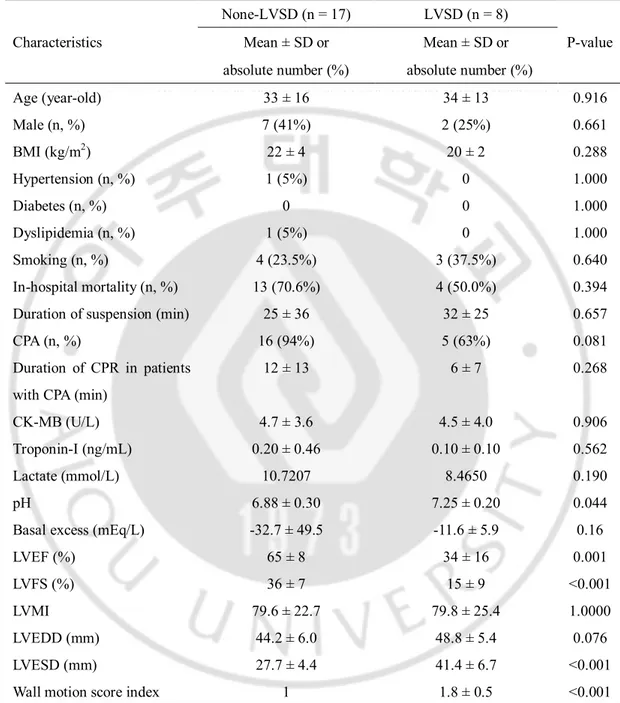
Total twenty-five patients were included, in which 17 patients had no LV systolic dysfunction and 8 patients had LV systolic dysfunction (Table 1). Mean age was 33 ± 16 years in LVSD and 34 ± 13 years in LVSD. Seven patients (41%) were male in none-LVSD and 2 patients (25%) were male in none-LVSD. There was no significant difference between both groups in duration of suspenstion, presence of CPA, duration of CPR, in-hospital mortality, CK-MB, troponin-I, lactate except in pH (6.89 ± 0.33 in none-LVSD vs. 7.29 ± 0.22 in LVSD). LVEF was lower in LVSD (34 ± 16%) than that in none-LVSD (65 ± 8%). LVESD and wall motion index score were also lower in LVSD.
6
-Table 1. Baseline characteristics
Characteristics
None-LVSD (n = 17) LVSD (n = 8)
P-value Mean ± SD or Mean ± SD or
absolute number (%) absolute number (%)
Age (year-old) 33 ± 16 34 ± 13 0.916 Male (n, %) 7 (41%) 2 (25%) 0.661 BMI (kg/m2) 22 ± 4 20 ± 2 0.288 Hypertension (n, %) 1 (5%) 0 1.000 Diabetes (n, %) 0 0 1.000 Dyslipidemia (n, %) 1 (5%) 0 1.000 Smoking (n, %) 4 (23.5%) 3 (37.5%) 0.640 In-hospital mortality (n, %) 13 (70.6%) 4 (50.0%) 0.394 Duration of suspension (min) 25 ± 36 32 ± 25 0.657
CPA (n, %) 16 (94%) 5 (63%) 0.081
Duration of CPR in patients with CPA (min)
12 ± 13 6 ± 7 0.268
CK-MB (U/L) 4.7 ± 3.6 4.5 ± 4.0 0.906
Troponin-I (ng/mL) 0.20 ± 0.46 0.10 ± 0.10 0.562
Lactate (mmol/L) 10.7207 8.4650 0.190
pH 6.88 ± 0.30 7.25 ± 0.20 0.044
Basal excess (mEq/L) -32.7 ± 49.5 -11.6 ± 5.9 0.16
LVEF (%) 65 ± 8 34 ± 16 0.001
LVFS (%) 36 ± 7 15 ± 9 <0.001
LVMI 79.6 ± 22.7 79.8 ± 25.4 1.0000
LVEDD (mm) 44.2 ± 6.0 48.8 ± 5.4 0.076
LVESD (mm) 27.7 ± 4.4 41.4 ± 6.7 <0.001
Wall motion score index 1 1.8 ± 0.5 <0.001
LVSD, left ventricular systolic dysfunction; SD, standard deviation; BMI, body mass index; CPA, cardiopulmonary arrest; CPR, cardiopulmonary resuscitation; LVEF, left ventricular ejection fraction; FS, LVFS, left ventricular fractional shortening; LVMI, left ventricular mass index; LVEDD, left ventricular end diastolic dimension; LVESD, left ventricular end systolic dimension.
7
-B. Patients with LV Dysfunction
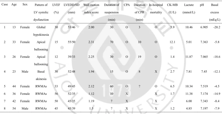
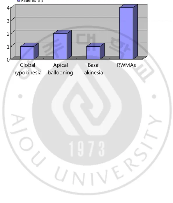
Eight patients had LV systolic dysfunction, in which 2 patients were male and 6 were female (Table 2). Five patients had CPA, in which only one case survived. Three patients didn’t have CPA and all of them survived. Patterns of LV systolic dysfunction were global hypokinesia in one, apical ballooning like Takotsubo cardiomyopathy in two, and basal akinesia in one, and four patients had non-specific RWMAs unmatched with coronary territories (Figure 1).
The follow-up echocardiography was performed in 3 patients. In one patient (female, 13 year-old), echocardiography revealed global hypokinesia with LVEF 30%. After 2 weeks, echocardiography was undergone again and it revealed completely restored LV function with LVEF 76%. In another pateint (female, 33 year-old), echocardiography revealed akinesia of the mid and apical segments of LV with sparing of the basal segments and LVEF was 15%, which was compatible with apical ballooning. Echocardiography performed after 4 days presented no significant change from previous one. In another pateint (female, 26 year-old), echocardiography presented apical ballooning pattern of LV systolic dysfunction with 12% of LVEF. Echocardiography performed after 9 days presented fully recovered LV function with 71% of LVEF.
8
-Table 2. Clinical parameters of echocardiography in patients with LV systolic dysfunction Case Age Sex Pattern of
LV systolic dysfunction LVEF (%) LVEDD/SD (mm) Wall motion index score Duration of suspension (min) CPA Duration of CPR (min) In-hospital mortality CK-MB (U/L) Lactate (mmol/L) pH Basal excess (mEq/L) 1 13 Female Global hypokinesia 30 53/46 2.00 30 O 3 O 5.9 10.46 6.905 -20.2 2 33 Female Apical ballooning 15 55/50 2.31 70 O 10 O 12.1 5.01 7.363 -5.8 3 26 Female Apical ballooning 12 39/33 2.25 30 O 19 O 1.4 11.87 7.065 -18.6 4 23 Male Basal akinesia 30 52/48 1.94 15 O 8 X 2.7 7.81 7.45 -12.1 5 44 Female RWMAs 33 49/45 2.12 60 O ? O 6.5 10.34 7.519 -4.5 6 36 Female RWMAs 56 52/35 1.12 10 X X 1.7 11.38 7.174 -14.9 7 42 Female RWMAs 50 45/35 1.19 7 X X - 6.00 7.343 -8.4 8 54 Male RWMAs 45 45/39 1.5 ? X X 1.2 4.85 7.197 -7.9
LV, left ventricle; LVEF, left ventricular ejection fraction; LVEDD/SD, left ventricular end diastolic dimension/systolic diminesion; CPA, cardiopulmonary arrest; CPR, cardiopulmonary resuscitation; RWMAs, regional wall motion abnormalities.
9 -0 1 2 3 4 Global hypokinesia Apical ballooning Basal akinesia RWMAs Patients (n)
- 10 -
IV. DISCUSSION
The cause of death in suicidal hangings usually results from variable mechanisms (Clément et al, 2010). The relative contribution of each theory to the mechanism of death in hanging (airways occlusion and asphyxia, occlusion of the neck vessels with interruption of cerebral blood flow, and cardiac inhibition by vagal stimulation) has not been fully determined (Clément et al, 2010; Boghossian et al, 2010). But, all of those mechanisms may be
associated with cardiac impairment in hanging injury (Mohammedi et al, 2005; Boghossian et al, 2010; Pajouh and Bahler, 2004; Ako et al, 2006).
Stress-induced cardiomyopathy involving transient LV systolic dysfunction in the absence of obstructive coronary artery disease has been issued in recent years (Pelliccia et al, 2014). It is also known as ‘Takotsubo syndrome’, ‘Takotsubo cardiomyopathy’ and ‘transient left ventricular apical ballooning syndrome.’ The reports of this condition have increased worldwide during the past decade, it has become clear that stress-induced cardiomyopathy has unique clinical features that can be distinguished from those of an acute coronary syndrome (Wittstein, 2008). In stress-induced cardiomyopathy, the LV systolic dysfunction is reversible in most cases and it is well known that this condition is more likely presented in elderly women and has a favorable prognosis (Bossone et al, 2013).
Etiopathologenesis of stress-induced cardiomyopathy is explained by triggers, pathogenic factors (catecholamine, coronary vasomotor abnormalities) and predisposing factors (cardiovascular risk factors, endothelial dysfunction, co-morbidities), which are likely to
- 11 -
interplay differently in different patients leading to the development of LV systolic
dysfunction (Pelliccia et al,2014). Triggers known as the mechanisms associated with stress-induced cardiomyopathy are emotional stress, physical stress and neurological triggers (Sharkey et al, 2010; Lee et al, 2006). In patients of hanging injury, all of these triggers could develop. Hanging attempt is a behavior may be accompanied by severe emotional stress. Acute hypoxia associated with asphyxia or pulmonary edema in hanging injury could be a physical stressor (Viswanathan et al, 2012; Clément et al, 2010). Uncommonly, severe trauma of the neck structures also could be a physical stressor (Gunnell et al, 2005; Wee et al, 2014). And acute brain damage due to brain ischemia with occlusion of neck vessels could be a neurological trigger (Clément et al, 2010; Boghossian et al, 2010; Ako et al, 2006; Lee et al, 2006).
Many cases of hanging injury presented with CPA and prognosis of them has been poor (Gunnell et al, 2005; Matsuyama et al, 2006; Deasy et al, 2013; Wee et al, 2014; Deasy et al, 2011; Davies et al, 2011). The mechanism by which hanging causes CPA is unique (Wee et al, 2014). Complete airway/arterial occlusion, carotid sinus stimulation with increased vagal tone and long anoxic time could be the mechanism of CPA in hanging cases (Wee et al, 2014). In hanging patient with CPA, CPA itself or drugs for CPR could be a cause of stress-induced cardiomyopathy (Copetti et al, 2013; Laínez et al, 2009; Amariles, 2011). However, in our study, 16 of 17 patients had CPA in none-LVSD group and 5 of 8 patients had CPA in LVSD group and there was no significant difference. So we think that the presence of CPA in hanging injury may not be associated with presence of LV systolic dysfuncction.
- 12 -
There are several previous reports about the cardiac impairment in hanging patient (Mohammedi et al, 2005; Gnanavelu and Sathiakumar, 2008; Sivanandan et al, 2009; Champion et al, 2013; Viswanathan et al, 2012; Chacko et al, 2011). The cases which performed follow-up echocardiography usually revealed full recovery of LV function. In two studies, echocardiography was performed in several patients. In one of those studies,
echocardiography was performed in 7 patients of hanging with pulmonary edema and 4 of them presented LV systolic dysfunction (Viswanathan et al, 2012). Another study reported that 5 patients performed echocardiography and 2 patients with LV systolic dysfunction, in which one revealed an apical ballooning pattern and another revealed basal hypokinesia (Champion et al, 2013). In our study, LV systolic dysfunction was presented in considerable rate of hanging patients who performed echocardiography (8 of 25 patients, 32%) and some of them presented very severe LV systolic dysfunction. The pattern of LV systolic
dysfunction was variable. The variable patterns of LV systolic dysfunction in hanging cases may be associated with the variable mechanisms of LV systolic dysfunction in hanging injury.
In stress-induced cardiomyopathy, it is known that most of LV function will recover
completely within a few weeks in the majority of cases (Singh et al, 2014). Three patients in our study performed follow-up echocardiography and two of them presented full recovery of LV function. One’s follow-up echocardiography presented no interval change. But it could be due to short duration of 4 days from initial echocardiography to follow-up
- 13 -
One study suggested that secondary stress-induced cardiomyopathy in a patient already suffering from a potentially life-threatening condition was associated with much higher mortality rate relative to primary stress-induced cardiomyopathy in the absence of such acute critical illness (Singh et al, 2014). In our study, hanging injury patients of LVSD group presented 50% of in-hospital mortality rate (4 deaths of 8 patients). But it was not
significantly different from that of none-LVSD group with 70% in-hospital mortality rate (13 deaths of 17 patients). Because of the small size of the study population, whether the presence of LV systolic dysfunction is associated with in-hospital mortality in hanging patients is not conclusive in this study. But as like the secondary stress-induced
cardiomyopathy from another etiology, severity of the underlying disease and condition of the patient may be the most important factor for in-hospital mortality.
There are several limitations in our study. Echocardiography was not performed all of the patients of haning injury, but the patients were selected by physicians of emergency department. And the duration from admission to performing echocardiography was variable from on arrival to 2 weeks. The small number of hanging patients who performed echocardiography is also a limitation of this study, even though about one-third of them presented LV systolic dysfunction. Then much of the results may not be conclusive. It is also a limitation that the number of pateints with follow-up echocardiography was small. And this is a retrospective observational study: some information may be missing or inaccurate.
- 14 -
V. CONCLUSION
Incidence of hanging attempts seems to have increased worldwide. The prognosis of patients with hanging injury is usually poor, especially in the patients with CPA, but the patients without CPA presented favorable prognosis.
Hanging injury often causes cardiac impairment with LV systolic dysfunction and sometimes it may be very severe. Because of unique pathophysiology of hanging injury, variable mechanisms are possibly involved: emotional stress, physical stress of hypoxia or trauma, neurological trigger by brain damage, CPA and drugs for CPR could be the cause of stress-induced cardiomyopathy. Cardiac inhibition by vagal stimulation also could be a mechanism of cardiac impairment.
Although it is not conclusive, the presence of LV systolic dysfunction could be associated with morbidity and mortality. So we suggest that echocardiographic evaluation at early stage should be performed for all of patients with hanging. And in patients with LV systolic dysfunction, follow-up echocardiographic evaluation should also be performed at least 2 weeks after initial study.
- 15 -
REFERENCE
1. Ako J, Sudhir K, Farouque HM, Honda Y, Fitzgerald PJ: Transient left ventricular dysfunction under severe stress: brain-heart relationship revisited. Am J Med. 119:10-17, 2006
2. Amariles P: Drugs as possible triggers of Takotsubo cardiomyopathy. Curr Clin
Pharmacol. 6:1-11, 2011
3. Baker SP, Hu G, Wilcox HC, Baker TD: Increase in suicide by hanging/suffocation in the U.S., 2000-2010. Am J Prev Med. 44:146-149, 2013
4. Boghossian E, Clément R, Redpath M, Sauvageau A: Respiratory, circulatory, and neurological responses to hanging: a review of animal models. J Forensic Sci. 55:1272-1277, 2010
5. Borgquist O, Friberg H: Therapeutic hypothermia for comatose survivors after near-hanging-a retrospective analysis. Resuscitation. 80:210-212, 2009
6. Bossone E, Savarese G, Ferrara F, Citro R, Mosca S, Musella F, Limongelli G, Manfredini R, Cittadini A, Perrone Filardi P: Takotsubo cardiomyopathy: overview.
Heart Fail Clin. 9:249-266, 2013
- 16 -
in suicide rates by hanging and/or suffocation and firearms among young persons aged 10-24 years in the United States: 1992-2006. J Adolesc Health. 46:503-505, 2010
8. Chacko J, Brar G, Elangovan A, Moorthy R: Apical ballooning syndrome after attempted suicidal hanging. Indian J Crit Care Med. 15:43-45, 2011
9. Champion S1, Spagnoli V, Deye N, Mégarbane B, Baud F: Cardiac impairment after hanging attempt: a preliminary descriptive study. Ann Cardiol Angeiol (Paris). 62:259-264, 2013
10. Clément R, Redpath M, Sauvageau A: Mechanism of death in hanging: a historical review of the evolution of pathophysiological hypotheses. J Forensic Sci. 55:1268-1271, 2010
11. Copetti R, Peric D, Amore G, Guglielmo N, Federici N, Cominotto F: Transient Tako-Tsubo cardiomyopathy after cardiopulmonary resuscitation: A causal role of adrenaline? Resuscitation. 84:e45-46, 2013
12. Davies D, Lang M, Watts R: Paediatric hanging and strangulation injuries: A 10-year retrospective description of clinical factors and outcomes. Paediatr Child
- 17 -
13. Deasy C, Bray J, Smith K, Bernard S, Cameron P: Hanging-associated out-of-hospital cardiac arrests in Melbourne, Australia. Emerg Med J. 30:38-42, 2013 14. Deasy C, Bray J, Smith K, Harriss LR, Bernard SA, Cameron P: Paediatric hanging
associated out of hospital cardiac arrest in Melbourne, Australia: characteristics and outcomes. Emerg Med J. 28:411-415, 2011
15. Gnanavelu G, Sathiakumar DB: Reversible left ventricular dysfunction in suicidal hanging. J Assoc Physicians India. 56:545-546, 2008
16. Gunnell D, Bennewith O, Hawton K, Simkin S, Kapur N: The epidemiology and prevention of suicide by hanging: a systematic review. Int J Epidemiol. 34:433-442, 2005
17. Höfer P, Rockett IR, Värnik P, Etzersdorfer E, Kapusta ND: Forty years of increasing suicide mortality in Poland: undercounting amidst a hanging epidemic?.
BMC Public Health. 12:644, 2012
18. Kaki A1, Crosby ET, Lui AC: Airway and respiratory management following non-lethal hanging. Can J Anaesth. 44:445-450, 1997
19. Laínez B, Ureña M, Alvarez V, Lezaun R: Iatrogenic Tako–Tsubo Cardiomyopathy Secondary to Catecholamine Administration. Rev Esp Cardiol. 62:1498-1499, 2009
- 18 -
20. Lang RM, Bierig M, Devereux RB, Flachskampf FA, Foster E, Pellikka PA, Picard MH, Roman MJ, Seward J, Shanewise JS, Solomon SD, Spencer KT, Sutton MS, Stewart WJ: Recommendations for Chamber Quantification: A Report from the American Society of Echocardiography’s Guidelines and Standards Committee and the Chamber Quantification Writing Group, Developed in Conjunction with the European Association of Echocardiography, a Branch of the European Society of Cardiology. J Am Soc Echocardiogr. 18:1440-1463, 2005
21. Lee VH, Oh JK, Mulvagh SL, Wijdicks EF: Mechanisms in neurogenic stress cardiomyopathy after aneurysmal subarachnoid hemorrhage. Neurocrit Care. 5:243-249, 2006
22. Matsuyama T, Okuchi K, Seki T, Murao Y: Prognostic factors in hanging injuries.
Am J Emerg Med. 22:207-210, 2004
23. Meel B: Epidemiology of suicide by hanging in Transkei, South Africa. Am J
Forensic Med Pathol. 27:75-78, 2006
24. Mohammedi I, Perret X, Argaud L, Le Vavasseur O, Martin O, Robert D: Hanging causing severe reversible left ventricular dysfunction. Intensive Care Med. 31:495, 2005
- 19 -
25. Pajouh M, Bahler RC: Hyperacute respiratory failure causing transient left ventricular dysfunction. Am J Med. 116:784-785, 2004
26. Pelliccia F, Greco C, Vitale C, Rosano G, Gaudio C, Kaski JC: Takotsubo Syndrome (Stress Cardiomyopathy): An Intriguing Clinical Condition in Search of its Identity.
Am J Med. pii: S0002-9343(14)00309-X, 2014
27. Penney DJ, Stewart AH, Parr MJ: Prognostic outcome indicators following hanging injuries. Resuscitation. 54:27-29, 2002
28. Sharkey SW1, Windenburg DC, Lesser JR, Maron MS, Hauser RG, Lesser JN, Haas TS, Hodges JS, Maron BJ: Natural history and expansive clinical profile of stress (tako-tsubo) cardiomyopathy. J Am Coll Cardiol. 55:333-341, 2010
29. Singh K, Carson K, Shah R, Sawhney G, Singh B, Parsaik A, Gilutz H, Usmani Z, Horowitz J: Meta-Analysis of Clinical Correlates of Acute Mortality in Takotsubo Cardiomyopathy. Am J Cardiol. 113:1420-8, 2014
30. Singh K, Carson K, Shah R, Sawhney G, Singh B, Parsaik A, Gilutz H, Usmani Z, Horowitz J: Review: transient left ventricular apical ballooning, broken heart syndrome, ampulla cardiomyopathy, atypical apical ballooning, or Tako-Tsubo cardiomyopathy. Am J Cardiol. 15:1420-1428, 2014
- 20 -
31. Sivanandan S, Sinha A, Juneja R, Lodha R: Reversible acute left ventricular dysfunction in accidental strangulation. Pediatr Crit Care Med. 10:e5-8, 2009 32. Statistics Korea. Available at http://kosis.kr/
33. Viswanathan S, Muthu V, Remalayam B: Pulmonary edema in near hanging. J
Trauma Acute Care Surg. 72:297-301, 2012
34. Wee JH, Park KN, Oh SH, Youn CS, Kim HJ, Choi SP: Outcome analysis of cardiac arrest due to hanging injury. Am J Emerg Med. 30:690-694, 2014
35. Wee JH1, Park JH, Choi SP, Park KN: Outcomes of patients admitted for hanging injuries with decreased consciousness but without cardiac arrest. Am J Emerg Med. 31:1666-1670, 2013
- 21 - - 국문요약 -
목맴 손상에 따르는 좌심실 수축 기능 이상
목적: 목맴 손상은 흔하지는 않으나 그 임상경과는 대개 심각한 손상을 동반하게 된다. 목맴 손상으로 내원하는 환자는 많은 경우 심폐소생술의 시행을 필요로 하나 목맴 손상이 심혈관계에 미치는 영향에 대해서는 충분히 잘 알려져 있지 않다. 본 연구에서는 목맴 손상으로 내원한 환자들의 심초음파 소견을 분석하여 그 양상에 대해 밝히고자 한다. 방법: 목맴 손상으로 내원하였고 그 중 내원후 2 주 이내에 심초음파검사를 시행한 환자 총 25 명(남자 9 명, 33 ±15 세)을 연구대상으로 하였다. 환자들의 임상적 및 인구학적 정보, 실험실 검사 결과, 심초음파 소견 등의 자료를 분석하였다. 결과: 총 환자 25 명 중 8 명(남자 2 명, 34 ± 13 세)에게서 좌심실 수축 기능 이상이 나타났으며 평균 좌심실 박출계수는 34 ± 16%이었다. 그 중 한 환자는 전체적 좌심실 부전을 보였다. 두 환자들에게서는 심실의 중간부 및 심첨부의 운동불능을 보이고 기저부는 정상 운동을 보였다. 심실 기저부의 운동불능 및 심첨부의 운동과다를 보이는 소견도 한 환자로부터 나타났다. 그 외 4 명의 환자에게서는 관상동맥의 분포와 일치하지 않는 국소벽운동이상이 나타났다.- 22 - 좌심실 수축 기능 이상의 여부에 따라 CPR 시간, 목맴 시간이 의미있는 차이를 보이지는 않았다. 결론: 이 연구는 목맴 손상으로 내원한 다수의 환자들에 대해서 심초음파 소견을 분석한 첫 번째 연구이다. 이를 통해서 목맴 손상으로 내원한 환자들에게서 다양한 양상의 좌심실 수축 기능 이상 소견이 나타남을 알 수가 있다. 핵심어 : 목맴, 목맴 손상, 좌심실 기능 이상, 심초음파