저작자표시-비영리-변경금지 2.0 대한민국 이용자는 아래의 조건을 따르는 경우에 한하여 자유롭게 l 이 저작물을 복제, 배포, 전송, 전시, 공연 및 방송할 수 있습니다. 다음과 같은 조건을 따라야 합니다: l 귀하는, 이 저작물의 재이용이나 배포의 경우, 이 저작물에 적용된 이용허락조건 을 명확하게 나타내어야 합니다. l 저작권자로부터 별도의 허가를 받으면 이러한 조건들은 적용되지 않습니다. 저작권법에 따른 이용자의 권리는 위의 내용에 의하여 영향을 받지 않습니다. 이것은 이용허락규약(Legal Code)을 이해하기 쉽게 요약한 것입니다. Disclaimer 저작자표시. 귀하는 원저작자를 표시하여야 합니다. 비영리. 귀하는 이 저작물을 영리 목적으로 이용할 수 없습니다. 변경금지. 귀하는 이 저작물을 개작, 변형 또는 가공할 수 없습니다.
Thesis for the Degree of Doctor of Philosophy
Physically Crosslinked and Chemically
Photocrosslinked Silk Hydrogel
Manipulated via Molecular Weight Control
분자량조절기법을 이용하여 제조한 물리적 가교 및 화
학적 광가교 실크 하이드로젤
February 2017
By
Kim, Hyung Hwan
Department of Biosystems & Biomaterials
Science and Engineering
Thesis for the Degree of Doctor of Philosophy
Physically Crosslinked and Chemically
Photocrosslinked Silk Hydrogel
Manipulated via Molecular Weight Control
February 2017
By
Kim, Hyung Hwan
Department of Biosystems & Biomaterials
Science and Engineering
Thesis for the Degree of Doctor of Philosophy
Physically Crosslinked and Chemically
Photocrosslinked Silk Hydrogel
Manipulated via Molecular Weight Control
A Thesis Submitted to the Faculty of Seoul National University
in Partial Fulfillment of the Requirements for
the Degree of Doctor of Philosophy
By
Kim, Hyung Hwan
Advisor : Professor Young Hwan Park, Ph.D.
February 2017
Department of Biosystems & Biomaterials
Science and Engineering
Department of Biosystems & Biomaterials
Science and Engineering
SEOUL NATIONAL UNIVERSITY
Supervisory Committee Approval
of Thesis submitted by Kim, Hyung Hwan
This thesis has been read by each member of the following
supervisory committee and has been found to be satisfactory.
Chairman of Committee :
Hyun, Jin Ho
Vice Chairman of Committee :
Park, Young Hwan
Member of Committee :
Lee, Ki Hoon
Member of Committee :
Lee, Han Sup
Member of Committee :
Physically Crosslinked and Chemically
Photocrosslinked Silk Hydrogel Manipulated
via Molecular Weight Control
분자량조절기법을 이용하여 제조한 물리적 가교 및 화
학적 광가교 실크 하이드로젤
지도교수 박 영 환
이 논문을 박사 학위논문으로 제출함
2016년 11월
서울대학교 대학원
바이오시스템·소재학부
바이오소재전공
김 형 환
김형환의 박사 학위논문을 인준함
2017년 1월
위 원 장 현 진 호 (인)
부위원장 박 영 환 (인)
위 원 이 기 훈 (인)
위 원 이 한 섭 (인)
위 원 엄 인 철 (인)
Abstract
In this study, alkaline hydrolysis was utilized using heat-alkaline treatment (HAT) method to manipulate the silk hydrogel properties. By regulating the hydrolysis time (10-180 min), a broad molecular weight range of silk fibroin (SF) could be obtained (77.2-258.6 kDa). The change of molecular weight of SF also greatly affected the physical properties (i.e., swelling ratio, shear modulus, transparency) of SF hydrogel. As a result of structural analysis, the molecular weight of SF played a crucial role in the construction of microstructure of SF hydrogel. These findings indicate that physically crosslinked SF hydrogels of variable physical properties can be fabricated based on molecular weight control. However, this manipulation could not improve the mechanical property (i.e., brittleness) of typical SF hydrogel in addition to a long gelation time. Chemical crosslinking of SF can overcome these problems by making strong covalent bond in a network within predictable gelation time. Therefore, new strategy was developed for making chemically photo-crosslinked SF hydrogel without using of fresh SF aqueous solution. By lowering the molecular weight of SF, the stability of SF aqueous solution could be enhanced and consequently, this allowed direct chemical modification of SF. Subsequently, photo-crosslinkable silk fibroin methacrylate (SFMA) was synthesized using the hydrolyzed SF and chemically photo-crosslinked SF hydrogel (SFMA hydrogel) could be fabricated with a rapid gel formation. The structural characteristics, physical properties, and performance of chemically crosslinked SF hydrogel were intensively examined on the effect of immobilized MA amount on SF and molecular weight of SF. It is expected that SFMA hydrogel has a high
ii
potential use in biomedical applications due to its excellent gel properties and performance (e.g., transparency, resiliency, and injectability).
Keywords: Silk fibroin, Methacrylate, Hydrogel, Alkaline hydrolysis, Physical crosslinking, Chemical crosslinking, Photo-crosslinking
iii
CONTENTS
I. INTRODUCTION ... 1
II. LITERATURE SURVEY ... 7
2.1.
S
ILK FIBROIN(SF)
AS A BIOMATEIRAL... 7
2.2.
D
ISSOLUTION AND REGENERATION OFSF ... 11
2.3.
SF
HYDROGEL... 15
2.3.1. General characteristics of hydrogel ... 15
2.3.2. Physically crosslinked SF hydrogel... 16
2.3.2.1.Gelation mechanism ... 16
2.3.2.2. Stretagies for hydrogel fabrication ... 16
2.3.3. Chemically crosslinked SF hydrogel... 24
2.3.3.1. General characteristics ... 24
2.3.3.2. Chemical crosslinkers ... 27
2.3.3.3. Photo-crosslinking system ... 30
2.3.4. Biomedical applications of SF hydrogel ... 32
2.3.4.1. Bone regeneration ... 32
2.3.4.2. Cell encapsulation ... 33
2.3.4.3. Drug delivery ... 34
2.3.4.4. Miscellaneous ... 36
III. MATERIALS AND METHODS ... 37
iv
3.2.
DISSOLUTION
AND
H
YDROLYSIS OFSF ... 38
3.3.
F
ABRICATION OFSF
HYDROGEL... 39
3.3.1. Physically crosslinked SF hydrogel... 39
3.3.2. Chemically photo-crosslinked SF hydrogel ... 42
3.4.
P
ROPERTY MEASUREMENT AND ANALYSIS... 45
3.4.1. Molecular weight of SF ... 45
3.4.2. Qualitative analysis of SFMA ... 46
3.4.3. Swelling behavior of SF hydrogel ... 47
3.4.3.1. Gel fraction ... 47
3.4.3.2. Swelling ratio ... 47
3.3.4. Rheological behavior of SF hydrogel ... 49
3.4.4.1. Gel point ... 49
3.4.4.2. Equilibrium shear elastic modulus ... 49
3.4.4.3. Degradation kinetic ... 50
3.4.4.4. Thixotropic property ... 50
3.4.5. Structural characterization of SF hydrogel ... 51
3.4.5.1. Fourier transform infrared spectroscopy ... 51
3.4.5.2. Thioflavin T assay ... 51
3.4.5.3. Visible light transmittance ... 51
3.4.5.4. Wide angle X-ray diffraction ... 52
3.4.5.5. Small angle X-ray scattering ... 52
3.4.5.6. Circular dichroism ... 53
3.4.5.7. Field emission scanning electron microscope ... 54
v
3.5.
B
IOLOGICAL EVALUATION OFSF
HYDROGEL... 56
3.5.1. Cell culture and sample preparation ... 56
3.5.1.1. Physically crosslinked SF hydrogel ... 56
3.5.1.2. Chemically photo-crosslinked SF hydrogel ... 56
3.5.2. Cell morphology ... 58
3.5.3. Metabolic activity ... 59
IV. RESULTS AND DISCUSSION ... 60
4.1.
P
HYSICALLY CROSSLINKEDSF
HYDROGEL... 60
4.1.1. Molecular weight control of SF ... 60
4.1.2. Gelation behavior of SF hydrogel ... 65
4.1.3. Physical properties of SF hydrogel ... 71
4.1.4. Microstructure of SF hydrogel ... 80
4.1.5. Cell adhesion behavior of SF hydrogel ... 86
4.2.
C
HEMICALLY PHOTO-
CROSSLINKEDSF
HYDROGEL... 89
4.2.1. Synthesis of SFMA ... 89
4.2.2. 1H NMR analysis of SFMA ... 95
4.2.3. Gelation behavior of SFMA hydrogel ... 101
4.2.4. Physical properties of SFMA hydrogel ... 106
4.2.4.1. Gel properties ... 106
4.2.4.2. Transparency ... 113
4.2.4.3. Thixotropic property ... 115
4.2.4.4. Resilience ... 119
vi
4.2.5. Microstructure of SFMA hydrogel ... 127
4.2.6. Cytotoxicity of SFMA hydrogel ... 131
V.
CONCLUSIONS ... 133
vii
List of Tables
Table 1. Amino acid composition of the heavy chain of silk fibroin... 25 Table 2. Sample ID and preparation conditions of alkali hydrolyzed SF solutions using LiBr-HAT method. ... 40 Table 3. Sample ID and preparation conditions of alkali hydrolyzed SF solutions using CaCl2-HAT method. ... 41
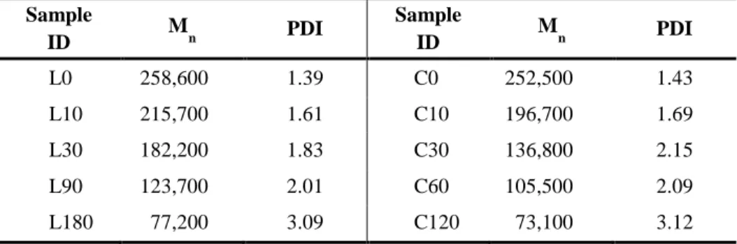
Table 4. Number average molecular weight (Mn) and polydispersity index
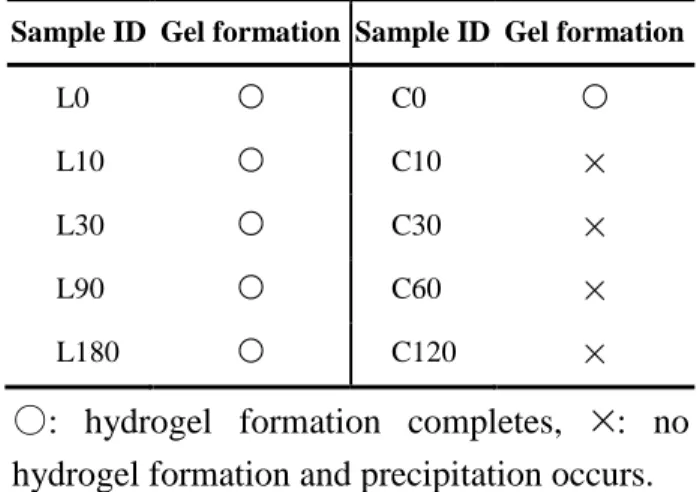
(PDI) of hydrolyzed SF aqueous solutions measured by GFC... 61 Table 5. Gel forming ability of ultra-sonicated SF aqueous solutions. ... 63 Table 6. Sample ID and preparation conditions of SFMA hydrogels. ... 94 Table 7. Amino acid composition (mol%) of un-hydrolyzed (pristine SF) and hydrolyzed SF. ... 98
viii
List of Figures
Figure 1. Electron micrograph and schematic illustration of silk fiber composed of silk fibroin (SF) and silk sericin (SS). ... 8
Figure 2. Typical regenerated forms of SF. ... 13
Figure 3. Schematic illustration of structural transition of SF molecular chains during gel formation. ... 17
Figure 4. Illustration of the formation of nanofibrillar network at different gelation stages. Inserted SEM images represent freeze-dried RSF hydrogel at different gelation states: (i) 0 hr, (ii) 1 hr, (iii) 3 hr, (iv) 20 hr after ultra-sonication. The scale bar presents 500 μm. ... 18
Figure 5. Chemical structures of most abundant reactive amino acids in SF. ( ): number of each residue among total number of amino acids, 5263. 26
Figure 6. Proposed crosslinking reaction pathways for tyrosyl residues in SF molecular chains. ... 29
Figure 7. Reaction scheme for the synthesis of hydrolyzed silk fibroin methacrylate (SFMA) using IEM. ... 43
Figure 8. Schematic of hydrolyzed silk fibroin methacrylate (SFMA) hydrogel formation under UV irradiation. ... 44
Figure 9. (A) Gel filtration chromatograms, and (B) number average molecular weight (Mn) and polydispersity index (PDI) of alkali
ix
hydrolyzed SF by HAT. ... 64
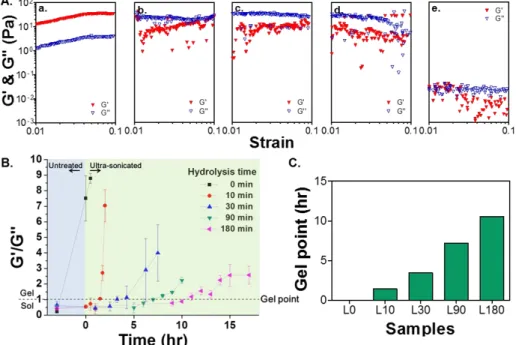
Figure 10. (A) Shear elastic and loss moduli (G' & G'') of un- (a), 10 min- (b), 30 min- (c), 90 min- (d), and , 180 min- (e) hydrolyzed 3% (w/v) SF solutions immediately after ultra-sonication. (B) G'/G'' ratio change of SF solutions with incubation time (a G′/G′′ ratio larger than 1 indicates gel state, mean ± SD, n = 3). (C) Gel point (time when G'/G'' = 1) of SF aqueous solutions formed of different molecular weights of SF. ... 66
Figure 11. (A) FT-IR spectra and (B) secondary structure contents of freeze-dried SF hydrogels formed of different molecular weights of SF. ... 68
Figure 12. Thioflavin T fluorescence profiles of SF hydrogels formed of different molecular weights of SF, red box indicates the enlarged image of initial profiles at 0-time (mean ± SD, n = 3). ... 69
Figure 13. (A) Gel fraction and (B) equilibrium swelling ratio of SF hydrogels formed of different molecular weights of SF (mean ± SD, n = 3). .. 72
Figure 14. Equilibrium shear elastic modulus of SF hydrogels formed of different molecular weights of SF (mean ± SD, n = 3, *p < 0.01). .. 73
Figure 15. Equilibrium shear elastic modulus of SF hydrogels with different ultra-sonication time (mean ± SD, n = 3). ... 75
Figure 16. (A) Images of SF hydrogels formed of different molecular weights of SF. The bottom letters depicts transparency of each hydrogel. (B)
x
Transmittance of SF hydrogels at different wavelength of visible light (mean ± SD, n = 3). ... 76
Figure 17. Two-theta scan profiles of WAXD analysis of SF hydrogels with different molecular weights of SF. ... 77
Figure 18. (A) SAXS curves, (B) Guinier plot curves, (C) Radius of gyration (derived from Guinier equation), and (D) Kratky plot curves of SF hydrogels with different molecular weights of SF... 81
Figure 19. Schematic of microstructures of un-/180 min-hydrolyzed SF hydrogels. ... 83
Figure 20. Cross-section image of freeze dried SF hydrogel (sample L0, un-hydrolyzed SF) observed by FE-SEM... 84
Figure 21. (A) Phase-contrast images of hMSC cultured on SF hydrogels of different hydrolysis times after 24-hour post-seeding (scale: 100 µm). (B) Metabolic activity of hMSC cultured on SF hydrogels of different molecular weights after 24-hour post-seeding (mean ± SD, n = 3). (C) Confocal images of hMSC on SF hydrogels after 24-hour post-seeding: (a) L0 and (b) L180 (F-actin: red, nucleus: blue)... 87
Figure 22. (A) Molecular weight distribution, (B) number average molecular weight (Mn), and polydispersity index (PDI) of the hydrolyzed SF
prepared by HAT method. ... 90
xi
SF and input IEM amount. ... 92
Figure 24. (A) 600 MHz 1H NMR spectra of 12 hr hydrolyzed SF (25_0) and SFMA (25_1.0) (D2O was used as a solvent) (B) Immobilized
amount of methacrylate group (MA) on SF and (C) reaction yield of SFMA (n = 3, mean ± SD). ... 96
Figure 25. 600 MHz 1H NMR spectra of SF and SFMA prepared with various molecular weight of SF (D2O was used as a solvent). ... 99
Figure 26. Minimum gel forming concentration of SFMA formed of different immobilized MA amount and molecular weight of SF... 102
Figure 27. In situ photo-rheometry results of SFMA precursor solutions. (A) 25_0.25, (B) 25_0.5, (C) 25_1.0, (D) 100_0.25, (E) 100_0.5, and (F) 100_1.0. UV light source was turned on at 60 s after the onset of measurements (same concentration: 12.5 wt%). ... 103
Figure 28. Gel points of SFMA hydrogels formed of different immobilized MA amount and molecular weight of SF (same concentration: 12.5 wt%) (n = 3, mean ± SD). ... 104
Figure 29. (A) Gel fraction and (B) swelling ratio of SFMA hydrogels formed of different immobilized MA amount and molecular weight of SF (same concentration: 12.5 wt%) (n = 3, mean ± SD). ... 107
Figure 30. Equilibrium shear elastic modulus (G′) of SFMA hydrogels formed of different immobilized MA amount and molecular weight of SF
xii
(same concentration: 12.5 wt%) (n = 3, mean ± SD). ... 108
Figure 31. Gel fraction of SFMA hydrogels with various concentrations. (A) 25 kDa SF, (B) 100 kDa SF (n = 3, mean ± SD). ... 110
Figure 32. Swelling ratio of SFMA hydrogels with various concentrations. (A) 25 kDa SF, (B) 100 kDa SF (n = 3, mean ± SD). ... 111
Figure 33. Equilibrium shear elastic modulus (G') of SFMA hydrogels with various concentrations. (A) 25 kDa SF, (B) 100 kDa SF (n = 3, mean ± SD). ... 112
Figure 34. Transmittance of SFMA hydrogels formed of different immobilized MA amount and molecular weight of SF at (A) 400 nm, (B) 600 nm, and (C) 800 nm of visible light wavelength (same concentration: 12.5 wt%) (n = 3, mean ± SD). ... 114
Figure 35. G′ and G′′ variation of SFMA hydrogels formed of different immobilized MA amount and molecular weight of SF at stress-sweep oscillatory mode for thixotropic property analysis (same concentration: 12.5 wt%). ... 116
Figure 36. Crossover point (G′ = G′′) of SFMA hydrogels formed of different immobilized MA amount and molecular weight of SF at stress-sweep oscillatory mode for thixotropic property analysis (same concentration: 12.5 wt%). ... 117
xiii
stress was applied (300 Pa) and released (0.1 Pa), alternatively. .... 118
Figure 38. Cyclic compression curves of SFMA hydrogels formed of different immobilized MA amount of SF and molecular weight of SF from 50 times of cyclic compressive test when 20% of compressive strain was applied (same G′ ~ 1.3 kPa). ... 120 Figure 39. S-S curve results from 3 times of cyclic compressive test (A) physically crosslinked and (B) chemically crosslinked (100_0.5 SFMA hydrogel sample) SF hydrogels. ... 121
Figure 40. Before and after images of chemically crosslinked SF hydrogel (100_0.5 SFMA hydrogel sample) when 50% of compressive strain was applied. ... 123
Figure 41. (A) (B) Changes of shear elastic modulus (G′) and (C) degradation rate constant (k′) of SFMA hydrogels formed of different immobilized MA amount on SF and molecular weight of SF (n = 3, mean ± SD). All the hydrogels were incubated in PBS at 37°C for 48 days. ... 125
Figure 42. (A) SAXS curves and Kratky plots of SFMA hydrogels formed of different immobilized MA amount on SF and molecular weight of SF. (A) 25 kDa SF, (B) 100 kDa. ... 128
Figure 43. Circular dichroism spectra of a precursor solution of SFMA formed of different immobilized MA amount on SF and molecular weight of SF. (A) 25 kDa SF, (B) 100 kDa. ... 130
xiv
Figure 44. Relative cell viability of NIH-3T3 cells cultured on SFMA hydrogel (sample ID: 25_1.0 and 100_1.0) extracts contained DMEM; (A) 25_1.0, (B) 100_1.0 (mean ± SD, n = 3). ... 132
1
I. INTRODUCTION
Silk fibroin (SF) is a major protein, which plays a critical role in both structural feature and mechanical property of silk cocoons. To date, it has been widely reported that SF can be used in biomedical applications due to excellent biocompatibility and superior mechanical property [1-6]. Besides, the facile recrystallization by alcohol treatment, inducing the physical crosslinking via intra- and inter-molecular hydrogen bonding of hydrophobic segments of SF, is generally exploited in the fabrication process for SF [7, 8]. Such a process is very useful in biomedical application fields (e.g., tissue engineering scaffold, drug carrier, etc.) since this process does not cause any harmful damaging to living organisms.
SF can be easily fabricated into various forms (e.g., film, nanofiber, sponge, hydrogel, etc.) [9]. Among various types and shapes of SF, numerous trials were progressed to develop the SF hydrogel for biomedical applications. It is because highly porous network structure of hydrogel retains a large amount of water with inter-connected porous structure, which can facilitate the encapsulation of inorganic particles, drugs, and even the living cells [6]. Moreover, structural feature of the hydrogel provides physiologically stable condition for cell survival with appropriate three-dimensional (3D) microenvironment that allows the cell survival with growth in vitro and in
vivo [10-12]. Therefore, SF hydrogel can exhibit excellent biological behavior
2
[13], poly(vinyl alcohol) [14]) as a 3D cell niche.
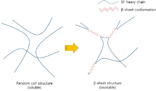
Another interesting structural feature of SF, compared to other polymeric materials, is that unique structural transition occurs during hydrogel network formation. It is known that major protein of SF chains in crystalline region is consisted of repeated glycine (G) -alanine (A) segment (GAGAGX, X = Ser or Tyr) that can build the insoluble anti-parallel β-sheet structure [15]. Meanwhile, in aqueous solution state, SF chains exist as a random-coil conformation [16]. However, it is thermodynamically unstable to maintain this structural conformation in aqueous solution. As a consequence, structural transition occurs to β-sheet conformation by accompanying with chain aggregation [17, 18]. On a macroscopic level, the SF chains finally construct physically crosslinked network structure of hydrogel via hydrogen bonding formation.
Due to its structural difference, properties of SF hydrogel are affected by a wide variety of processing parameters, such as concentration of SF solution [19, 20], incubation temperature [19], vortexing time [21], and ultra-sonic power [22]. The concentration is a major factor to determine physical properties of SF hydrogel. Generally, higher concentration results in stiffer hydrogels with a shorter gelation time. The gelation behavior is also largely dependent on the incubation temperature [19]. For example, the SF hydrogel formed at a higher temperature (~50°C) mostly has a higher Young’s modulus than that formed at a lower temperature (~4°C). The effect of amplitude of
3
shear force was also investigated. X. Wang et al. showed that the amplitude of ultra-sonication affected the mechanical properties of SF hydrogel [22]. T. Yucel et al. reported the effect of vortexing time on the shear modulus of SF hydrogel [21]. Although many studies for manipulating SF hydrogel properties were reported, none of processing variables could elicit the change of a wide range in mechanical as well as physical properties except the concentration of the precursor SF solution.
Generally, the molecular weight of polymer composing network structure of hydrogel is a crucial factor that determines the gel properties (i.e., modulus, swelling ratio, permeability) [23-25]. Both crosslinking density and mesh size of hydrogel are largely dependent on the polymer chain length regardless of crosslinking methods (i.e., chemical or physical crosslinking) [26, 27]. Nevertheless, the effect of molecular weight of SF on hydrogel fabrication has not been explored. Hence, in this study, the heat-alkaline treatment (HAT) was conducted during SF dissolution step to control the molecular weight of SF as a one-pot process. This method is not only simple but also suitable to obtain a high yield of hydrolyzed SF. In contrast, proteinase treatment needs relatively delicate process including enzyme removing process and causes severe mass loss of product in spite of high efficiency [28].
After the HAT, hydrolyzed SF was used for the fabrication of hydrogel using ultra-sonication method. Then, physical and mechanical property analyses were performed in order to investigate the effect of molecular weight of SF on
4
the hydrogel formation. By controlling the molecular weight of SF, it was possible to manipulate the structure, performance and physical properties of physically crosslinked SF hydrogel in a wide range for their proper applications.
Nevertheless, poor mechanical property of typical SF hydrogel, which being vulnerable to shear deformation (e.g., bending, twisting, etc.), was pointed out as a limitation of real applications in tissue engineering. It is because hydrogen bonding formed in physical crosslinking is inherently weak against shear deformation. Also, crystallinity of physically crosslinked SF hydrogel is too low even though β-sheet crystalline structure formed. Regenerated SF generally shows a lower degree of crystallinity compared to natural SF fibers [29]. In case of regenerated SF film, it becomes brittle after insolubilization and crystallization treatment, in which SF chains are transformed into β-sheet crystalline conformation. Most of regenerated SF forms exhibit poor mechanical behavior unless chemical treatment is carried out.
To enhance the mechanical property of SF hydrogel, introduction of chemical crosslinking might be an alternative strategy for its better performance. Many chemical crosslinkers were investigated for the linkage of hydroxyl or amine groups in SF [30-34]. Enzymes (e.g., horseradish peroxidase [35], tyrosinase [36], etc.) can also be utilized for chemical crosslinking of SF in aqueous solution state via di-tyrosine linkage formation. This enzyme treatment is considerably biocompatible compared to usage of
5
chemical crosslinkers. Recently, M.B. Applegate et al. reported that photo-initiator, riboflavin, could be used for photo-crosslinking of SF hydrogel via inter-covalent bond formation between tyrosine groups in SF and this SF hydrogel showed high resilience property [37]. However, chemical crosslinking strategies still have some limitations in an application of SF hydrogel because fresh SF aqueous solution is required for hydrogel fabrication. Substantially, SF aqueous solution is difficult to manipulate and hard to maintain its pure state due to innate instability.
Here, a new fabrication method was developed for making chemically photo-crosslinked SF hydrogel with excellent physical properties and high performance. First, the alkaline hydrolysis was conducted to manipulate a molecular weight of SF, which can make enhancing a stability of SF in aqueous solution. Consequently, this strategy is feasible for the application as ready-to-use SF hydrogel without an instant preparation of fresh SF aqueous solution. In addition, this allowed direct chemical modification of SF without any precipitation during chemical reaction and dialysis process. It has been reported that due to the instability of SF, it was hard to modify the SF chemically in aqueous solution state except hydrophilic silk sericin [38].
In this study, photo-crosslinking method was utilized by introducing methacrylate (MA) groups onto SF, with a use of photo-initiator. Using the hydrolyzed SF, MA-immobilized SF (silk fibroin methacrylate, SFMA) was synthesized and photo-crosslinkable SF hydrogel was then fabricated. The
6
structure, physical properties, and performance of the chemically photo-crosslinked SF hydrogel (SFMA hydrogel) were evaluated on the effect of immobilized MA amount on SF.
Therefore, in the first part, the physically crosslinked SF hydrogels with various molecular weights of SF were fabricated and the effect of its molecular weight on microstructure and hydrogel properties was investigated using a novel hydrolysis technique (HAT method). For the second part, the chemically photo-crosslinked SF hydrogel was fabricated using the hydrolyzed (molecular weight controlled) SFMA and evaluated its microstructure and properties. In order to find out its excellent physical property and performance, gelation behavior, gel property, transparency, thixotropic property, resilience, and degradation behavior of the photo-crosslinked SF hydrogel were intensively studied for determining its feasible use in biomedical applications.
7
II. LITERATURE SURVEY
2.1. SILK FIBROIN (SF) AS A BIOMATERIAL
Silk fibroin (SF) is a major fibrous protein, which plays a critical role in both structural feature and mechanical property of silk cocoons, which are produced by silkworms (Bombyx mori). In raw silk fibers, two SF strands are covered with a gum-like adhesive protein, known as silk sericin (SS) (Figure 1) [39], which occupies 25-30 wt% of total silkworm cocoon. The SF consists of a heavy chain (Mw ~ 390 kDa) and a light chain (Mw ~ 25 kDa) linked by a
disulfide bond [9, 40]. In silkworm gland, the SF exists as a sol state with a random coil conformation. During spinning process of fiber formation, the SF becomes concentrated, pH of the solution is changed to acidic condition, and shear stress is applied [41, 42]. These environmental changes tend to occur the structural transition of hydrophobic repeating unit of SF (Gly-Ala-Gly-Ala-Gly-X, X = Ser or Tyr) into a crystalline solid state with insolubilization. The β-sheet conformation of macromolecular chains is a well-known secondary structure of SF fiber. When silk fiber is spun from the silkworm spinneret with stretching, the β-sheet crystal domains are highly aligned to longitudinal direction of fiber axis. Consequently, raw silk fiber shows excellent tensile property due to its highly oriented crystalline structure. Ultimate tensile strength of B.mori silk fibers is 740 MPa. While, collagen and poly (lactic acid) (PLA) has ultimate tensile strength of 0.9-7.4 MPa and 28-50 MPa, respectively [43].
8
Figure 1. Electron micrograph and schematic illustration of silk fiber composed of silk fibroin (SF) and silk sericin (SS) [39].
9
SF attracts lots of attention as a biomaterial due to its unique structural characteristics and properties. Most protein materials used for biomedical applications are derived from tissues of allogeneic or xenogeneic origins. Such proteins have a potential risk for the infection from animal sources. On the other hand, SF protein, which is originated from insects, cocoon or spider web, might be safer than other animal protein sources. It has a good biocompatibility with minimal immunogenicity. It has been reported that the SF could be degraded into non-toxic products when implanted in vivo [44]. Most of all, unique structural characteristics and properties of SF, such as highly crystalline β-sheet structure and excellent mechanical property with high tensile strength can give better performance for end uses as a biomaterial. Potential risks of chemicals used for biomaterials fabrication might be a trouble when they are implanted into patient. Compared to other biopolymers, SF can be easily fabricated to various forms and shapes with insolubilization and crystallization simply by alcohol treatment, in which the alcohol is removed completely by washing and drying process. Therefore, the SF has a numerous potential in applications of biomedical fields.
In an economical point of view, cost of SF material is very low compared to other natural or synthetic biopolymers used in biomedical applications. Especially, in case of collagens, the raw material is very expensive due to its high cost and complexity of isolation and purification process. The SF can be simply purified from silkworm cocoons by weak alkaline treatment which
10
removes SS and other impurities. The processing cost for biomedical grade products is relatively low compared with that of other animal proteins. In addition, the SF annually has produced 1,000 metric tons per year in a world and the raw materials of SF can be easily obtained in a form of cocoons or fibers in a cheap price [2]. Such an economical advantage is favorable for a mass production of SF biomedical products.
11
2.2. DISSOLUTION AND REGENERATION OF SF
In general, SS is removed from silkworm cocoon by degumming process prior to use of silk fiber. This process is well known to textile industry for producing degummed silk fiber. Even when silk protein is used in biomedical applications, the soluble SS, which is coated on SF, should be removed before next regeneration step. Adequate removal of contaminating SS from silkworm silk may be required to avoid biocompatibility problems. It was reported that remained SS caused the immune response in vivo [43]. The SS can be removed easily by weak alkaline boiled solution. Sodium carbonate (Na2CO3)
is mainly used for weak alkali reagent but sodium oleate (CH3(CH2)7CH=CH(CH2)7COONa) is added to Na2CO3 in order to prevent
the molecular weight decrease of SF. In severe conditions of degumming process, degradation of SF chain occurs and properties of SF is changed. Especially, SF can be damaged by long degumming process so that degumming time should be carefully controlled for a reproducibility of SF. When the degumming process is not properly carried out for a uniform degumming ratio, there are many difficulties in processing and the final SF products show different properties and performance.
The degummed SF has a well ordered crystalline structure with high crystallinity and orientation. Due to the hierarchical order in supramolecular structure and organization with high molecular weight, the SF is insoluble in water and most organic solvents. Truly, it is hard to dissolve the SF (usually in
12
a fiber form) without degradation. However, it has been known that highly concentrated salt solution (e.g., LiBr, CaCl2,CaNO3, etc.) were used for the
dissolution of SF. The dissolution mechanism is that at a certain concentration condition, the salt effectively penetrates crystal regions of SF and disrupts hydrogen bonding of β-sheet structure [16]. After sufficient dialysis process, pure SF aqueous solution can be obtained and SF chains exist as a random coil conformation in the solution. It was reported that LiBr solvent system rarely damaged the SF molecular chains during dissolution process while CaCl2 solvent system severely cleaved the SF chains [45]. In this reason, short
dissolution time (within 3 min) is recommended for CaCl2-based solvent
system.
After dissolution and dialysis process, clear SF aqueous solution can be acquired. In general, freeze drying is conducted to make a sponge form of regenerated SF for further processing of SF into various forms. Because this regenerated SF is amorphous and consists of random coil conformation, it can be dissolved completely in acidic solvent (e.g., formic acid, trifluoroacetic acid, etc.) or fluorinated alcohol (e.g., 1,1,1,3,3,3-hexafluoro-2-propanol, etc.) [15, 46, 47]. Using suitable solvent systems, various types of regenerated SF, such as powder, particle, fiber, film, hydrogel, membrane, web, etc., can be fabricated for a proper application in biomedical field (Figure 2) [48]. Sponge type SF, which is fabricated by freeze drying of SF aqueous solution, is highly porous, providing a favorable environment in 3D for cell spreading,
13
14
migration, and proliferation [49-51]. SF hydrogel is also easily fabricated using SF aqueous solution by forming of crosslinked network structure [6]. The hydrogel has a hydrophilic nature and highly porous structural environment in a wet condition, which can facilitate the 3D cell culture and cell encapsulation.
SF regenerated fiber can be fabricated by typical wet spinning process. When the SF dope solution is directly spun into a coagulation bath (precipitation in alcohol containing bath), several micron sized fiber can be obtained after stretching and drawing process [52, 53]. Electrospinning method can provide the fibers of several tens to hundreds of nanometer size in diameter, depending on the electrospinning conditions [54-56]. Especially, SF nanofiber web has a highly porous structure with interconnected fiber network. Porous structural features of this nanofibrous electrospun SF web can provide a favorable environment in cell culture and therefore, it has a high potential for a use in tissue engineering scaffold. Film or membrane can also be fabricated. After solvent casting and evaporation process, thin and transparent SF film were prepared and analyzed [57-59]. Solvent emulsion and electrospraying method were used for a fabrication of SF microsphere which had a small diameter within several tens of micron, being able to apply in drug delivery system [60-62].
15
2.3. SF HYDROGEL
2.3.1. General characteristics of hydrogel
Hydrogel is composed of 3D polymeric network which retains a large amount of water inside. According to a crosslinking mechanism, it is classified into two categories, physically crosslinked and chemically crosslinked hydrogel [6]. Polymeric network of physically crosslinked hydrogel is held together by secondary forces such as hydrogen bond, ionic bond, and hydrophobic interaction. On the contrary, chemically crosslinked hydrogel is linked by covalent bonds. Due to these polymeric network structures, hydrogel is basically elastic and possesses tremendous interconnected pores, in which lots of water molecules are entrapped. The main reason for wide applications of the hydrogel is close resemblance of its structural features and physical properties to those of biological tissues [63]. Highly porous 3D structure provides physiologically similar environment to cells that it has a potential in tissue engineering field.
16
2.3.2. Physically crosslinked SF hydrogel
2.3.2.1. Gelation mechanism
Compared to other polymeric materials, SF chains can make physical crosslinking in solution state via hydrogen bonding formation, which is attributed to the structural transition of SF into insoluble β-sheet structure (Figure 3) [17, 18]. In solution state, a random coil conformation of SF is thermodynamically unstable, so that it prefers to be changed into stable β-sheet conformation. The structural transition is irreversible and hydrogel once formed cannot be recovered to original solution state. In addition to structural transition, aggregation and entanglement of SF molecular chains also plays a critical role in hydrogel formation. As a result of molecular chain association, it was reported that several tens of nanometer diameter of SF particles could be formed [64]. As gelation proceeds, these particles gradually aggregated into larger size by forming hydrogel network structure (Figure 4) [18, 29]. On a macroscopic level, the SF chains finally construct physically crosslinked network structure of hydrogel, which consists of β-sheet crystalline structure of SF. Due to its crystalline nature, SF hydrogel exhibits opaque optically [17].
2.3.2.2. Strategies for hydrogel fabrication
Alcohols (e.g., methyl alcohol, ethyl alcohol, etc.) are mainly used for insolubilization of SF, which is attributed to promote its crystallization. When SF aqueous solution is treated with alcohols, water molecules are removed
17
Figure 3. Schematic illustration of structural transition of SF molecular chains during gel formation.
18
Figure 4. Illustration of the formation of nanofibrillar network at different gelation stages. Inserted SEM images represent freeze-dried RSF hydrogel at different gelation states: (i) 0 hr, (ii) 1 hr, (iii) 3 hr, (iv) 20 hr after ultra-sonication. The scale bar presents 500 μm [29].
19
temporarily from the SF chains (known as dehydration), which can facilitate the β-sheet structural transition of SF [65]. Such a transition occurs instantly in a very short time that alcohol induced SF hydrogel forms heterogeneous network structure mostly. Acetone was also used for a preparation of SF hydrogel in order to facilitate structural transition [66]. When the organic solvents are used for SF hydrogel fabrication, there might be a potential risk of cytotoxicity due to residual solvent remained.
Gelation temperature is an important factor for the gel formation. Gelation of SF aqueous solution can be promoted at high temperature condition. Because hydrophobic regions of SF tend to be separated from its hydrophilic regions at a relatively high temperature, the formation of aggregate becomes more preferable [64]. Such an aggregate formation is also affected by a concentration of SF aqueous solution. It is because possibility of molecular chain associations increase with an increment of concentration and aggregated/entangled regions are easily formed for gel formation [17, 19]. Another important parameter for affecting gel formation is pH. Physical crosslinking of SF is known to be facilitated at acidic condition [17]. At neutral pH condition (pH ~ 7), SF particles in aqueous solution have negative surface charges, which can interrupt the aggregation of SF due to a negative charge repulsion. On the other hand, possibility of chain aggregation increases when the pH of SF aqueous solution lowers to an isoelectric point of SF (pI ~ 4.3). Hence, the hydrogel formation of SF aqueous solution is preferable at an
20
acidic pH condition. Indirect method for controlling the pH of SF aqueous solution were proposed by M.L. Floren et al. and R.R. Mallepally et al [67, 68]. To acidifying the SF aqueous solution, they developed carbon dioxide (CO2) gas bubbling technique for making SF hydrogel with unique structural
characteristics. When CO2 is applied to SF aqueous solution, CO2 reacts with
water to form carbonic acid (H2CO3). Protons (H +
) produced from H2CO3
finally lower the pH of the solution. Here, the applied CO2 can acidify the
whole part of SF aqueous solution, and as a result, homogeneous network structure of SF hydrogel is obtained.
In general, gelation conditions, such as temperature, concentration, pH, etc., should be adjusted for a proper gelation of SF. At ambient conditions, however, it takes several days or weeks for the gel forming. Therefore, it is necessary to accelerate gelation time so that gel formation can be done within 1 hour. It is well-known that hydrogel formation can be accelerated by external physical stimulation at same gelation conditions. Shear stress applying method, simply by tilting or agitating solution, is a mostly utilized strategy for shortening the gelation time of SF hydrogel. This stimulation can induce the aggregation of SF chains and promote the gel formation [21]. By controlling the amplitude and applying time of shear stress, both gelation time and gel strength of SF hydrogel were manipulated. However, a precise control of gelation behavior and physical property of hydrogel was impossible even though vortexing induced shear stress applying method used. It is because SF
21
chains prefer not a gel formation but a precipitation during vortexing treatment.
Ultra-sonication method is suggested as an alternative for making homogenous SF hydrogel structure. Not only ultra-sonication quickly induces gel transition of SF aqueous solution but also it can build up hydrogel network structure without precipitation [22, 69]. Similar to shear stress treatment, ultra-sonic wave highly enhances a frequency of molecule movement, which facilitates the aggregation of SF molecules. In addition, ultra-sonic wave can be distributed to whole parts of SF aqueous solution evenly so that this hydrogel has a homogeneous network structure with high mechanical strength and shear modulus. The ultra-sonication method also has a good reproducibility for hydrogel fabrication.
Electronic stimulation method was also introduced for the formation of physically crosslinked SF hydrogel [70, 71]. When the electric field applied to SF aqueous solution, a random coil conformation of SF changed to metastable state of α-helix conformation. Contrary to the shear stress applying method, such a structural transition can be temporarily recovered into a solution state if the applied electric field is removed. When the high voltage is applied to metastable state of SF, SF chains become elongated to the direction of current flow, resulting that α-helical conformation of SF changed to β-sheet conformation. This structural transition behavior finally constructs the physically crosslinked network of SF hydrogel.
22
In other way, additives can be used to induce and promote hydrogel formation. It was reported that poly(ethylene oxide) [19], poloxamer [72], N-lauroyl sarcosinate [73], glycerol [74], and sodium dodecyl sulfate [75, 76] were used as additives for SF hydrogel fabrication in order to promote the gelation of SF aqueous solution. The optimum amounts of additives exist for facilitating the gel transition of SF. This method has a limitation to be used because additional washing is required for removing residual additives from the hydrogel.
All the physical crosslinking methods utilized an innate structural characteristic of SF polymer in which hydrogel network structure was formed by self-aggregation of SF chains and structural transition of β-sheet conformation. Additional chemical modification or chemical crosslinker treatment are not required for the hydrogel formation. Therefore, only physically crosslinked SF hydrogel has a big advantage in biomedical applications due to its high biocompatibility. Nevertheless, there are some limitations remained for the use of this hydrogel. A brittleness, which is vulnerable to shear deformation (e.g., bending, twisting, etc.), is pointed out as one of drawbacks for the SF hydrogel. Poor mechanical property of physically crosslinked SF hydrogel may be due to inherently weak intermolecular hydrogen bonding against shear deformation in addition to a low degree of crystallinity of regenerated SF hydrogel [29]. To enhance the mechanical properties of SF hydrogel markedly, physical crosslinking is not
23
sufficient and consequently, introduction of chemical crosslinkers should be necessary for the better performance of the SF hydrogel.
24
2.3.3. Chemically crosslinked SF hydrogel
2.3.3.1. General characteristics
Generally, chemically crosslinked hydrogel has more rigid and resilience mechanical property than physically crosslinked one. Besides, chemical crosslinking method can easily manipulate physical property of hydrogel in a wide range. It is because its property is mainly governed by crosslinking density of hydrogel. Such a crosslinking density can be modulated by controlling the input amount of crosslinker or amount of crosslinkable functional group of polymer. Many chemical crosslinking methods were proposed for the hydrogel fabrication of natural polymers. In case of SF hydrogel, SF polymer contains reactive functional moieties of hydroxyl (i.e., serine, tyrosine, and threonine), carboxyl (i.e., aspartic acid and glutamic acid), and amine (i.e., arginine and lysine) groups (Table 1) [1, 77]. Figure 5 shows major functional groups of SF, which can be involved in chemical crosslinking reaction.
Compared to physical crosslinking, chemical crosslinking method has noticeable advantages for better performance of hydrogel. This method has a potential risk of cytotoxicity, however mechanical properties of chemically crosslinked hydrogel are superior due to network structural formation via intermolecular covalent bonds. Moreover, a gelation time can be controlled and predictable in chemical crosslinking. When this chemically crosslinked
25
Table 1. Amino acid composition of the heavy chain of silk fibroin [77].
Amino acid # Residues Mol %
Ala (A) 1593 30.3 Arg (R) 14 0.3 Asn (N) 20 0.4 Asp (D) 25 0.5 Cys (C) 5 0.1 Gln (G) 10 0.2 Glu (E) 30 0.6 Gly (G) 2415 45.9 His (H) 5 0.1 Ile (I) 13 0.2 Leu (L) 7 0.1 Lys (K) 12 0.2 Met (M) 4 0.1 Phe (F) 29 0.6 Pro (P) 14 0.3 Ser (S) 635 12.1 Thr (T) 47 0.9 Trp (W) 11 0.2 Tyr (Y) 277 5.3 Val (V) 97 1.8
Total number (#) of amino acids: 5263 Molecular weight: 391,593 Da
26
Figure 5. Chemical structures of most abundant reactive amino acids in SF. ( ): number of each residue among total number of amino acids, 5263.
27
hydrogel is used for biomedical applications, its biocompatibility should be evaluated and suitable fabrication method of this hydrogel should be also developed.
2.3.3.2. Chemical crosslinkers
Among many chemical crosslinking agents, aldehyde type crosslinkers, such as formaldehyde, glyoxal, and glutaraldehyde, are mostly used for the crosslinking of proteins. It has been reported that amine groups of SF are participated in the crosslinking formation with high reaction yield when glutaraldehyde crosslinker was used in alkaline condition [32]. Even though the glutaraldehyde is frequently used for the crosslinking of SF, it is hard to make highly crosslinked network structure of SF hydrogel due to a low content of amine group (0.5 mol%) in SF. In acidic condition, on the contrary, hydroxyl groups, which are largest amount of reactive groups in SF protein (14.2 mol%), might be involved in chemical crosslinking by forming ether bond bridges between these groups for SF hydrogel fabrication. However, proper buffer solution is required to avoid precipitation of SF during crosslinking reaction.
Epoxide type crosslinkers are also popular for the chemical crosslinking formation of polymeric materials. Among them, epichlorohydrin and diglycidyl ether showed a high reactivity to link both of amine group and hydroxyl group of SF for hydrogel fabrication [30, 31, 33, 78]. Even though
28
these crosslinkers can easily make chemical crosslinked SF hydrogel, residual chemicals might have a toxicity problem and give a limitation of use in biomedical application. Moreover, this type of chemical reaction can lower the both of cell viability and drug activity when the cells and drugs are encapsulated in hydrogel network because of non-specific crosslinking reaction.
Often the chemical crosslinking method requires severe biological evaluations for the safety. Most of chemical crosslinkers are toxic and restricted to be used in biomedical fields. Nowadays, bioactive functional materials from natural sources tend to be used for the higher cytocompatibility. Genipin, which is a natural protein extracted from gardenia seeds (Gardenia
Jasminoides Ellis), was introduced as a biocompatible crosslinker. W.H.
Elliott et al reported a possible use of genipin, which could interlink amine groups of protein by covalent bond, for the fabrication of SF hydrogel [34]. The genipin crosslinker showed much lower cytotoxicity and its use in biomedical application was proposed. However, this type of crosslinking reaction requires long reaction time for hydrogel network formation, which makes some limitations to use, for example, in cell encapsulation.
Enzyme can be used for the crosslinking formation. In case of SF, tyrosinase-induced crosslinking pathways were proposed (Figure 6) [79]. SF contains about 5 mol% of tyrosine and this moiety can be crosslinked by tyrosine-based enzyme. When tyrosinase is added to SF aqueous solution,
29
Figure 6. Proposed crosslinking reaction pathways for tyrosyl residues in SF molecular chains [79].
30
tyrosine residue of SF subsequently oxidized to dopa and dopaquinone. Final oxidized product, dopaquinone, can be reacted with amine group via Schiff-base reaction or same tyrosine group via Michael reaction by covalent bonding linkage. Such an enzyme-based chemical reaction is occurred at physiological condition, which makes an advantage in biomedical field. Moreover, crosslinking density can be controlled by amount of enzyme and dissolved oxygen. However, this enzyme-induced crosslinking still requires a longer reaction time for the hydrogel formation. Recently, B.P. Partlow et al. used horseradish peroxidase enzyme for chemical crosslinking of silk protein and developed highly tunable elastomeric functional SF hydrogel [35]. By increasing the concentration of dissolved oxygen, the chemical reaction could be completed within several minutes. Consequently, the gelation time was remarkably reduced and as a result, it was feasible to use the chemically crosslinked SF hydrogel in cell encapsulation.
2.3.3.3. Photo-crosslinking system
As shown in enzyme-based chemical reaction methods, tyrosine residue is the mostly reactive functional group for the chemical crosslinking of SF. In the same way, photo-crosslinking method can be applied to the SF due to active photo-reaction of tyrosine groups. J.L. Whittaker et al. proposed facile and rapid ruthenium mediated photo-crosslinking of Bombyx mori SF for the fabrication of photo-crosslinkable SF hydrogel [80]. They used tris(2,2-bipyridyl) dichloro ruthenium (II) hexahydrate (Ru(II)(bpy)3
2+
31
and ammonium persulfate as an electron acceptor, respectively. Under visible light, Ru(II)(bpy)3
2+
makes tyrosine radical. Subsequently, this radical reacts with another tyrosine and make di-tyrosine linkage. Covalent crosslinked SF hydrogel was successfully prepared but this hydrogel showed red light color due to the presence of Ru(II)(bpy)3
2+
. Recently, M.B. Applegate et al. reported new method for SF hydrogel fabrication via di-tyrosine linkage using photo-initiator [37]. Radicals produced from photo-photo-initiator, riboflavin, made a covalent bonding between tyrosine residues of SF and this photo-crosslinked hydrogel showed highly transparent optical property, which can make possible to use for ocular prostheses. However, this reaction required longer reaction time compared to other photo-crosslinking reaction systems (more than 1 hr).
32
2.3.4. Biomedical applications of SF hydrogel
2.3.4.1. Bone regeneration
SF hydrogel has been majorly developed as bone regenerative scaffold due to its rigid characteristic. M. Fini et al. reported a possible use of SF hydrogel as a bone scaffold [81]. They implanted the SF hydrogel in critical size cancellous defects of rabbit for 12 weeks and found new bone formation in SF hydrogel without immune reaction. P.H.G. Chao et al. also reported that SF hydrogel could promote the proliferation and differentiation of chondrocytes [82]. Compared to agarose hydrogel, SF hydrogel promoted the production of glycosaminoglycan and type II collagen from chondrocytes. To improve the resilience of SF hydrogel, M. Parkes et al. used poly (ethylene diglycidyl ether) for crosslinking of SF aqueous solution [33]. This crosslinker enhanced the mechanical property of SF hydrogel, which is sufficient for application in cartilage regeneration.
Bioactive materials were also blended with SF hydrogel for enhancing the bone regeneration. VEGF (vascular endothelial growth factor) and BMP-2 (bone morphogenic protein-2) were delivered to injectable sonication-induced silk hydrogel for regeneration of maxillary sinus floor [83]. These additives effectively promoted new bone formation. Bone major ingredient, hydroxyapatite, was also incorporated with SF hydrogel for bone regeneration. For a mineralization of hydroxyapatite, SF hydrogel was soaked in calcium
33
ion (Ca2+) and phosphate ion (PO4
2-) containing solution, alternatively [84]. After repeated soaking treatment, hydroxyapatite layer was coated on SF hydrogel and this could promote the proliferation and differentiation of human osteoblast like cells (MG-63). Hydroxyapatite nanoparticle containing SF composite hydrogels were also developed and evaluated for the performance [85, 86]. Homogeneous mixing or coating of nano-hydroxyapatites onto silk hydrogel is important for fabricating the composite material and this composite SF hydrogel showed an enhanced osteogenic induction and differentiation for bone regeneration.
2.3.4.2. Cell encapsulation
SF hydrogel can be applied as a cell encapsulation matrix only if gelation time is controlled within several minutes for hydrogel formation. Many studies were carried out for potential application of SF hydrogel in the use of cell encapsulation. Ultra-sonication is the easiest and most popular gelation facilitating method for fabricating physically crosslinked SF hydrogel. X. Wang et al. encapsulated mesenchymal stem cells (MSCs) in SF hydrogel matrix using ultra-sonication with high cell viability [22]. The MSCs were also co-encapsulated with splenocytes in SF hydrogel for enhancing the secretion of islet proteins [87]. W. Sun et al. successfully encapsulated neural stem cells (NSCs) in IKVAV peptide immobilized SF hydrogel [88]. It was found that laminin derived peptide, IKVAV, effectively promoted the growth of NSCs. Enzyme-based crosslinking method is another route for fabricating
34
the SF hydrogel and possible use in cell encapsulation was studied for this hydrogel matrix. B.P. Partlow et al. confirmed that MSCs could be survived during enzyme-induced SF hydrogel network formation, indicating that oxygen radicals rarely affect the viability of MSCs [35].
Other studies were also investigated for the use of SF hydrogel as stem cell culture matrix by blending with natural biopolymers in gel formation. According to Z. Gong et al. report, injectable thixotropic hydrogel comprising SF and hydroxypropyl cellulose (HPC) could be successfully fabricated for the use of cell encapsulation [89]. During gel formation (temperature transition), the cells were entrapped well in hydrogel network without any cell death. This is based on that HPC have a thermosensitive property and SF aqueous solution can form hydrogel at above temperature of lower critical solution temperature (LCST). K. Ziv et al. reported the alginate blended SF hydrogel scaffold for stem cell culture and transplantation [90]. When the SF/alginate blended aqueous solution was exposed to calcium ions, interpenetrating polymer network (IPN) structure was formed and embryonic stem cells (ESCs) could be encapsulated in SF/alginate hydrogel with a good cell viability.
2.3.4.3. Drug delivery
Due to its unique structures and physical properties, the hydrogel is frequently used in drug delivery system. During gel formation, drugs can be
35
easily loaded on the hydrogel network. There were several reports for the evaluation of SF hydrogels for drug delivery [91-93]. Crosslinking density of hydrogel is greatly affected by a concentration of precursor solution. In general, hydrogel made from high concentration precursor solution has dense hydrogel network structure, which interrupts the releasing of drug from the matrix. Lyophilized SF hydrogel was fabricated for the sustained local delivery of therapeutic monoclonal antibodies [92] and doxorubicin-loaded SF hydrogel for the focal treatment of primary breast cancer [94]. In case of doxorubicin-loaded SF hydrogel, it was injectable and showed the toxicity toward human breast cancer cells (MCF-7).
Compared to other polymeric hydrogels, the SF hydrogel is rarely swelled in water. Swelling property is the important factor for drug delivery matrix because entrapped drugs can be released well in loose (swelled) network structure. In this reason, synthetic polymers, such as poly (acrylamide) and poly (vinyl alcohol), were blended with SF for hydrogel formation [95, 96]. These blended SF hydrogel showed interpenetrating network structure for controlled drug release and the release rate could be increased due to enhancing swelling property of the hydrogel. It was also reported that SF/copolymer composite hydrogel was fabricated and evaluated. PLA-PEG-PLA tri-block copolymer was blended with SF for controlling release rate of hydrophilic/hydrophobic drugs from the composite hydrogel network [97].
36
2.3.4.4. Miscellaneous
It was reported that gold nanoparticles embedded SF hydrogel (known as inorganic/organic hybrid hydrogel) was developed for treatment of skin infection [98]. When the gold nanoparticle was irradiated by laser beam, it produced the heat up to 60°C, which killed both of gram positive (+) (Staphlococcus aureus) and gram negative (-) (Escherichia coli) bacteria in subcutaneous. Therefore, focal infection treatment was achieved by using laser-mediated heating of injectable silk hydrogel with gold nanoparticles.
Recently, several trials were progressed to apply the SF hydrogel as ocular prostheses. When anti vascular endothelial growth factor (VEGF) loaded SF hydrogel was implanted to damaged cornea, it could suppress the vascularization without immune reaction [99]. In case of photo-crosslinked SF hydrogel prepared using riboflavin, it showed highly transparent optical property and greatly assisted the regeneration of cornea [37].
37
III. MATERIALS AND METHODS
3.1. MATERIALS
Bombyx mori (B.mori) silkworm cocoons were obtained from NIAS, RDA
(National Institute of Agricultural Sciences, Rural Development Administration) of Korea. B.mori silkworms were grown at Yeongdeok Taeyang Farm in Korea for obtaining medical grade silkworm cocoons.
Sodium oleate and calcium chloride were purchased from Tokyo Chemical Industry and Yakuri, respectively. Lithium bromide 1-hydrate and 2-isocyanatoethyl methacrylate were purchased from Kanto Chemical. The other chemicals were obtained from Sigma-Aldrich. All the reagents were used without further purification.
38
3.2. DISSOLUTION AND HYDROLYSIS OF SF
To remove SS, silkworm cocoons were boiled in 0.3% (w/v) sodium oleate and 0.2% (w/v) sodium carbonate cocktail solution at 100°C for 1 hr. The SF aqueous solutions were obtained by using two different dissolving methods in this study. Especially, the hydrolysis and molecular weight control of SF were carried out using HAT method during the SF dissolution step. For LiBr-dissolution method, the degummed (SS removed) cocoons were dissolved in 9.3 M LiBr solution at 80°C for 30 min. To hydrolyze SF, 0.6 M sodium hydroxide aqueous solution was directly added to the SF solution at a volume ratio of 1-to-5. Then, the final concentration of sodium hydroxide became 0.1 M in the SF solution. To control a molecular weight of SF, hydrolysis time was varied from 10 to 180 min, followed by subsequent dialysis against de-ionized water using cellulose acetate dialysis tube (MWCO: 12-14 kDa) for 3 days.
On the other hand, in case of CaCl2-dissolution method, the degummed
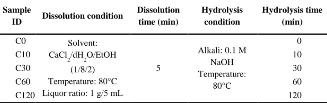
cocoons were dissolved in a ternary solvent of a CaCl2/dH2O/EtOH (molar
ratio 1/8/2) at 80°C for 5 min. The hydrolysis was directly performed at 80°C for 10 to 120 min, followed by subsequent dialysis against de-ionized water using cellulose acetate dialysis tube (MWCO: 12-14 kDa) for 3 days.
39
3.3. FABRICATION OF SF HYDROGEL
3.3.1. Physically crosslinked SF hydrogel
After dialysis, SF solutions were centrifuged at 3,000 g for 10 min to remove insoluble aggregates. The final concentration of SF solution was in the range of 3.5-4% (w/v) and each solution was diluted to 3% (w/v) concentration. The prepared SF solutions were stored at 4°C prior to gel fabrication process.
To initiate the gelation of SF solution, ultra-sonication was performed on 3% (w/v) SF aqueous solution of different molecular weights using an ultra-sonic processor (VCX-130, SONICS) at 32.5 W amplitude for 3 min. The treatment was conducted in an ice chamber to prevent the heat elevation during ultra-sonication. Then, ultra-sonicated SF solution was filtered and incubated at 60°C for 2 days for preparing physically crosslinked SF hydrogel. The detail preparation conditions and sample ID were listed in Table 2 and Table 3.
40
Table 2. Sample ID and preparation conditions of alkali hydrolyzed SF solutions using LiBr-HAT method.
Sample ID Dissolution condition Dissolution time (min) Hydrolysis condition Hydrolysis time (min) L0 Solvent: 9.3 M LiBr Temperature: 80°C Liquor ratio: 1 g/ 5mL 30 Alkali: 0.1 M NaOH Temperature: 80°C 0 L10 10 L30 30 L90 90 L180 180
41
Table 3. Sample ID and preparation conditions of alkali hydrolyzed SF solutions using CaCl2-HAT method.
Sample ID Dissolution condition Dissolution time (min) Hydrolysis condition Hydrolysis time (min) C0 Solvent: CaCl 2/dH2O/EtOH (1/8/2) Temperature: 80°C Liquor ratio: 1 g/5 mL 5 Alkali: 0.1 M NaOH Temperature: 80°C 0 C10 10 C30 30 C60 60 C120 120
42
3.3.2. Chemically photo-crosslinked SF hydrogel
After dialysis, SF solutions were centrifuged at 15,000 g for 20 min to remove insoluble aggregates. Then, supernatant was freeze-dried and stored at 4°C prior to chemical modification of SF. To synthesize SFMA, previously reported method for methacrylate-functionalized SS was applied [38]. Briefly, the hydrolyzed SF was dissolved in 1 M lithium chloride (LiCl) dissolved dimethyl sulfoxide (DMSO). Subsequently, 2-isocyanatoethyl methacrylate (IEM) was directly added to the SF solution at 60°C for 5 hr under nitrogen atmosphere (Figure 7). The final concentrations of SF and IEM in the reaction solution were 5 wt% and 0.25-2 mmol per 1 g of SF, respectively. Then, the solution was diluted with 10 × volume de-ionized water and dialyzed against de-ionized water at room temperature for 3 days using cellulose acetate tube (MWCO: 12-14 kDa). After dialysis, SFMA solutions were centrifuged at 15,000 g for 20 min to remove insoluble aggregates. Then, supernatant was freeze-dried and stored at 4°C until gel fabrication.
For preparing precursor solutions, SFMA and 1mM lithium phenyl-2,4,6-trimethylbenzoylphosphinate (LAP) were dissolved in pH 7.4 phosphate buffered saline (PBS) with varying SFMA concentrations (10 to 20 wt%). After vortexing, the solution was placed in the gap between two glass slides (gap size: 1 mm), followed by UV light irradiation (5 mW/cm2, 365 nm) for 5 min (Figure 8). Finally, SFMA hydrogel slabs were punched out by an 8 mm biopsy punch.
43
Figure 7. Reaction scheme for the synthesis of hydrolyzed silk fibroin methacrylate (SFMA) using IEM.
44
Figure 8. Schematic of hydrolyzed silk fibroin methacrylate (SFMA) hydrogel formation under UV irradiation.
![Figure 1. Electron micrograph and schematic illustration of silk fiber composed of silk fibroin (SF) and silk sericin (SS) [39]](https://thumb-ap.123doks.com/thumbv2/123dokinfo/4719956.9015/28.774.228.570.312.683/figure-electron-micrograph-schematic-illustration-composed-fibroin-sericin.webp)
![Table 1. Amino acid composition of the heavy chain of silk fibroin [77].](https://thumb-ap.123doks.com/thumbv2/123dokinfo/4719956.9015/45.774.208.570.167.779/table-amino-acid-composition-heavy-chain-silk-fibroin.webp)
![Figure 6. Proposed crosslinking reaction pathways for tyrosyl residues in SF molecular chains [79]](https://thumb-ap.123doks.com/thumbv2/123dokinfo/4719956.9015/49.774.201.575.117.823/figure-proposed-crosslinking-reaction-pathways-tyrosyl-residues-molecular.webp)