Lithospermic acid B (LAB) protects pancreatic beta
cells from cytokine-induced apoptosis by both
alleviating apoptotic pathways and activating
anti-apoptotic pathways of Nrf2–HO-1 and Sirt1
SungWan Chun
Department of Medicine
Lithospermic acid B (LAB) protects pancreatic beta
cells from cytokine-induced apoptosis by both
alleviating apoptotic pathways and activating
anti-apoptotic pathways of Nrf2–HO-1 and Sirt1
SungWan Chun
Department of Medicine
Lithospermic acid B (LAB) protects pancreatic beta
cells from cytokine-induced apoptosis by both
alleviating apoptotic pathways and activating
anti-apoptotic pathways of Nrf2–HO-1 and Sirt1
Directed by
Professor Byung-Wan Lee
The Doctoral Dissertion
submitted to the Department of Medicine,
the Graduate School of Yonsei University
in partial fulfillment of the requirements for the degree of Doctor of
Philosophy of Medicine
SungWan Chun
This certifies that the Doctoral
Dissertation of SungWan Chun is
approved.
---
Thesis Supervisor: Byung-Wan Lee
---
Thesis Committee Member: HyunChul Lee
---
Thesis Committee Member: Chunsik Park
---
Thesis Committee Member: Hoguen Kim
---
Thesis Committee Member: Joo-Young Kim
The Graduate School
Yonsei University
ACKNOWLEDGEMENTS
This graduation thesis is not my work and has not been possible without the heartfelt support of many generous people. First of all, I would like to express my deep gratitude to my saviors, Professor Byung-Wan Lee and Professor HyunChul Lee, whose endless support from start to finish enabled me to complete this task. I am also indebted to Professor ChunSik Park, without his excellent intellect and experiences this research would not have been successful. I also thank Darren Williams and SeungJin Han, who offered valuable ideas and data throughout the research. Special thanks go to SuHyun Kim, whose technical assistances are greatly appreciated. I would also like to thank the members of my supervisory committee for their excellent advice: Professor Hoguen Kim and Professor Joo-Young Kim. Most importantly, I wish to express my love to my parents, wife and son for their steadfast faith despites many shames. Lastly, I offer my blessings to anyone who supported me in hide and I will pay back for my lifetime.
<TABLE OF CONTENTS>
ABSTRACT ··· 1
I. INTRODUCTION··· 3
II. MATERIALS AND METHODS ··· 6
1. Animals ··· 6
2. Cell culture··· 6
3. Cytotoxicity assessed by MTT and cleaved caspase-3 expression ·· 7
4. Preparation of nuclear and cytoplasmic extracts ··· 8
5. Western blotting··· 9
6. Knockdown and inhibition of heme oxygenase-1 ··· 10
7. Oral glucose tolerance test ··· 11
8. Histologic examination ··· 11
9. Statistical analysis··· 11
III. RESULTS··· 13
1. Effects of LAB on the cytokine-induced caspase-3 activity and viability of INS-1 cells··· 13
2. LAB inhibits p38 and JNK phosphorylation in cytokine-induced INS-1 cell toxicity ··· 15
3. LAB activates the Nrf2-HO-1 pathway in INS-1 cells··· 19
4. LAB ameliorates hyperglycemia and protects pancreatic beta cell mass in type 2diabetic OLETF rats ··· 24
5. Effects of LAB on beta cell mass ··· 26
References ··· 34
LIST OF FIGURES
Figure 1. Effects of LAB on cytokines-induced caspase-3
activity and viability of INS-1 cells ··· 14
Figure 2. Effect of LAB on MAP kinase pathway in
cytokine-treated INS 1 cells ··· 18
Figure 3. LAB induces expression of Nrf2-HO-1 and Sirt1 in
INS-1 cells ··· 20
Figure 4. Effects of knockdown and inhibition of heme
oxygenase-1 ··· 23
Figure 5. Oral glucose tolerance test in OLETF diabetic rats with
and without LAB treatment and LETO control rats ··· 25
1 ABSTRACT
Lithospermic acid B (LAB) protects pancreatic beta cells from cytokine-induced apoptosis by both alleviating apoptotic pathways
and activating anti-apoptotic pathways of Nrf2–HO-1 and Sirt1
SungWan Chun
Department of Medicine
The Graduate School, Yonsei University
(Directed by Professor Byung-Wan Lee)
Lithospermic acid B (LAB) has been reported to protect Otsuka
Long-Evans Tokushima Fatty (OLETF) rats, an established type 2 diabetic animal model, from the development of diabetes-related vascular complications.
We investigated whether magnesium LAB has a protective role under cytokine-induced apoptosis in INS-1 cells in vitro and whether it slows the development of diabetes in OLETF rats in vivo.
2
Pretreatment with 50 μM LAB significantly reduced the 1000 U/mL INF-γ plus 100 U/mL IL-1β-induced INS-1 cell death. LAB significantly alleviated cytokine-induced phosphorylations of p38 and c-Jun N-terminal kinase (JNK) in accordance with a decrease in cleaved caspase-3 activity in beta cells. LAB also protected against the cytokine-induced caspase-3 apoptotic pathway via significant activation of nuclear factor E2-related factor 2 (Nrf2) - heme-oxigenase-1 (HO-1) and silent information regulator T1 (Sirt1) expression. OLETF rats treated with 40 mg/kg/day LAB showed a significant improvement in glucose tolerance compared to untreated OLETF control rats in vivo.
This study suggested that the protective effects of LAB on pancreatic beta cells were related with both alleviating apoptotic pathways and activating anti-apoptotic pathways of Nrf2–HO-1 and Sirt1.
.
Key words: lithospermic acid B, pancreatic beta cell,
3
Lithospermic acid B (LAB) protects pancreatic beta cells from cytokine-induced apoptosis by both alleviating apoptotic pathways
and activating anti-apoptotic pathways of Nrf2–HO-1 and Sirt1
SungWan Chun
Department of Medicine
The Graduate School, Yonsei University
(Directed by Professor Byung-Wan Lee)
I. INTRODUCTION
The beta cell dysfunction and beta cell loss in type 2 diabetes are caused by a marked increase in beta cell apoptosis, as shown in human pancreas autopsy specimens and in isolated islets1. Although the primary cause of the apoptosis is not yet clear, glucolipotoxicity, islet amyloid polypeptide (IAPP), and cytokines, especially IL-1β, have been implicated, and these are pathogenic factors to beta cells and beta cell
4
lines in vitro2. These pathogenic factors are known to induce oxidative stress leading to apoptosis of the beta cell3,4. In accordance, treatment with antioxidant agents such as N-acetylcisteine, aminoguanidine, and α-lipoic acid showed protective effects on beta cell dysfunction and apoptotic beta cell loss triggered by hyperglycemia and hydrogen peroxide3,4.
Magnesium lithospermate B (LAB), an active component of Danshen is known to be a potent antioxidant with other beneficial effects, including free-radical scavenging, inhibition of lipid peroxidation5,6, activation of the Nrf2–antioxidant response element (ARE) – NAD(P)H:quinone oxidoreductase1 (NQO1) transcriptional pathway in vitro7,8, and hypouricemic and anti-inflammatory effects in vivo8. Such variety of multiple actions of LAB has been shown to be associated with preventing the development of diabetes-induced nephropathy9,10 and injury-induced neointimal formation7,11. Theoretically, if decreased apoptosis of beta cells could be achieved by reducing the apoptotic pathway of reactive oxygen species and/or activating antioxidant or anti-apoptotic pathways12, the development of beta cell apoptosis could be prevented. In particular, the anti-apoptotic effects and mechanism of action of LAB have not yet been studied in pancreatic beta cells. To test
5
this hypothesis, we investigated whether LAB has a protective effect under cytokine-induced apoptosis in INS-1 cells in vitro and whether it slows the development of diabetes in OLETF rats in vivo, an established diabetic strain with features of hyperphagia-induced obesity due to loss of cholecystokinin-1 (CCK1) receptor via a spontaneous genetic mutation13.
6 II. MATERIALS AND METHODS
1. Animals
Ten-week-old male OLETF rats and age-matched control Long– Evans Tokushima Otsuka (LETO) male rats were supplied by the Tokushima Research Institute (Otsuka Pharma, Tokushima, Japan). All rats were fed standard rat chow (Samyang, Seoul, South Korea) and allowed free access to tap water. OLETF rats were randomly treated with 40 mg/kg/day (n=4) LAB, which was isolated from dried S. miltiorrhiza roots as previously described9 and placebo (distilled water in the same volume as LAB, n=4). All agents were given orally every day for 42 weeks. All experiments in this study conformed to Yonsei University College of Medicine's guidelines for care and use of laboratory animals.
2. Cell culture.
INS-1 cells were grown at 37 °C with 5% CO2 in a regular
RPMI-1640 medium (Gibco-BRL-Life Technologies, Grand Island, NY) at 11 mM glucose supplemented with 10% heat-inactivated fetal bovine serum, 10 mM HEPES (pH 7.4), 1 mM sodium pyruvate, and 50 μM
β-7
mercaptoethanol (complete medium). To measure cytokine-induced cell death, the cells were seeded at a density of 4×10⁴ cells/cm2 and grown for 48 hr in complete RPMI. Following culture establishment, the cells were pretreated with 50 μM LAB for 24 or 48 hr in complete RPMI containing 10% fetal bovine serum, followed by 1000 U/mL INF-γ and 100 U/mL IL-1β (R&D Systems, Minneapolis, MN) treatment at 11 mM glucose in RPMI incubation medium. To elucidate the possible pathway of LAB, the pancreatic beta cell line was pretreated with Akt or MAP kinase family inhibitor. INS-1 cells were pretreated with 20 nM wortmanin, 20 μM U0126 (Cell signaling, Danvers, MA), 10 μM SB203580, or 10 nM SP600123 (Calbiochem, Darmstadt, Germany) for 1 hr and then incubated in the presence of 1000 U/mL INF-γ plus 100 U/mL IL-1β for 18 hr.
3. Cytotoxicity assessed by MTT and cleaved caspase-3 expression.
The INS–1 cells were dispensed in 24-well plates at a density of 5×10⁴ cells/well. The INS-1 cells were treated with 0.5 mg/mL MTT [3-(4,5-dimethylthiazol-2-yl)-2,5-diphenyl-tetrazolium bromide] for 2 hr at 37 °C, and then dissolved in dimethyl sulfoxide (Sigma, St. Louis, MO). After 30 min at room temperature, absorbance was measured at
8
570 nm using a microplate reader (Bio-Rad, Hercules, CA). The absorbance of five wells for each treatment was averaged and expressed as a percentage of the mean of control untreated wells. Expression of cleaved caspase-3 was assessed by Western blotting with caspase-3 antibody (Cell signaling, Danvers, MA). The experiments were performed 6 and 4 times for MTT and western blotting, respectively, under identical conditions.
4. Preparation of nuclear and cytoplasmic extracts.
INS-1 cells were washed and incubated for 10 min with ice-cold PBS, scraped into PBS with 1 mM phenymethylsulphonyfluoride (PMSF), and pelleted by centrifugation (500 g) at 4 °C for 10 min. Cell pellets were resuspended in buffer A (20 mM HEPES, pH 7.9, 10 mM KCl, 0.1mM ethylene diamine tetraacetic acid (EDTA), 1 mM ethylene glycol tetraacetic acid (EGTA), 1 mM sodium orthovanadate, 50 mM NaF, 1 mM PMSF, 20 mg/mL aprotinin, 20 mg/mL antipain, 20 mg/mL leupeptin and 10 mg/mL pepstatin A) and incubated on ice for 10 min. Nonidet P-40 was added to a final concentration of 0.2% and the cell suspension was passed through a 26-gauge needle to break open the cells. After centrifugation (12000 g, in a microcentrifuge) at
9
4 °C for 1 min, supernatants were collected as cytoplasmic extracts, and the pellets (crude nuclei) were resuspended in buffer B (20 mM HEPES, pH 7.9, 0.4 M NaCl, 1 mM EDTA, 1 mM EGTA, 1 mM dithiothreitol, 1 mM sodium orthovanadate, 50 mM NaF and the same protease inhibitors as in buffer A). After incubating for 60 min on ice, insoluble materials were pelleted by centrifugation (15000 g) at 4 °C for 10 min and the supernatants were collected as nuclear extracts.
5. Western blotting.
Whole lysated and nuclear protein extracts were separated by 12% SDS-PAGE, blotted onto polyvinylidene difluoride membranes, and detected using the following primary antibodies: Sirt1, Nrf2 (Santa-Cruz, Delaware Avenue, CA), Nf-кB (p65) for nuclear extract and HO-1, total p44/p42 mitogen-activated protein kinase(MAPK) (ERK 1/2), and phospho-p44/p42 MAPK (Thr202/Tyr204) (p-ERK 1/2), p38 MAPK, JNK, cleaved caspase-3 (Cell signaling, Danvers, MA) for whole lysate and β-actin (Sigma, St. Louis, MO) were used as primary antibodies, diluted 1:1000. Horseradish peroxidase-conjugated goat anti-mouse IgG, anti-rabbit IgG and anti-goat IgG (Santa-Cruz, Delaware Avenue, CA) were used as secondary antibodies (at dilution
10
of 1:5000). The development of the Western blot was performed using an enhanced chemiluminescence Western blotting analysis system (ECL, Intron, South Korea). The experiment was performed four times under identical conditions.
6. Knockdown and inhibition of heme oxygenase-1.
For knockdown of HO-1 expression, we transfected INS-1 cells with small interference RNA (siRNA) molecules against HO-1 and then we incubated in the presence or absence of cytokines and LAB. SiRNA against mouse HO-1 and nonsilencing siRNA were purchased from Qiangen (Hilden, Germany) to inhibit HO-1 expression. The sense and antisense sequences of mouse HO-1 siRNA were as follows: sense, 5′-GGCUUUAAGCUGGUGAUGGTT-3′; and antisense, 5′-CCAUCACCAGCUUAAAGCCTT-3′. The scrambled siRNA and HO-1 HO-100 nmol siRNA were transiently transfected into differentiated INS-1 cells with Lipofectamine 2000 according to the manufacturer's instructions (Invitrogen, Carlsbad, CA) for 48 hr. For enzymatic inhibition of HO-1 expression, HO-1 competitive inhibitor, Zn(II)PPIX (Sigma, St. Louis, MO), was dissolved in 0.1 M NaOH to the final stock concentration of 2, 5, and 10 mM. Changes in the viability and
11
expression of HO-1 and cleaved caspase-3 were determined by MTT and immunoblots using anti HO-1 antibody in both experiments.
7. Oral glucose tolerance test.
At 52 weeks of age, animals were fasted overnight and glucose (2 g/kg) was orally administered at 08:00 AM. Blood samples were obtained from the tail vein at 0, 30, 60, 90, and 120 min after glucose load. Blood glucose levels were measured using a glucose analyzer (SureStep, Lifescan, Inc., Milpitas, CA).
8. Histologic examination.
At 52 weeks of age, the animals were euthanized and the pancreases fixed with 10% neutral buffered formalin, embedded in paraffin, sectioned into 3-μm-thick slices, and stained with hematoxylin–eosin. Immunostaining was done in the pancreas sections using a rabbit anti-insulin polyclonal antibody (Cell Signaling, Danvers, MA) followed by avidin–biotin peroxidase complex visualization (DAKO, Carpinteria, CA).
12
Statistical analysis was performed using the PRISM (GraphPad Software Inc., San Diego, CA). Results are expressed as mean± SD. One-way analysis of variance (ANOVA) was used for comparisons involving more than two groups. Statistical significance was defined as
13 III. RESULTS
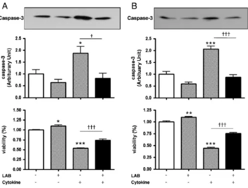
1. Effects of LAB on the cytokine-induced caspase-3 activity and viability of INS-1 cells
INS-1 cells exposed to 1000 U/mL INF-γ plus 100 U/mL IL-1β for 12 hr
showed significantly decreased viability as assessed by MTT and activated
apoptotic signal caspase-3 by cleavage blotting assay (Fig. 1). Compared to
the untreated controls, INS-1 cells treated with 50 μM LAB for 24 (1.00±0.03
vs. 1.10±0.07, p<0.05) and 48 hr (1.00±0.03 vs. 1.10±0.03, p<0.01) showed
statistically increased viability. Pretreatment with 50 μM LAB for 24 and 48
hr before cytokine exposure significantly reduced cytokine induced INS-1 cell
viability, respectively (0.540±0.003 vs. 0.744±0.061, p<0.001 and
0.445±0.004 vs. 0.753±0.055; cytokine vs. LAB and cytokines, p<0.001). The
expression of cleaved caspase-3 was also reduced (1.868±0.576 vs.
0.813±0.405, p<0.05 and 2.056±0.258 vs. 0.870±0.195 ; cytokine vs. LAB
14
Figure 1. Effects of LAB on cytokines-induced caspase-3 activity and viability of INS-1 cells. INS-1 cells exposed to 1000 U/mL INF-γ plus 100
U/mL IL-1β for 12 hr with and without pretreatment with 50 μM LAB for 24
hr (A) and 48 hr (B). The bars represent relative value to the untreated control
neither with cytokines nor LAB (value 1.0). Results are shown as mean ±
SEM. *p<0.05, **p<0.01, ***p<0.001: differences between groups treated
with LAB vs. control without treatment with LAB and cytokines. †p<0.05,
†††p<0.001: differences between groups treated with cytokine alone vs.
15
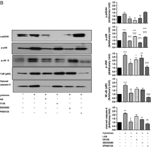
2. LAB inhibits p38 and JNK phosphorylation in cytokine-treated INS-1 cells
To elucidate the possible effects of LAB in the anti-apoptotic pathway, we
investigated the effects of LAB on the MAP kinase pathway involved in
INS-1 cells treated either LAB or cytokines. We evaluated the effects of cytokines
or LAB each alone on the phosphorylation of p42/44, p38, JNK and
expression of Nf-кB of INS-1 cells by Western blot. Under unstressed control
conditions, 48 hr exposure to LAB alone had negligible effects on
phosphorylation of MAP kinase pathways and Nf-кB expression (Fig. 2A).
Eighteen hours of exposure of INS-1 cells to 1000 U/mL INF-γ plus 100
U/mL IL-1β significantly increased nuclear translocation of Nf-кB subunit
p65 (2.15±0.45, p<0.001) and increased in p38 (2.14±0.10, p<0.001) and JNK
phosphorylation (2.72±0.48, p<0.001). Pretreatment of INS-1 cells with 50
μM LAB, however, significantly reduced the cytokine-induced
phosphorylation of p 38 and JNK, the nuclear translocation of Nf-кB subunit
p65 and the level of cleaved caspase-3 (Fig. 2B). Cytokine-induced apoptosis
using the p38 and JNK pathways were also blocked by 50 μM LAB. The
reactive oxygen species (ROS) levels after cytokine treatment were
significantly attenuated by pretreatment with 50 μM LAB.
Pretreatment of INS-1 cells with specific inhibitors of p38 (SB203580) and
JNK (SP600125) before exposure to 1000 U/ml INF-γ plus 100 U/ml IL-1β
16
and 1.11± 0.43 compared to the value without pretreatment of the inhibitor,
respectively. The expression of cleaved caspase-3 also decreased to 1.38±0.53
and 0.79±0.18, respectively. Of the MAP kinase inhibitors, only the JNK
inhibitor, SP600125, completely blocked the translocation of nuclear Nf-кB
subunit p65.
Taken together, cytokine-induced oxidative stress has been implicated in the
cross-talk between phosphorylation of JNK and transnuclear activation of
18
Figure 2. Effect of LAB on MAP kinase pathways in cytokine-treated INS-1 cells. INS-1 cells exposed to 50 μM LAB or cytokines for 48 hr (A). A
representative immunoblot of phosphorylations of p38, JNK and cleaved
caspase-3 activity in cytokine-treated INS-1 cells (on the left) and summary of
six experiments (on the right). Similar experiments with LAB as well as MAP
kinase inhibitors (B). *p<0.05, **p<0.01, ***p<0.001 vs. untreated control
19
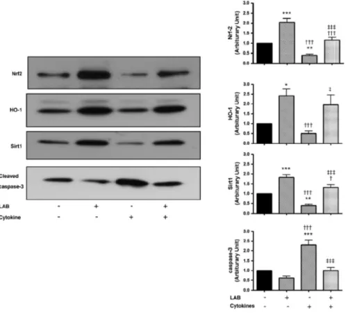
3. LAB activates the Nrf2–HO-1 pathway in INS-1 cells
In order to identify the survival signaling cascade induced by LAB, we
investigated whether LAB protected against cytokine-induced caspase-3
apoptotic pathway via Nrf2–HO-1 and Sirt1 activation. Immunoblot analysis
of the nuclear extract for Nrf2 and Sirt1 showed significantly increased
nuclear translocation of Nrf2 (vs. untreated control; 2.04±0.56, p<0.001) and
Sirt1 (vs. untreated control; 2.39±1.00, p<0.001). In parallel fashion, there
was a decrease in cleaved caspase-3 activity in LAB-treated INS-1 cells in
both with and without treatment with cytokines under both normal and
20
Figure 3. LAB induces expression of Nrf2–HO-1 and Sirt1 in INS-1 cells.
Immunoblot analysis of nuclear extract for Sirt1, Nrf2 and cleaved caspase-3
activity in INS-1 cells under the conditions with and without LAB. A
representative immunoblot (left) and summary of 4 experiments (right)
*p<0.05, **p<0.01, ***p<0.001 vs. untreated control cells. †††p<0.01 vs.
LAB-treated cells. ‡p<0.05, ‡‡‡p<0.001 vs. cytokine-treated cells.
21
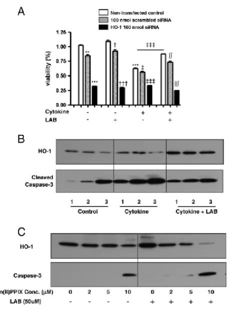
INS-1 cell apoptosis via the Nrf2–HO-1 pathway, we investigated its effects
under HO-1 knockdown and HO-1 competitive inhibition (Zn(II)PPIX)
conditions. We transfected INS-1 cells with siRNA against HO-1 and then
treated them with in the presence or absence of cytokines and LAB. In
non-transfected INS-1 cells, exposure to cytokines showed significantly
deacreased (1.03±0.01 vs. 0.62±0.01, p<0.001), and LAB partly restored
viability which was reduced with cytokine treatment (Fig 4A;(0.62±0.01 vs.
0.87±0.01, p<0.001)), in consistence with the results shown in Fig. 1.
Knockdown siRNA against a murine HO-1 significantly decreased cell
viability in all conditions (absence of cytokine and LAB, presence of LAB
alone or cytokine alone and both cytokine and LAB) (Fig. 4A).
Knockdown siRNA against a murine HO-1 significantly decreased the
expression of HO-1 in the control and cytokine-treated conditions, but its
expression of HO-1 was maintained in cells treated with LAB at the level
even greater than that of the untreated with cytokines (Fig. 4A, third column).
HO-1 knockdown increased expression of cleaved caspase-3 in all conditions
(Fig. 4B). LAB reduced the increased activity of caspase-3 by HO-1
knockdown regardless of cytokine treatment (Fig. 4B).
Inhibition of HO-1 activity with Zn(II)PPIX exerted an inhibitory effects on
the expression level of HO-1 as well as an expression of cleaved caspase-3 at
its concentration of 10 μM Zn(II)PPIX in INS-1 cells (Fig. 4C). Similar
22
caspase-3 were observed in LAB-pretreated INS-1 cells.
Taken together, our results suggest that LAB increased cell viability through
activation of the Nrf2–HO-1 pathway, which appears to be one of the possible
23
Figure 4. Effects of knockdown and inhibition of heme oxygenase-1.
Scrambled siRNA and HO-1 100 nmol siRNA were transfected into INS-1
cells with Lipofectamine 2000. The viabilities of transfected cells assessed by
24
HO-1 and cleaved caspase-3 activity were shown in lanes 1 (non-transfected
control), 2 (100 nmol scrambled siRNA) and 3 (HO-1 100 nmol siRNA
transfected cells) (B). After 24 hr of incubation, INS-1 cells were exposed to
different doses of Zn(II)PPIX, an HO-1 competitive inhibitor, from 2 to 10
mM for 1 hr (C). **p<0.01, ***p<0.001 vs. non-transfected control cells in
the absence of cytokine and LAB. †p<0.05, †††p<0.001 vs. non-transfected
control cells in the presence of LAB. ‡p<0.05, ‡‡‡p<0.001 vs.
non-transfected control cells in the presence of cytokine. ∫∫p<0.01, ∫∫∫p<0.001 vs.
non-transfected control cells in the presence of both cytokine and LAB.
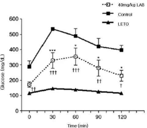
4. LAB ameliorates hyperglycemia and protects pancreatic beta cell mass in type 2 diabetic OLEFT rats
At 52 weeks of age with treatment of 40 mg/kg/day LAB for 42 weeks, 2
g/kg oral glucose tolerance test (OGTT) was performed. Blood glucose levels
of the LETO (control) group were significantly lower than those in the
placebo or LAB-treated groups of OLETF rats at all time points after oral
administration. The LAB-treated OLETF group showed significantly lower
glucose values than the OLETF control group at 30 (p<0.001), 60 (p<0.05),
90 (p<0.05) and 120 (p<0.05) min after glucose loading (Fig. 5). OLETF rats
treated with LAB showed significant improvement in glucose tolerance but
their blood glucose levels were still significantly higher than that of LETO
25
Figure 5. Oral glucose tolerance test in OLETF diabetic rats with and without LAB treatment and LETO control rats. Oral glucose tolerance tests were performed at 52 weeks. The blood glucose levels of OLETF rats treated with 40 mg/kg/day LAB (n=6, blank box), OLETF control rats (n=4, black box), LETO rats (n=7, triangle) during oral glucose change test were shown. Control: OLETF rats treated with distilled water in the same volume as LAB; 40 mg/kg: OLETF rats treated with 40 mg/kg/day LAB; LETO: nondiabetic control rats. ***p<0.001 vs. OLETF rat controls. †p<0.05, ††p<0.01, †††p<0.001 vs. LETO.
26
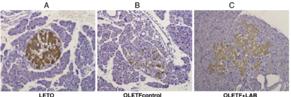
5. Effects of LAB on beta cell mass
The beta cell mass of pancreatic islets were evaluated by insulin
immunohistochemistry in LETO, untreated and LAB-treated OLETF rats at
52 weeks of age. Compared to the morphology of the LETO rat, that of
untreated OLETF rats showed extremely atrophic islet architecture and scanty
insulin-positive pancreatic beta-cells (Fig. 6). In the LAB-treated OLETF rats,
beta cell mass was relatively conserved but was still lower than that of the
LETO rats.
Figure 6. Effects of LAB on pancreas histology. Pancreatic islets masses were stained by insulin immunohistochemistry in LETO (A), untreated (B) and LAB-treated OLETF rats (C) at 52 weeks of age.
27 IV. DISCUSSION
It is well known that magnesium lithospermate B (LAB) scavenged
superoxide radicals directly in the system β-NADH/PMS and inhibited the
production of superoxide in human neutrophils stimulated by PMA and fMLP
8
. In view of the fact that activation of the Nrf2–ARE–NQO1 transcriptional
pathway and superoxide scavenging effects of LAB, it is believed to have
therapeutic effects in various pathologic conditions by attenuating the
apoptotic signaling cascades and/or promoting antiapoptic signaling cascades.
We previously demonstrated the protective roles of LAB on a balloon injured
vascular rat model and diabetic nephropathy in the OLETF rat7,8,9,10,
suggesting a potential therapeutic application of LAB in atherosclerosis and
restenosis after angioplasty and prevention of nephropathy in type 2 diabetes.
Although antioxidants are known to exert a protective effect against oxidative
stress-induced cell death in various cells and tissues4,14,15, the effects of LAB
on the apoptotic and/or antiapoptotic signaling cascades in pancreatic beta
cells have not been elucidated.
In the present study, our attention was focused on the potential protective
effects of LAB under the condition of cytokine-induced beta cell apoptosis
and the mechanism of its action. Concerning cytokine-induced beta cell
apoptosis, IL-1β (in rat) on its own or combined with INF-γ (human and
mouse) was reported very toxic to an isolated islet in vitro, killing the
28
viability of INS-1 cells treated with 1000 U/ml INF-γ alone (1.00±0.08) or
100 U/ml IL-1β alone (0.938±0.07) for 48 hr was not significantly affected
as assessed by MTT. But combining of 1000 U/ml INF-γ plus 100 U/ml IL-1β
significantly decreased cell viability (Fig. 1A and Fig. 4B) (1.00±0.08 vs.
0.587±0.06, p<0.001). Thus, we adopted 1000 U/mL INF-γ plus 100 U/mL
IL-1β for beta cell apoptosis inducer. Our results are consistent with the result
that prolonged combined exposure to IL-1β plus IFN-γ, but not to either
cytokine alone evolved to beta cell death2,3.
First, we observed that the viability of INS-1 cells treated with 50 μM LAB
for 24 hr and 48 hr was significantly improved, at that concentration it was
found to have protective effects on atherosclerosis and diabetic
nephropathy7,10. Interestingly, LAB improved INS-1 cell viability in both with
and without cytokine treatment (Fig. 1; Fig. 4A). Pretreatment with 50 μM
LAB for 24 hr and 48 hr significantly decreased the INF-γ plus IL-1β-induced
INS-1 cell apoptosis (Fig. 1, Fig. 2B, Fig. 3). Hence, LAB improve INS-1 cell
viability at least in part by reducing apoptotic signaling pathway.
Second, we investigated the effects of LAB on the apoptotic signaling
pathways. Pro-inflammatory cytokines are known to induce the transcription
factors Nf-кB and STAT1 leading to affect multiple and distinct
transcriptional factors and gene networks16,17, and to upregulate the apoptotic
pathways of JNK and p38 mitogen-activated protein kinase (MAPK)2. In
29
both increased expression of Nf-кB and phosphorylation of p-38 and JNK,
resulting in increased expression of cleaved caspase-3 (Fig. 2). To find the
specificity of LAB to cytokine-induced INS-1 cell apoptosis, we also
investigated the effects of LAB alone on phosphorylation of the apoptotic
pathways of p42/44, p38, and JNK, and found that LAB showed negligible
effects under unstressed conditions. With regards to Nf-кB on the beta cell
apoptotic pathway, expression of both Nf-кB and p-JNK phosphorylation
increased in the presence of cytokines, and these increases were profoundly
decreased in the presence of the JNK inhibitor SP600125 in similar manner as
LAB did (Fig. 2). Based on previous reports2,16, these findings implicated a
cross-talk between Nf-кB and p-JNK and induce different kinds of gene
responses with pro-apoptosis of beta cells. Therefore, as we and others have
found, pro-inflammatory cytokines might exert their main effects through the
Nf-кB pathway2,16,18,19.
Third, we investigated the induction of LAB-induced anti-apoptotic
pathways in cytokine induced apoptotic beta cells. In this study, treatment
with 50 μM LAB alone statistically improved the viability of INS-1 cells, but
had negligible effects on the phosphorylation of apoptotic pathways. A battery
of genes encoding detoxifying enzymes such as glutathione S-transferases
(GSTs) and NQO1 and antioxidative stress proteins such as HO-1 and
γ-glutamylcysteine synthetase (γ-GCS), which are regulated through a
30
of oxidative stress20. One of the most important antioxidant genes protecting
against oxidative stress is mediated by the transcription factor Nrf2 21,22.
Briefly, Keap1 binds to actin cytoskeleton and retains Nrf2 in the cytoplasm
under basal conditions. Upon exposure of cells to oxidative stress or
chemopreventive compounds, Nrf2 dissociates from Keap1, translocates to
the nucleus, forms a heterodimer with its obligatory partner Maf, and
ultimately activates ARE-dependent gene expression. Besides its antioxidant
function, Nrf2 is known to be a key factor regulating a battery of genes that
protect cells against deleterious environmental insults23. Nrf2–ARE signaling
is also known to be mainly responsible for the up-regulation of HO-1 gene
expression and hence constitutes a crucial cellular response to environmental
stresses24.
As expected, LAB increased both Nrf2 and HO-1 expression with or without
the presence of cytokines. Because HO-1 was the most important survival
protein and ubiquitously induced antioxidant by various stimulants, we
hypothesized that LAB might play a more crucial role in inducing HO-1 to a
severe stressful condition. To test this hypothesis, HO-1 expression of INS-1
cells was knocked down by siRNA and enzymatically inhibited by
Zn(II)PPIX. When comparing control cells, similar over-expression of
cleaved caspase-3 and attenuated expression of HO-1were found in INS-1
cells cultured in the presence of cytokines (Figs.3 and 4). The knockdown
31
profound in the control and cytokine-treated condition as assessed by Western
blotting. It is noteworthy that knockdown of HO-1 markedly decreased cell
viability in INS-1 cells with and without cytokine treatment pointing to a
crucial role of HO-1 in cell survival in general (Fig. 4A). However, LAB
treatment of INS-1 cells with HO-1 knockdown exposed to cytokine increased
HO-1 expression far greater than the level of the untranfected control ,but
failed to reduce the cleaved caspase-3 below the untransfected control (Fig.
4B). Seemingly contradictory to the results of HO-1 knockdown with siRNA
under which expression of HO-1 by LAB was recovered to the level above
control but HO-1 inhibitor Zn(II)PPIX at 10 uM almost completely abrogated
the effect of LAB (Fig. 4C). Despite such peculiarity, LAB increased HO-1
expression and decreased cleaved caspase-3 level in both INS-1 cells with
HO-1 knockout and reduced HO-1 activity with its inhibitor at its
concentration lower than 10 uM (Fig. 4). These results could support our
hypothesis that HO-1 induction is an important mechanism for protection in
cytokine-induced beta cell apoptosis.
Finally, we tried to confirm the in vitro effectiveness of LAB on INS-1 cells
in animal models of obesity-related diabetic rats in vivo. We showed that
LAB-treated OLETF rats showed a significant improvement in postprandial
glucose tolerance compared to OLETF control rats. Pancreatic pathology in
placebo-treated OLETF rats showed destructive islet morphology and scant
32
relatively less destructive islet architecture with positive insulin staining.
These results suggest that LAB prevents beta cell death in the islets and
delays the development of diabetes in vivo. Taken together, LAB has
beneficial effects on pancreatic beta cell in vitro as well as in vivo.
There are some limitations to this study. First of all, we did not measure
insulin concentration during oral glucose tolerance, because levels of insulin
are essential for evaluating whether the anti-diabetic effects of LAB are a
direct effect on pancreas beta cells or the indirect effect of improving insulin
sensitivity. Also, its anti-apoptotic roles were demonstrated only to the
cytokine-induced pathway, not to all possible pathways involved in the
development of type 2 diabetes, including glucolipotoxicity, and
immune-mediated and inflammatory reactions. There was no direct results for the
effect of LAB and HO-1 inhibitor in the setting of cytokine-induced apoptosis
even though the experiment was additional (Fig. 4C). However, this study is
the first to examine the effects of LAB on glucose homeostasis and beta cell
survival.
In summary, this study observed that LAB had an anti-apoptotic effect on
INF-γ plus IL-1β-induced apoptotic pancreatic beta cells in vitro and a
preventive effect on the development of type 2 diabetes in an OLEFT animal
model in vivo. In this study, the anti-apoptotic effects of LAB were
demonstrated by the down-regulation of the apoptotic pathways of c-JNK and
Nrf2/HO-33
1 and Sirt1 expression. Further clinical studies are needed to evaluate the use
34 REFERENCES
1. Butler, A.E., Janson, J., Bonner-Weir, S., Ritzel, R., Rizza, R.A.,
Butler, P.C., 2003. Beta-cell deficit and increased beta-cell apoptosis
in humanswith type 2 diabetes. Diabetes 52, 102–10.
2. Cnop, M., Welsh, N., Jonas, J.C., Jorns, A., Lenzen, S., Eizirik, D.L.,
2005. Mechanisms of pancreatic beta-cell death in type 1 and type 2
diabetes: many differences, few similarities. Diabetes 54 (Suppl 2),
S97–107.
3. Eizirik, D.L., Mandrup-Poulsen, T., 2001. A choice of death—the
signal-transduction of immune-mediated beta-cell apoptosis.
Diabetologia 44, 2115–33.
4. Hambrock, A., deOliveira Franz, C.B., Hiller, S., Grenz, A.,
Ackermann, S., Schulze, D.U., Drews, G., Osswald, H., 2007.
Resveratrol binds to the sulfonylurea receptor (SUR) and induces
apoptosis in a SUR subtype-specific manner. J. Biol. Chem. 282,
35
5. Wu, X.J., Wang, Y.P., Wang, W., Sun, W.K., Xu, Y.M., Xuan, L.J.,
2000. Free radical scavenging and inhibition of lipid peroxidation by
magnesium lithospermate B. Acta Pharmacol. Sin. 21, 855–8.
6. Soung, D.Y., Rhee, S.H., Kim, J.S., Lee, J.Y., Yang, H.S., Choi, J.S.,
Yokozawa, T., Han, Y.N., Chung, H.Y., 2003. Peroxynitrite
scavenging activity of lithospermate B from Salvia miltiorrhiza. J.
Pharm. Pharmacol. 55, 1427–32.
7. Hur, K.Y., Kim, S.H., Choi, M.A., Williams, D.R., Lee, Y.H., Kang,
S.W., Yadav, U.C., Srivastava, S.K., Jung, M., Cho, J.W., Kim, S.G.,
Kang, E.S., Lee, E.J., Lee, H.C., 2010. Protective effects of
magnesium lithospermate B against diabetic atherosclerosis via
Nrf2–ARE–NQO1 transcriptional pathway. Atherosclerosis 211, 69–
76.
8. Liu, X., Chen, R., Shang, Y., Jiao, B., Huang, C., 2008. Lithospermic
acid as a novel xanthine oxidase inhibitor has anti-inflammatory and
hypouricemic effects in rats. Chem. Biol. Interact. 176, 137–42.
9. Lee, G.T., Ha, H., Jung, M., Li, H., Hong, S.W., Cha, B.S., Lee, H.C.,
36
experimental diabetic renal injury. J. Am. Soc. Nephrol. 14, 709–20.
10. Kang, E.S., Lee,G.T., Kim, B.S. Kim,C.H., Seo, G.H., Han,S.J., Hur,
K.Y., Ahn,C.W., Ha,H., Jung,M., Ahn, Y.S., Cha, B.S., Lee, H.C.,
2008. Lithospermic acid B ameliorates the development of diabetic
nephropathy in OLETF rats. Eur. J. Pharmacol. 579, 418–25.
11. Hur, K.Y., Seo, H.J., Kang, E.S., Kim, S.H., Song, S., Kim, E.H.,
Lim, S., Choi, C., Heo, J.H., Hwang, K.C., Ahn, C.W., Cha, B.S.,
Jung, M., Lee, H.C., 2008. Therapeutic effect of magnesium
lithospermate B on neointimal formation after balloon-induced
vascular injury. Eur. J. Pharmacol. 586, 226–33.
12. Kim, M.K., Jung, H.S., Yoon, C.S., Ko, J.H., Jun, H.J., Kim, T.K.,
Kwon, M.J., Lee, S.H., Ko, K.S., Rhee, B.D., Park, J.H., 2010. The
effect of glucose fluctuation on apoptosis and function of INS-1
pancreatic beta cells. Korean Diabetes J 34, 47–54.
13. Schroeder, M., Zagoory-Sharon, O., Shbiro, L., Marco, A., Hyun, J.,
Moran, T.H., Bi, S., Weller, A., 2009. Development of obesity in the
Otsuka Long–Evans Tokushima Fatty rat. Am. J. Physiol. Regul.
37
14. Targonsky, E.D., Dai, F., Koshkin, V., Karaman, G.T.,
Gyulkhandanyan, A.V., Zhang, Y., Chan, C.B., Wheeler, M.B., 2006.
alpha-Lipoic acid regulates AMP-activated protein kinase and inhibits
insulin secretion from beta cells. Diabetologia 49, 1587–98.
15. Lee, B.W., Kwon, S.J., Chae, H.Y., Kang, J.G., Kim, C.S., Lee, S.J.,
Yoo, H.J., Kim, J.H., Park, K.S., Ihm, S.H., 2009. Dose-related
cytoprotective effect of alpha-lipoic acid on hydrogen
peroxide-induced oxidative stress to pancreatic beta cells. Free Radic. Res. 43,
68–77.
16. Thomas, H.E., McKenzie, M.D., Angstetra, E., Campbell, P.D., Kay,
T.W., 2009. Beta cell apoptosis in diabetes. Apoptosis 14, 1389–404.
17. Yang, S.R., Wright, J., Bauter, M., Seweryniak, K., Kode, A.,
Rahman, I., 2007. Sirtuin regulates cigarette smoke-induced
proinflammatory mediator release via RelA/p65 NF-kappaB in
macrophages in vitro and in rat lungs in vivo: implications for chronic
inflammation and aging. Am. J. Physiol. Lung Cell. Mol. Physiol.
38
18. Papa, S., Bubici, C., Zazzeroni, F., Pham, C.G., Kuntzen, C., Knabb,
J.R., Dean, K., Franzoso, G., 2006. The NF-kappaB-mediated control
of the JNK cascade in the antagonism of programmed cell death in
health and disease. Cell Death Differ. 13, 712–29.
19. Lee, J.H., Song, M.Y., Song, E.K., Kim, E.K., Moon, W.S., Han,
M.K., Park, J.W., Kwon, K.B., Park, B.H., 2009. Overexpression of
SIRT1 protects pancreatic beta-cells against cytokine toxicity by
suppressing the nuclear factor-kappaB signaling pathway. Diabetes
58, 344–51.
20. Itoh, K., Tong, K.I., Yamamoto, M., 2004. Molecular mechanism
activating Nrf2–Keap1 pathway in regulation of adaptive response to
electrophiles. Free Radic. Biol. Med. 36, 1208–13.
21. Itoh, K., Chiba, T., Takahashi, S., Ishii, T., Igarashi, K., Katoh, Y.,
Oyake, T., Hayashi, N., Satoh, K., Hatayama, I., Yamamoto, M.,
Nabeshima, Y., 1997. An Nrf2/small Maf heterodimer mediates the
induction of phase II detoxifying enzyme genes through antioxidant
response elements. Biochem. Biophys. Res. Commun. 236, 313–22.
39
Phosphatidylinositol 3-kinase regulates nuclear translocation of
NF-E2-related factor 2 through actin rearrangement in response to
oxidative stress. Mol. Pharmacol. 62, 1001–10.
23. Zhang, D.D., 2006. Mechanistic studies of the Nrf2–Keap1 signaling
pathway. Drug Metab. Rev. 38, 769–89.
24. Surh, Y.J., Kundu, J.K., Li, M.H., Na, H.K., Cha, Y.N., 2009. Role
of Nrf2-mediated heme oxygenase-1 upregulation in adaptive
survival response to nitrosative stress. Arch. Pharm. Res. 32, 1163–
40 ABSTRACT (IN KOREAN)
췌장 베타세포의 사이토카인 유발
자멸반응에 대한 LAB의 보호효과와 기전
<지도교수 이병완> 연세대학교 대학원 의학과 전 성 완 LAB는 제2형 당뇨병 동물모델에서 혈관손상에 대한 보호효과가 확인된 바 있다. 기존 연구에서 LAB를 투여한 쥐에서 췌장세포의 손상이 완화되는 현상이 관찰된 바 있어 본 연구자는 췌장세포에 대한 LAB의 작용을 연구하고자 하였다.INS-1 세포 배양액에 50 μM LAB를 전처리하면 1000 U/mL INF-γ
41
LAB는 사이토카인으로 인한 p38과 JNK의 인산화를 의미있게 감소
시키며 이에 따라 cleaved caspase 3 활성을 낮추었다. 또한,
Nrf2–HO-1 활성과 SirtNrf2–HO-1 발현을 증가시켜 사이토카인 자극에 의한 caspase 3
세포자멸 기전을 억제했다. LAB를 42주간 40 mg/kg/day 경구 투여한
52주령의 OLETF 쥐는 LAB를 투여하지 않은 OLETF 쥐와 비교하여
경구당부하검사 상 뚜렷한 개선양상을 보였고, 췌도의 구조적인 변 형이 완화함을 관찰하였다. 본 연구는 LAB가 세포자멸 기전 자체를 억제할 뿐 아니라 Nrf2– HO-1 과 Sirt1을 통한 세포자멸 억제기전을 활성화시켜 결과적으로 세포 및 생체 수준에서 의미있는 췌도 베타세포 보호효과를 나타냄 을 보여주었다.
핵심되는 말:lithospermic acid B, pancreatic beta cell,