www.jpis.org
pISSN 2093-2278 eISSN 2093-2286 Copyright © 2012 Korean Academy of PeriodontologyThis is an Open Access article distributed under the terms of the Creative Commons Attribution Non-Commercial License (http://creativecommons.org/licenses/by-nc/3.0/).
Comparative evaluation of roughness of titanium
surfaces treated by different hygiene instruments
Otgonbayar Unursaikhan†, Jung-Seok Lee†, Jae-Kook Cha, Jung-Chul Park, Ui-Won Jung, Chang-Sung Kim,
Kyoo-Sung Cho, Seong-Ho Choi*
Department of Periodontology, Research Institute for Periodontal Regeneration, Yonsei University College of Dentistry, Seoul, Korea
Purpose: The use of appropriate instruments to clean surfaces with minimal change, is critical for the successful maintenance of a dental implant. However, there is no consensus about the type and methodology for such instruments. The aim of this study was to characterize changes in the roughness of titanium surfaces treated by various scaling instruments.
Methods: Thirty-seven identical disks (5 mm in diameter) were investigated in this study. The specimens were divided into eight groups according to the types of instrumentation and the angle of application. Ultrasonic scaling systems were applied on a titanium disk to simulate standard clinical conditions. The equipment included a piezoelectric ultrasonic scaler with a newly developed metallic tip (NS group), a piezoelectric ultrasonic scaler with a conventional tip (CS group), a piezoelectric root planer ultrasonic scaler with a conventional tip (PR group), and a plastic hand curette (PH group). In addition, the sites treated using piezoelectric ultrasonic scaler systems were divided two sub-groups: 15 and 45 degrees. The treated titanium surfaces were observed by scanning electron microscopy (SEM), and the average surface roughness (Ra) and mean roughness profile depth (Rz) were measured with a profilometer.
Results: SEM no significant changes in the titanium surfaces in the NS group, regardless of the angle of application. The PH group also showed no marked changes to the titanium surface, although some smoothening was observed. All CS and PR sites lost their original texture and showed irregular surfaces in SEM analysis. The profilometer analysis demonstrated that the roughness values (Ra and Rz) of the titanium surfaces increased in all, except the PH and NS groups, which showed roughness decreases relative to the untreated control group. The Ra value differed significantly between the NS and PR groups (P<0.05). Conclusions: The results of this study indicated that changes in or damage to titanium surfaces might be more affected by the hardness of the scaler tip than by the application method. Within the limitations of this study, the newly developed metal-lic scaler tip might be especially suitable for peri-implant surface decontamination, due to its limited effects on the titanium surface.
Keywords: Dental implants, Dental instruments, Peri-implantitis, Periodontal debridement.
INTRODUCTION
Over the past few decades, dental implants have become one of the most effective treatments following tooth loss. However, there has been controversy about marginal bone
loss around dental implants since screw-type implants were first introduced in 1971-specifically, whether the bone loss is caused by pathologic change or a normal biologic response.
Peri-implant diseases were recently classified into two groups at the 6th European Workshop on Periodontology in
Received: Jan. 19, 2012; Accepted: May 20, 2012
*Correspondence: Seong-Ho Choi
Department of Periodontology, Yonsei University College of Dentistry, 50 Yonsei-ro, Seodaemoon-gu, Seoul 120-752, Korea E-mail: [email protected], Tel: +82-2-2228-3189, Fax: +82-2-392-0398
the data available about the treatment of periodontitis [7]. The rationale is to keep the bacterial load below the individ-ual’s threshold for disease. To achieve this goal, mechanical debridement using hand or ultrasonic devices, adjunctive use of chemical agents, and laser application have been used for nonsurgical treatment of implant mucositis and peri-implantitis [8]. However, although peri-implant mucositis was found to be reversible by nonsurgical therapy [9], peri-implantitis remained problematic [1]. A previous study evalu-ating the effects of combined surgical and antimicrobial in-terventions on peri-implantitis found that 7 out of 26 implants were lost despite repeated treatment [10].
Developments in surface modifications have allowed for enhanced osseointegration of implants, but they might also enhance biofilm formation [7], so the exposed and contami-nated rough surface of these implants could not be restored completely by conventional treatments. For these reasons, the prevention of peri-implant disease (including periodon-titis) is more important than the treatment of disease prog-ress. The principal objective of preventive therapy for peri-odontal/peri-implant diseases is suppressing bacterial colo-nization via mechanical plaque control, and ultrasonic devic-es have commonly been used in clinical applications for periodontal preventive treatment due to their effectiveness. However, while conventional mechanical instruments can plane a diseased irregular root surface, they also roughen the surface of a titanium implant surface [11,12]. In order to over-come some of these limitations, non-metallic devices or oth-er altoth-ernative methods have been developed for the mainte-nance treatment of dental implants, including [13-17] plastic
ultrasonic scaler that is softer than titanium so as to avoid damaging the implant surface. The objectives of this study were to 1) determine the effects of the various ultrasonic scal-ing systems with different degrees of hardness on changes to the titanium surface, and 2) estimate the changes to titanium surfaces according to the angle at which the instruments are applied.
MATERIALS AND METHODS
Materials and study design
Thirty-seven pure titanium disks 5 mm in diameter (Den-tium, Seoul, Korea) were used in this study. They were divid-ed into the following 5 groups according to the different in-struments used (Table 1, Fig. 1):
1) Piezoelectric ultrasonic scaler with a newly developed metallic tip (NS group)
2) Piezoelectric ultrasonic scaler with a conventional tip (CS group)
3) Piezoelectric root planer/ultrasonic scaler with a conven-tional tip (PR group)
4) Plastic hand curette (PH group) 5) Control
Groups 1, 2, and 3 were further subdivided into two groups in which two tip angles (15 and 45 degrees) were applied. This produced, a total of eight groups (including the control group), each of which included five disks.
Table 1. Group allocation according to scaler type.
Group Scaling procedure Product name and manufacturer Angle (˚) NS Piezoelectric ultrasonic scaler with a newly developed metallic tip Yoshida, Tokyo, Japan Tip: B&L Biotech, Seoul, Korea 15, 45 CS Piezoelectric ultrasonic scaler with a conventional tip Yoshida, Tokyo, Japan Tip: EMS, Nyon, Switzerland 15, 45 PR Piezoelectric root planer ultrasonic scaler with a conventional tip Satelec, Merignaccedex, France Tip: EMS, Nyon, Switzerland 15, 45 PH Plastic hand curette Universal implant scaler Columbia 4L/4R design; Hu-Friedy 45 C Control No treatment
Figure 1. Scaler tips used in this study. (A) Piezoelectric ultrasonic scaler with a newly developed metallic tip, (B) piezoelectric ultrason-ic scaler with a conventional tip, (C) root planner ultrasonultrason-ic scaler, (D) hand scaler.
A B
Experimental procedure
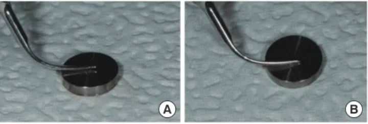
Two small lines were scribed as orientation marks onto each surface: the lines were approximately 2 mm long and separated by 2 mm (Fig. 2). The titanium surfaces were treat-ed and then examintreat-ed in this area. The titanium surfaces were cleaned with a solvent wash of acetone for 1 minute be-fore and after each hygiene treatment, and then air-dried and wrapped in 2×2 gauze. The specimens were oriented hori-zontally on a table, and the piezoelectric conventional scalers were used at moderate finger pressure for 30 seconds at 15 and 45 degrees, 25 kHz, and at their highest power. The piezo-electric root planer was applied at 27 to 32 kHz at its highest power (Fig. 3). The plastic hand curette was used at finger pressure for 30 strokes at 45 degrees. Untreated titanium sur-faces served as controls. All experimental procedures were performed by the same investigator.
Scanning electron microscopy
Titanium specimens were examined by scanning electron microscopy (SEM; S-300OH, Hitachi, Tokyo, Japan) operat-ing at 10 kV and photographed at a magnification of ×200. Before examination, the specimens were coated with an elec-troconductive layer of gold, which was evaporated by an ion sputter coater (E 101, Hitachi).
Profilometer
After scaling, the surface roughness of the specimens was assessed with a profilometer (CS 3100, Mitutoyo, Tokyo, Japan). This instrument measures high-frequency surface irregulari-ties and not widely-spaced irregulariirregulari-ties caused by waviness or curvature.
The average surface roughness (Ra) and the mean rough-ness profile depth (Rz) were measured. In each case, the measurement was performed with a 0.25 mm cutoff and over an assessment length of 1.25 mm. Each specimen was
measured three times at 0.5 mm intervals lengthwise and widthwise, from which the average for each specimen was calculated.
Statistical analysis
Individual mean values were calculated. A one-way analysis of variance was used to evaluate the differences between the titanium surfaces, and the post-hoc Scheffe’s test was used to evaluate differences between groups. A P-value of <0.05 was considered significant. SPSS ver. 12.0.0 (SPSS Inc., Chicago, IL, USA) was used for all of the statistical analysis.
RESULTS
SEM observations
The characteristics of the titanium surfaces in the obtained SEM images varied with the applied treatment. It was evident that the untreated disks did not have smooth surfaces with circumferential milling marks being evident (Fig. 4A).
The use of the plastic hand curette (PH group) (Fig. 4B) and the newly developed metallic scaler tip (NS group) (Fig. 4C and D) did not appear to markedly affect the titanium surfaces, although some smoothening occurred. The surface roughness did not differ among the NS, control, and PH groups.
The use of the piezoelectric conventional scaler (CS group) (Fig. 4G and H) and piezoelectric root planer (PR group) (Fig. 4E and F) clearly resulted in scraping of the titanium surfaces and loss of their original texture, leading to increased surface roughness.
In addition, the SEM images demonstrated that the chang-es in the surface texture were lchang-ess extensive when the proce-dure was performed at 45 degrees than at 15 degrees. How-ever, there were no statistically significant differences be-tween these two experimental groups.
Profilometer analysis
The roughness parameters in the experimental and control surfaces (mean values of Ra and Rz) are reported in Fig. 5 and Table 2.
All of the procedures increased the roughness parameters,
Figure 3. Description of experimental methods. The ultrasonic scal-ers were used with moderate finger pressure, for 30 seconds, at 15 (A) and 45 degrees (B) at 25 kHz set on highest power.
A B
except for treatment with the plastic hand curette (PH group: 0.20±0.04 μm, mean±standard deviation) and the newly de-veloped metallic scaler tip (NS group: 0.22±0.05 μm for the 15 degree group and 0.23±0.06 μm for the 45 degree group), which showed a lower Ra value than the control one (0.24± 0.07 μm). The Ra values for the titanium surfaces increased following treatments with the piezoelectric conventional scaler tip (CS group) and root planer (PR group) at 15 and 45
degrees (0.34±0.06 μm or 0.34±0.03 μm and 0.39±0.02 μm or 0.36±0.04 μm). There were statistically significant differ-ences between the NS and PR groups (P<0.05) (Table 2).
Most of the procedures increased Rz (1.59±0.54 μm, 2.54± 0.79 μm, 2.82±0.78 μm in the NS, CS, and PR group): the ex-ception was treatment with the plastic hand curette (PH group: 1.38±0.51 μm). Rz was lower in the PH group than in the control group (1.49±0.50 μm) (Fig. 5, Table 2). There were no statistically significant differences.
Figure 4. Scanning electron microscopy images of the titanium surfaces with various treatments showed differences depending upon the given treatment. The control group clearly showed the machined grooves (A). The plastic curette and a newly developed metallic tips did not appear to significantly affect the titanium surface, especially after treatment. However some smoothening of the titanium surface ap-pears to have occurred (B-D). In addition, the difference in angles did not appear to affect the outcome of the experiment. The groups using conventional scaler tip clearly showed the damages induced by the tips, both at 15 and 45 degrees. Also, the images showed that circumfer-ential milling on the titanium surface have been scraped (E, F). Other groups using piezoelectric root planer also showed similar results to groups using conventional scaler tip (G, H).
A E B F C G D H
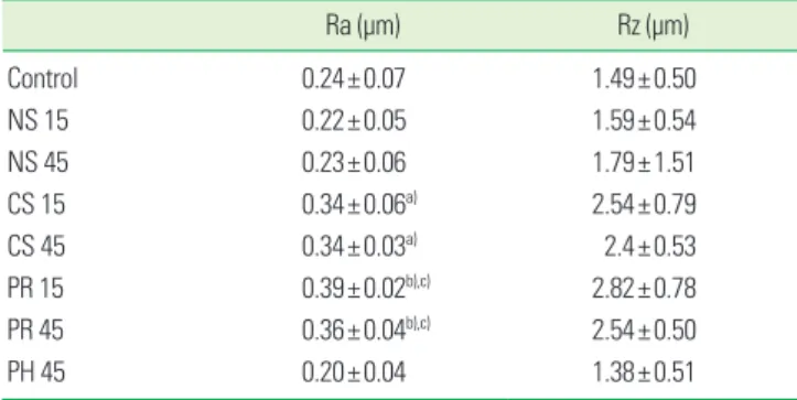
Table 2. Mean and standard deviation values of surface roughness.
Ra (μm) Rz (μm) Control 0.24±0.07 1.49±0.50 NS 15 0.22±0.05 1.59±0.54 NS 45 0.23±0.06 1.79±1.51 CS 15 0.34±0.06a) 2.54±0.79 CS 45 0.34±0.03a) 2.4±0.53 PR 15 0.39±0.02b),c) 2.82±0.78 PR 45 0.36±0.04b),c) 2.54±0.50 PH 45 0.20±0.04 1.38±0.51 Ra: average surface roughness, Rz: mean roughness profile depth, NS: piezoelectric ultrasonic scaler with a newly developed metallic tip, CS: piezoelectric ultrasonic scaler with a conventional tip, PR: piezoelectric root-planer/ultrasonic scaler with a conventional tip, PH: plastic hand curette. a)Statistically significant difference from PH45 (P<0.05). b)Statistically significant difference from NS15 (P<0.05). c)Statistically significant difference from NS45 (P<0.05).
Figure 5. Results from profilometer analysis. Graph showing the av-erage surface roughness (Ra) and mean roughness profile depth (Rz) in each group. Ra and Rz were similar in the NS, PH and control groups, but increased in the CS and PR groups, indicating that the conventional ultrasonic scaler and the root planer had significant effects on the titanium surface. NS: piezoelectric ultrasonic scaler with a newly developed metallic tip, CS: piezoelectric ultrasonic scaler with a conventional tip, PR: piezoelectric root-planer/ultra-sonic scaler with a conventional tip, PH: plastic hand curette.
3.5 3.0 2.5 2.0 1.5 1.0 0.5 0 Contr ol NS15 NS45 CS15 CS45 PR15 PR45 PH45 Rz Ra
Compromised oral hygiene resulting in plaque accumula-tion is a major risk factor that can contribute to the failure of dental implants, and hence good oral hygiene is one of the prerequisites for the long-term success of implants [21,22]. The principal objective of treatment of peri-implantitis is complete removal of all calcified and bacterial deposits from implant surfaces in order to stop disease progression. Many studies have investigated the effects of using different hy-giene instruments on dental implant surfaces [12,13,23-26]. Routine prophylactic procedures might cause damage to im-plant surfaces over time and lead to changes in the surface topography that can increase the potential for plaque accu-mulation.
Quirynen et al. [27] reported that any metal-to-metal con-tact might lead to a damaged implant surface during profes-sional cleaning [28]. This has led to many instruments being developed that aim at less damage to the titanium surface: these incorporate Teflon-coated, plastic, or other types of nonmetallic tips [26,29,30]. However, the application of non-metallic instruments has been reported to be inadequate for eliminating bacteria from roughened implant surfaces [11,24]. Air-powder abrasive systems have also been introduced for cleaning contaminated implant surfaces [11,31], but their ap-plications are limited by being associated with an increased risk of emphysema [32]. Schwarz et al. [18] also reported that a nonmetallic carbon fiber tip (Vector ultrasonic scaler) was not suitable for decontaminating titanium surfaces. Titani-um surfaces treated with the Vector system showed conspic-uous surface damage and deposits of the used carbon fibers, with a reduction in the cell density on all implant surfaces.
The present study used an ultrasonic scaler with a newly developed metallic scaler tip composed of copper alloy and plated with 99.99% silver. Because the newly developed scaler tip (89HV) was softer than the titanium fixture (200 to 280HV) and conventional scaler tip (610HV), its use would reduce or even avoid damage to the implant surface during treatment of peri-implantitis [33]. Therefore, the present study compared the effects on titanium surfaces of the newly developed scal-er tip with those of a plastic hand curette and piezoelectric ultrasonic scalers.
In a result of this study, the effects on the titanium surface were minor in the PS and NS groups but significant in the CS and PR groups (Fig. 4). The SEM analysis revealed that the plastic hand curette (PS group) and the newly developed me-tallic scaler tip (NS group) produced no changes to the titani-um surface topography (Fig. 4B-D). SEM showed that the plastic hand curette (PH group) produced similar results to those of previous studies that have employed plastic scalers
ment by the conventional scaler tip (CS group) and piezoelec-tric root planer (PR group) (Fig. 4E-H), which is in accordance with the result of studies by Cross-Poline et al. [14] and Hall-mon et al. [15]. Samples in the PR group, in which a root plan-er with a conventional scalplan-er tip was used, showed deep sur-face abrasions on the titanium sursur-faces in SEM analysis (Fig. 4E and F).
A profilometer is a direct-reading instrument for measur-ing the average roughness height down to the micron levels. Two basic amplitude parameters were used in this study to characterize the implant surface roughness: Ra and Rz. Ra is universally recognized and the mostly commonly used pa-rameter of roughness: it corresponds to the arithmetic mean of the absolute deviation of the roughness profile from the mean line determined by fitting a least square line of nomi-nal form through the primary profile. Rz corresponds to the maximum peak-to-valley height of the profile over the as-sessment length.
In this study, the roughness increased in the CS and PR ex-perimental groups compared with the control group, while there was a small reduction in the Rz value when using a plastic curette or the newly developed metallic scaler tip (Fig. 5). These results are in accordance with previous studies find-ing that the Ra and Rz values after usfind-ing nonmetallic scaler tips were similar to or lower than those in the control groups, whereas the use of the metallic scaler tip increased both Ra and Rz [16,28,29].
Another objective of the present study was to identify an appropriate application method of a scaling system for treat-ing peri-implantitis, which was achieved by measurtreat-ing chang-es in the titanium surface according to the anglchang-es between the scaler tip and the surface. There were no significant dif-ferences between the results of SEM and profilometer analy-ses. The changes in or damage to titanium surfaces might be affected more by the hardness of the scaler tip than by the application method.
CONFLICT OF INTEREST
No potential conflict of interest relevant to this article was reported.
ACKNOWLEDGEMENTS
This study was supported by Basic Science Research Pro-gram through the National Research Foundation of Ko rea (NRF) funded by the Ministry of Education, Science and Technology, and by a grant of the Korea Health technology R&D Project, Ministry of Health & Welfare, Republic of Korea
of the Sixth European Workshop on Periodontology. J Clin Periodontol 2008;35(8 Suppl):282-5.
2. Mombelli A, van Oosten MA, Schurch E Jr, Land NP. The microbiota associated with successful or failing osseoin-tegrated titanium implants. Oral Microbiol Immunol 1987; 2:145-51.
3. Leonhardt A, Renvert S, Dahlen G. Microbial findings at failing implants. Clin Oral Implants Res 1999;10:339-45. 4. Costerton JW, Stewart PS, Greenberg EP. Bacterial
bio-films: a common cause of persistent infections. Science 1999;284:1318-22.
5. Lamont RJ, Jenkinson HF. Subgingival colonization by Porphyromonas gingivalis. Oral Microbiol Immunol 2000;15:341-9.
6. Fransson C, Lekholm U, Jemt T, Berglundh T. Prevalence of subjects with progressive bone loss at implants. Clin Oral Implants Res 2005;16:440-6.
7. Renvert S, Persson GR. Periodontitis as a potential risk factor for peri-implantitis. J Clin Periodontol 2009;36 Sup-pl 10:9-14.
8. Renvert S, Roos-Jansaker AM, Claffey N. Non-surgical treatment of peri-implant mucositis and peri-implantitis: a literature review. J Clin Periodontol 2008;35(8 Suppl): 305-15.
9. Lang NP, Berglundh T; Working Group 4 of Seventh Eu-ropean Workshop on Periodontology. Periimplant diseas-es: where are we now? Consensus of the Seventh Europe-an Workshop on Periodontology. J Clin Periodontol 2011; 38 Suppl 11:178-81.
10. Leonhardt A, Dahlen G, Renvert S. Five-year clinical, mi-crobiological, and radiological outcome following treat-ment of peri-implantitis in man. J Periodontol 2003;74: 1415-22.
11. Augthun M, Tinschert J, Huber A. In vitro studies on the effect of cleaning methods on different implant surfaces. J Periodontol 1998;69:857-64.
12. Thomson-Neal D, Evans GH, Meffert RM. Effects of vari-ous prophylactic treatments on titanium, sapphire, and hydroxyapatite-coated implants: an SEM study. Int J Peri-odontics Restorative Dent 1989;9:300-11.
13. Barnes CM, Fleming LS, Mueninghoff LA. SEM evalua-tion of the in-vitro effects of an air-abrasive system on various implant surfaces. Int J Oral Maxillofac Implants 1991;6:463-9.
16. Sato S, Kishida M, Ito K. The comparative effect of ultra-sonic scalers on titanium surfaces: an in vitro study. J Peri-odontol 2004;75:1269-73.
17. Kawashima H, Sato S, Kishida M, Ito K. A comparison of root surface instrumentation using two piezoelectric ul-trasonic scalers and a hand scaler in vivo. J Periodontal Res 2007;42:90-5.
18. Schwarz F, Rothamel D, Sculean A, Georg T, Scherbaum W, Becker J. Effects of an Er:YAG laser and the Vector ultrason-ic system on the biocompatibility of titanium implants in cultures of human osteoblast-like cells. Clin Oral Implants Res 2003;14:784-92.
19. Schwarz F, Papanicolau P, Rothamel D, Beck B, Herten M, Becker J. Influence of plaque biofilm removal on reestab-lishment of the biocompatibility of contaminated titani-um surfaces. J Biomed Mater Res A 2006;77:437-44. 20. Schwarz F, Ferrari D, Popovski K, Hartig B, Becker J.
Influ-ence of different air-abrasive powders on cell viability at biologically contaminated titanium dental implants sur-faces. J Biomed Mater Res B Appl Biomater 2009;88:83-91. 21. Berglundh T, Lindhe J, Marinello C, Ericsson I, Liljenberg
B. Soft tissue reaction to de novo plaque formation on im-plants and teeth. An experimental study in the dog. Clin Oral Implants Res 1992;3:1-8.
22. Salonen MA, Oikarinen K, Virtanen K, Pernu H. Failures in the osseointegration of endosseous implants. Int J Oral Maxillofac Implants 1993;8:92-7.
23. Kwan JY, Zablotsky MH, Meffert RM. Implant maintenance using a modified ultrasonic instrument. J Dent Hyg 1990; 64:422, 424-5, 430.
24. Fox SC, Moriarty JD, Kusy RP. The effects of scaling a tita-nium implant surface with metal and plastic instruments: an in vitro study. J Periodontol 1990;61:485-90.
25. Rapley JW, Swan RH, Hallmon WW, Mills MP. The surface characteristics produced by various oral hygiene instru-ments and materials on titanium implant abutinstru-ments. Int J Oral Maxillofac Implants 1990;5:47-52.
26. Gantes BG, Nilveus R. The effects of different hygiene in-struments on titanium surfaces: SEM observations. Int J Periodontics Restorative Dent 1991;11:225-39.
27. Quirynen M, van der Mei HC, Bollen CM, Schotte A, Marechal M, Doornbusch GI, et al. An in vivo study of the influence of the surface roughness of implants on the mi-crobiology of supra- and subgingival plaque. J Dent Res
28. Mengel R, Meer C, Flores-de-Jacoby L. The treatment of uncoated and titanium nitride-coated abutments with different instruments. Int J Oral Maxillofac Implants 2004; 19:232-8.
29. Bailey GM, Gardner JS, Day MH, Kovanda BJ. Implant surface alterations from a nonmetallic ultrasonic tip. J West Soc Periodontol Periodontal Abstr 1998;46:69-73. 30. Ruhling A, Kocher T, Kreusch J, Plagmann HC. Treatment
of subgingival implant surfaces with Teflon-coated sonic and ultrasonic scaler tips and various implant curettes. An
31. Parham PL Jr, Cobb CM, French AA, Love JW, Drisko CL, Killoy WJ. Effects of an air-powder abrasive system on plasma-sprayed titanium implant surfaces: an in vitro evaluation. J Oral Implantol 1989;15:78-86.
32. Van de Velde E, Thielens P, Schautteet H, Vanclooster R. Subcutaneous emphysema of the oral floor during clean-ing of a bridge fixed on an IMZ implant. Case report. Rev Belge Med Dent (1984) 1991;46:64-71.
33. Davis JR. Metals handbook: desk edition, 2nd ed. Materi-als Park: ASM International; 1998. p. 1510-1.