P1-120 / Y. J Park
• IMID 2009 DIGEST
Abstract
Using gelatin and acacia as wall and electronic ink as core substance, microcapsules were prepared by complex coacervation to fabricate a flexible electronic paper display. In order to obtain the microcapsules in a narrow dispersed distribution, we focus on the interfacial tension between the hydrophobic electrophoretic ink and an aqueous polyelectrolyte solution, through controlling sodium dodecyl sulphate (SDS) concentration and stirring rate. The existence of anionic surfactant of SDS not only decreases the droplet diameters, but also reduces the diameter size distribution. And, as the stirring rate is increased, the average size of microcapsule is also decreased.
1. Introduction
Electro-optic response time τ of the microencapsulated electrophoretic display(EPD) in given by τ ≈ d2/µν, where µ in the electrophoretic mobility ν in the applied voltage across the electronic ink, and d in a diameter of the electronic ink microcapsules. The electronic ink microcapsule diameter and the particle mobility inside microcapsule largely determine the electro-optic response time of the EPD [1]. For microencapsulated electric inks, the microcapsules with regular morphology and smooth wall may endow the materials with good optical properties when they are used as display materials.
To prepare the microencapsulated electronic ink, different kinds of wall materials were used. Among them, the gelatin-acacia complex coacervation method has been widely used for the microencapsulation of many hydrophobic materials, because of the merit of non-toxic, biodegradable and good optical performance. Complex formation between gelatin and
oppositely charged surfactant is well studied in the literature [2-4]. The fact that the charge on gelatin is pH-dependent makes this method of preparation of microcapsules extremely pH-dependent and hence easy to control.
The formation and properties of microcapsules are investigated to be formed by a two-stage process ; emulsification in the presence of an anionic surfactant and addition of negatively charged gelatin which was followed by cross-linking with and aldehyde.
Emulsification stability usually sketches as polymer/surfactant interactions. The association, i.e. formation of wall aggregates starts at a defined concentration called critical aggregation concentration (CAC), which is well below the critical micelle concentration (CMC) of surfactant. In anionic surfactant, the Sodium dodecyl sulfate(SDS) is well characterized in the literature, moreover, it is involved in many polymer/surfactant complexes.
So, some parameters are varied in the present study such as the concentration of surfactant, stirring rate, adding rate of cross-linking agent, effect of pH, concentration of polyelectrolyte. It appears that these parameters have an important influence on good morphology, high contrast ratio, and large reflectivity, and the wall thickness of microcapsules.
The purpose of this study is to investigate the effect of surfactant and stirring rate on the microencapsulation in the gelatin-gum Arabic complex wall formation. Then what we try to do in to reduce the response of the electrophoretic display.
2. Experimental
The electronic ink was used as core substance,
Preparation of the Narrow-dispersed Microcapsules containing
Electronic Ink : Influence of Surfactant and Stirring Rate
Youn-jung Park
1, 2, Chul Am Kim
1, Kyung-Soo Suh
1, Seung-Youl Kang
1, Soo-Min Park
21Electronics ans Telecommunications Research Institute, Daejeon, Korea
Tel.:82-42-860-1467, E-mail: [email protected]
2Pusan National University, Busan, Korea
P1-120 / Y. J. Park
IMID 2009 DIGEST •
gelatin and acacia as wall materials, Sodium dodecyl sulfate (SDS) as surfactant, and glutaric aldehyde as cross-linking agent. There were two kinds of core electronic ink in this study ; one-particle system and dual–particles system. The electronic ink suspensions were prepared by suspending the TiO2/PS composite
particles, as the charged white pigment in a blue dyed dielectric fluid for one-particle system ; the white composite particle and the blue pigment particle in a clear dielectric fluid. Stock solution-slurry was prepared by motar-mixer. The procedure was to first dissolve the charge control agent in the suspending liquid. The white pigment was then added, and the mixture was grinded for several hours to make stock solution-typed suspension. The concentrated black suspensions were prepared the same method. To make the electrophoretic ink suspension, the concentrated white stock solution and the black stock solution were mixed, and then it was diluted to the fixed concentration and was exposed to sonication. The method used for preparing the microcapsules was complex coacervation. In the O/W emulsion, the gelatin-acacia was coacervated at pH 4.3 [5-6].
In this work, we used various concentrations of SDS and stirring rate. After the preparation of the electronic ink microcapsule reaction, the resulting capsule slurry is sieved to remove the by-products. The resulting capsule slurry is mixed with the aqueous binder. The slurry mixture is coated using a doctor blade onto a ITO sputtered polycarbonate film of 150µm thick to fabricate the EPD image sheet.
3. Results and discussion
The microcapsules prepared in this study are spherical with the Gaussian distribution and the surface in transparent and smooth. The average microcapsule diameters are controlled by the addition of the anionic surfactant and stirring rate.
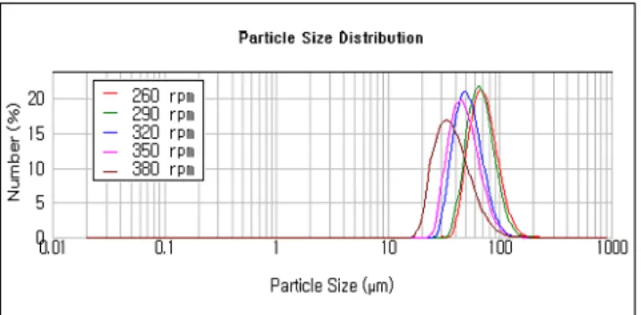
Fig. 1, it can be seen that the influence of stirring rate. As the stirring rate is increased from 260 to 380rpm, the trend of average microcapsule diameter descended. The average diameter of the microcapsules prepared with 260rpm of stirring rate is approximate 70µm, however, in the 380 rpm, the average diameter of the microcapsules is 36µm. As Shown in the Fig. 1, the size distributions of the microcapsules span out with the stirring speed. It is due to the unstable emulsion state in the emulsification process.
Fig. 1 Particle size distribution of microcapsules as function of stirring rate.
Fig. 2 shows the microcapsule diameter change behavior with the various SDS concentrations at 260rpm. As the surfactant addition is increased, the average microcapsule diameter descended. The average diameter of the microcapsules prepared with 0.2mM SDS is approximate 68µm, that with 0.01mM SDS is ~74µm, and that with 0 mM SDS is ~ 77µm. The negative slope up to the 0.2mM concentration of SDS is from the reduction of interfacial energy with the surfactant concentrations under the CMC. The positive slope of the capsule diameter could be attributed from the aggregation of coacervates [7].
Fig. 2 Average diameter of microcapsules and SDS concentration in the microencapsulation reaction.
Fig. 3 is the optical microscopy of microcapsules with different SDS concentrations. There results confirm the surfactant effect on the microcapsule size : there is the coacervate’s aggregation point of the SDS.
P1-120 / Y. J Park
• IMID 2009 DIGEST
Fig. 3 The optical microscopy of capsules with different concentration of SDS
(a)0 (b)0.01 (c)0.1 (d)0.2 (e)1 mM
The Fig. 4 shows photograph of white and blue EPD capsule image sheet using average 70µm microcapsule slurry. This displays showed white and blue state by applying voltage +30 and -30V to a front electrode respectively.
Fig. 4 Photograph of white/blue microcapsulated EPD under electric field. (a) is applied to +10V on the front electrode, and (b) is applied to -10 V.
4. Summary
In this study, we investigated the relation between narrow dispersed electronic ink microcapsules and two kinds of factor, that is surfactant and stirring rate. From the experiment, the existence of the anionic surfactant of SDS, which can make the polyelectrolyte coacervating layer easy to deposit on the electronic ink emulsions. Moreover, we found out about optimum condition of surfactant and stirring rate.
Acknowledgement
This work was supported by the Korea Ministry of Knowledge Economy.
5. References
1. S. Inoue, H. Kawai, S. Kando, T. Saeki, and T. Shimoda, IEEE Trans Electron. Dev., 49(8), pp1532 (2002)
2. J. B. Vincent and D. W. Flick, U.S. Patent, 6639709 (2003)
3. U. Bach, D. Corr, D. Lupo, F. Pichot, and M. Ryan, Adv. Mater., 14, pp845 (2002)
4. R.A. Hayes and B.J. Feenstra, Nature, 425, pp283 (2003)
5. K. S. Mayya, A. Bhattacharyya and J.-F. Argillier, Polym. Inter., 52. pp644 (2003)
6. D. W Wang, X. P. Zhao, J. Microencapsulation, 26(1), pp37-45 (2009)
7. H. Guo, X. Zhao, J. Microencapsulation, June, 25(4), pp 221–227(2008)
(a) (b)
(c) (d) (e)