321 A 66-year-old man presented with fever and headache that
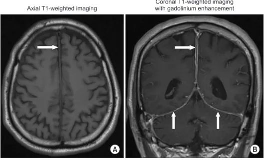
had lasted for 3 months. The headache had a continuous pressing quality and bilateral involvement. He had a history of undifferentiated arthritis, for which he had undergone a treatment with chroloquine, leading to disease remission 2 years prior. He also had long term hypertension and diabe-tes. Current medications included enalapril, metformin, and chroloquine. He was febrile with body temperature of 38.5°C. Neither nuchal rigidity nor lymphadenopathy was detected. The findings of other general and neurological examinations were unremarkable. Magnetic resonance imaging (MRI) of the brain revealed thickening of the falx cerebri with
dif-fuse pachymeningeal enhancement (Figure 1). A lumbar puncture revealed an opening pressure of 200 mm H2O. Ce-rebrospinal fluid (CSF) analysis showed a white blood cell count of 785 cells/mm3 with 62% lymphocytes and 38% neu-trophils, a protein level of 72 mg/dL, and a glucose level of 80 mg/dL (concurrent a blood glucose level of 182 mg/dL). The results of Gram and Ziehl-Neelsen staining, India ink preparation, cryptococcal antigen, tuberculosis polymerase chain reaction, and culture for bacteria were negative. The CSF culture, which was subsequently reported, was also negative for Mycobacterium tuberculosis. The adenosine deaminase (ADA) level in CSF, which was determined by an
Diffuse Hypertrophic Pachymeningeal
Tuberculosis
Verajit Chotmongkol, M.D. and Sittichai Khamsai, M.D.
Department of Medicine, Faculty of Medicine, Khon Kaen University, Khon Kaen, Thailand
Address for correspondence: Sittichai Khamsai, M.D.
Department of Medicine, Faculty of Medicine, Khon Kaen University, 123 Mitraparp Road, Khon Kaen 40002, Thailand
Phone: 66-43-363664, Fax: 66-43-348399, E-mail: Sittikh@kku.ac.th
Received: Jun. 3, 2020, Revised: Jul. 17, 2020, Accepted: Aug. 6, 2020, Published online: Aug. 10, 2020
cc It is identical to the Creative Commons Attribution Non-Commercial License (http://creativecommons.org/licenses/by-nc/4.0/).
IMAGES OF INTEREST
https://doi.org/10.4046/trd.2020.0055ISSN: 1738-3536(Print)/2005-6184(Online) • Tuberc Respir Dis 2020;83:321-323
Copyright © 2020
The Korean Academy of Tuberculosis and Respiratory Diseases.
Figure 1. Magnetic resonance imaging of a patient with diffuse hypertrophic pachymeningeal tuberculosis (before treatment). T1-weighted magnetic resonance imaging and with contrast enhancement showed thickened falx cerebri (arrow in A) with diffuse dural enhancement (arrows in B).
A B
Axial T1-weighted imaging
Coronal T1-weighted imaging with gadolinium enhancement
V Chotmongkol et al.
322 Tuberc Respir Dis 2020;83:321-323 www.e-trd.org
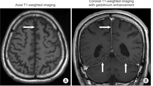
automated method, was 11.1 U/L. Blood serological study for Venereal Disease Research Laboratory was nonreactive. The result of a chest X-ray was within the normal limits. The patient denied performing the meningeal biopsy. Diffuse hypertrophic pachymeningeal tuberculosis was suspected due to the patient having symptoms of prolonged fever and headache, lymphocytic CSF pleocytosis with elevated pro-tein level and low CSF-to-serum glucose ratio, and a high level of CSF-ADA. The patient was treated with combined antituberculous drugs [isoniazid (I), rifampin (R), pyrazin-amide (Z), ethambutol (E)], for 2 months, followed by IR (isoniazid, rifampin) for 4 months1. The fever and headache gradually disappeared. A follow-up MRI of the brain revealed a marked decreased in the thickening of the falx cerebri and pachymeningeal enhancement (Figure 2). The results of repeat CSF analysis and the level of CSF-ADA were within normal ranges.
Hypertrophic pachymenigitis is a rare disease and consists of localized or diffuse thickening of the dura mater. It can involve the cranial or the spinal dura or both. Neurological manifestations include chronic headache, cranial nerve pal-sies, and progressive other neurological deficits arising due to compression of neural structures by the enlarged dura mater. The etiologies of this disorder divide into primary (idiopathic) and secondary causes; including infections (tu-berculosis, fungal infection, cysticercosis, syphilis), systemic disorders, and malignancies2. Initial diagnosis depends on radiological findings which reveals focal or diffuse thicken-ing of the dura. Dural tuberculosis is rare and usually pres-ents with localized involvement of the pachymeninges3. In this patient, the diagnosis of pachymeningeal tuberculosis was confirmed by clinical response to antituberculous treat-ment4.
This report also demonstrates the clinical utility of
CSF-ADA as a one of diagnostic tests in the diagnosis of central nervous system tuberculosis5.
Authors’ Contributions
Conceptualization: Chotmongkol V, Khamsai S. Methodolo-gy: Chotmongkol V, Khamsai S. Formal analysis: Chotmongkol V. Data curation: Chotmongkol V, Khamsai S. Software: Chot-mongkol V, Khamsai S. Validation: ChotChot-mongkol V, Khamsai S. Writing - original draft preparation: Chotmongkol V. Approval of final manuscript: all authors.
Conflicts of Interest
No potential conflict of interest relevant to this article was reported.
Funding
No funding to declare.
References
1. Chotmongkol V. Treatment of tuberculous meningitis with 6-month course of chemotherapy. Southeast Asian J Trop Med Public Health 1991;22:372-4.
2. Tariq R, Ahmed R. Tuberculous hypertrophic pachymenin-gitis presenting as visual blurring and headaches. J Pak Med Assoc 2012;62:966-8.
3. Goyal M, Sharma A, Mishra NK, Gaikwad SB, Sharma MC.
Figure 2. Magnetic resonance imaging of a patient with diffuse hypertrophic pachymeningeal tuberculosis (after treatment). T1-weighted magnetic re-sonance imaging and with contrast en-hancement revealed a marked decrease in the thickening of the falx cerebri (ar-row in A) and pachymeningeal enhance-ment (arrows in B), compared with Figure 1.
A B
Axial T1-weighted imaging
Coronal T1-weighted imaging with gadolinium enhancement
Diffuse hypertrophic pachymeningeal tuberculosis
https://doi.org/10.4046/trd.2020.0055 323
www.e-trd.org
Imaging appearance of pachymeningeal tuberculosis. AJR Am J Roentgenol 1997;169:1421-4.
4. Parney IF, Johnson ES, Allen PB. “Idiopathic” cranial hy-pertrophic pachymeningitis responsive to antituberculous therapy: case report. Neurosurgery 1997;41:965-71.
5. Feres MC, Martino MC, Maldijian S, Batista F, Gabriel Ju-nior A, Tufik S. Laboratorial validation of an automated assay for the determination of adenosine deaminase activ-ity in pleural fluid and cerebrospinal fluid. J Bras Pneumol 2008;34:1033-9.