Vol. 16, 348 –357, January 2005
The Major Target of the Endogenously Generated Reactive
Oxygen Species in Response to Insulin Stimulation Is
Phosphatase and Tensin Homolog and Not
Phosphoinositide-3 Kinase (PI-3 Kinase) in the PI-3
Kinase/Akt Pathway
□
DJi Hae Seo,* Younghee Ahn,
†Seung-Rock Lee,
†‡Chang Yeol Yeo,* and
Kyu Chung Hur*
§*Department of Biology and
†Center for Cell Signaling Research, Ewha Women’s University, Seoul 120-750,
Korea; and
‡Department of Biochemistry, Chonbuk National University Medical School, Chonju 561-182,
Korea
Submitted May 6, 2004; Revised October 22, 2004; Accepted October 27, 2004 Monitoring Editor: Carl-Henrik Heldin
Phosphoinositide-3 kinase (PI-3 kinase) and its downstream signaling molecules PDK-1 and Akt were analyzed in SK-N-SH and SK-N-BE(2) human neuroblastoma cell lines. When cells were stimulated with insulin, PI-3 kinase was activated in both cell lines, whereas the translocation of PDK-1 to the membrane fraction and phosphorylated Akt were observed only in SK-N-SH cells. Analyses of the insulin-mediated reactive oxygen species (ROS) generation and Phosphatase and Tensin homolog (PTEN) oxidation indicate that PTEN oxidation occurred in SK-N-SH cells, which can produce ROS, but not in SK-N-BE(2) cells, which cannot increase ROS in response to insulin stimulation. When SK-N-SH cells were pretreated with the NADPH oxidase inhibitor diphenyleneiodonium chloride before insulin stimulation, insulin-mediated translocation of PDK-1 to the membrane fraction and phosphorylation of Akt were remarkably reduced, whereas PI-3 kinase activity was not changed significantly. These results indicate that not only PI-3 kinase activation but also inhibition of PTEN by ROS is needed to increase cellular level of phosphatidylinositol 3,4,5-trisphosphate for recruiting downstream signaling molecules such as PDK-1 and Akt in insulin-mediated signaling. Moreover, the ROS generated by insulin stimulation mainly contributes to the inactivation of PTEN and not to the activation of PI-3 kinase in the PI-3 kinase/Akt pathway.
INTRODUCTION
Reactive oxygen species (ROS), and especially H2O2, are rapidly and transiently increased by a variety of external signals that include cytokines, peptide growth factors, and agonists of seven-transmembrane domain, or G protein-coupled receptors (GPCRs) (Ohba et al., 1994; Kimura et al., 1995; Krieger-Brauer and Kather, 1995; Sundaresan et al., 1995; Bae et al., 1997; Patterson et al., 1999). Previous studies have demonstrated that the binding of peptide growth fac-tors to their specific recepfac-tors induces a transient increase in the intracellular concentration of ROS (Krieger-Brauer and Kather, 1995; Lo and Cruz, 1995; Sundaresan et al., 1996; Bae
et al., 1997; Sattler et al., 1999). This growth factor-mediated
transient increase of ROS is an integral constituent of down-stream signal transduction (Finkel, 1998). The ROS-medi-ated modulation of growth factor signaling was achieved via
the inactivation of PTPases through oxidation of the cysteine residue in the active site sequence motif (Zhang, 1998).
ROS generation in a variety of nonphagocytic cells is much lower than in phagocytes, although the ROS genera-tion systems in nonphagocytic cells are considered similar (Babior, 1999; Bokoch and Diebold, 2002). ROS generation in phagocytes is catalyzed by NADPH oxidase, which consists of two transmembrane subunits, p22phoxand gp91phox, and at least three cytosolic subunits, p47phox, p67phox, and Rac (Babior, 1999; Banfi et al., 2003). Recently, six gp91phox ho-mologues (NOX1, NOX3, NOX4, NOX5, DUOX1, and DUOX2) have been identified in different mammalian tissue (Banfi et al., 2003). Although the mechanism of the growth factor-mediated ROS generation in nonphagocytotic cells has not been completely understood, several recent reports (Bae et al., 2000; Bokoch and Diebold, 2002) have demon-strated that the growth factor mediated-activation of phos-phoinositide-3 kinase (PI-3 kinase) and Rac is needed for ROS generation.
The PI-3 kinase pathway is implicated in controlling a variety of cellular events, including proliferation, growth, and survival (Franke et al., 1997; Shepherd et al., 1998; Cantrell, 2001; Cantley, 2002). PI-3 kinase can be activated through tyrosine kinase receptors or GPCRs and generates phosphatidylinositol 3,4,5-trisphosphate [PtdIns(3,4,5)P3] and phosphatidylinositol 3,4-bisphosphate (Stephens et al.,
Article published online ahead of print. Mol. Biol. Cell 10.1091/ mbc.E04 – 05– 0369. Article and publication date are available at www.molbiolcell.org/cgi/doi/10.1091/mbc.E04 – 05– 0369.
□D The online version of this article contains supplemental material at MBC Online (http://www.molbiolcell.org).
1993; Vanhaesebroeck et al., 1997). These 3-phosphorylated phosphatidylinositols (PIs) can recruit serine/threonine ki-nases such as PI-dependent kiki-nases (PDKs) and Akt (PKB) via interactions with a pleckstrin homology (PH) domain in these proteins (Leevers et al., 1999). Once recruited, Akt is phosphorylated at two sites, Thr308 and Ser473, by one or more PDKs, and these phosphorylations activate the kinase and release it from the membrane (Alessi et al., 1997; Stokoe
et al., 1997).
Phosphatase and Tensin homolog (PTEN), originally iden-tified as a tumor suppressor gene mutated in a large per-centage of human cancers, is considered a key negative regulator of PI-3 kinase (Cantley and Neel, 1999; Sulis and Parsons, 2003). Studies of phosphatidylinositol metabolism indicate that PTEN dephosphorylates PtdIns(3,4,5)P3at the D3 position and reduces cellular levels of PtdIns(3,4,5)P3 (Maehama and Dixon, 1998; Myers et al., 1998). Because PTEN has a critical role in antagonizing PI-3 kinase signal-ing pathways, it might be expected that PTEN would be the target of complex control mechanisms. Only limited studies have addressed this issue, but evidence is now emerging for functional regulation of PTEN at the level of protein stability and localization, as well as transcription of the PTEN gene (Adey et al., 2000; Vazquez et al., 2000, 2001; Kurose et al., 2001; Bastola et al., 2002).
It has been demonstrated that the activity of PI-3 kinase, the level of 3-phosphorylated PIs, and the activity of Akt are increased in cells exposed to H2O2(Konishi et al., 1997; Shaw
et al., 1998; Sonoda et al., 1999; Tanaka et al., 1999; Qin et al.,
2000). This H2O2-mediated activation of PI-3 kinase/Akt pathway is considered to be the result of reversible oxida-tion and inactivaoxida-tion of protein tyrosine phosphatase (PTP) family protein such as PTEN that have a critical cysteine residue in the active site (Denu and Tanner, 1998; Lee et al., 1998; Meng et al., 2002). Moreover, recent studies have indi-cated that inactivation by oxidation could be a physiological mechanism of regulation of PTP family proteins both by oxidative stress and by transiently increased ROS in cells stimulated with growth factors (Meng et al., 2002). Possible roles for the oxidation and inactivation of PTEN in regulat-ing PI-3 kinase/Akt pathway have been suggested because this enzyme is a PTP family member. A recent report has revealed that PTEN can be oxidized and inactivated by formation of a disulfide bond between the active site Cys124 and Cys71in cells treated with H2O2(Lee et al., 2002). It has been demonstrated that either H2O2 or endogenous ROS production in macrophages inactivates a fraction of PTEN and that this is associated with an oxidant-dependent acti-vation of downstream signaling (Leslie et al., 2003). More-over, the increase of cellular PtdIns(3,4,5)P3levels and Akt activation by oxidative stress does not occur in cells lacking PTEN. These results suggest that the regulation of PTEN plays a pivotal role in PI-3 kinase/Akt signaling.
In this report, we have demonstrated that the generation ROS and the resultant inactivation of PTEN is needed for insulin-mediated Akt activation by using two neuroblas-toma cell lines, SK-N-SH, which can produce ROS, and SK-N-BE(2), which cannot produce ROS. Moreover, the ROS generated in response to insulin stimulation primarily con-tributes to the inactivation of PTEN and not to the activation of PI-3 kinase.
MATERIALS AND METHODS
Materials
Antibodies specific for phosphotyrosine (PY20), phospho-Akt (Ser473), phos-pho-PTEN (Ser 380, Thr 382/383), PDK-1, and Akt were obtained from Cell
Signaling Technology (Beverly, MA). Phosphotyrosine (4G10), p85 (regula-tory subunit of PI-3 kinase), and PTEN antibodies were purchased from Upstate Biotechnology (Waltham, MA). Protein A- or G-conjugated Sepha-rose and enhanced chemiluminescence (ECL) reagent were purchased from Amersham Biosciences (Piscataway, NJ), and 2⬘,7⬘-dichlorodihydrofluorers-cein diacetate (H2DCFDA) was from Molecular Probes (Eugene, OR). All the
media, sera, and antibiotic–antimycotic mixtures were from Invitrogen (Carls-bad, CA), and all other reagents including Na3VO4, LY294002,
diphenylenei-odonium chloride (DPI), N-acetylcysteine (NAC), phosphatidylinositol, insu-lin, and epidermal growth factor (EGF) were purchased from Sigma-Aldrich (St. Louis, MO). [␥-32P]ATP (1000 Ci/mmol) was obtained from PerkinElmer
Life and Analytical Sciences (Boston, MA). Methods
Cell Culture. SK-N-BE(2)C (Biedler et al., 1978) and SK-N-SH cells were cultured in DMEM supplemented with 10% calf serum in the presence of 5% CO2at 37°C. Fresh media were supplied every 2 days. Insulin (100 nM) or
H2O2(1 mM) was added for appropriate periods, depending on the purpose
of the experiment, to cells that had been incubated in serum-free media for the previous 12 h (serum-starved cells). To measure the effect of DPI, an inhibitor of NADPH oxidase, serum-starved SK-N-SH cells were incubated in DPI (10 M) for 30 min, and then treated with insulin for 15 min. To inhibit PI-3 kinase activity, serum-starved cells were incubated in 10M LY294002 for 30 min before insulin stimulation.
Immunoprecipitation and Immunoblotting. Cells that had reached ⬃80% confluence on 100-mm plastic dishes were serum starved, treated with insu-lin, and washed with ice-cold phosphate-buffered saline. The washed cells were lysed with 1 ml of ice-cold lysis buffer (50 mM Tris-Cl, pH 8.0, 150 mM NaCl, 1% NP-40, 100g/ml phenylmethylsulfonyl fluoride [PMSF], 1 g/ml leupeptin, 1g/ml aprotinin, 1 mM Na3VO4, 1 mM EDTA, 1 mM EGTA, 1
mM NaF, and 1 mM benzamidine). The cell lysate was centrifuged for 5 min at 13,000⫻ g, and the resulting supernatant (1 mg of protein) was incubated with an appropriate amount (⬃5g) of antibodies at 4°C for 1 h. A 50-l aliquot of protein A-Sepharose (10% suspension) was added and after a 30-min incubation, the antigen–antibody complexes were pelleted by a brief centrifugation. The pellet was washed three times with ice-cold lysis buffer by centrifugation at 13,000⫻ g for 30 s, dissolved in 20l of Laemmli’s sample buffer, and fractionated on 10% SDS-polyacrylamide gels. The fractionated proteins were electrophoretically transferred to nitrocellulose. Antibody la-beling of protein bands was detected with ECL reagents according to the manufacturer’s protocol.
Measurement of PI-3 Kinase Activity.Anti-phosphotyrosine or anti-p85 im-munoprecipitates were obtained from the cell lysates (1 mg) treated with insulin (100 nM), 1 mM H2O2, insulin plus DPI (10M), or insulin plus H2O2,
for the indicated time depending on the purpose of the experiment. The pellet was washed three times with a buffer containing 20 mM Tris-Cl, pH 7.4, 100 mM NaCl, 10 mM MgCl2, and 0.5 mM EGTA. The pellet was incubated in 50
l of reaction buffer containing 20 mM Tris-Cl, pH 7.4, 100 mM NaCl, 10 mM MgCl2, 0.5 mM EGTA, 100M phosphatidylinositol, 120 M adenosine, 20
M ATP, and 2 Ci of [␥-32P]ATP for 30 min at 25°C. The reaction was
terminated by adding 100l of 1 N HCl. Phospholipids were extracted with 200l of chloroform/methanol (CHCl3/MeOH) (1:1), centrifuged at 10,000⫻
g for 1 min, and then the lower phase (organic phase) was transferred to a new tube. The organic phase was dried under nitrogen gas and resuspended in 20 l of a CHCl3/MeOH (1:1) and resolved by thin layer chromatography (TLC)
by using a silica gel plate coated with 1% potassium oxalate in a running buffer of CHCl3/MeOH/NH4OH/H2O (60:47:11.3:2). The32P-labeled
phos-phatidylinositol phosphate (PIP) was visualized and quantitated using a PhosphorImager (BAS 2000; Fuji, Tokyo, Japan).
Translocation of PDK-1 to the Membrane Fraction.Cells treated with either insulin or H2O2for 10 min were lysed in 100l of hypotonic buffer (20 mM
Tris-Cl, pH 7.4, 20 mM-mercaptoethanol, 1 mM EDTA, 1 mM EGTA, 1 mM Na3VO4, 0.5 mM PMSF, 1g/ml leupeptin, 1 g/ml aprotinin, and 1 mg/ml
benzamidine) by repeated quick freezing and thawing by using liquid nitro-gen, and then homogenized with a glass homogenizer after addition of 900l of same buffer. The homogenate was centrifuged at 3000⫻ g for 10 min to remove unbroken cells and nuclei. The supernatant was centrifuged for 30 min at 15,000⫻ g, and the pellet (50g of protein) was fractionated on a 10% SDS-polyacrylamide gel. Proteins were electrophoretically transferred to ni-trocellulose. PDK-1 was visualized using an anti-PDK-1 antibody.
Measurement of ROS Generation.Intracellular ROS production was mea-sured by a previously described method (Bae et al., 1997). Serum-starved cells were treated with insulin for the indicated time, and then washed with phenol red-free minimal essential medium and incubated in the dark for 5 min in DMEM containing 5 mM H2DCFH-DA at 37°C. Culture dishes were
trans-ferred to a confocal microscope (LSM 510; Carl Zeiss, Jena, Germany), and the production of ROS was measured by DCF fluorescence (DCFH, which is
produced from DCFH-DA by intracellular esterases, is converted to the highly fluorescent dye DCF in the presence of H2O2). DCF fluorescence was
detected using excitation at 488 nm and emission at 515–540 nm. To avoid photooxidation of DCFH, the fluorescent image was collected by a single rapid scan by using identical parameters (e.g., contrast and brightness) for all samples.
Analysis of Reduced and Oxidized Forms of PTEN by Nonreducing SDS-PAGE and Immunoblotting.After appropriate stimulation, cells were imme-diately frozen in liquid nitrogen, and lysed with 1 ml of ice-cold buffer containing 50 mM Tris-Cl, pH 8.0, 150 mM NaCl, 1% NP-40, 50 mM N-ethylmaleimide (NEM), 100g/ml PMSF, 1 g/ml leupeptin, 1 g/ml apro-tinin, 1 mM Na3VO4, 1 mM EDTA, 1 mM EGTA, and 1 mM NaF. Cell lysates
were centrifuged at 15,000⫻ g for 15 min. The resulting supernatant was fractionated on 10% nonreducing SDS-polyacrylamide gels followed by im-munoblot assay by using the anti-PTEN antibody.
Measurement of PTEN Phosphatase Activity.Substrate for PTEN phospha-tase activity assay was made as described previously (Walker et al., 2001) by using purified PI-3 kinase ␥. PtdIns(3,4,5)P3phosphatase assay was
per-formed for 30 min at 25°C in 100l of 40 mM Tris-HCl, pH 8, containing immunoprecipitated PTEN, 10 mM dithiothreitol (DTT), 1 mM EDTA, 20l of lipid vesicle consisting of phosphatidylserine (0.05 mg/ml) and diacylglyc-erol (0.005 mg/ml), and PtdIns(3,4,5)P3(0.025 mg/ml) containing⬃80,000
cpm of [32P]PtdIns(3,4,5)P
3. The reaction was terminated by the addition of
200l of 1 N HCl and sequential addition of 200 l of methanol and 200 l of chloroform. Released [32P]Pi in the aqueous phase was pooled, and the
radioactivity was counted. Anti-PTEN immunoprecipitates were obtained from the cell lysate (5g antibody/1 mg protein) that were prepared from the cells stimulated with either insulin or H2O2by lysis in 1 ml of ice-cold lysis
buffer containing 50 mM NEM to block free sulfhydryl groups in PTEN.
RESULTS
Insulin-mediated Activation of PI-3 Kinase Cascade
A previous report in our laboratory demonstrated that the tyrosine-phosphorylated components and the degree of ty-rosine phosphorylation of each component in SK-N-BE(2) human neuroblastoma cells were relatively lower than those in PC12 cells in response to insulin or nerve growth factor (Hwang et al., 1995). To address whether the diminished levels of insulin-stimulated tyrosine phosphorylation ob-served in SK-N-BE(2) cells affected activation of PI-3 kinase cascade, we measured in vitro PI-3 kinase activity in anti-phosphotyrosine immunoprecipitates from insulstimu-lated SK-N-BE(2) and SK-N-SH cells (Figure 1A). An in-crease in PI-3 kinase activity occurred within 5 min after insulin stimulation, reached maximal level⬃15–30 min, and declined after 60 min in both SK-N-SH and SK-N-BE(2) cells. The insulin-mediated activation of PI-3 kinase always oc-curred more quickly and declined more slowly in SK-N-SH cells compared with SK-N-BE(2) cells, although the maximal level of activation was very similar between these two cell lines.
To confirm the observed insulin-mediated increase of PI-3 kinase activity in anti-phosphotyrosine immunoprecipitates sufficiently reflects the change of PI-3 kinase activity in the cell, we measured PI-3 kinase activity in anti-p85 immuno-precipitates from insulin-stimulated SH and SK-N-BE(2) cells for 15 min (Figure 1B). The insulin-mediated increase of PI-3 kinase activity in both cells seemed very similar with that observed in anti-phosphotyrosine immu-noprecipitates. These results indicate that PI-3 kinase can be activated in SK-N-BE(2) cells by insulin stimulation.
Next, we measured whether PDK-1 can be recruited to the membrane fraction (Figure 2A) and whether Akt can be activated by insulin stimulation (Figure 2B). Pilot experi-ments that measured the time-dependent change of Akt phosphorylation revealed that the phosphorylation of Akt was observed within 5 min after insulin stimulation and maintained up to 30 min in SK-N-SH cells but did not occur in SK-N-BE(2) cells (our unpublished data). Based on these
data, we examined the translocation of PDK-1 to the mem-brane fraction and Akt phosphorylation in the cells stimu-lated with insulin for 10 min. The translocation of PDK-1 to the membrane fraction was observed when SK-N-SH cells were stimulated with insulin. In contrast, the insulin-medi-ated translocation of PDK-1 to the membrane fraction was not observed in SK-N-BE(2) cells (Figure 2A). The lack of membrane association of PDK-1 in insulin stimulated SK-N-BE(2) cells could be due either to a defect in the PI-3 kinase/ PtdIns(3,4,5)P3 generation pathway or to a defect in the ability of PDK-1 to bind PtdIns(3,4,5)P3. To address this possibility, we applied H2O2, which is known to recruit PDK-1 to the plasma membrane and to activate Akt, to SK-N-BE(2) cells. The translocation of PDK-1 to the mem-brane fraction was apparent when SK-N-BE(2) cells were stimulated with H2O2, although the amount of PDK-1 found in the membrane fraction was less than that observed in insulin-stimulated SK-N-SH cells (Figure 2A). We measured the phosphorylation status of Akt in whole cell lysates from Figure 1. Insulin stimulation increases PI-3 kinase activity in SK-N-SH and SK-N-BE(2) cells. Cells treated with insulin (100 nM) for the indicated time, were lysed and immunoprecipitated with either anti-phosphotyrosine (PY20, 5g) (A) or anti-p85 (B). Kinase activ-ity was measured as described under Materials and Methods by using phosphatidyl inositol as substrate, and the product PIP was re-solved by TLC. Incorporation of 32P into PIP was imaged and
quantitated using a PhosphorImager. Each bar represents the quan-titated averageⴞ SE for three independent experiments normalized to the value of unstimulated SK-N-SH cells. The anti-p85 blot (B) was obtained from the protein A beads used for PI-3 kinase activity assay.
insulin-stimulated cells by using an antibody specific for Akt phosphorylated at Ser473 (Figure 2B). In agreement with PDK-1 translocation to the membrane fraction, stimulation of cells with insulin revealed that the phosphorylation of Akt increased in SK-N-SH cells but not in SK-N-BE(2) cells. An increased phosphorylation of Akt also was observed in SK-N-BE(2) cells in response to exogenous H2O2(Figure 2B). Although the increase of PDK-1 recruitment to the mem-brane fraction and Akt phosphorylation occurred in SK-N-BE(2) cells stimulated with exogenous H2O2, the level of increase was lower than that in SK-N-SH cells stimulated with insulin. This could be due to not enough activation of PI-3 kinase. To address this possibility, we compared the level of phosphorylated Akt in both cells treated with insu-lin, H2O2, or insulin plus H2O2(Figure 2C). Akt phosphor-ylation was increased in insulin-, H2O2-, or insulin plus H2O2-stimulated SK-N-SH cells. The level of Akt
phosphor-ylation in insulin-stimulated cells was higher than that in H2O2-stimulated cells. But, Akt phosphorylation was not increased further by stimulation with insulin plus H2O2 compared with insulin alone. The comparison of Akt phos-phorylation between SK-N-SH and SK-N-BE(2) cells re-vealed that insulin stimulation along with exogenous H2O2 restored the level of Akt phosphorylation in SK-N-BE(2) cells comparable with that in insulin- or insulin plus H2O2 -stimulated SK-N-SH cells. The increase of Akt phosphory-lation in exogenous H2O2-stimulated SK-N-SH cells was very similar to that in SK-N-BE(2) cells.
These results indicate that the lack of PDK-1 translocation to the membrane fraction and Akt activation in insulin stim-ulated SK-N-BE(2) cells is due to a defect in PI-3 kinase’s ability to produce PtdIns(3,4,5)P3and thereby recruit PDK-1/Akt to the plasma membrane. These results suggest that activation of PI-3 kinase is not sufficient to increase PtdIns(3,4,5)P3 level to recruit PDK-1 and Akt in insulin-stimulated SK-N-BE(2) cells.
Measurement of Insulin-mediated ROS Generation
The reported roles of ROS on the growth factor-mediated signaling and the observed increase of PDK-1 recruitment to the plasma membrane and Akt phosphorylation in SK-N-BE(2) cells stimulated with H2O2suggest that the defect in insulin-mediated signaling could be related to ROS genera-tion. To address this possibility, we analyzed whether insu-lin can induce the transient generation of ROS in SK-N-SH and SK-N-BE(2) cells (Figure 3). Cells loaded with the redox-sensitive fluorophore H2DCFH-DA were treated with insu-lin, and the production of ROS was measured by DCF flu-orescence with a confocal microscope. In SK-N-SH cells, the DCF fluorescence rapidly increased, reached a maximal level within 15 min, and then declined in response to insulin stimulation. In contrast, an insulin-mediated increase of DCF fluorescence was not detected in SK-N-BE(2) cells. In-stead, DCF fluorescence was decreased by insulin stimula-tion. Application of other growth factors, such as EGF or platelet-derived growth factor (PDGF), produced similar re-sults (our unpublished data). However, the basal DCF fluo-rescence in SK-N-BE(2) cells was always higher than that in SK-N-SH cells. These results indicated not only that the SK-N-BE(2) cell line cannot generate ROS in response to insulin stimulation but also that the lack of insulin-mediated ROS production could be responsible for the defective sig-naling in these cells.
Insulin-mediated Modification of PTEN and PTEN Activity
Studies of phosphatidylinositol metabolism indicate that PTEN has a critical role as a negative regulator of PI-3 kinase because PTEN dephosphorylates PtdIns(3,4,5)P3 at the D3 position and reverses the action of PI-3 kinase (Maehama and Dixon, 1998; Myers et al., 1998). Recent studies indicate that PTEN can be inactivated by H2O2, and this inactivation was correlated with a shift in the electrophoretic mobility of PTEN consistent with its oxidation both in vitro and in cultured cells (Lee et al., 2002). Moreover, either H2O2 or endogenous ROS production in macrophages inactivates a fraction of PTEN (Leslie et al., 2003). Our results, along with these earlier observations suggest that the inactivation of PTEN by endogenously produced ROS is required to pro-duce a net increase in PtdIns(3,4,5)P3 levels in response to insulin stimulation. To address this possibility, we examined PTEN oxidation in insulin-treated SK-N-SH and SK-N-BE(2) cells by using nonreducing SDS-PAGEs and immunoblot analysis as described previously (Lee et al., 2002). Both SK-Figure 2. Effect of insulin or H2O2on the PDK-1 recruitment to the
membrane fraction (A) and Akt activation (B and C) in SK-N-SH and SK-N-BE(2) cells. Membrane fractions (A) or whole cell lysates (B and C) (50g of protein) prepared from cells treated with insulin (100 nM), H2O2 (1 mM), or insulin plus H2O2 for 10 min were
analyzed with anti-PDK-1 (A), or anti-phospho-Akt (Ser473) (B and C). The same blots were reprobed with anti-actin (A) and anti-Akt (B and C), respectively, to confirm equal protein loading.
N-SH and SK-N-BE(2) cells were incubated for various pe-riods of time with insulin or H2O2. Cell lysates were pre-pared with a buffer containing NEM to block free sulfhydryl groups, and lysates were then subjected to immunoblot by using anti-PTEN antibodies (Figure 4). When SK-N-SH cells were treated with insulin, the oxidized form of PTEN, based on the increased electrophoretic mobility, occurred 2 min after insulin stimulation. Oxidized PTEN increased with time of incubation, reached a maximum at 10 min, and decreased gradually thereafter. In addition, application of DPI before insulin stimulation of the cells abolished the oxidized form of PTEN that occurred in response to insulin stimulation (Figure 4A). Only a minor portion of PTEN (⬃10% at a maximal level) occurred in the oxidized form in insulin-treated cells. However, the majority of PTEN was observed to be in the oxidized form when cells were treated Figure 3. Effect of insulin on ROS generation in SK-N-SH and SK-N-BE(2)C cells. The generation of ROS was assayed on the basis of DCF fluorescence as described in Materials and Methods. Confocal micrographs (A) of DCF fluorescence from the SH and SK-N-BE(2) cells either unstimulated or stimulated with insulin (100 nM) for 15 min. Time course of the ROS generation by insulin in SK-N-SH (black bar) and SK-N-BE(2) (gray bar) cells. The relative fluorescence value of each time point was normalized to the value of unstimulated SK-N-SH cells. Each bar represents the average⫾ SE of three independent experiments.
Figure 4. Oxidation of PTEN and recovery of PTEN activity from oxidative inactivated PTEN in the cells stimulated either insulin or H2O2. The oxidation of PTEN was analyzed in SK-N-SH cells
stim-ulated with insulin (A) or treated with DPI (10 M) for 30 min before insulin stimulation (B) and in SK-N-BE(2) cells stimulated with insulin (C) or H2O2(D). SK-N-SH and SK-N-BE(2) cells were
stimulated for the indicated times with insulin (100 nM), DPI (10 M) plus insulin, or H2O2(1 mM) and processed by the procedure
described in Materials and Methods. Proteins, 200g (A and B), 400 g (C), or 50 g (D), were fractionated on a 10% nonreducing SDS-PAGE. The redox state of PTEN was analyzed using anti-PTEN antibody immunoblotting to identify shifts in PTEN mobility. The recovery of PTEN phosphatase activity from oxidative inactivated PTEN was measured in anti-PTEN immunoprecipitates from the cells stimulated with insulin or H2O2for 15 min (E). Free cysteine
residues of PTEN were blocked with irreversible alkylating reagent NEM immediately after cells were stimulated with insulin or H2O2
as described in Materials and Methods. The oxidized fraction of PTEN in anti-PTEN immunoprecipitates was converted to active form by reducing with DTT. The phosphatase activity was measured using
32P-labeled PtdIns(3,4,5)P
3 as substrates. Each bar represents the
with 1 mM H2O2for 10 min. In contrast, the oxidized form of PTEN was not observed in SK-N-BE(2) cells treated with insulin, even when double the amount of protein was loaded on the gel (Figure 4B). However, application of 1 mM H2O2 in SK-N-BE(2) cells induced oxidation of the majority of PTEN within 5 min, and oxidized PTEN was maintained up to 60 min and then returned to the reduced form after 90 min.
To address whether the oxidized PTEN in insulin-stimu-lated SK-N-SH cells is correinsulin-stimu-lated with the oxidative inhibi-tion of PTEN activity, we examined phosphatase activity in PTEN immunoprecipitates from the cells stimulated with insulin or H2O2(Figure 4E). Previous reports (Lee et al., 2002; Meng et al., 2002; Leslie et al., 2003) demonstrated that PTEN is reversibly inactivated by oxidation, like other members of PTP family, and the oxidized cysteine residues in PTEN are protected from alkylation. To measure the portion of inacti-vated PTEN activity by oxidation, cells were lyzed in a buffer containing NEM immediately after stimulation to pre-vent further oxidation of PTEN and to block free sulfhydryl groups in the unoxidized fraction of PTEN before anti-PTEN immunoprecipitation. Thus, the phosphatase activity in anti-PTEN immunoprecipitates reflects the fraction of inactivated PTEN by oxidation in the cells because their oxidation is reversible. The released [32P]phosphate was 661 ⫾ 68, 1049⫾ 80, or 2415 ⫾ 352 cpm in the anti-PTEN immuno-precipitates from the cells unstimulated, stimulated with insulin, or stimulated with 1 mM exogenous H2O2, respec-tively. To measure what fraction of total PTEN is oxidized by insulin stimulation, the phosphatase activities of PTEN from insulin- or H2O2-stimulated cells were compared after subtracting the basal level of phosphatase activity observed from unstimulated cells. The phosphatase activity of H2O2 -stimulated cells is considered as total PTEN activity, because almost all PTEN in cells was oxidized by H2O2. About 16% of the total PTEN is oxidized by insulin stimulation. The higher value of insulin-mediated PTEN oxidation observed in the phosphatase activity assay (⬃16%) compared with the value observed in the gel-shift assay (⬃10%) may reflect the difference of sensitivity between these two methods, or it may reflect the fact that intermediate form of oxidized PTEN, which did not form intramolecular disulfide bonds, is included in the phosphatase activity assay.
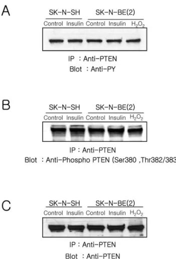
A recent report demonstrated that activated Src reduces PTEN activity to in micelles, promotes Akt translocation to the plasma membrane, and increases tyrosine phosphoryla-tion of PTEN (Lu et al., 2003). To test whether the status of PTEN phosphorylation was changed, we studied PTEN phosphorylation in anti-PTEN immunoprecipitates from in-sulin-treated cells by using anti-phosphotyrosine (4G10) and an antibody specific Ser/Thr phosphorylated PTEN (Figure 5). The level of tyrosine phosphorylated (Figure 5A) and Ser/Thr phosphorylated (Figure 5B) PTEN did not change in either cell type in response to insulin or H2O2treatment for 15 min. Similar amounts of PTEN were loaded on the gel as confirmed by reblotting with anti-PTEN (Figure 5C).
These results indicate that when cells were stimulated with insulin, PTEN can be oxidized and inactivated by en-dogenously produced ROS in SK-N-SH cells, but not in SK-N-BE(2) cells. Moreover, the modification of PTEN by phosphorylation is not related to in this early event of PI-3 kinase/Akt signaling.
Effect of DPI and H2O2on PI-3 Kinase Activity and Akt
Phosphorylation
SK-N-BE(2) cells, which have a defective response to insulin, are unable to generate ROS and inactivate PTEN. SK-N-SH
cells, which respond normally to insulin, have increased ROS and oxidized, inactivated PTEN. These dada suggest that the PTEN inactivation via oxidation together with the activation of PI-3 kinase are both required to increase the cellular PtdIns(3,4,5)P3level. To address this possibility and to confirm that the defect in PI-3 kinase/Akt signaling ob-served in SK-N-BE(2) is due to lack of ROS generation, we examined the effect of DPI, an inhibitor of NADPH oxidase, on the PI-3 kinase activity and Akt phosphorylation in SK-N-SH cells stimulated with insulin (Figure 6A). The PI-3 kinase activity was increased 1.8⫾ 0.2- and 1.6 ⫾ 0.3-fold in SK-N-SH and SK-N-BE(2) cells, respectively, in response to 1 mM H2O2. When SK-N-SH cells were pretreated with DPI before insulin stimulation, the increase of PI-3 kinase activ-ity (7.0 ⫾ 0.5-fold) was almost identical with that (7.5 ⫾ 0.8-fold) in the cells treated with insulin alone. Moreover, the increase of PI-3 kinase activity in SK-N-BE(2) was very similar in cells stimulated with insulin alone (5.4⫾ 0.2) or insulin plus 1 mM H2O2(5.8⫾ 0.1). The lower increase of PI-3 kinase activity in N-BE(2) cells compared with SK-N-SH cells in response to insulin stimulation is due to higher basal activity of PI-3 kinase in SK-N-BE(2) cells.
Figure 5. Analyses of the phosphorylation status of PTEN. Cells were stimulated with insulin (100 nM) or H2O2(1 mM) for 10 min,
lysed, and immunoprecipitated using anti-PTEN (5g) as described in Materials and Methods. The anti-PTEN immunoprecipitates were fractionated on a 10% SDS-PAGE, transferred to nitrocellulose, and blotted with anti-phosphotyrosine (4G10) (A), anti-phospho-PTEN (B), and anti-PTEN.
In contrast, the amount of PDK-1 in the membrane frac-tion markedly decreased in SK-N-SH cells pretreated with DPI or LY294002 before insulin stimulation, compared with
levels in the cells stimulated with insulin alone (Figure 6B). Consistent with the recruitment of PDK-1 to the membrane fraction, phosphorylated Akt also was decreased in the SK-N-SH cells pretreated with DPI or LY294002 before insulin stimulation (Figure 6B). An increase of Akt phosphorylation in response to exogenous H2O2was abolished by LY294002 in both SK-N-SH and SK-N-BE(2) cells (Figure 6C). More-over, the examination of phosphorylated Akt in human embryonic kidney 293T cells stimulated with insulin or epi-dermal growth factor (EGF) revealed that the level of either insulin- or EGF-mediated Akt phosphorylation was de-creased by DPI pretreatment (our unpublished data).
These results together with the data obtained from SK-N-BE(2) cells indicate that both activation of PI-3 kinase as well as inhibition of PTEN by endogenously produced ROS are needed to increase cellular PtdIns(3,4,5)P3levels enough to recruit downstream target molecules such as PDK-1 and Akt to the membrane fraction via their PH domains. Moreover, the ROS generated by insulin stimulation mainly contributes to the inactivation of PTEN and not to the activation of PI-3 kinase.
DISCUSSION
A large body of compelling evidence demonstrates that peptide growth factors induce a transient increase in the intracellular concentration of ROS, especially H2O2(Ohba et
al., 1994; Kimura et al., 1995; Krieger-Brauer and Kather,
1995; Sundaresan et al., 1995; Bae et al., 1997; Patterson et al., 1999). This growth factor-mediated transient increase of ROS is considered an integral component of downstream signal transduction (Finkel, 1998). In this report, we have shown that the activation of PI-3 kinase as well as inactiva-tion of PTEN, via oxidainactiva-tion by transiently increased ROS, are both needed for insulin-mediated Akt activation by us-ing two different neuroblastoma cell lines, SK-N-SH and SK-N-BE(2), which can and cannot produce ROS, respec-tively, in response to insulin stimulation. Moreover, the insulin-mediated increase of ROS contributes mainly to the inactivation of PTEN and not to the activation of PI-3 kinase to cause a net increase cellular PtdIns(3,4,5)P3levels.
Analysis of PI-3 kinase/Akt pathway in insulin-stimu-lated SK-N-BE(2) cells, which cannot increase insulin-medi-ated cellular ROS levels, demonstrates that recruitment of PDK-1 to the membrane fraction and activation of Akt do not occur despite PI-3 kinase activation. Moreover, translo-cation of PDK-1 to the membrane fraction and activation of Akt can be induced by application of exogenous H2O2to this cell line. These results indicate that the insulin-mediated activation of PI-3 kinase is not sufficient to produce the levels of cellular PtdIns(3,4,5)P3 needed to recruit PDK-1 and activate Akt and suggest that the transient increase of ROS may play a pivotal role in increasing PtdIns(3,4,5)P3 levels. Consistent with this possibility, our data demonstrate that inhibition of insulin-mediated ROS production in SK-N-SH cells by using DPI results in a decrease of PDK-1 translocation to the membrane fraction and reduced Akt activation without significant effect on PI-3 kinase activity, although several reports showed that the subcellular local-ization of PDK-1 seem to be growth factor insensitive (Yamada et al., 2002; Kim et al., 2003) and that PDK-1 is activated through a PI-3 kinase-independent pathway (Kim
et al., 2003). However, a recent report demonstrated that Akt
cannot be activated by insulin-like growth factor-1 in ho-mozygous knockin embryonic stem cells expressing a form of PDK-1 with a mutation in its PH domain that abolishes Figure 6. PI-3 kinase/Akt pathway regulation. (A) Effect of the
NADPH oxidase inhibitor DPI and exogenous H2O2on PI-3 kinase/Akt
pathway. The PI-3 kinase activity was measured in anti-phosphotyrosine immunoprecipitates from cells treated for 10 min with insulin, H2O2,
insulin plus DPI, or insulin plus H2O2. Each bar represents the mean
value⫾ SE of three independent experiments normalized to the value of unstimulated SK-N-SH cells. (B) Effect of DPI and LY294002 on the insu-lin-mediated translocation of PDK-1 and Akt activation in SK-N-SH cells. Membrane fractions and lysates were prepared from cells stimulated with insulin, insulin plus DPI, or insulin plus LY294002. Samples were fraction-ated (50g of protein) by 10% SDS-PAGE and transferred to nitrocellu-lose. PDK-1 and activated Akt were visualized using anti-PDK-1 and anti-phospho-Akt. The same blots were reprobed with anti-actin and anti-Akt, respectively, to confirm equal protein loading. (C) Effect of PI-3 kinase inhibitor on the Akt activation in response to exogenous H2O2.
Proteins (50g) from cells stimulated with H2O2or H2O2plus LY294002,
were fractionated on two 10% SDS-PAGs and transferred to nitrocellulose. One blot was used for visualizing phosphorylated Akt by using anti-phospho-Akt, and the other blot was used for visualizing Akt by using anti-Akt to confirm equal protein loading.
PtdIns(3,4,5)P3 binding, without affecting catalytic activity (McManus et al., 2004).
A previous report has revealed that PTEN was reversibly oxidized and inactivated in the cells stimulated with H2O2 through the formation of a disulfide bond between Cys 124 at the active site and Cys 71 (Lee et al., 2002). Moreover, endogenous oxidant production in macrophages inactivates a fraction of the cellular PTEN, and this is associated with an oxidant-dependent activation of downstream signaling (Le-slie et al., 2003). These results, together with our observations identifying the insulin-mediated oxidized form of PTEN, translocation of PDK-1 to the membrane fraction, and Akt activation in SK-N-SH cells, but not in the SK-N-BE(2) or SK-N-SH cells preincubated with DPI, strongly suggest that insulin-stimulated ROS generation plays a key role in the PI-3 kinase/Akt pathway through the reversible oxidation and inactivation of PTEN. Although only a small fraction of PTEN (⬍10%) is oxidized in insulin-stimulated SK-N-SH cells this might be enough to increase cellular PtdIns(3,4,5)P3 levels to recruit PDK-1 and Akt. The relevant fraction of PTEN may be closely associated with the signaling complex
where ROS generation most likely regulates the
PtdIns(3,4,5)P3 level in the insulin-mediated signaling. In fact, a previous study has revealed that only⬃10% of PTEN was oxidized in stimulated macrophages (Leslie et al., 2003). It has been demonstrated that PtdIns(3,4,5)P3 and the NADPH oxidase complex are found in the plasma mem-brane (Babior, 1999) and that the activated PDGF receptor recruits PI-3 kinase to the plasma membrane (Nave et al., 1996; Clark et al., 1998). Thus, it is likely that ROS is pro-duced near activated receptors and that the PTEN localized at these sites can be the target of oxidative inactivation.
In contrast to PDGF, studies on the localization of insulin-mediated signaling complex have revealed that the majority of IRS-1 and PI-3 kinase occur in the low-density membrane fractions that contain GLUT4 vesicles (Nave et al., 1996; Inoue et al., 1998). However, a more detailed evaluation of the components in the low-density fractions revealed that PI-3 kinase and IRS-1 are associated with a cytoskeletal structure that is distinct from the microsomal membrane (Clark et al., 1998). In addition, a recent report has demon-strated that plasma membrane-associated IRS-1 is needed for insulin-mediated full activation of Akt in a cell-free system (Murata et al., 2003). These results suggest that IRS-1 and PI-3 kinase must be in close opposition to the cell surface and accessible to the insulin receptor and other signaling molecules. Moreover, it has been revealed that the insulin receptor possesses a conserved tyrosine autophos-phorylation site (YxxM motif) in the C terminus its -sub-unit and recruits PI-3 kinase directly to this site (Levy-Toledano et al., 1994; Van Horn et al., 1994).
Our data revealed that SK-N-BE(2) cells, which cannot increase ROS, and SK-N-SH cells, in which ROS generation was inhibited using DPI, are not able to activate Akt in response to insulin, whereas PI-3 kinase activation is normal in these cells. It is difficult to rule out the possibility that our data derived from the results of the oxidative inhibition of the protein phosphatase or of other lipid phosphatases, which control the redox-dependant PI metabolism. How-ever, our data together with a previous report that has demonstrated that PTEN-null glioblastoma cells increase cellular PtdIns(3,4,5)P3levels and activate Akt in response to PDGF stimulation but not exogenous H2O2 (Leslie et al., 2003), indicate that ROS generated by insulin stimulation mainly contributes to the inactivation of PTEN and not to the activation of PI-3 kinase. In fact, we also observed that Akt is activated by either insulin or PDGF stimulation but
not by exogenous H2O2 in PTEN-null glioblastoma cells (U87MG) (our unpublished data).
It has been postulated that the increased production of intracellular oxidants may contribute to enhancing tyrosine phosphorylation-dependent signaling in response to growth factors by transiently suppressing activities of the members of PTP family (Denu and Tanner, 1998; Lee et al., 1998; Meng
et al., 2002). However, it is not clear how widely this can be
applied across a variety of tyrosine phosphorylation-depen-dent signaling pathways. The examination of PI-3 kinase activity has demonstrated that the increase of PI-3 kinase activity in both SK-N-SH and SK-N-BE(2) cells is much lower in response to exogenous H2O2(⬍2-fold) than insulin (⬎7-fold) as noted in a previous report (Van der Kaay et al., 1999). This result suggests that exogenous oxidant is less effective than insulin stimulation in the activation of PI-3 kinase. However, it has been demonstrated that application of exogenous H2O2activates receptor tyrosine kinases such as EGF receptors which in turn activate PI-3 kinase, resulting in production of PtdIns(3,4,5)P3and in the activation of Akt (Knebel et al., 1996; Wang et al., 2000). Our data show that the insulin-mediated increase of PI-3 kinase activity is indepen-dent of ROS production and suggest that PI-3 kinase is not the major target of ROS. Moreover, these results suggest that the binding motifs of the p85 regulatory subunit of PI-3 kinase located in the insulin receptor or IRS are more resis-tant to the member of PTP family, the known target of the reversible inactivation by ROS, than other tyrosine phos-phorylation sites.
A role for ROS in insulin-mediated signaling has been suggested by the observation that insulin stimulation in-duces the rapid production of ROS and that H2O2can mimic some of the action of insulin (Czech et al., 1974; Krieger-Brauer et al., 1997). Recent reports (Mahadev et al., 2001a; Mahadev et al., 2001b) have demonstrated that insulin-stim-ulated ROS generation modinsulin-stim-ulated the steady-state tyrosine phosphorylation of the insulin receptor and its cellular sub-strates by reversible inhibition of PTPases. In addition, the transient increase of ROS in response to insulin regulates the activity of PI-3 kinase/Akt and cellular glucose transport. Our results demonstrating that Akt is activated in SK-N-SH, which can produce ROS, but not in SK-N-BE(2) cells, which cannot produce ROS in response to insulin stimulation, in-dicate the importance of ROS generation in insulin-mediated signaling. However, why SK-N-BE(2) cells decrease ROS levels in response to insulin stimulation is difficult to explain until the growth factor-mediated ROS generation system in this cell line is completely understood.
Although the ROS generation in a variety of nonphago-cytic cells is much lower than in phagocytes, the ROS gen-eration system itself is thought to be similar (Bokoch and Diebold, 2002). Although the mechanism of the growth fac-tor-mediated ROS generation in nonphagocytic cells is still rather poorly understood, several recent reports (Bae et al., 2000; Bokoch and Diebold, 2002) have indicated that growth factor mediated-activation of PI-3 kinase and Rac is needed for ROS generation. However, a recent report has shown that the insulin-mediated ROS production is independent of PI-3 kinase activity in 3T3-L1 adipocytes (Mahadev et al., 2001a). Our results demonstrating that SK-N-BE(2) cells do not produce ROS in response to EGF or PDGF, although this cell line has receptors for these growth factors (our unpub-lished data), suggest that the defect in the ROS generation system in SK-N-BE(2) cells could be located at a common point shared by both insulin and EGF signaling pathway.
In conclusion, the data in this report show that the gen-eration of ROS has a pivotal role in insulin-mediated
acti-vation of PI-3 kinase/Akt signaling through the reversible oxidation and inactivation of PTEN, which occurs indepen-dently of PI-3 kinase activation. Moreover, our results sug-gest that there must be a signal amplification mediated by the transiently increased ROS. Insulin-stimulated ROS pro-duction oxidizes and inactivates PTEN, thereby allowing PI-3 kinase to increase the cellular level of PtdIns(3,4,5)P3 enough to recruit PDK and Akt.
ACKNOWLEDGMENTS
This work was supported by Korea Science and Engineering Foundation grants and a BK21 grant, Ministry of Education. We thank Dr. Nick R. Leslie (University of Dundee, Dundee, United Kingdom) for providing purified PI3-kinase␥.
REFERENCES
Adey, N. B., Huang, L., Ormonde, P. A., Baumgard, M. L., Pero, R., Byreddy, D. V., Tavtigian, S. V., and Bartel, P. L. (2000). Threonine phosphorylation of the MMAC1/PTEN PDZ binding domain both inhibits and stimulates PDZ binding. Cancer Res. 60, 35–37.
Alessi, D. R., James, S. R., Downes, C. P., Holmes, A. B., Gaffney, P. R., Reese, C. B., and Cohen, P. (1997). Characterization of a 3-phosphoinositide-depen-dent protein kinase which phosphorylates and activates protein kinase Bal-pha. Curr. Biol. 7, 261–269.
Babior, B. M. (1999). NADPH oxidase: an update. Blood 93, 1464 –1476. Bae, Y. S., Kang, S. W., Seo, M. S., Baines, I. C., Tekle, E., Chock, P. B., and Rhee, S. G. (1997). EGF-induced generation of hydrogen peroxide. Role in EGF receptor-mediated tyrosine phosphorylation. J. Biol. Chem. 272, 217–221. Bae, Y. S., Sung, J. Y., Kim, O. S., Kim, Y. J., Hur, K. C., Kazlauskas, A., and Rhee, S. G. (2000). PDGF-induced H2O2production requires the activation of
phosphatidylinositol 3-kinase. J. Biol. Chem. 275, 10527–10531.
Banfi, B., Clark, R. A., Steger, K., and Krause, K. H. (2003). Two novel proteins activate superoxide generation by the NADPH oxidase NOX1. J. Biol. Chem. 278, 3510 –3513.
Bastola, D. R., Pahwa, G. S., Lin, M. F., and Cheng, P. W. (2002). Downregu-lation of PTEN/MMAC/TEP1 expression in human prostate cancer cell line DU145 by growth stimuli. Mol. Cell. Biochem. 236, 75– 81.
Biedler, J. L., Roffler-Tarlov, S., Schachner, M., and Freedman, L. S. (1978). Multiple neurotransmitter synthesis by human neuroblastoma cell lines and clones. Cancer Res. 38, 3751–3757.
Bokoch, G. M., and Diebold, B. A. (2002). Current molecular models for NADPH oxidase regulation by Rac GTPase. Blood 100, 2692–2696. Cantley, L. C. (2002). The phosphoinositide 3-kinase pathway. Science 296, 1655–1657.
Cantley, L. C., and Neel, B. G. (1999). New insights into tumor suppression: PTEN suppresses tumor formation by restraining the phosphoinositide 3-ki-nase/AKT pathway. Proc. Natl. Acad. Sci. USA 96, 4240 – 4245.
Cantrell, D. A. (2001). Phosphoinositide 3-kinase signalling pathways. J. Cell Sci. 114, 1439 –1445.
Clark, S. F., Martin, S., Carozzi, A. J., Hill, M. M., and James, D. E. (1998). Intracellular localization of phosphatidylinositide 3-kinase and insulin recep-tor substrate-1 in adipocytes: potential involvement of a membrane skeleton. J. Cell Biol. 140, 1211–1225.
Czech, M. P., Lawrence, J. C., Jr., and Lynn, W. S. (1974). Evidence for the involvement of sulfhydryl oxidation in the regulation of fat cell hexose transport by insulin. Proc. Natl. Acad. Sci. USA 71, 4173– 4177.
Denu, J. M., and Tanner, K. G. (1998). Specific and reversible inactivation of protein tyrosine phosphatases by hydrogen peroxide: evidence for a sulfenic acid intermediate and implications for redox regulation. Biochemistry 37, 5633–5642.
Finkel, T. (1998). Oxygen radicals and signaling. Curr. Opin. Cell. Biol. 10, 248 –253.
Franke, T. F., Kaplan, D. R., and Cantley, L. C. (1997). PI3K: downstream AKTion blocks apoptosis. Cell 88, 435– 437.
Hwang, J. J., Kwon, J. H., Lee, K. Y., and Hur, K. C. (1995). Effet of nerve growth factor, insulin, and extracellular proteins on the neurite outgrowth of SK-N-BE(2) human neuroblastoma cells. Mol. Cell 5, 501–507.
Inoue, G., Cheatham, B., Emkey, R., and Kahn, C. R. (1998). Dynamics of insulin signaling in 3T3–L1 adipocytes. Differential compartmentalization and
trafficking of insulin receptor substrate (IRS)-1 and IRS-2. J. Biol. Chem. 273, 11548 –11555.
Kim, D. W., et al. (2003). RET/PTC (rearranged in transformation/papillary thyroid carcinomas) tyrosine kinase phosphorylates and activates phosphoi-nositide-dependent kinase 1 (PDK1): an alternative phosphatidylinositol 3-ki-nase-independent pathway to activate PDK1. Mol. Endocrinol. 17, 1382–1394. Kimura, T., Okajima, F., Sho, K., Kobayashi, I., and Kondo, Y. (1995). Thyro-tropin-induced hydrogen peroxide production in FRTL-5 thyroid cells is mediated not by adenosine 3⬘,5⬘-monophosphate, but by Ca2⫹ signaling followed by phospholipase-A2 activation and potentiated by an adenosine derivative. Endocrinology 136, 116 –123.
Knebel, A., Rahmsdorf, H. J., Ullrich, A., and Herrlich, P. (1996). Dephosphor-ylation of receptor tyrosine kinases as target of regulation by radiation, oxidants or alkylating agents. EMBO J. 15, 5314 –5325.
Konishi, H., Matsuzaki, H., Tanaka, M., Takemura, Y., Kuroda, S., Ono, Y., and Kikkawa, U. (1997). Activation of protein kinase B (Akt/RAC-protein kinase) by cellular stress and its association with heat shock protein Hsp27. FEBS Lett. 410, 493– 498.
Krieger-Brauer, H. I., and Kather, H. (1995). The stimulus-sensitive H2O2
-generating system present in human fat-cell plasma membranes is multire-ceptor-linked and under antagonistic control by hormones and cytokines. Biochem. J. 307, 543–548.
Krieger-Brauer, H. I., Medda, P. K., and Kather, H. (1997). Insulin-induced activation of NADPH-dependent H2O2 generation in human adipocyte
plasma membranes is mediated by Galphai2. J. Biol. Chem. 272, 10135–10143. Kurose, K., Zhou, X. P., Araki, T., Cannistra, S. A., Maher, E. R., and Eng, C. (2001). Frequent loss of PTEN expression is linked to elevated phosphorylated Akt levels, but not associated with p27 and cyclin D1 expression, in primary epithelial ovarian carcinomas. Am. J. Pathol. 158, 2097–2106.
Lee, S. R., Kwon, K. S., Kim, S. R., and Rhee, S. G. (1998). Reversible inacti-vation of protein-tyrosine phosphatase 1B in A431 cells stimulated with EGF. J. Biol. Chem. 273, 15366 –15372.
Lee, S. R., Yang, K. S., Kwon, J., Lee, C., Jeong, W., and Rhee, S. G. (2002). Reversible inactivation of the tumor suppressor PTEN by H2O2. J. Biol. Chem.
277, 20336 –20342.
Leevers, S. J., Vanhaesebroeck, B., and Waterfield, M. D. (1999). Signalling through phosphoinositide 3-kinases: the lipids take centre stage. Curr. Opin. Cell Biol. 11, 219 –225.
Leslie, N. R., Bennett, D., Lindsay, Y. E., Stewart, H., Gray, A., and Downes, C. P. (2003). Redox regulation of PI 3-kinase signalling via inactivation of PTEN. EMBO J. 22, 5501–5510.
Levy-Toledano, R., Taouis, M., Blaettler, D. H., Gorden, P., and Taylor, S. I. (1994). Insulin-induced activation of phosphatidyl inositol 3-kinase. Demon-stration that the p85 subunit binds directly to the COOH terminus of the insulin receptor in intact cells. J. Biol. Chem. 269, 31178 –31182.
Lo, Y. Y., and Cruz, T. F. (1995). Involvement of reactive oxygen species in cytokine and growth factor induction of c-fos expression in chondrocytes. J. Biol. Chem. 270, 11727–11730.
Lu, Y., et al. (2003). Src family protein-tyrosine kinases alter the function of PTEN to regulate phosphatidylinositol 3-kinase/AKT cascades. J. Biol. Chem. 278, 40057– 40066.
Maehama, T., and Dixon, J. E. (1998). The tumor suppressor, PTEN/MMAC1, dephosphorylates the lipid second messenger, phosphatidylinositol 3,4,5-trisphosphate. J. Biol. Chem. 273, 13375–13378.
Mahadev, K., Wu, X., Zilbering, A., Zhu, L., Lawrence, J. T., and Goldstein, B. J. (2001a). Hydrogen peroxide generated during cellular insulin stimulation is integral to activation of the distal insulin signaling cascade in 3T3–L1 adipocytes. J. Biol. Chem. 276, 48662– 48669.
Mahadev, K., Zilbering, A., Zhu, L., and Goldstein, B. J. (2001b). Insulin-stimulated hydrogen peroxide reversibly inhibits protein-tyrosine phospha-tase 1b in vivo and enhances the early insulin action cascade. J. Biol. Chem. 276, 21938 –21942.
McManus, E. J., Collins, B. J., Ashby, P. R., Prescott, A. R., Murray-Tait, V., Armit, L. J., Arthur, J. S., and Alessi, D. R. (2004). The in vivo role of PtdIns(3,4,5)P3binding to PDK1 PH domain defined by knockin mutation.
EMBO J. 23, 2071–2082.
Meng, T. C., Fukada, T., and Tonks, N. K. (2002). Reversible oxidation and inactivation of protein tyrosine phosphatases in vivo. Mol. Cell 9, 387–399. Murata, H., Hresko, R. C., and Mueckler, M. (2003). Reconstitution of phos-phoinositide 3-kinase-dependent insulin signaling in a cell-free system. J. Biol. Chem. 278, 21607–21614.
Myers, M. P., Pass, I., Batty, I. H., Van der Kaay, J., Stolarov, J. P., Hemmings, B. A., Wigler, M. H., Downes, C. P., and Tonks, N. K. (1998). The lipid
phosphatase activity of PTEN is critical for its tumor supressor function. Proc. Natl. Acad. Sci. USA 95, 13513–13518.
Nave, B. T., Haigh, R. J., Hayward, A. C., Siddle, K., and Shepherd, P. R. (1996). Compartment-specific regulation of phosphoinositide 3-kinase by PDGF and insulin in 3T3–L1 adipocytes. Biochem. J. 318, 55– 60.
Ohba, M., Shibanuma, M., Kuroki, T., and Nose, K. (1994). Production of hydrogen peroxide by transforming growth factor-beta 1 and its involvement in induction of egr-1 in mouse osteoblastic cells. J. Cell Biol. 126, 1079 –1088. Patterson, C., Ruef, J., Madamanchi, N. R., Barry-Lane, P., Hu, Z., Horaist, C., Ballinger, C. A., Brasier, A. R., Bode, C., and Runge, M. S. (1999). Stimulation of a vascular smooth muscle cell NAD(P)H oxidase by thrombin. Evidence that p47(phox) may participate in forming this oxidase in vitro and in vivo. J. Biol. Chem. 274, 19814 –19822.
Qin, S., Stadtman, E. R., and Chock, P. B. (2000). Regulation of oxidative stress-induced calcium release by phosphatidylinositol 3-kinase and Bruton’s tyrosine kinase in B cells. Proc. Natl. Acad. Sci. USA 97, 7118 –7123. Sattler, M., Winkler, T., Verma, S., Byrne, C. H., Shrikhande, G., Salgia, R., and Griffin, J. D. (1999). Hematopoietic growth factors signal through the forma-tion of reactive oxygen species. Blood 93, 2928 –2935.
Shaw, M., Cohen, P., and Alessi, D. R. (1998). The activation of protein kinase B by H2O2or heat shock is mediated by phosphoinositide 3-kinase and not by
mitogen-activated protein kinase-activated protein kinase-2. Biochem. J. 336, 241–246.
Shepherd, P. R., Withers, D. J., and Siddle, K. (1998). Phosphoinositide 3-ki-nase: the key switch mechanism in insulin signalling. Biochem. J. 333, 471– 490.
Sonoda, Y., Watanabe, S., Matsumoto, Y., Aizu-Yokota, E., and Kasahara, T. (1999). FAK is the upstream signal protein of the phosphatidylinositol 3-ki-nase-Akt survival pathway in hydrogen peroxide-induced apoptosis of a human glioblastoma cell line. J. Biol. Chem. 274, 10566 –10570.
Stephens, L. R., Jackson, T. R., and Hawkins, P. T. (1993). Agonist-stimulated synthesis of phosphatidylinositol(3,4,5)-trisphosphate: a new intracellular sig-nalling system? Biochim. Biophys. Acta 1179, 27–75.
Stokoe, D., Stephens, L. R., Copeland, T., Gaffney, P. R., Reese, C. B., Painter, G. F., Holmes, A. B., McCormick, F., and Hawkins, P. T. (1997). Dual role of phosphatidylinositol-3,4,5-trisphosphate in the activation of protein kinase B. Science 277, 567–570.
Sulis, M. L., and Parsons, R. (2003). PTEN: from pathology to biology. Trends Cell Biol. 13, 478 – 483.
Sundaresan, M., Yu, Z. X., Ferrans, V. J., Irani, K., and Finkel, T. (1995). Requirement for generation of H2O2for PDGF signal transduction. Science
270, 296 –299.
Sundaresan, M., Yu, Z. X., Ferrans, V. J., Sulciner, D. J., Gutkind, J. S., Irani, K., Goldschmidt-Clermont, P. J., and Finkel, T. (1996). Regulation of reactive-oxygen-species generation in fibroblasts by Rac1. Biochem. J. 318, 379 –382. Tanaka, K., et al. (1999). Evidence that a phosphatidylinositol 3,4,5-trisphos-phate-binding protein can function in nucleus. J. Biol. Chem. 274, 3919 –3922. Van der Kaay, J., Beck, M., Gray, A., and Downes, C. P. (1999). Distinct phosphatidylinositol 3-kinase lipid products accumulate upon oxidative and osmotic stress and lead to different cellular responses. J. Biol. Chem. 274, 35963–35968.
Van Horn, D. J., Myers, M. G., Jr., and Backer, J. M. (1994). Direct activation of the phosphatidylinositol 3⬘-kinase by the insulin receptor. J. Biol. Chem. 269, 29 –32.
Vanhaesebroeck, B., Leevers, S. J., Panayotou, G., and Waterfield, M. D. (1997). Phosphoinositide 3-kinases: a conserved family of signal transducers. Trends Biochem. Sci. 22, 267–272.
Vazquez, F., Grossman, S. R., Takahashi, Y., Rokas, M. V., Nakamura, N., and Sellers, W. R. (2001). Phosphorylation of the PTEN tail acts as an inhibitory switch by preventing its recruitment into a protein complex. J. Biol. Chem. 276, 48627– 48630.
Vazquez, F., Ramaswamy, S., Nakamura, N., and Sellers, W. R. (2000). Phos-phorylation of the PTEN tail regulates protein stability and function. Mol. Cell. Biol. 20, 5010 –5018.
Walker, S. M., Downes, C. P., and Leslie, N. R. (2001). TPIP: a novel phos-phoinositide 3-phosphatase. Biochem. J. 360, 277–283.
Wang, X., McCullough, K. D., Franke, T. F., and Holbrook, N. J. (2000). EGF receptor-dependent Akt activation by oxidative stress enhances cell survival. J. Biol. Chem. 275, 14624 –14631.
Yamada, T., Katagiri, H., Asano, T., Tsuru, M., Inukai, K., Ono, H., Kodama, T., Kikuchi, M., and Oka, Y. (2002). Role of PDK1 in insulin-signaling pathway for glucose metabolism in 3T3–L1 adipocytes. Am. J. Physiol. 282, E1385– E1394.
Zhang, Z. Y. (1998). Protein-tyrosine phosphatases: biological function, struc-tural characteristics, and mechanism of catalysis. Crit. Rev. Biochem. Mol. Biol. 33, 1–52.