Toll like receptor 4 initiates an innate
immune response to lipopolysacccharide
in human conjunctival epithelial cells
So-Hyang Chung
Department of Medicine
Toll like receptor 4 initiates an innate
immune response to lipopolysacccharide
in human conjunctival epithelial cells
Directed by Professor Eung Kweon Kim
Doctoral Dissertation
submitted to the Department of Medicine,
the Graduate School of Yonsei University
in partial fulfillment of the requirements
for the degree of Doctor of Philosophy
So-Hyang Chung
This certifies that the Doctoral Dissertation
of So-Hyang Chung is approved.
---
Eung Kweon Kim
---
Sung Chul Lee
---
In-Hong Choi
---
Do-Hyung Lee
---
Hyung Keun Lee
The Graduate School
Yonsei University
ACKNOWLEDGEMENTS
I am deeply grateful to Professor Eung Kweon Kim, who generously guided me during this work. His guidance, generous support, and encouraging interest have been of valueless support during this work.
I am greatly indebted to Professor In-Hong Choi for her interest, and fruitful discussion given to this study.
To Professor Hyung Keun Lee, I wish to express my sincere gratitude for his continuous support and constructive criticism in all phases of this study.
For generous support and encouraging interest, I would like to thank Professor Sung Chul Lee, and Do-Hyung Lee.
Prof. Hong H Kim is greatly acknowledged for providing meNF-κB-luciferase reporter plasmids and a construct of a dominant negative TRAF6.
I would further like to thank Dr. Mi-Na Kweon for her generous support in all phases of my research.
Finally, my deepest appreciation goes to my family, especially my mother for their encouraging support and understanding.
i
<TABLE OF CONTENTS>
ABSTRACT………..1
I. INTRODUCTION……….3
II. MATERIALS AND METHODS
1.
Human conjunctival epithelial cell cultures and experimental protocols….…62.
RT-PCR…...………..73.
Real-time quantitative PCR………..84.
ELISA……..……….…85.
Transfection and NF- κB–driven luciferase reporter assay……..………96.
Flow cytometric analysis...…...………9III. RESULTS
1.
Cultured human conjunctival epithelial cells (HCECs) express TLR4 and MD2 mRNA. ……….112.
Cultured human conjunctival epithelial cells express TLR4 intracellularly and on the cell surface. ………113.
Cultured human conjunctival epithelial cells respond to LPS. …...………….144. Inflammatory cytokine production in response to LPS is NF-κB- dependent..16
5. Blocking TLR4 and TRAF6 activity prevents the activation of inflammatory pathways induced by LPS in HCECs. ………...……….17
6. LPS does not induce the gene expression, intracellular expression and surface expression of TLR4 in HCECs. ………..………..19
IV. DISCUSSION………..………....21
V. CONCLUSION…………...………..25
VI. REFERENCES……..……….…..26
ii
<LIST OF FIGURES>
LIST OF FIGURES
Figure 1.
Normal human conjunctival epithelial cells and cultured human
conjunctival epithelial cells express TLR4 and MD2-specific mRNA.
………
...………..………..12
Figure 2.
TLR4 is expressed intracellularly and on the cell surface of humanconjunctival epithelial cells.…
………....13
Figure 3.
Cultured human conjunctival epithelial cells treated with LPS produce IL-6 and IL-8. ………..15
Figure 4.
Treatment of cultured human conjunctival epithelial cells activates NF- κB. …………...16
Figure 5.
LPS-induced IL-6 and IL-8 expressions are abolished by blocking TLR4or TRAF6 activity in cultured human conjunctival epithelial cells. ...
…...18
Figure 6.
TLR4 expression is not induced on cultured human conjunctival1
<ABSTRACT>
Toll like receptor 4 initiates an innate immune response to
lipopolysacccharide in human conjunctival epithelial cells
So-Hyang Chung
Department of Medicine
The Graduate School, Yonsei University
(Directed by Professor Eung Kweon Kim)
Conjunctival epithelial cells serve as a first line of defense against pathogens presented to the innate immune system. The inflammatory response to Gram-negative bacteria is initiated by Toll-like receptor 4 (TLR4). The purpose of our study was to investigate whether a TLR4 ligand induces production of inflammatory cytokines in human conjunctival epithelial cells (HCECs) through nuclear factor
kappa-B (NF-κB).
HCECs were stimulated with various concentrations of lipopolysaccharide (LPS). HCECs were evaluated for TLR4 expression by reverse transcriptase polymerase chain reaction (RT-PCR) and flow cytometric analysis. The innate immune response was quantified by measuring expression of the inflammatory cytokines 6 and IL-8. Functional NF-κB activation was examined using a luciferase reporter assay.
Expression of TLR4-specific mRNA as well as its corresponding protein was observed both intracellularly and on the cell surface of HCECs. Incubation of
2
HCECs with LPS led to activation of the NF-κB transcription factor and secretion of IL-6 and IL-8 in a dose dependent manner. Blockade of TLR4 and TRAF6 activity abolished induction of the inflammatory response to LPS in HCECs. LPS did not induce the expression of TLR4 in HCECs. This study demonstrated that surface expression of TLR4 in human conjunctival epithelial cells was able to elicit a TLR4-mediated innate immune response and contribute to an inflammatory environment on the ocular surface.
--- Key words: conjunctival epithelial cells, innate immune response, IL-6, IL-8, toll-like receptor 4
3
<본문>
Toll like receptor 4 initiates an innate immune response to
lipopolysacccharide in human conjunctival epithelial cells
So-Hyang Chung
Department of Medicine
The Graduate School, Yonsei University
(Directed by Professor Eung Kweon Kim)
I. INTRODUCTION
On the ocular surface, the surface epithelium serves a critical function in front-line defense of the mucosal innate immune system.1 When presented with a challenge, the epithelial cells lining the mucosal surface play a pivotal role in innate immunity by secreting chemokines and other immune mediators. Human conjunctival epithelial cells (HCECs) participate in the host defense mechanism by maintaining a tear film and initiating an inflammatory response via release of proinflammatory cytokines.
The innate immune system recognizes microbial pathogens using Toll-like receptors (TLRs), a family of innate immune-recognition receptors that provide an initial triggering signal for induction of antimicrobial immune responses.2 A total of 11 mammalian TLRs have been described, and recent studies have revealed that a striking feature of TLRs is their ability to discriminate among different classes of pathogen-associated molecules.3,4 Among them, TLR4 recognizes
4
lipopolysaccharide (LPS), a constituent of the cell wall of Gram-negative bacteria.5,6 For binding of LPS to TLR4 requires three additional proteins, referred to as the LPS receptor complex.7 LPS recognition is mainly initiated by LBP (LPS-binding protein) whose function is to extract LPS monomers from aggregated endotoxin structures for their subsequent delivery to CD14.8,9 CD14 transfers LPS to the TLR4/MD-2 transmembrane coreceptor.10-12 Binding of a ligand to a TLR leads to activation of a complex signaling cascade that includes the activation of
transcription factor NF-κB and increased expression of inflammatory cytokines.13-15 It has also been reported that several TLRs are expressed in the mucosal epithelia.16-22 In the human conjunctiva, one group reported expression of TLR2, 4 and 9 in both healthy and allergic human conjunctiva, including the epithelium.23 Another group reported that TLR1, 2, 3, 4, 5, 6, and 9 are expressed in the conjunctival epithelia at both mRNA and protein levels.24 Conjunctival epithelial cells are continuously exposed to large numbers of biologically active microbial products such as LPS and PGN. The response capabilities of TLR across the conjunctival surface may be expected to cover pathogen signals. Although the incidence of negative bacterial conjunctivitis is less common than Gram-positive bacterial conjunctivitis, their consequences are severe.25,26 However, previous reports looking at the responsiveness of HCECs to Gram-negative bacterial cell wall components demonstrated that HCECs do not respond to TLR4 ligands even though both TLR4 mRNA and protein is present in cultured conjunctival epithelial cells.24,27 Further, respiratory and bladder epithelial cells are capable of
5
responding to LPS.18,20 In human corneal epithelial cells, an early study from Song et
al. clearly showed LPS-stimulated IL-6 and IL-8 secretion.28 On the contrary, a lack of LPS-induced inflammatory responses in human corneal epithelial cells has been observed by Ueta et al.29 In the case of intestinal and oral epithelial cells, controversial results have also been reported.16,17,21,22
In this study, we demonstrated constitutive expression of TLR4 in multi-layered human conjunctival epithelial cells. Our cultured conjunctival epithelial cells
activated NF-κB and thus were able to generate the inflammatory cytokines IL-6 and IL-8 in the presence of LPS, which was specific to TLR4.
6 II. MATERIALS AND METHODS
1. Human conjunctival epithelial cell cultures and experimental protocols Methods detailing the culturing of primary epithelial cells have been described.30 Briefly, three conjunctival biopsy specimens(3 x 3 mm) were obtained from the superior temporal bulbar conjunctiva of patients undergoing routine cataract surgery. Each patient provided written informed consent, and the study protocol was approved by theInstitutional Review Board of the Ethics Committee of the Inje University College of Medicine in Seoul, Korea, in accordance with the Declaration of Helsinki. Conjunctival specimens were enzymatically digested overnight. Epithelial cells were then gently scraped, dispersed into a single-cell suspension, and cultured in bronchial epithelial growth medium (BEGM; Clonetics Corp, Walkerville, MD, USA). Passage 3 human conjunctival epithelial cells (HCECs) were seeded onto Costar® Transwell-clear 3450 culture inserts (Corning, NY, USA) and cultured in a 1:1 mixture of BEGM:DMEM. Cells reached confluencey within five days and were then cultured using an air-liquid interface (ALI).
To stimulate TLR4, cells were treated with different concentrations of LPS from
Pseudomonas aeruginosa (0.1, 1, or 10 ug/ml; Sigma-Aldrich, St Louis, MO, USA)
for 24 hours after one week of ALI culture. To establish the role of TLR4 signaling in LPS-induced activation of HCECs, an inhibitory mAb for TLR4 (20 ug/ml; eBioscience, San Diego, CA, USA) was used in the cytokine production experiments. HCECs were preincubated with the inhibitory mAb for one hour prior
7
to addition of LPS to ensure complete blockage; cytokine production was assessed 24 hours after stimulation. For abrogate TRAF6 activity, transfection of a dominant negative TRAF6 was performed before adding LPS.
2. RT-PCR
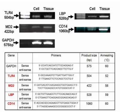
Total RNA from HCECs and human conjunctival tissue was isolated using TRIzol reagent. Synthesis of cDNA was performed using 3 µg total RNA (SuperScript III Reverse Transcriptase; Gibco-Invitrogen, NY, USA), according to the manufacturers' suggested protocol. Residual genomic DNA from the samples was eliminated by a DNase I digestion of the RNA preparation. Primers specific for TLR4, MD2, LBP, and CD14 were designed to produce PCR products 504 bp,29 422 bp,27 528 bp,31 and 1060 bp31 in size, respectively. The amplification protocol consisted of 38 (35 for MD2, LBP, and CD14) cycles of denaturation at 94°C for 1 min, annealing at 52°C (56°C for MD2, 59°C for LBP, and 60°C for CD14) for 1 min, and extension at 72 °C for 1 min, with a final extension at 72 °C for 10 minutes. Amplification using specific primers for GAPDH (glyceraldehyde-3-phosphate dehydrogenase) was used as an endogenous reference to determine the integrity of the mRNA in each sample. Amplified samples (10 µL) were electrophoresed on 1.4% agarose gels containing ethidium bromide and were photographed (UVIpro gel documentation system; UVITEC, Cambridge, UK). Results are representative of at least three independent experiments.
8 3. Real-time quantitative PCR
Real-time PCR amplification was performed in the presence of double-labeled fluorogenic probes for IL-6 and IL-8 (TaqMan probes; Applied Biosystems, Foster City, CA, USA). For each amplification reaction, 200 ng of cDNA in a total volume of 50 µL was used (TaqMan chemistry). Assays were performed using an ABI Prism 7500 Sequence Detection System (Applied Biosystems). The average threshold cycle (CT) values for GAPDH were used as an internal calibrator to correct for
differences in the integrity and amount of total RNA added to each reaction. For relative quantification, we used the 2-∆∆CTmethod.32 A non-template control was included in all experiments to eliminate the possibility of DNA contamination of the reagents used for amplification. None of the non-template controls in our experiments resulted in a positive signal, which indicated that there was no DNA contamination in the RNA used for the assays.Results were represented as the mean ± SD of three independent experiments.
4. ELISA
To quantify the cytokine secretion, human conjunctival epithelial cells were plated in Costar® Transwell-clear 3450 culture inserts (1 x 10⁵ cells/well). After one week of ALI culturing, the cells were either left untreated or were exposed to 0.1, 1, or 10 ug/ml LPS from Pseudomonas aeruginosa (Sigma-Aldrich) for 24 h from both apical and basolateral sides. The culture supernatants were harvested, and the levels of IL-6 and IL-8 were determined using commercially available ELISA kits (R&D
9
systems, Minneapolis, MN, USA). Results were represented as the mean ± SD of three independent experiments.
5. Transfection and NF- κB–driven luciferase reporter assay
HCECs cells were transfected with 3 µg NF-κB-luciferase reporter plasmids using lipofectamine 2000 according to the manufacturer's suggested protocol (Invitrogen). After 2 h incubation with DNA-lipofectamine mixtures, cells were maintained in fresh medium for another 24 h before LPS treatment. Cells were stimulated with 0.1, 1, or 10 ug/ml LPS for 6 h and then washed twice with PBS and lysed with reporter lysis buffer (Promega, Madison, WI, USA). After vortexing and centrifugation at 12,000×g for 1 min at 4 C, the supernatant was stored −70 C until assayed for luciferase activity. A total of 50 µl of the cell extract was mixed with 50 µl of the luciferase assay reagent at room temperature, and luciferase activity was measured with a luminometer (LMax II384, Molecular Devices, Sunnyvale, CA, USA).
6. Flow cytometric analysis
Human conjunctival epithelial cells were treated with 0.02% EDTA. Cell surface expression of TLR4 was examined by flow cytometry. Cells were incubated with the PE-conjugated mouse anti-human TLR4 (HTA125) mAb (eBioscience) or isotype control mouse IgG2a (BD Biosciences, San Jose, CA, USA) for 1h at room temperature. For intracellular FACS, the cell fixation/permeabilization kit (BD
10
Biosciences) was used. Cells were fixed with Cytofix/Cytoperm and then stained with PE-conjugated mouse anti-human TLR4 (HTA125) mAb, as described above, in Perm/Wash solution for one hour at room temperature. Stained cells were analyzed with a FACSCalibur (BD Biosciences), and data were analyzed using CellQuest software (BD Biosciences).
11 III. RESULTS
1. Cultured human conjunctival epithelial cells (HCECs) express TLR4, MD2, LBP and CD14-specific mRNA.
Among all members of the TLR family, TLR4 has a pattern recognition receptor for targeting Gram-negative (e.g., LPS) bacteria. First, we elucidated whether HCECs and normal human conjunctival epithelial cells harbor specific mRNA for
TLR4. TLR4-specific mRNA was present in both HCECs and normal human
conjunctival epithelial cells. MD2, LPB, and CD14 are an accessory molecule that is required for LPS recognition and signaling. HCECs also express MD2, LBP, and
CD14-specific mRNA (Fig. 1).
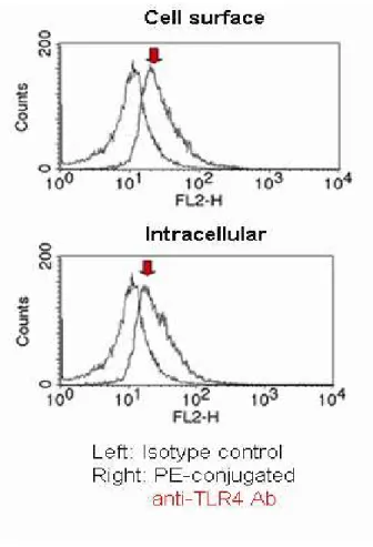
2. Cultured human conjunctival epithelial cells express TLR4 intracellularly and on the cell surface.
The next logical step was to investigate whether HCECs express TLR4 on their cell surface.To make this determination, we examined the cell surface expressionof TLR4 on HCECs. FACS analysis showed that HCECs expressed TLR4 on the cell surface as well as intracellulary (Fig. 2).
12
Figure 1. Normal human conjunctival epithelial cells and cultured human conjunctival epithelial cells (HCECs) express TLR4, MD2, LBP, and CD14-specific mRNA. Representative RT-PCR profiles from three experiments showed the mRNA expression of TLR4, MD2, LBP, and CD14. GAPDH was used as an internal control in normal human conjunctival epithelial cells and HCECs. Ethidium bromide-stained 1.4% agarose gels showing amplified products for TLR4, MD2, LBP, CD14, and GAPDH.
13
Figure 2. TLR4 is expressed intracellularly and on the cell surface of human conjunctival epithelial cells (HCECs). Cell surface and intracellular expression of TLR4 in HCECs was examined by flow cytometric analysis. Cells were incubated with PE-conjugated anti-human TLR4 (HTA125) mAb or an isotype control. Histogram data are representative of three separate experiments.
14
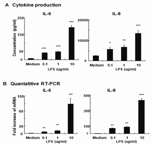
3. Cultured human conjunctival epithelial cells respond to LPS.
Next, we examined whether HCECs respond to the TLR4 ligand LPS. We first examined the production of inflammatory cytokines by HCECs after exposure to different doses of LPS. As shown in Fig. 3A, LPS stimulation induced the secretion of IL-6 and IL-8. Therefore, levels of IL-6 and IL-8 production in the treated supernatants significantly increased over those in un-stimulated NHCE. The higher concentration of LPS resulted in a greater induction of the secretion of IL-6 and IL-8. These findings demonstrate that human conjunctival epithelial cells were capable of responding to exogenous microbial stimuli (e.g., LPS).
This finding was further confirmed at the level of mRNA. After in vitro incubation of HCECs with various concentrations of LPS, quantitative RT-PCR was performed for the respective cytokines. The levels of IL-6- and IL-8-specificmRNA were elevated in HCECs stimulated with LPS (Fig.3B). NHCE responded to LPS in a dose-dependent mannerfor the enhancement of IL-6- and IL-8-specific mRNA (Fig. 3B).Taken together, these results showed that human conjunctival epithelial cells were able to respond to LPS from P. aeruginosa.
15
Figure 3. Cultured human conjunctival epithelial cells (HCECs) are responsive to LPS. To quantify inflammatory cytokine secretion, HCECs were left untreated or were exposed to 0.1, 1, and 10 ug/ml LPS from P. aeruginosa for 24 h. The culture supernatants were harvested for measurements of IL-6 and IL-8 (A). Quantitative RT-PCR was used to measure the expression of IL-6 and IL-8 mRNA in HCECs after treatment with LPS. The quantification data were normalized to the expression of the housekeeping gene GAPDH. The y-axis shows an increase in specific mRNA
over unstimulated samples (B). Data represent the mean ± SD from an experiment done in triplicate. *, p < 0.05, **, p < 0.01, ***, p < 0.001.
16
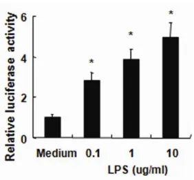
4. Inflammatory cytokine production in response to LPS is NF-κκκκB-dependent In epithelial cells, the transcription factor NF-κB plays a central role in regulating genes associated with mucosal immune responses.Since the activation of NF-κB by LPS can induce expression of proinflammatory mediators,13-15 we investigated the
effects of LPS on NF-κB activity using luciferase assays. NHCEs were stimulated with LPS for 6 h. The expression of an NF-κB reporter construct was measured by relative luciferase activity. As shown in Fig. 4, LPS significantly enhanced the
NF-κB activity.
Figure 4. To characterize NF-κB activation, cultured human conjunctival epithelial cells (HCECs) were transfected with NF-κB-luciferase reporter plasmids and were left untreated or were exposed to LPS (10 ug/ml) for 6 h. After stimulation, the
NF-κB assay was performed using a luciferase reporter assay. Data represent the mean ± SD from an experiment with triplicate wells. *, p < 0.05
17
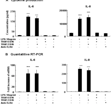
5. Blocking TLR4 and TRAF6 activity prevents the activation of inflammatory pathways induced by LPS in HCECs
In order to assess the role of TLR4 in IL-6 and IL-8 production by HCECs, we pretreated HCECs with TLR4-blocking antibody. Human conjunctival epithelial cells pretreated with 20 ug/ml TLR4 blocking antibody (HTA 125) for 1 h failed to respond to LPS. The magnitude of cytokine production at both mRNA and protein levels was reduced significantly compared with those seen from not pretreated cells (Fig. 5). TRAF6 has been implicated in the TLR4 signaling pathway and has been shown to complex with IRAK and IRAK-2 downstream of the receptor signaling complex.13,14,33 We therefore determined whether a dominant-negative version of TRAF6 could act to inhibit TLR4-induced inflammatory cytokine production. Dominant-negative TRAF6, but not dominant-negative TRAF2, which served as a control, significantly impaired TLR4-induced inflammatory cytokine production (Fig. 5), suggesting that TRAF6 may act as a downstream mediator of the TLR4-induced signaling cascade.
18
Figure 5. LPS-induced IL-6 and IL-8 expressions are abolished by blocking TLR4 or TRAF6 activity in cultured human conjunctival epithelial cells (HCECs). Cells were left untreated or were exposed to LPS (10 ug/ml) for 24 h. In some experiments, cells were pre-incubated with anti-TLR4 (20 ug/ml)-neutralizing Abs or transfected with a dominant negative TRAF6 before treatment. The cultured supernatants were harvested for measurements of IL-6 and IL-8 (A). Quantitative RT-PCR was used to measure the expression of IL-6 and IL-8 mRNA in HCECs after treatment with LPS. The quantification data were normalized to the expression
of the housekeeping gene GAPDH. (B). Data represent the mean ± SD from an experiment done in triplicate. ***, p < 0.001.
19
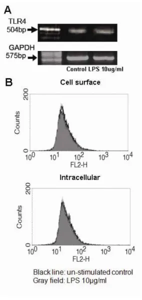
6. LPS does not induce the gene expression, intracellular expression and surface expression of TLR4 in HCECs
We examined whether TLR4-specific mRNA was inducible in HCECs by the TLR4 agonist LPS. As shown in Fig. 6, TLR4-sepcific mRNA was not induced in HCECs stimulated with an optimal concentration of 10 ug/ml LPS. Furthermore, Intracellular and cell-surface expression of TLR4 was also not increased by LPS (Fig. 6). Taken together, these findings demonstratedthat stimulation of HCECs with LPS failed to induce TLR4 expression.
20
Figure 6. TLR4 expression is not induced on cultured human conjunctival epithelial cells (HCECs) by LPS stimulation. RT-PCR showed TLR4 gene expression was not augmented with 10 ug/ml LPS-stimulated HCECs. GAPDH was used as an internal control (A). Flow cytometric analysis demonstrated that cell-surface and intracellular expression of TLR4 was also not inducible by LPS (B).
21 IV. DISCUSSION
Interestingly, our results indicated that conjunctival epithelial cells, which are an important component of the mucosal immune system, express functional TLR4. Incubation with lipopolysaccharide (LPS) induced the secretion by human conjunctival epithelial cells (HCECs) of inflammation-associated cytokines such as
IL-6 and IL-8. Further, NF-κB activation was up-regulated by the stimulation of HCECs with LPS. These results showed that our cultured HCECs were capable of responding to LPS from P. aeruginosa. To support this finding, we subsequently used FACS to show that human conjunctival epithelial cells express TLR4 intracellularly and on the cell surface. These findings suggest the interesting possibility that the human conjunctival epithelium may serve as both a critical immunological barrier against invasion by Gram-negative bacteria as well as a physical barrier.
When invaded by pathogens, mucosal epithelial cells elicit pro-inflammatory gene expression, secretion of cytokines and chemokines, and recruitment of inflammatory cells to the site of infection.13-15 These findings suggest that epithelial cells play a major role in the innate immune response, and have likely evolved as a means to limit the frequency and intensity of infection by pathogenic bacteria at the invasion site. To this end, it has been shown that several TLRs, including TLR4, are expressed in the mucosal epithelium of the human trachebronchia.18 After exposure to LPS, human trachebronchial epithelial cells are activated to produce increased levels of hBD2 mRNA. Bladder epithelial cells have also been reported to express
22
TLR4 as well as increased levels of proinflammatory cytokines following incubation with LPS.34 Similarly, our findings suggest that the conjunctival epithelia expresses TLR4 and is inducible with respect to inflammatory cytokine following exposure to LPS.
The findings in this study contradict earlier reports which demonstrate that conjunctival epithelial cells are not activated by LPS.24,27 Previous work by Talreja
et al. demonstrated that deficiency of MD2 contributes to a lack of LPS responsiveness in immortalized conjunctival cell lines,27 as MD2 is required for the binding of LPS to TLR4.10-12 An early study from Li et al. showed that TLR4-mediated LPS-induced proinflammatory responses do not exist in primary cultured conjunctival epithelial cells, even when the cells are positive for TLR4 mRNA and protein expression.24 Our results convincingly demonstrate that conjunctival epithelial cells express TLR4 as well as MD2, and respond to LPS, as evidenced by the induction of inflammatory cytokine production and mRNA expression. One possible explanation for our data could be that the conclusions of our study were based on the basis of differentiated human conjunctival epithelial cells with multi-layered features similar to an in vivo situation.30
Our study also presents a novel finding that cultured human conjunctival epithelial cells express TLR4 intracellularly and on the cell surface. The human conjunctival epithelium responds to bacteria via TLRs in order to initiate the innate immune response. TLR4 on conjunctival epithelial cell surface interacted with LPS and subsequently induced LPS-associated inflammatory mediators. Our experiments
23
further showed that LPS did not up-regulate TLR4 expression at mRNA and protein levels as well as on the cell surface (Fig. 6). In macrophages, TLR4 expression is not inducible by TLR4 ligands, although such stimuli can successfully induce TLR3
expression through autocrine IFN-β induction.35 In human corneal epithelial cells, LPS does not induce TLR4 expression and is incapable of inducing expression of
IFN-β.36
After ligand binding, TLRs/IL-1Rs dimerize and undergo a conformational change required in order for the newly formed complex to recruit downstream signaling molecules, including the adaptor molecule MyD88, IL-1R-associated
kinase (IRAK), TNFR-associated factor 6 (TRAF6), and NF-κB-inducing kinase.3,13,14 The recruitment of these molecules triggers the stimulation of downstream kinases, including MAPKs such as ERK1/2, p38 MAPK, and stress-activated protein kinase (SAPK)/JNK, as well as activation of the transcriptional
factors NF-κB and AP-1.3,13,14 The activation of these transcriptional factors leads to the induction of genes encoding cytokines and inflammatory mediators. The findings in this study demonstrated that LPS, a TLR4 ligand, was able to stimulate human conjunctival epithelial cells and induce rapid activation of the TLR4/IL-1RI signal transduction pathways. Thus, a key indication of activation of these receptors following LPS stimulation is activation of the subsequent downstream transcription
factor NF-κB (Fig. 4). Consistent with the hypothesis that LPS mediates inflammatory events by activating TLR4/IL-1RI, our data demonstrated that LPS-induced inflammatory cytokine production in human conjunctival epithelial cells
24
was abolished by blocking the activation of TLR4 with a neutralizing Ab (Fig. 5). Our results also demonstrated that by blocking TRAF6 activity, LPS-induced inflammatory cytokine production was inhibited both at the mRNA and protein levels. These data suggest that LPS stimulates inflammatory cytokine production in human conjunctival epithelial cells via TLR4.
25 V. CONCLUSION
The data presented in this study demonstrate that human conjunctival epithelial cells respond to LPS due to their ability to express TLR4 on their cell surface. We provide evidence for the gene and surface expression of TLR4 in human conjunctival epithelial cells and suggest that expressed TLR4 is functionally active and is involved in the secretion of the inflammatory mediators IL-6 and IL-8. Thus, we concluded that LPS could induce the secretion of inflammatory mediators by human conjunctival epithelial cells. These findings suggest that human conjunctival epithelial cells play a vital role in the initiating the TLR4-mediated innate immunity on the ocular surface.
26 VI. REFERENCES
1. Haynes RJ, Tighe PJ, Dua HS. Antimicrobial defensin peptides of the human ocular surface. Br J Ophthalmol 1999; 83:737-41.
2. Medzhitov R, Janeway C. Innate immune recognition: mechanisms and
pathways. Immunol Rev 2000; 173:89-97.
3. Takeda K, Kaisho T, Akira S. Toll-like receptors. Annu Rev Immunol 2003;
21:335-76.
4. Aderem A, Ulevitch RJ. Toll-like receptors in the induction of the innate
immune response. Nature 2000; 406:782-7.
5. Takeuchi O, Hoshino K, Kawai T, Sanjo H, Takada H, Ogawa T, et al.
Differential roles of TLR2 and TLR4 in recognition of Gram-negative and Gram-positive bacterial cell wall components. Immunity 1999; 11:443-51. 6. Hoshino K, Takeuchi O, Kawai T, Sanjo H, Ogawa T, Takeda Y, et al. Toll-like
receptor-4 deficient mice are hyporesponsive to lipopolysaccharide. Evidence for TLR4 as the Lps gene product. J Immunol 1999; 162:3749-52.
7. Gioannini TL, Teghanemt A, Zhang D, Coussens NP, Dockstader W, Ramaswamy S, et al. Isolation of an endotoxin-MD-2 complex that produces Toll-like receptor 4-dependent cell activation at picomolar concentrations. Proc Natl Acad Sci U S A 2004; 101:4186–91.
8. Pugin J, Schurer-Maly CC, Leturcq D, Moriarty A, Ulevitch RJ, Tobias PS. Lipopolysaccharide lipopolysaccharide-binding protein and soluble CD14. Proc Natl Acad Sci U S A 1993; 90:2744-8.
27
9. Tobias PS, Soldau K, Gegner JA, Mintz D, Ulevitch RJ. Lipopolysaccharide binding protein-mediated complexation of lipopolysaccharide with soluble CD14. J Biol Chem 1995; 270:10482–8.
10. Nagai Y, Akashi S, Nagafuku M, Ogata H, Iwakura Y, Akira S, et al. Essential
role of MD-2 in LPS responsiveness and TLR4 distribution. Nat Immunol 2002; 3:667-72.
11. Shimazu R, Akashi S, Ogata H, Nagai K, Fukudome K, Miyake K, et al.
MD-2, a molecule that confers lipopolysaccharide responsiveness on Toll-like receptor 4. J Exp Med 1999; 189:1777-82.
12. Miyake K. Endotoxin recognition molecules, Toll-like receptor 4-MD-2.
Semin Immunol 2004; 16:11-6.
13. Kawai T, Akira S. TLR signaling. Semin Immunol 2007; 19:24-32.
14. Guha M, Mackman N. LPS induction of gene expression in human monocytes.
Cell Signal 2001; 13:85–94.
15. Chow JC, Young DW, Golenbock DT, Christ WJ, Gusovsky F. Toll-like receptor-4 mediates lipopolysaccharide-induced signal transduction. J Biol Chem 1999; 274:10689–92.
16. Abreu MT, Vora P, Faure E, Thomas LS, Arnold ET, Arditi M. Decreased expression of Toll-like receptor-4 and MD-2 correlates with intestinal epithelial cell protection against dysregulated proinflammatory gene expression in response to bacterial lipopolysaccharide. J Immunol 2001; 167:1609-16.
28
17. Cario E, Brown D, McKee M, Lynch-Devaney K, Gerken G, Podolsky DK. Commensal-associated molecular patterns induce selective Toll-like receptor-trafficking from apical membrane to cytoplasmic compartments in polarized intestinal epithelium. Am J Pathol 2002; 160:165-73.
18. Becker MN, Diamond G, Verghese MW, Randell SH. CD14-dependent
lipopolysaccharide-induced beta-defensin-2 expression in human tracheobronchial epithelium. J Biol Chem 2000; 275:29731-6.
19. Wolfs TG, Buurman WA, van Schadewijk A, de Vries B, Daemen MA,
Hiemstra PS, et al. In vivo expression of Toll-like receptor 2 and 4 by renal epithelial cells: IFN-gamma and TNF-alpha mediated up-regulation during inflammation. J Immunol 2002; 168:1286-93.
20. Backhed F, Soderhall M, Ekman P, Normark S, Richter-Dahlfors A. Induction
of innate immune responses by Escherichia coli and purified lipopolysaccharide correlate with organ- and cell-specific expression of Toll-like receptors within the human urinary tract. Cell Microbiol 2001; 3:153-8. 21. Uehara A, Sugawara S, Takada H. Priming of human oral epithelial cells by
interferon-gamma to secrete cytokines in response to lipopolysaccharides, lipoteichoic acids and peptidoglycans. J Med Microbiol 2002; 51:626-34. 22. Krisanaprakornkit S, Kimball JR, Weinberg A, Darveau RP, Bainbridge BW,
Dale BA. Inducible expression of human beta-defensin 2 by Fusobacterium nucleatum in oral epithelial cells: multiple signaling pathways and role of commensal bacteria in innate immunity and the epithelial barrier. Infect
29 Immun 2000; 68:2907-15.
23. Bonini S, Micera A, Iovieno A, Lambiase A, Bonini S. Expression of Toll-like receptors in healthy and allergic conjunctiva. Ophthalmology 2005; 112:1528-34.
24. Li J, Shen J, Beuerman RW. Expression of toll-like receptors in human limbal
and conjunctival epithelial cells. Mol Vis 2007; 13:813-22.
25. Aswad MI, John T, Barza M, Kenyon K, Baum J. Bacterial adherence to
extended wear soft contact lenses. Ophthalmology 1990; 97:296-302.
26. Doyle A, Beigi B, Early A, Blake A, Eustace P, Hone R. Adherence of bacteria
to intraocular lenses: a prospective study. Br J Ophthalmol 1995; 79:347-9.
27. Talreja J, Dileepan K, Puri S, Kabir MH, Segal DM. Human Conjunctival
Epithelial Cells Lack Lipopolysaccharide Responsiveness Due to Deficient Expression of MD2 but Respond After Interferon-gamma Priming or Soluble MD2 Supplementation. Inflammation 2005; 29:170-81.
28. Song PI, Abraham TA, Park Y, Zivony AS, Harten B, Edelhauser HF, et al. The expression of functional LPS receptor proteins CD14 and toll-like receptor 4 in human corneal epithelial cells. Invest Ophthalmol Vis Sci 2001; 42:2867-77.
29. Ueta M, Nochi T, Jang MH, Park EJ, Igarashi O, Hino A, et al. Intracellularly expressed TLR2s and TLR4s contribution to an immunosilent environment at the ocular mucosal epithelium. J Immunol 2004; 173:3337-47.
30
primary human conjunctival epithelial cells producing MUC5AC. Exp Eye Res 2007: 85:226-33.
31. Blais DR, Vascotto SG, Griffith M, Altosaar I. LBP and CD14 Secreted in Tears by the Lacrimal Glands Modulate the LPS Response of Corneal Epithelial Cells. Invest Ophthalmol Vis Sci 2005; 46:4235-44.
32. Livak KJ, Schmittgen TD. Analysis of relative gene expression data using
real- time quantitative PCR and the 2-∆∆CTmethod. Methods 2001; 25:402-8. 33. Mukaida N, Ishikawa Y, Ikeda N, Fujioka N, Watanabe S, Kuno K, et al.
Novel insight into molecular mechanism of endotoxin shock: biochemical analysis of LPS receptor signaling in a cell-free system targeting NF-kappaB and regulation of cytokine production/action through beta 2 integrin in vivo. J Leukoc Biol 1996; 59:145–51.
34. Schilling JD, Mulvey MA, Vincent CD, Lorenz RG, Hultgren SJ. Bacterial
invasion augments epithelial cytokine responses to Escherichia coli through a lipopolysaccharide-dependent mechanism. J Immunol 2001; 166:1148-55. 35. Doyle SE, O’Connell R, Vaidya SA, Chow EK, Yee K, Cheng G. Toll-like
receptor 3 mediates a more potent antiviral response than Toll-like receptor 4. J Immunol 2003; 170: 3565-71.
36. Ueta M, Hamuro J, Kiyono H, Kinoshita S. Triggering of TLR3 by polyI:C in human corneal epithelial cells to induce inflammatory cytokines. Biochem Biophys Res Commun 2005; 331:285-94.
31
< ABSTRACT(IN KOREAN)>
인체결막상피세포에서 lipopolysacccharide에 대해 toll-like receptor 4로
유도되는 선천면역반응
<지도교수 김응권>
연세대학교 대학원 의학과
정 소 향
결막상피는 선천면역체계의 한 구성원으로써 침입한 병균에 대한 일차 방어선으로 작용한다. 침입한 병균에 대한 선천면역은 Toll-like receptor (TLRs)에 의해 시작되며, 그 중 TLR4은 그람 음성 세균의 구성 성분인 lipopolysaccharide (LPS)를 인지하여, 염증성 cytokine을 생성하 는 것으로 알려져 있다. 본 연구에서는 중층으로 배양된 결막상피세포에 서 TLR4의 ligand가 NF-kappa B의 활성화를 통하여 염증성 cytokine을 생 성할 수 있는지 알아보기로 하였다.중층으로 배양한 결막상피세포를 다양한 농도의 LPS 로 처리하였다. 우선 결막상피세포에 TLR4 가 존재하는지 RT-PCR 과 FACS 로 확인하였다. 선천면역반응의 측정은 IL-6 및 IL-8 유전자 발현과 상층액에서 IL-6 및 IL-8 생성으로 측정하였다. LPS 처리 후 NF-kappa B 가 활성화되는지 알아보기 위하여 luciferase reporter assay 를 시행하였다.
32
중층으로 분화시킨 인체결막상피세포에서 TLR4 유전자 발현이 RT-PCR로 확인되었으며, FACS 시행 결과 세포표면 및 세포내에 TLR4가 존재함이 밝혀졌다. LPS를 농도별로 처리한 결과, NF-kappa B 가 활성화되었으며, IL-6 및 IL-8 의 발현 및 생성이 증가하였다. TLR4와 TRAF6 활성화를 억제한 결과 결막상피세포에서 LPS에 의한 염증 반응의 유도가 억제되었다. 또한 LPS는 결막상피세포에서 TLR4의 발현을 증가시키지는 않았다. 본 연구결과는 인체결막상피세포 표면에 TLR4가 존재하며, 결막상피세포가 침입한 그람음성세균에 대하여 TLR4에 의해 매개되는 선천면역을 유도하여 안구표면의 염증성 환경을 이루는 데 중요한 역할을 함을 보여준다. --- 핵심되는 말: 결막상피세포, 선천면역반응, IL-6, IL-8, toll-like receptor 4