The Korean Journal of Helicobacter and Upper Gastrointestinal Research
Vol. 12, No. 3, 141-144, September 2012 ∙ http://dx.doi.org/10.7704/kjhugr.2012.12.3.141
141
위식도 접합부 암의 특성
Adenocarcinoma of the Esophagogastric Junction:
Clinicopathological Characteristics
Jie-Hyun Kim
Department of Internal Medicine, Gangnam Severance Hospital, Yonsei Uni-versity College of Medicine, Seoul, Korea
The incidence of adenocarcinoma of the esophagogastric junction (AEG) is dramatically increasing in Western countries, while it is not increasing in Eastern countries. Siewert type I tumors are observed less frequently in Eastern countries in comparison to Western countries. On the other hand, other clinicopathological features of AEG, including age, male-to-female ratio, pathological grade, tumor progression, and prognosis, are similar in Western and Eastern countries. Many previous studies have demonstrated that the characteristics of type II tumors are more like those of type III tumors than those of type I tumors. Further investigation of the adequacy of esophageal classification for AEG of the seventh American Joint Committee on Cancer TNM classification should be necessary. (Korean J Helicobacter Up
Gastro-intest Res 2012;12:141-144)
Key Words: Esophagogastric junction; Stomach neoplasms; Cardia;
Esopha-geal neoplasms; Classification
연세대학교 의과대학 강남세브란스병원 내과학교실 김지현 접수일:2012년 8월 14일 승인일:2012년 8월 23일 연락처:김지현 서울시 강남구 언주로 211 (도곡동 146-92) 우편번호: 135-720 연세대학교 의과대학 강남세브란스병원 내과 Tel: 02-2019-3505 Fax: 02-3463-3882 E-mail: [email protected]
서 론
서양에서 증가하고 있는 위식도 접합부 암은 위의 다른 부위에 발생하는 선암과 비교하여 예후나 림프절 전이 등 임상병리학적 특성에 차이가 있다고 알려져 왔다.1,2 반면 우리나라를 비롯한 동양에서는 위식도 접합부 암을 근위부 위선암의 일부로 종종 간주하여 왔으며,3,4 보고마다 차이는 있지만 일부 연구에서는 서양과 비슷하게 동양에서도 증가 되는 경향을 보여 주었다.3,4 위식도 접합부 암은 최근 2010년도에 개정된 Union for International Cancer Control (UICC)/American Joint Commit-tee on Cancer (AJCC) 병기 체계 7판에서 기존에 위암으로 분류되었던 병변이 위식도 접합부 침범 여부에 따라 식도 암의 병기로 분류됨으로써 논란의 대상이 되고 있다. 따라서, 위식도 접합부 암에 대해 살펴보는 것이 현 시점 에서 의미가 있을 것으로 생각되며 본 고에서는 위식도 접 합부 암의 분류 및 임상병리학적 특성에 대해 검토하겠다.본 론
1. 분류 및 기원(classification and origin)
위식도 접합부 암의 분류는 해부학적 위치에 근거한 Siewert 분류가 일반적으로 사용되며 다음과 같은 세가지로 분류된다.5 Type I (adenocarcinoma of the distal esophagus)은 병변이 위식도 접합부를 침범하든 안하든 병변의 중심 (epicenter)이 위식도 접합부 상방 1∼5 cm 이내에 위치하는 원위부 식도선암이며, type II (true cardia cancer)는 위식도 접합부를 침범한 병변으로 중심이 위식도 접합부 상방 1 cm에서 하방 2 cm 이내에 위치하는 위 분문암이고, type III (subcardial cancer)도 역시 위식도 접합부를 침범한 병변으 로, 중심이 위식도 접합부 하방 2∼5 cm에 위치하는 분문하
142 Korean J Helicobacter Up Gastrointest Res:제 12 권 제 3 호 2012
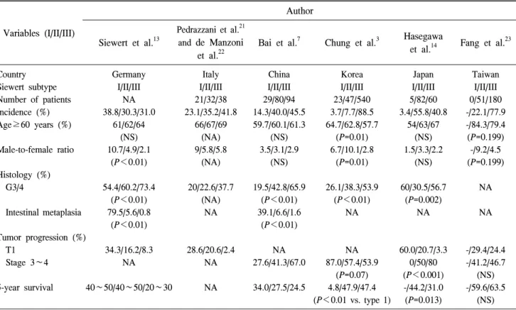
Table 1. Differences in the Incidence and Clinicopathological Features of Adenocarcinoma of the Esophagogastric Junction between Western and Eastern Countries
Variables (I/II/III) Author Siewert et al.13 Pedrazzani et al.21 and de Manzoni et al.22
Bai et al.7 Chung et al.3 Hasegawa
et al.14 Fang et al.
23
Country Germany Italy China Korea Japan Taiwan
Siewert subtype I/II/III I/II/III I/II/III I/II/III I/II/III I/II/III Number of patients NA 21/32/38 29/80/94 23/47/540 5/82/60 0/51/180 Incidence (%) 38.8/30.3/31.0 23.1/35.2/41.8 14.3/40.0/45.5 3.7/7.7/88.5 3.4/55.8/40.8 -/22.1/77.9 Age≥60 years (%) 61/62/64 (NS) 66/67/69 (NA) 59.7/60.1/61.3 (NS) 64.7/62.8/57.7 (P=0.01) 54/63/67 (NS) -/84.3/79.4 (P=0.199) Male-to-female ratio 10.7/4.9/2.1 (P<0.01) 9/5.8/5.8 (NA) 3.5/3.1/2.9 (NS) 6.7/10.1/2.8 (P=0.01) 1.5/3.3/2.2 (NS) -/9.2/4.5 (P=0.199) Histology (%) G3/4 54.4/60.2/73.4 (P<0.01) 20/22.6/37.7 (NA) 19.5/42.8/65.9 (P<0.01) 26.1/38.3/53.9 (P<0.01) 60/30.5/56.7 (P=0.002) NA Intestinal metaplasia 79.5/5.6/0.8 (P<0.01) NA 39.1/6.6/1.6 (P<0.01) NA NA NA Tumor progression (%) T1 34.3/16.2/8.3 28.6/20.6/2.4 NA NA 60.0/20.7/3.3 -/29.4/24.4 Stage 3∼4 NA NA 27.6/41.3/67.0 87.0/57.4/53.9 (P=0.07) 0/50/80 (P<0.001) -/41.2/46.7 (NS) 5-year survival 40∼50/40∼50/20∼30 NA 34.0/27.5/24.5 4.8/47.9/47.4 (P<0.01 vs. type 1) -/44.2/31.0 (P=0.013) -/59.6/63.5 (NS) G3/4, grade 3/4 undifferentiated histology; NA, not available; NS, no significant difference. (Adapted from the article of Hasegawa and Yoshikawa. Gastric Cancer 2010;13:63-735).
위선암이다. 일반적으로 동양인의 경우 서양인에 비해 위 의 소만 길이가 짧다고 알려졌기 때문에, 위식도 접합부 하 방 5 cm라는 길이 기준 적용의 적합성에 대해서는 추후 연
구가 필요할 수 있다.6
이론적으로 type I 병변은 식도선상피(esophageal glandular epithelium) 또는 특수 장상피화생(specialized intestinal meta-plasia, so called Barrett’s esophagus)에서 발생한 것으로 특히 특수 장상피화생의 경우 위식도 접합부 암 발생과 밀접한 관계가 있다고 알려져 있다. 바렛식도의 유병률은 서양이든
동양이든 type I에서 type II/III 보다 더 높다고 보고된다.7
Type II 병변은 접합부위에서 기원한 진정한 위식도 접합
부 암으로,5 위분문 상피 또는 장상피화생에서 기원한다.8
Type II 병변의 기원은 일부는 type I 병변과 같을 수 있고
일부는 type III 병변과 일치할 수 있다고 보고되는데,5 많은
이전 연구에서 type II 병변은 type I 보다는 type III 병변의 특성과 비슷하여 기원 또한 type I 보다는 type III와 일치할
것으로 보고하고 있다.9-11 Type II 병변의 기원과 관련된 특 수 장상피화생에 있어서 한 연구에서는 수술 조직을 대상 으로 전향적 병리 분석을 시행하였을 때 type II 병변의 약 28%에서만 goblet 세포가 관찰되었고 대부분의 type II 병변 에서는 관찰할 수 없기 때문에 type II 병변의 대부분은 위 점막에서 기원한 위선암임을 보고하였다.9
Type III 병변은 위점막에서 기원하며 이는 Helicobacter pylori와 위축성 위염과 연관이 있을 것으로 보고되고 있 다.5,12 병변의 중심이 위식도 접합부 아래 2∼5 cm에 있다고 하더라도 위식도 접합부를 침범하지 않으면 위식도 접합부 암이 아닌 분문하 위선암으로 분류되며, 병변의 중심이 위 식도 접합부 아래 2∼5 cm에 있으면서 위식도 접합부를 침 범한 경우에만 위식도 접합부 암으로 분류된다. 따라서, 같 은 분문하 위선암이라고 하더라도 위식도 접합부를 침범한 병변, 즉 위식도 접합부 암으로 분류된 병변의 크기가 더
크게 된다.5 Type III 병변은 UICC/AJCC 병기 체계 7판에서
식도암으로 분류되어 논란이 되고 있지만, 대부분의 기관 에서 진성 위암으로 간주하고 이에 준하여 치료를 진행하
여 왔다.8
2. 임상병리학적 특징
김지현:위식도 접합부 암의 특성 143 서양 사이에 큰 차이가 있으며, type I 병변은 동양보다 서양 에서 더 자주 발생한다.5 Type I 병변인 원위부 식도선암의 위험인자가 위식도역류, 바렛식도, 비만이며, Helicobacter pylori가 보호인자로 작용할 수 있다는 점에서 이와 같은 유 병률 차이는 예측되는 결과이다. Table 1은 동양과 서양에 서 보고된 위식도 접합부 암 각각의 분류에 따른 임상병리 학적 특징들을 요약한 것이다.5 각 분류에 따른 유병률을 제외하고 대부분의 임상병리학적 특징은 동서양 사이에 비 슷하다. Type II는 동서양 모두 type I과 III의 중간적 특징을 보여주는 경향이 있으나, 동양에서는 type II와 III가 좀 더 비슷한 경향의 임상적 특징을 보여주고 있다.
위식도 접합부 암의 평균 나이는 60세 전후로 type I, II, III의 세 분류 사이에 큰 차이는 없다. 세 분류 모두 남성 비율이 높으나 type I에서 더 두드러지며, type III가 가장 덜 두드러진다. 한 연구에서는 여성에 대한 남성의 비율을 type I, II, III에서 각각 10.7, 4.9, 2.2로 보고하였다.13 분화암(장형)은 type I에서 더 자주 관찰되며, 저분화/미 분화암(미만형)은 type III에서 더 관찰된다. 특수 장상피화 생, 즉 바렛식도의 경우 type I에서 두드러지게 관찰된다. Type III의 경우 type I과 II에 비해 더 진행성 암의 특징을 보여주며 이는 동서양이 비슷한 양상으로 관찰되는데, 이 는 분문하 위암인 type III가 다른 분류의 병변에 비해 크기
가 큰 특징을 고려할 때 당연한 결과라고 하겠다.14
우리나라를 비롯한 동양에서는 type I 병변은 흔하지 않 기 때문에 주로 type II, III 병변의 위식도 접합부 암을 근위 부 위선암의 일부로 간주하여 임상적 특성을 분석하는 경 우가 많다. 이들 연구에 따르면 근위부 위선암의 경우 원위 부 위선암과 비교하여 진행성 병변인 경우가 많고, 림프절 전이나 원격전이가 더 흔하고, 예후도 나쁜 것으로 보고하 고 있다.15-17 그러나, 최근 한 연구에서는 type II/III 병변과 이외의 위선암을 비교하였을 때 T 병기나 N 병기가 위식도 접합부 암인 type II/III 병변에서 더 높았지만, 수술 후 생존 율에 있어서는 의미 있는 차이를 보여주지 않음을 보고하 면서 위식도 접합부 암이 다른 위선암에 비해 진행성 병변 일 때 발견되기 때문에 예후가 안 좋다고 알려져 있는 것이 며, 위식도 접합부 암이 다른 위선암과 구별되는 병변일 가 능성이 적음을 보고하였다.6 또한, 이 연구에서는 위식도 접 합부 암과 다른 근위부 위선암의 림프절 전이 형태를 비교 하여 보았는데, 위식도 접합부 암에서는 종양 주변과 위소 만 부위의 림프절 전이가 주로 관찰되었으며, 근위부 위선 암에 비해 대만 부위의 림프절 전이는 덜 관찰되었다.6 하지 만, celiac axis 주변의 림프절 전이가 위식도 접합부 암에서 도 무시할 수 없을 정도로 관찰되었고 이는 위식도 접합부 암의 수술적 치료에 있어 이 부위의 림프절 제거가 필요함 을 시사한다고 하겠다. 3. 위식도 접합부 암의 UICC/AJCC 병기 분류 최근 2010년도에 개정된 UICC/AJCC 병기 체계 7판에서 는 하부식도, 위식도 접합부에서 발생한 암은 물론, 위식도 접합부에서 5 cm 이내의 근위부 위에서 발생한 선암도 암 의 일부가 위식도 접합부를 침범하였다면 식도암의 병기를 따르는 것으로 분류하고 있다. 즉 위식도 접합부 암은 모두 식도암의 병기를 따르는 것으로 분류된다는 것인데, 암의 기원에 대한 고려 및 기존의 많은 연구들에서 type II 병변 이 위선암의 특징에 좀 더 가까움을 보여주었다는 점에서 이와 같은 병기 분류는 논란의 여지가 많다. 또한, type III 병변의 경우 같은 분문하 위선암의 경우에 있어서 위식도 접합부를 침범할 경우 식도암의 병기로 분류하라는 것인 데, 이와 같은 분류가 의미가 있기 위해서는 분문하 위선암 에서 위식도 접합부 침범 여부에 따른 임상적 특징에 차이 가 있어야 할 것이다. 같은 분문하 위선암에서 위식도 접합부를 침범한 경우 (type III)와 침범하지 않은 경우를 비교한 연구를 보았을 때,6 위식도 접합부를 침범한 경우 병변의 크기가 크고, T 병기나 N 병기가 더 높았으나, 이는 앞에서도 언급한 것과 같이 종양의 중심(epicenter)이 같은 분문하 위선암에서 위 식도 접합부를 침범하기 위해서는 병변의 크기가 클 수밖 에 없고, 이에 따른 좀 더 진행된 병기가 될 수밖에 없기 때문에 관찰되는 임상적 차이일 수 있다. 중요한 것은 두 병변 사이의 림프절 전이 양상일 수 있는데, 두 병변의 림프 절 전이 양상은 차이가 없이 종양 주변, 위의 소만 부위, cel-iac axis 주변에서 관찰되었다.6 또한, 방사성동위원소 주입 하여 림프절 전이 패턴을 분석한 연구들에서도 type II 병변 에 해당하는 분문암의 경우 대부분 복강 내 림프절 전이로 관찰되었음을 보고하였으며, 다른 몇몇 연구에서도 type II/III 병변의 경우 celiac axis 림프절 전이가 많이 관찰된 것
을 보고하였다.18,19 이러한 림프절 전이의 특성에 따라 type II 위식도 접합부 암의 근치적 수술 범위에 식도절제를 포 함하는 경우가 생존률의 향상에 기여하는 바가 없었던 연 구의 결과도 type II의 분류가 식도암이 아닌 위암에 합당하 다는 것을 시사한다.20 또한, T 병기를 보정하였을 때 위식도 접합부 침범 여부 에 따라 생존율에도 차이가 없었다.6 따라서, 같은 분문하 위선암에서 위식도 접합부 침범 여부에 따라 식도암 또는 위암으로 병기를 분류하는 것은 재고되어야 할 수 있다. 중 요한 것은 위식도 접합부를 침범하였느나 침범하지 않았느 냐가 아니라 위식도 접합부 암 내에서 각 분류에 따른 암의 기원이나 임상병리학적 특징일 것이다.
144 Korean J Helicobacter Up Gastrointest Res:제 12 권 제 3 호 2012 위식도 접합부 암 중 type II/III 병변을 식도암으로 병기 분류를 한 경우와 위암으로 병기 분류한 경우를 비교한 최 근 연구에서는 위암 병기로 분류하였을 때 각 병기별 생존 율 차이가 좀 더 명확하게 관찰됨을 보고하였다.6
결 론
위식도 접합부 암의 Siewert 분류에 따른 임상병리학적 특징은 동양과 서양에 있어 큰 차이를 보이지는 않는다. 위 식도 접합부 암의 각 분류에 따른 기원 및 임상병리학적 특징의 올바른 분석과 이해는 추후 적합한 병기 분류와 치 료 적용에 도움이 될 것으로 생각한다. 위식도 접합부 암과 다른 위선암에 대한 종양생물학적 연구 및 임상적 비교 연 구가 위식도 접합부 암의 완전한 이해 및 분류를 위하여 필요할 것이다.참 고 문 헌
1. Devesa SS, Blot WJ, Fraumeni JF Jr. Changing patterns in the incidence of esophageal and gastric carcinoma in the United States. Cancer 1998;83:2049-2053.
2. Botterweck AA, Schouten LJ, Volovics A, Dorant E, van Den Brandt PA. Trends in incidence of adenocarcinoma of the oeso-phagus and gastric cardia in ten European countries. Int J Epidemiol 2000;29:645-654.
3. Chung JW, Lee GH, Choi KS, et al. Unchanging trend of esoph-agogastric junction adenocarcinoma in Korea: experience at a sin-gle institution based on Siewert's classification. Dis Esophagus 2009;22:676-681.
4. Okabayashi T, Gotoda T, Kondo H, et al. Early carcinoma of the gastric cardia in Japan: is it different from that in the West? Cancer 2000;89:2555-2559.
5. Hasegawa S, Yoshikawa T. Adenocarcinoma of the esophago-gastric junction: incidence, characteristics, and treatment strategies. Gastric Cancer 2010;13:63-73.
6. Suh YS, Han DS, Kong SH, et al. Should adenocarcinoma of the esophagogastric junction be classified as esophageal cancer? A comparative analysis according to the seventh AJCC TNM classi-fication. Ann Surg 2012;255:908-915.
7. Bai JG, Lv Y, Dang CX. Adenocarcinoma of the Esophagogastric Junction in China according to Siewert's classification. Jpn J Clin Oncol 2006;36:364-367.
8. Marsman WA, Tytgat GN, ten Kate FJ, van Lanschot JJ. Differences and similarities of adenocarcinomas of the esophagus
and esophagogastric junction. J Surg Oncol 2005;92:160-168. 9. Siewert JR, Feith M, Stein HJ. Biologic and clinical variations of
adenocarcinoma at the esophago-gastric junction: relevance of a topographic-anatomic subclassification. J Surg Oncol 2005;90:139- 146.
10. Siewert JR, Stein HJ. Classification of adenocarcinoma of the oe-sophagogastric junction. Br J Surg 1998;85:1457-1459. 11. Stein HJ, Feith M, Siewert JR. Cancer of the esophagogastric
junction. Surg Oncol 2000;9:35-41.
12. Hansen S, Vollset SE, Derakhshan MH, et al. Two distinct aetiol-ogies of cardia cancer; evidence from premorbid serological mark-ers of gastric atrophy and Helicobacter pylori status. Gut 2007; 56:918-925.
13. Siewert JR, Stein HJ, Feith M. Adenocarcinoma of the esoph-ago-gastric junction. Scand J Surg 2006;95:260-269.
14. Hasegawa S, Yoshikawa T, Cho H, Tsuburaya A, Kobayashi O. Is adenocarcinoma of the esophagogastric junction different be-tween Japan and western countries? The incidence and clinic-opathological features at a Japanese high-volume cancer center. World J Surg 2009;33:95-103.
15. Park JC, Lee YC, Kim JH, et al. Clinicopathological features and prognostic factors of proximal gastric carcinoma in a population with high Helicobacter pylori prevalence: a single-center, large-volume study in Korea. Ann Surg Oncol 2010;17:829-837. 16. Siewert JR, Böttcher K, Stein HJ, Roder JD, Busch R. Problem of proximal third gastric carcinoma. World J Surg 1995;19: 523-531.
17. Sakaguchi T, Watanabe A, Sawada H, et al. Characteristics and clinical outcome of proximal-third gastric cancer. J Am Coll Surg 1998;187:352-357.
18. Cense HA, Sloof GW, Klaase JM, et al. Lymphatic drainage routes of the gastric cardia visualized by lymphoscintigraphy. J Nucl Med 2004;45:247-252.
19. Ott K, Weber WA, Fink U, et al. Fluorodeoxyglucose-positron emission tomography in adenocarcinomas of the distal esophagus and cardia. World J Surg 2003;27:1035-1039.
20. Rüdiger Siewert J, Feith M, Werner M, Stein HJ. Adenocarcinoma of the esophagogastric junction: results of surgical therapy based on anatomical/topographic classification in 1,002 consecutive patients. Ann Surg 2000;232:353-361.
21. Pedrazzani C, de Manzoni G, Marrelli D, et al. Lymph node in-volvement in advanced gastroesophageal junction adenocarcinoma. J Thorac Cardiovasc Surg 2007;134:378–385.
22. de Manzoni G, Pedrazzani C, Pasini F, et al. Results of surgical treatment of adenocarcinoma of the gastric cardia. Ann Thorac Surg 2002;73:1035–1040.
23. Fang WL, Wu CW, Chen JH, et al. Esophagogastric junction ad-enocarcinoma according to Siewert classifi cation in Taiwan. Ann Surg Oncol 2009;16:3237–44.