http://crossmark.crossref.org/dialog/?doi=10.14474/ptrs.2020.9.3.201&domain=pdf&date_stamp=2020-9-25
Received: 31 August, 2020 Revised: 22 September, 2020 Accepted: 24 September, 2020 Corresponding author: Yijung Chung (ORCID https://orcid.org/0000-0002-2431-8895)
Department of Physical Therapy, College of Health Science and Social Welfare, Sahmyook University, 815 Hwarang-ro, Nowon-gu, Seoul 01795, Republic of Korea Tel: 82-2-3399-1637 Fax: 82-2-3399-1639 E-mail: [email protected]
This is an Open-Access article distributed under the terms of the Creative Commons Attribution Non-Commercial License (http://creativecommons.org/licenses/ by-nc/4.0) which permits unrestricted non-commercial use, distribution, and reproduction in any medium, provided the original work is properly cited.
Copyright © 2020 Korean Academy of Physical Therapy Rehabilitation Science https://doi.org/10.14474/ptrs.2020.9.3.201
pISSN 2287-7576 eISSN 2287-7584
Phys Ther Rehabil Sci 2020, 9 (3), 201-208 www.jptrs.org
The effects of different V-sit positions on abdominal muscle
activation
Jina Seoa , Yijung Chungb
aPhysical Therapy Clinic, Seoul Hospital, Seoul, Republic of Korea
bDepartment of Physical Therapy, College of Health Science and Social Welfare, Sahmyook University, Seoul, Republic of Korea
Objective: This study aimed to identify the effects of performing shoulder and hip abduction during the V-sit exercise on
abdomi-nal muscle activity.
Design: Cross-sectional study.
Methods: Thirty healthy adults volunteered for this experiment. The participants randomly performed 6 types of V-sit exercises,
including V-sit alone (hip 0°, shoulder 0°), V-sit with hip abduction 0° and shoulder abduction 15°, V-sit with hip abduction 0° and shoulder abduction 30°, V-sit with hip abduction 15° and shoulder abduction 0°, V-sit with shoulder and hip abduction 15°, and V-sit with shoulder abduction 30° and hip abduction 15°. EMG data were recorded from the rectus abdominis (RA), external obli-que (EO), and internal obliobli-que (IO) muscles of both sides. All abdominal EMG data during the six types of V-sit exercises were measured for 5 seconds, three times, and recorded for the middle 3 seconds excluding the 1 second at the start and end.
Results: V-sit with shoulder abduction 30° resulted in significantly greater muscle activity of both RA, EO compared to shoulder
abduction 0°, shoulder abduction 15° (p<0.05) and V-sit with shoulder abduction 15° showed significantly greater muscle activa-tion of the RA compared with shoulder abducactiva-tion 0° (p<0.05). The muscle activity of both EO and IO in the V-sit with hip abduc-tion 15° was significantly greater than hip abducabduc-tion 0° in all shoulder condiabduc-tions (p<0.05).
Conclusions: Greater angles of shoulder and hip abduction produced more abdominal muscle activity increases during the V-sit
exercises. Shoulder abduction affected the RA, EO muscle activation and hip abduction affected the EO, IO muscle activation. This study showed that shoulder and hip abduction during V-sit exercises enabled effective activation of the trunk muscles.
Key Words: Abdominal muscles, Electromyography, Hip joint, Posture, Shoulder joint
Introduction
Stability refers to the function of the musculoskeletal sys-tem to maintain equilibrium against disturbance of kine-matic control [1]. At this time, the stabilization of the trunk is performed through three sub-systems of passive, active, and neural regulation. The active regulation system is com-posed of the core muscle system, providing dynamic stabili-zation to the proximal part of the spine and limbs, as well as the neural regulation system, and conveys information about movement [2]. In other words, it can be said that the active
muscle power produced by the core muscles play a major role in maintaining trunk stability against the external load imposed on our body [3]. At this time, abdominal strength-ening exercises that improve trunk stability have a positive effect on reducing low back pain, improving upper limb function in patients with breast cancer, and contribute to im-proving lower limb function in patients with hip and total knee arthroplasty, as well as improve capacity and exercise performance in athletes [4].
Therefore, abdominal strengthening exercises are widely practiced not only in physical therapy, but also in sports and
physical education [5].
In general, abdominal strengthening exercises are com-monly used in a supine position, such as sit-ups, and trunk flexion exercises are commonly used [6]. In this case, sev-eral studies have suggested that in the case of V-sit exercise, more effective abdominal muscle activation can be expected than the sit-up exercise by simultaneously moving the upper and lower bodies [7]. This can be seen from the results of a study that compared the abdominal muscle activity of the sit-up and V-sit posture where the V-sit posture showed rela-tively greater muscle activity in the lower rectus abdominis (RA) and external oblique muscles [8]. In addition, the addi-tional limb movements can cause predictable perturbation in our body and promote the activation of abdominal muscles to maintain body stability [9]. Based on the results of a pre-vious study comparing the abdominal muscle activity ac-cording to the hip joint movement of the bridge exercise, the abdominal muscle activity of the RA and the internal ab-dominal oblique muscle was greater when the hip joint was abducted during the bridge exercise than when the hip was not abducted [10]. In addition, in a study comparing abdomi-nal muscle activity when one hip was abducted and not ab-ducted during a straight leg raise, the muscle activity of the ipsilateral external oblique (EO) muscle was greater as a re-sult of abducting the hip joint [11].
Since the abdominal muscles have different directions of movement of the various muscle fibers, it is believed that the response to the load applied in different postures will be dif-ferent [11]. In a previous study, it was reported that shoulder flexion induces back muscle activation while shoulder ab-duction induces abdominal muscle activation [12].
This can be seen from the results of a study that measured the abdominal muscle activity according to the movement of the limb in a sitting position in patients with stroke and the muscle activity of the abdominal muscles was greater than that with shoulder flexion during shoulder abduction [13]. In addition, in a study comparing abdominal muscle activity when standing in normal subjects, in the case of the trans-verse abdominal muscle, there was no difference in muscle activity according to the movement direction of the limb, and although it was activated first in all directions, the IO muscle was activated more preferentially during shoulder abduction than in the sagittal movement such as shoulder flexion and extension [14].
Among the abdominal muscles, the lateral fibers of the abdominal and EO muscles are considered to be the main mobilizers of trunk movement, while the internal and
trans-verse abdominal muscles are considered to be the main sta-bilizing muscles of trunk movement.
At this time, the stabilizing muscles can be further classi-fied into primary and secondary stabilizing muscles. Primary stabilizing muscles include muscles that cannot cause joint movement relatively, such as the transverse ab-dominal muscles [2]. The secondary stabilizing muscles in-clude the medial fibers of the internal and EO muscles. These muscles also contribute to stabilization, but also act to move the spinal joints.
At this time, the RA, EOs, and IOs are activated differ-ently according to the direction of movement before the limb movement to stabilize the spine, whereas the transverse ab-dominal muscles act before movement in all directions re-gardless of the movement direction of the limb and are in-volved in maintaining posture.
In addition, the IO muscle is attached to the lumbar spine and contributes to spinal stabilization through the control of intervertebral stiffness [15].
Existing studies on the V-sit posture were limited to com-paring abdominal muscle activity according to the angle of lifting the leg from the ground [16] and comparing abdomi-nal muscle activity with the most clinically used abdomiabdomi-nal strength training exercises [8].
Therefore, studies analyzing the change of abdominal muscle activity according to the change of upper or lower limb posture in V-sit posture are insufficient.
Therefore, this study attempted to present basic data for future exercise regimens by applying postural change to V-sit exercises in healthy subjects and examining the effect of postural change on abdominal muscle activity.
Methods
Study participants
This study included 30 subjects who had met the subject selection criteria among healthy adults. Before proceeding with the experiment, after explaining the experiment proce-dure and conditions to the study subjects, the study partic-ipants agreed to volunteer and signed the consent form.
The selection criteria for subjects were those who could maintain a certain posture for more than 30 seconds after lift-ing the trunk or both lower limbs [17], and the exclusion cri-teria were those who had experienced low back pain in the last 6 months, had severe scoliosis or congenital spine. Those with deformities, those who were unable to perform exercise due to cancer or pregnancy, spinal disorders such as
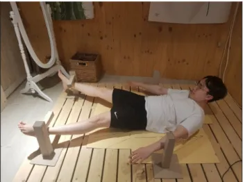
Figure 2. Shoulder abduction, hip abduction of V sit position. Figure 1. V-sit position.
herniated discs [18], a history of neurological disorders and spinal surgery [19], pain in the lower back or abdomen, a wound or injury to the stomach due to surgery or trauma were excluded [8].
Research procedure
In this study, 30 healthy adults who met the subject se-lection criteria were selected as the study participants and the investigation was conducted through a cross-sectional study design. Prior to the start of the experiment, the proce-dure was fully explained to the subjects, and after the sub-jects fully understood and signed the consent form, the ex-periment was conducted. The study was approved by the Institutional review board of Sahmyook University (2-7001793-AB-N-012018052HR).
For normalization of muscle activity, electrodes were at-tached to both RA, external and internal rectus muscles, and then the maximum voluntary isometric contraction (MVIC) was measured [20]. When performing the V-sit posture, muscle activity was repeatedly measured 3 times and the average was obtained for each movement by applying bi-lateral hip 0° abduction and 15° abduction to each condition of bilateral shoulder 0° abduction, 15° abduction, and 30°. In order to minimize muscle fatigue in each posture, a 1-minute rest period was provided for each movement [21].
Measurement and evaluation method
In this study, in the V-sit position, bilateral hip joint 0° ab-duction, bilateral shoulder joint 0° abab-duction, 15° abab-duction, and 30° abduction were performed. Abduction was per-formed to measure the muscle activity of the target muscles.
Before the experiment, the V-sit posture and the upper and lower limb postures were sufficiently explained to the study subject, and the study was conducted after preliminary prac-tice so that the study subject could fully understand the ex-perimental movements.
The starting position for each movement entailed having the subjects placing their to both sides of the trunk in the su-pine position.
When performing the V-sit posture, subjects flexed their head, shoulders and trunk so that their scapulae did not touch the ground [8] and the cervical spine was flexed as much as possible [22]. At the same time, both lower limbs were lifted until the knee joint was fully extended and the hip joint flex-ion reached 10°. The hip joint was flexed until it reached the wooden model (Figure 1) [23].
At the time of measurement, subjects were asked to lift their head, shoulders, trunk, and both lower limbs for 1 sec-ond with the bell sound of the experimenter, and after hold-ing this for 3 seconds, they slowly lowered their upper and lower bodies within 1 second to return to the starting position. Shoulder and hip abduction movements were made by applying the guidelines that were prepared on four boards. For the shoulder joint, the axis of the guideline was placed at the center point of the back of the shoulder joint. In the case of the hip joint, after placing the center point under the subject’s superior iliac spine a wooden model was set up at the limit of the angle of abduction of each limb, so that the subject could recognize when the abduction movement ex-ceeded the limit [24]. Shoulder and hip abduction move-ments (Figure 2) were performed simultaneously for 1 sec-ond by lifting the head, shoulders, trunk, and both legs.
Similarly, after holding for 3 seconds, both the upper and lower limbs were to return to the starting position within 1 second. During all movements, a metronome was set for 60 times per minute to measure the execution time, and when the inspector found an error in the tree model, the measured value was deleted. Each movement was performed in ran-dom order using a ranran-dom number table and each condition was measured three times. In addition, in order to minimize muscle fatigue of the study subjects, they were given a 1-mi-nute break after each movement [21].
To measure muscle activity in each motion, the surface telemetry EMG system (TELEmyo 2400T G2; Noraxon, Scottsdale, AZ, USA, 2011) was used to measure the muscle activities of bilateral RA, EOs, and the IO muscles (inferior fibers of the IO), and the sampling rate of the EMG signal was set to 1,500 HZ, and the frequency bandwidth was set to 20-500 HZ. In this study, the measured muscle EMG signals were processed by full wave rectification using the MyoResearch XP Master edition software (Noraxon, 2011), and then root mean square 250 milliseconds was taken [25]. In the case of the IO muscle, the electrodes were attached in parallel with the muscle fiber in the lower part of the upper anterior iliac spine, and in the case of the EO muscles, the electrodes were attached parallel to the muscle fibers. Also, in the case of the rectus abdominal muscle, the electrodes were attached in parallel with the muscle fibers at a distance of 2 cm outward from the midline and 3 cm above the belly button [9]. Skin resistance was minimized by removing body hair before attaching the electrode and cleaning the electrode site with an alcohol cotton [26].
To assess the MVIC of each muscle, electrodes were at-tached to both internal, EO muscles and the rectus abdomi-nal muscles, and the measurement posture of each muscle was performed based on the manual muscle test for each re-spective muscle. The rectus abdominal muscle was meas-ured by flexing the knee to 90° in a supine position, fixing both feet, and applying resistance to the shoulder in the di-rection of trunk extension when the trunk was maximally flexed. The IO and EO muscles were measured by applying resistance to the shoulder in the opposite direction of rota-tion while the trunk was maximally flexed in the same pos-ture as the abdominal rectus muscle and the trunk was ro-tated left and right [20].
As for the normalization method, the EMG signal ex-tracted from the center of the 3 second section in the static state was divided by the MVIC value and normalized to the %MVIC value with the exclusion of the 1 second time
peri-od before and after the movement occurred during each 5 second exercise [9].
Statistical analysis
For all work and statistics in this study, the mean and standard deviation values were calculated using the IBM SPSS Statistics for Windows, Version 19.0 (IBM Co., Armonk, NY, USA), and descriptive statistics were used for the general characteristics of the subjects.
To investigate the effect of V-sit posture on abdominal muscle activity, there were 3 upper extremity postural con-ditions of shoulder joint 0° abduction, 15° abduction, and 30° abduction, and the hip joint 0° abduction and 15° abduction. A two-way repeated measure ANOVA was per-formed for a total of six movements with the conditions applied. Post-hoc verification was performed using the least significant difference, and the statistical significance level (p) of all data was set to 0.05.
Results
The muscle activity of the bilateral RA was significantly affected by the change of all upper limb postures (F=8.427,
p<0.001; F=9.459, p<0.001), but the main effect of the
change of lower limb posture and the interaction effect of the change of upper and lower limb posture were not significant. As a result of post-hoc analysis, the muscle activity of the bilateral RA was significantly greater in all hip abduction conditions in the shoulder 30° abduction condition, and it was significantly greater than in the shoulder joint 0° and 15° abduction conditions (p<0.05).
The muscle activity of both EO muscles was significantly affected by the changes in all upper extremity postures (F=4.900, p=0.009; F=4.137, p=0.018) and the main effects according to lower limb posture changes (F=19.921,
p<0.001; F=10.270, p=0.002) The interaction effect
accord-ing to the posture of upper and lower limbs was not significant. As a result of post-hoc analysis, the muscle ac-tivity of both EO muscles was significantly greater in the 15° hip abduction condition and the 30° shoulder abduction con-dition than in the shoulder joint 0° abduction (p<0.05), and in all hip abduction conditions, muscle activity was sig-nificantly greater in the shoulder 30° abduction condition than in the 15° abduction of the shoulder joint (p<0.05).
In addition, in all shoulder abduction conditions, muscle activity was significantly greater in hip 15° abduction than 0° abduction (p<0.05). The effects of all lower limb posture
Table 1. Muscle activation of abdominal muscles (N=30) Muscle activation Main effect(upper) Main effect(lower) (upper*lower)Interaction
S0 S15 S30 F (p) F (p) F (p) Rt.RA (%MVIC) 8.427 (<0.001) 2.394 (0.124) 0.369 (0.692) H0 61.84 (11.45) 65.30 (12.13)a 71.40 (11.20)ab H15 64.48 (12.38) 68.70 (12.17)a 72.18 (11.92)ab Lt.RA (%MVIC) 9.459 (<0.001) 2.479 (0.117) 0.167 (0.846) H0 56.46 (12.54) 62.44 (13.39)a 68.53 (14.28)ab H15 60.78 (11.65) 66.03 (13.44)a 70.10 (14.01)ab Rt.EO (%MVIC) 4.900 (0.009) 19.921 (<0.001) 2.137 (0.121) H0 60.29 (12.38) 60.23 (10.07) 62.23 (11.01)ab H15 64.61 (11.37)c 66.33 (11.97)c 74.81 (10.94)abc Lt.EO (%MVIC) 4.137 (0.018) 10.270 (0.002) 1.247 (0.290) H0 55.89 (17.62) 56.32 (19.05) 59.71 (17.14)ab H15 60.23 (16.84)c 63.09 (17.13)c 72.93 (15.18)abc Rt.IO (%MVIC) 2.308 (0.103) 35.645 (<0.001) 0.069 (0.933) H0 49.19 (9.11) 50.26 (8.20) 52.62 (9.95) H15 57.69 (9.78)c 59.89 (11.73)c 62.42 (12.33)c Lt.IO (%MVIC) 0.832 (0.437) 19.956 (<0.001) 0.143 (0.867) H0 52.79 (12.92) 54.55 (12.30) 55.04 (11.88) H15 61.68 (16.53)c 63.08 (14.33)c 66.14 (15.60)c
Values are presented as mean (SD).
%MVIC: %maximum voluntary isometric contraction, Rt: right, Lt: left, S0: shoulder abduction 0°, S15: shoulder abduction 15°, S30: shoulder abduction 30°, H0: hip abduction 0°, H15: hip abduction 15°.
RA: rectus abdominis, EO: external oblique, IO: internal oblique.
aSignificant difference with S0 (p<0.05), bSignificant difference with S15 (p<0.05), cSignificant difference with H0 (p<0.05).
changes were significant (F=35.645, p<0.001; F=19.956,
p<0.001) on the muscle activity of the bilateral IO muscles,
but the effect according to the changes in upper limb posture and the interaction effect the changes in upper and lower limb postures were not significant. As a result of post-hoc analysis, the muscle activity of both IO muscles in all shoulder abduction conditions was significantly greater in the hip 15° abduction than in the 0° abduction (p<0.05; Table 1).
Discussion
Son et al. [16] compared abdominal muscle activity ac-cording to the hip joint flexion angle in the V-sit posture and found that the abdominal muscle activity was the highest be-tween 5° and 24°. Therefore, the V-sit posture in this study was performed by setting the hip flexion angle to 10°. At this time, Hong et al. [27] measured the distance of the sub-acromial space according to the shoulder abduction angle in healthy subjects. When the shoulder joint was abducted to 30°, the average of the subacromial space distance was 9.40
mm, but when shoulder joint was abducted by 60°, the aver-age was 8.01 mm, which was less than that of patients with rotator cuff disease.
Therefore, this study compared abdominal muscle activ-ity according to the changes in upper limb posture during the V-sit posture by setting the shoulder abduction angles to 15° and 30° within 60°.
Park et al. [10] compared abdominal muscle activity for basic bridge exercises and lower limb postural changes by abducting the hip joint by 20° during the bridge exercise. Abdominal muscle activity due the postural change was compared. Park [11] compared the abdominal muscle activ-ity against changes in lower extremactiv-ity posture by abducting the hip joint by 15° and 30° when lifting the leg.
Rutkowska and Szpala [5] argued that the change in upper limb posture changes the center of mass of the trunk, and this change in the center of mass of the trunk changes the mo-ment from the axis of rotation of the upper body to the center of mass, thereby changing the load that the abdominal mus-cles must cope with for trunk stabilization. That is, as the load due to the increase of the moment arm gradually
in-creased, the muscle activity of the abdominal muscles for posture maintenance increased [6]. Therefore, as the should-er abduction angle, which is a change in the uppshould-er limb pos-ture, increases, the center of mass moves toward the head (cranial position), and the load that the abdominal muscles have to resist when the trunk flexion is increased. However, according to Crommert et al. [6], the transverse abdominal muscle, the innermost muscle among the abdominal mus-cles, does not directly act on trunk flexion due to a lack of bone attachment points, but acts as an aid. Therefore, it is considered that the transverse abdominal muscle was not af-fected by the external load due to the changes in upper limb posture compared to other muscles.
Park et al. [10] compared the muscle activity of the RA muscle and the internal abdominal oblique muscle during hip joint abduction during bridge exercises in healthy subjects. The ratio of the internal abdominal and rectus ab-dominal muscles was also significantly greater during the abduction of one and both hip joints than the basic bridge ex-ercise (p<0.05).
Park [11] compared the muscle activity of the abdominal rectus, EO, and IO muscles according to hip abduction posi-tions in the supine position in healthy adults. Muscle activity was significantly greater 30° abduction than that of 0° ab-duction (p<0.05).
Kim et al. [15] compared abdominal muscle activity ac-cording to hip adduction during plank exercise in normal subjects. The abdominal rectus, EO, and IO muscles all showed significantly greater muscle activity with hip adduc-tion than the basic plank exercise with hip adducadduc-tion on one side. Except for the rectus abdominis, the case of the ex-ternal and IO muscle activity was significantly greater with single hip adduction compared to bilateral hip adduction (p<0.05).
In this study, according to the changes in lower limb pos-ture in the V-sit pospos-ture, there was a significant difference in muscle activity between the external and IO muscles, but not in the case of bilateral RA. At this time, both external and IO muscles showed significantly greater muscle activity in the 15° hip abduction condition than the 0° hip abduction (p<0.05).
Since abdominal muscles do not pass through the hip joint, they are not directly involved in hip joint flexion [28], but in supine position, hip joint flexion acts as an external re-sistance that breaks the static stability of the body by the weight of the leg [11]. Also, at this time, the change in the position of the lower limbs creates a load by the change of
the moment arm by changing the center of mass of the body by the weight of the leg, which also occurs with the change in the position of the upper limbs [5].
In addition, according to Park [11], hip abduction acts as a force that causes the opposite pelvis and trunk to rotate ip-silaterally, and in the case of the external and IO muscles, which are involved in rotating the trunk, they work together to produce counter rotation due to the weight of the leg.
In terms of functional anatomy, abdominal muscles are divided into global muscles and local muscles. The large muscles include the RA and the EO muscles, and the local muscles include the transverse abdominal muscles and the IO muscles [19].
At this time, Moon and Goo [29] suggested that the trans-verse abdominal muscle contraction occurs more effectively with positional changes of the lower extremity than the up-per extremity, because the lumbar spine and the thighs are in the same nerve dominant area.
In other words, the hip abduction muscles are connected through the local muscles of the abdomen and the superior anterior iliac spine of the pelvis, so the hip abduction con-tributes to the transfer of force to the local muscles of the ab-domen [30]. As a result, it is thought that the contraction of the lower extremity complex affected the activation of trunk stabilizing muscles such as the transverse abdominis and the IO muscles [31,32].
The limitation of this study is that the size of the sample group is small, and since the experiment was conducted on healthy adults, it is difficult to generalize to groups with spe-cific characteristics such as low back pain and patients with stroke.
Therefore, in future studies, in-depth studies will be need-ed for groups with characteristics of specific diseases.
Therefore, although the V-sit posture differed in the types of abdominal muscles that were effective according to changes in the upper and lower limb posture, it is thought that changes in upper and lower extremity postures such as shoulder abduction and hip abduction in the V-sit posture can induce muscle activity of the abdominal muscles more effectively by increasing the load on the muscles that the ab-dominal muscles must counter.
Acknowledgements
This paper was supported by the Korean Academy of Women’s Health in Physical Therapy and the Institute of Women’s Health in Physical Therapy of Sahmyook
University.
Conflict of Interest
The authors declared no potential conflicts of interest with respect to the authorship and/or publication of this article.
References
1. Granata KP, Lee PE, Franklin TC. Co-contraction recruitment and spinal load during isometric trunk flexion and extension. Clin Biomech (Bristol, Avon) 2005;20:1029-37.
2. Huxel Bliven KC, Anderson BE. Core stability training for in-jury prevention. Sports Health 2013;5:514-22.
3. Hubley-Kozey CL, Vezina MJ. Muscle activation during ex-ercises to improve trunk stability in men with low back pain. Arch Phys Med Rehabil 2002;83:1100-8.
4. Keays KS, Harris SR, Lucyshyn JM, MacIntyre DL. Effects of Pilates exercises on shoulder range of motion, pain, mood, and upper-extremity function in women living with breast cancer: a pilot study. Phys Ther 2008;88:494-510.
5. Rutkowska-Kucharska A, Szpala A. Electromyographic muscle activity in curl-up exercises with different positions of upper and lower extremities. J Strength Cond Res 2010;24:3133-9. 6. Crommert ME, Bjerkefors A, Tarassova O, Ekblom MM.
Abdominal muscle activation during common modifications of the trunk curl-up exercise. J Strength Cond Res 2018. doi: 10.1519/JSC.0000000000002439 [Epub ahead of print] 7. Monfort-Pañego M, Vera-García FJ, Sánchez-Zuriaga D,
Sarti-Martínez MA. Electromyographic studies in abdominal ex-ercises: a literature synthesis. J Manipulative Physiol Ther 2009; 32:232-44.
8. Willett GM, Hyde JE, Uhrlaub MB, Wendel CL, Karst GM. Relative activity of abdominal muscles during commonly pre-scribed strengthening exercises. J Strength Cond Res 2001;15: 480-5.
9. Marshall P, Murphy B. The validity and reliability of surface EMG to assess the neuromuscular response of the abdominal muscles to rapid limb movement. J Electromyogr Kinesiol 2003; 13:477-89.
10. Park HJ, Oh DW, Kim SY. Effects of integrating hip movements into bridge exercises on electromyographic activities of selected trunk muscles in healthy individuals. Man Ther 2014;19:246-51. 11. Park MC. The effects of hip abduction angles on abdominal mus-cle activity during leg raising. J Korean Soc Phys Med 2012;7: 165-71.
12. Mullington CJ, Klungarvuth L, Catley M, McGregor AH, Strutton PH. Trunk muscle responses following unpredictable loading of an abducted arm. Gait Posture 2009;30:181-6. 13. Lee DK, Kang MH, Kim JW, Kim YG, Park JH, Oh JS. Effects
of non-paretic arm exercises using a tubing band on abdominal muscle activity in stroke patients. NeuroRehabilitation 2013;33: 605-10.
14. Hodges PW, Richardson CA. Inefficient muscular stabilization of the lumbar spine associated with low back pain. A motor control
evaluation of transversus abdominis. Spine 1996;21:2640-50. 15. Kim SY, Kang MH, Kim ER, Jung IG, Seo EY, Oh JS.
Comparison of EMG activity on abdominal muscles during plank exercise with unilateral and bilateral additional isometric hip adduction. J Electromyogr Kinesiol 2016;30:9-14.
16. Son NJ, Jun HJ, Yi KO. Angular differences between the lower extremity and the ground that express maximum core muscle ac-tivation according to core-strengthening exercises. Korean J Sport Biomech 2017;27:247-55.
17. Kim HD, Jeon DM, Bae HW, Kim JG, Han N, Eom MJ. Changes in activation of abdominal muscles at selected angles during trunk exercise by using ultrasonography. Ann Rehabil Med 2015;39:950-6.
18. Dias JM, Menacho Mde O, Mazuquin BF, Obara K, Mostagi FQ, Lima TB, et al. Comparison of the electromyographic activity of the anterior trunk during the execution of two Pilates exercises - teaser and longspine - for healthy people. J Electromyogr Kinesiol 2014;24:689-97.
19. Imai A, Kaneoka K, Okubo Y, Shiina I, Tatsumura M, Izumi S, Shiraki H. Trunk muscle activity during lumbar stabilization ex-ercises on both a stable and unstable surface. J Orthop Sports Phys Ther 2010;40:369-75.
20. Kendall FP, McCreary EK, Provance PG, Rodgers MM, Romani WA. Muscles: testing and function with posture and pain. 5th ed. Baltimore (MD): Lippincott Williams & Wilkins; 2005. 21. D’hooge R, Hodges P, Tsao H, Hall L, Macdonald D, Danneels L.
Altered trunk muscle coordination during rapid trunk flexion in people in remission of recurrent low back pain. J Electromyogr Kinesiol 2013;23:173-81.
22. Shirado O, Ito T, Kaneda K, Strax TE. Electromyographic analy-sis of four techniques for isometric trunk muscle exercises. Arch Phys Med Rehabil 1995;76:225-9.
23. Choi SA, Cynn HS, Yi CH, Kwon OY, Yoon TL, Choi WJ, et al. Isometric hip abduction using a Thera-Band alters gluteus max-imus muscle activity and the anterior pelvic tilt angle during bridging exercise. J Electromyogr Kinesiol 2015;25:310-5. 24. Kang SY, Jeon HS, Kwon O, Cynn HS, Choi B. Activation of the
gluteus maximus and hamstring muscles during prone hip ex-tension with knee flexion in three hip abduction positions. Man Ther 2013;18:303-7.
25. Suehiro T, Mizutani M, Watanabe S, Ishida H, Kobara K, Osaka H. Comparison of spine motion and trunk muscle activity be-tween abdominal hollowing and abdominal bracing maneuvers during prone hip extension. J Bodyw Mov Ther 2014;18:482-8. 26. Criswell E, Cram JR. Cram’s introduction to surface electromyo-graphy. 2nd ed. Sudbury (MA): Jones and Bartlett Publishers; 2011.
27. Hong YT, Lee DY, Yu JH, Kim JS, Hong JH. Changes in the sub-acromial space according to the angle of shoulder abduction. Indian J Sci Technol 2015;8:1-6.
28. Andersson EA, Nilsson J, Ma Z, Thorstensson A. Abdominal and hip flexor muscle activation during various training exercises. Eur J Appl Physiol Occup Physiol 1997;75:115-23. 29. Moon HJ, Goo BO. The effect of change in transversus
abdomi-nis thickness using ultrasound image during a hip adductor contraction. J Korean Soc Phys Med 2011;6:287-92.
30. Myers TW. Anatomy trains: myofascial meridians for manual and movement therapists. Edinburgh: Churchill Livingstone;
2001.
31. Page P, Frank CC, Lardner R. Assessment and treatment of mus-cle imbalance: the Janda approach. Champaign (IL): Human Kinetics; 2010.
32. Tarnanen SP, Ylinen JJ, Siekkinen KM, Mälkiä EA, Kautiainen HJ, Häkkinen AH. Effect of isometric upper-extremity exercises on the activation of core stabilizing muscles. Arch Phys Med Rehabil 2008;89:513-21.