81
-Vol. 13, No. 2(December), 2016 The Journal of Medicine and Life Science
Sudden hemiparesis is a typical symptom of acute stroke. The patients who were suspected as acute stroke should undergo image study to diagnose whether hemorrhagic or ischemic stroke. In ischemic stroke, physicians try to treat with thrombolytic agents urgently for better outcome. However thrombolytic therapy might not be helpful, if the vascular obstruction is not related with the thrombus. Physicians who care the patients with acute ischemic stroke must evaluate not only bleeding events and thrombogenic risk factors, but also the other obstructing causes except thrombus. We experienced a patient who was afflicted with the infected embolic myxoma.
A 64-year-old man who has hypertension and diabetes visited emergency department (ED) with dysarthria and right side weakness. At the arrival, he was not able to communicate with others and so anxious. His wife and daughter said there was no traumatic event in several weeks, and he had no headache at all. They denied his cough, sputum, short of breath, chest pain and febrile
sense. He is a 20 pack-years ex-smoker, and daily 360 ml of 20% alcohol drinker.
His initial vital signs were as follows: blood pressure 94/59 mmHg, pulse rate 110 /min, respiratory rate 22 /min, body temperature 36.2℃. On examination, he was 168 cm in height, with a weight of 80 kg. Global aphasia was observed. His pupils were isocoric and prompt, however he had right side gazing limitation. The facial expression and sensory change were not clear. The motor power of right extremities was grade one while that of left extremity was grade five.
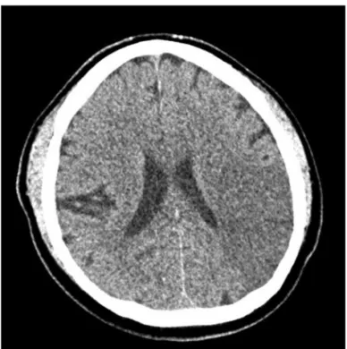
With the impression of acute stroke, he underwent brain computed tomography (CT). The CT image demonstrated diffuse hypodense lesion in the left middle cerebral artery territory (Figure1). He was diagnosed with acute ischemic stroke, and prepared to undergo thrombolysis.
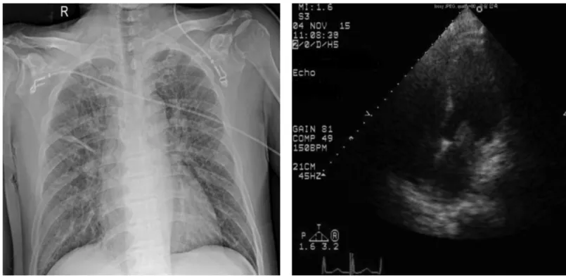
Electrocardiogram (ECG), chest X-ray, and blood sampling were performed to evaluate the thromboembolic and bleeding risks (Figure 2, 3). Serum laboratory tests revealed the following: arterial blood pH, 7.495; pCO2, 29.5 mmHg; pO2, 62.8 mmHg; HCO3, 24.6 mmol/L; SaO2, 91.8%; white blood cell, 26,400 /mm ; hemoglobin (Hb), 10.8 g/dl; platelet, 73,000 /μL; C-reactive protein, 21.19 mg/dL; procalcitonin, 1.10 ng/ml; blood urea nitrogen, 38.1 mg/dL; creatinine (Cr), 1.7 mg/dL; aspartate aminotransferase (AST), 67 IU/L;
Infected Cardiac Myxoma Presenting as an Acute Stroke in the Emergency Department
Hyun-Soo Park
Jeju National University School of Medicine, Department of Emergency Medicine (Received November 1, 2016; Revised November 8, 2016; Accepted November 15, 2016)
The intra-cardiac mass is observed in 0.05 percent of routine postmortem examinations. The difference diagnosis includes benign, malignant primary, secondary metastatic cardiac tumor, or thrombus. Myxomas is the most common primary cardiac tumors. The clinical features of myxomas are determined by their location, size, and mobility. Most patients present with one or more of the triad of embolism, intra-cardiac obstruction and constitutional symptoms. We present a 65-year-old man who visited the emergency department, complaining of sudden onset dysarthria and right side weakness. After initial brain image, he was diagnosed with acute stroke, however hypotension and elevated inflammatory marker were observed. He underwent emergent echocardiography, and 2 * 2 cm sized cardiac myxoma in his left atrium was detected. (J Med Life Sci 2016;12(2):81-84)
Key Words
: Myxoma, Stroke, Septic ShockIntroduction
Case
Abstract
Correspondence to : Hyun-Soo Park
Department of Emergency Medicine, Jeju National University School of Medicine, Aran 13gil 15, Jeju-si, Jeju Special Self-governing Province, 63241, Republic of Korea
E-mail : [email protected]
alanine aminotransferase, 112 IU/L; protein, 6.8 g/dL; albumin, 2.9g/dL; lactate, 33.7 mg/dL. Thus, elevated inflammatory marker, leukocytosis, and hypoxia were observed. Thrombolysis therapy was withheld, and emergency echocardiography was performed in emergency department (Figure 4). Trans-thoracic echocardiography revealed a round mass (2.1X1.9 cm) attached to the anterior mitral valve leaflet, which obstructed the mitral inflow. Tricuspid regurgitation also observed in Doppler image, and pulmonary hypertension (RVSP 51 mmHg) was calculated. Otherwise, there was no wall motion abnormality, and the chamber size was normal.
82
-Hyun-Soo Park
Figure 1. Brain computed tomography
Figure 2. Electrocardiogram
During the evaluation, his vital signs became unstable (blood pressure 80/65 mmHg, pulse rate 126 /min, respiratory rate 32 /min). Despite oxygen treatment via 8 L/min facial mask, follow-up arterial blood gas analysis revealed pCO2, 26.2 mmHg; pO2, 50.6 mmHg; SaO2, 85%. He was intubated and then transferred to intensive care unit (ICU) to manage pulmonary congestion and infection which were caused by the infected myxoma. Thoracic surgeons disagreed with the operative treatment for removal the mass and repair the valve, owing to severe hypotension and septic condition. After 5 days ICU care, he expired from hypoxia and multi-organ failure despite ventilator care and continuous renal replacement therapy.
Primary tumors of the heart are rare, with the incidence between 0.007 and 0.19 in unselected patients’ autopsy1-5). Three quarters of the tumors are benign, and nearly half the benign heart tumors are myxoma1 6-8). Before 1951, the diagnosis of intra-cardiac tumors was made only at postmortem examination; in that year the diagnosis of an intracavitary left atrial tumor was confirmed by angiocardiography9). The introduction of echocardiography has greatly facilitated the ante mortem diagnosis of cardiac tumors10).
The classical clinical manifestations of cardiac myxoma
can be triad with constitutional, obstructive and embolic symptoms. (Goodwin’s triad) Constitutional disturbances, such as fatigue, fever, rash, arthralgia, myalgia, and laboratory abnormalities have been observed in many patients. The production and release of the cytokine interleukin 6 by tumor may be responsible for these inflammatory and autoimmune manifestations11,12). Depending on their size and mobility, myxoma commonly give rise to signs of obstructed filling of the left or right ventricle with subsequent dyspnea, recurrent pulmonary edema, and right-heart failure. These signs mimic the clinical picture of mitral- or tricuspid- valve stenosis. Embolism occurs in 30 to 40 % of patients with myxoma10,13). In the majority of the embolism cases, the cerebral arteries, including the retinal arteries, are affected.
There is no clear distinction between infected and uninfected cardiac myxoma, however, infected myxoma is associated with more febrile symptoms (97.3% and 92%) and a higher risk of embolic events. (27.0% and 12%)14 In 1998 and 2015, Revankar and Yuan presented a literature collection of infected cardiac myxoma with 40 and 39 cases respectively14,15). Most of infected cardiac myxoma patients (38/39) had received a surgical treatment except patient who wasrapidly deteriorated did not undergo any surgical procedure14). The timing for surgical operation was reported in 26 patients, 4 of them were operated on an urgent basis and 22 had a delay of 14 (median; range 3~42) days (n =
83
-Infected Cardiac Myxoma Presenting as an Acute Stroke in the Emergency Department
Discussion
Figure 3. Chest X-ray (anterior-posterior) Figure 4. Echocardiogram (apical 4 chamber view)
21) after admission. Preoperative antibiotic treatment was described in 26 patients. The frequently used antibiotics included vancomycin (7 patients), penicillin (4 patients) and ampicillin (3 patients). Sometimes, they were used along with gentamicin. Of the 33 patients who underwent surgical resection, only 2 died. One of these deaths occurred 2 months postoperatively, due to intractable congestive heart failure. Operative mortality was low. (1/33 cases; 3%) During a follow-up period of 8.5 (median; range 0.1~58) months (n=16), 37 (92.6%) patients survived and 2 (7.4%) died. One patient died of rupture of a right lung abscess 3 weeks after admission and the other died of disseminated intravascular coagulation on postoperative day 10. Overall mortality was 5.1% and the operative mortality was 2.6%. The overall mortalities of infected cardiac myxoma in the literature are 21% in 1998, and 5.1% in 2015 (8/38 cases, 2/39 cases). In general, the outcome is good if diagnosis is made early before serious emboli or cardiopulmonary complications occur.
Revankar defined the infected cardiac myxoma in three levels based on clinical and pathological findings of the myxoma. According to the definition, our patient who had cardiac mass observed by transthoracic echocardiography and positive blood culture result for Staphylococcus lugdunensiswas diagnosed the possible infected cardiac myxoma. He initially looked like an acute ischemic stroke caused by thromboembolism. In such an acute stroke case, physicians make haste to treat with thrombolytics, however the embolus was septic emboli from cardiac myxoma. Physicians should be aware of the various cause of cerebral obstruction, even if the patient presents with typical symptoms. Despite we could diagnose in the early phase, he expired by pulmonary congestion and septic condition without any operative treatment. The operation could be possible if we utilized the cardiopulmonary supporting device which is recently used in resuscitation field to maintain vital organs.
1) Benjamin H. Primary fibromyxoma of the heart. Arch Pathol 1939;27(950):360.
2) Straus R. Primary tumor of the heart. Arch Pathol 1945;39:74-78.
3) Heath D. Pathology of cardiac tumors. The American journal of cardiology 1968;21(3):315-27.
4) Wold LE, Lie J. Cardiac myxomas: a clinicopathologic profile. The American journal of pathology 1980;101(1):219. 5) Schanz U. Endokardmyxome: neue Aspekte zur
Histophathogenese. Schwabe, 1984.
6) Griffiths G. A review of primary tumors of the heart. Progress in cardiovascular diseases 1965;7(5):465-79. 7) McAllister HA. Tumors of the cardiovascular system. 1978 8) Silverman NA. Primary cardiac tumors. Annals of surgery
1980;191(2):127.
9) Goldberg HP, GLENN F, DOTTER CT, et al. Myxoma of the Left Atrium Diagnosis Made during Life with Operative and Postmortem Findings. Circulation 1952;6(5):762-67.
10) Atrial myxomas: a review of clinical experience in 40 patients. Mayo Clinic Proceedings; 1980.
11) Saji T, Yanagawa E, Matsuura H, et al. Increased serum interleukin-6 in cardiac myxoma. American heart journal 1991;122(2):579-80.
12) Seino Y, Ikeda U, Shimada K. Increased expression of interleukin 6 mRNA in cardiac myxomas. British heart journal 1993;69(6):565-67.
13) Blondeau P. Primary cardiac tumors--French studies of 533 cases. The Thoracic and cardiovascular surgeon 1990;38:192-95.
14) Yuan S-M. Infected Cardiac Myxoma: an Updated Review. Revista Brasileira de Cirurgia Cardiovascular 2015;30(5):571-78.
15) Revankar SG, Clark RA. Infected Cardiac Myxoma: Case Report and Literature Review. Medicine 1998;77(5):337-44.
84
-Hyun-Soo Park