저작자표시-비영리-변경금지 2.0 대한민국 이용자는 아래의 조건을 따르는 경우에 한하여 자유롭게 l 이 저작물을 복제, 배포, 전송, 전시, 공연 및 방송할 수 있습니다. 다음과 같은 조건을 따라야 합니다: l 귀하는, 이 저작물의 재이용이나 배포의 경우, 이 저작물에 적용된 이용허락조건 을 명확하게 나타내어야 합니다. l 저작권자로부터 별도의 허가를 받으면 이러한 조건들은 적용되지 않습니다. 저작권법에 따른 이용자의 권리는 위의 내용에 의하여 영향을 받지 않습니다. 이것은 이용허락규약(Legal Code)을 이해하기 쉽게 요약한 것입니다. Disclaimer 저작자표시. 귀하는 원저작자를 표시하여야 합니다. 비영리. 귀하는 이 저작물을 영리 목적으로 이용할 수 없습니다. 변경금지. 귀하는 이 저작물을 개작, 변형 또는 가공할 수 없습니다.
Epigenetic regulation by G protein
alpha12 in human cancer cells and
vascular endothelial cells
HYEONJEONG LEE
Department of Medical Science
The Graduate School, Yonsei University
Epigenetic regulation by G protein
alpha12 in human cancer cells and
vascular endothelial cells
Directed by Professor Eun Jig Lee
The Doctoral Dissertation
submitted to the Department of Medical Science,
the Graduate School of Yonsei University
in partial fulfillment of the requirements for the degree of
Doctor of Philosophy
HYEONJEONG LEE
June 2015
This certifies that the Doctoral
Dissertation of Hyeon Jeong Lee is
approved.
이 은 직
Thesis Supervisor: Eun Jig Lee
안 철 우
Thesis Committee Member#1: Chul Woo Ahn
윤 호 근
Thesis Committee Member#2: Ho Geun Yoon
김 형 표
Thesis Committee Member#3: Hyoung-Pyo Kim
서 미 란
Thesis Committee Member#4: MiRan Seo
The Graduate School
Yonsei University
TABLE OF CONTENTS
Abstract……….………....1
Part
Ⅰ. Up-regulation of XAF1 by G
a12 knock down induces cell
death in A549 lung cancer cell line ………...…….…3
1. INTRODUCTION………..………..………....3
2. MATERIALS AND METHODS ………..…..….……7
A.
Cell culture ……….……..…..…...7
B.
Small interfering RNA transfection ………... …...7
C.
Western blot analysis.………...………... …..7
D.
RNA isolation and RT-PCR……….……...8
E. Immunofluorescence……….…...9
F. Immunoprecipitation ……….…....9
G.
TUNEL assay ………...10
H.
Annexin V staining………...10
I. Methylation-specific PCR (MSP) ………...….11
J. Methylated DNA Immunoprecipitation (MeDIP) ….……...11
K.
DNMT Activity Assay ……….…..…...12
L. Statistical analysis……….….……..….12
3. RESULTS ……….……...13
A.
Expression of XAF1 was regulated by Ga12………….……...13
B. Restoration of XAF1 by Ga12 down-regulation was appeared
in NSCSCs…...……….……...………...…15
C. DNA methylation of XAF1 controlled by Ga12 in A549 cells
………..…...…………...………...17
D.
DNMT1 mediate DNA methylation change by Ga12 knock
down………..………...…...19
E. Effect of the up-regulation of XAF1 expression by Ga12
knockdown on XIAP expression and localization…...….…...21
F. Effect of Ga12 knockdown on apoptosis………...………23
4. DISCUSSION……….…..…..26
Part
Ⅱ. Up-regulation of p16 by siGa12 inhibits cellular proliferation
of HepG2 cells ………...…..30
1. INTRODUCTION………...…...30
2. MATERIALS AND METHODS ………..……...…31
A.
Cell culture ………..…....31
B.
Small interfering RNA transfection………...…...31
C.
Western blot analysis.………...………...…...31
D.
RNA isolation and RT-PCR ………...……..32
E. Immunofluorescence………...32
F. Methylation-specific PCR (MSP) ………33
G.
Methylated DNA Immunoprecipitation (MeDIP)…….…… ...33
H.
DNMT Activity Assay ………...34
I. Cell cycle analysis……….…..…..34
J. Cell Growth Assay……….……..….35
K.
Soft Agar Assay……….…...…….35
L. Statistical analysis……….……..……..35
3. RESULTS ………..……....…..36
A.
Effect of Ga12 knockdown on p16 expression in various
Cancer cell lines………...…36
D.
Down-regulation of Ga12 decreases DNMT1- mediated
p16 methylation……….……..………....…....42
E. Regulation of Histone modification by Ga12 ……..……..…44
F. Effect of Ga12 knockdown on HepG2 cells proliferation..…46
4. DISCUSSION………...…………..49
Part III. G
a12 inhibits serum deprivation induced- apoptosis of
endothelial cells by miRNA regulation……….…...52
1. INTRODUCTION……….……….………....52
2. MATERIALS AND METHODS ……….………...…54
A.
Cell culture ……….…...……..……….54
B. Small interfering RNA transfection……….……..…..…...54
C. Western blot analysis.……….……….……...…..…54
D.
TUNEL assay ………..…………....…...55
E. miRNA qRT-PCR detection and quantification…...…...55
F. miRNA mimic, inhibitor transfection……….……...56
G.
Statistical analysis………..………..……...56
3. RESULTS ……….………..…...57
A.
Ga12 siRNA augments serum withdrawal-induced apoptosis
of HUVEC………...…..57
B. Ga12 is related to the regulation of microRNAs expression
……….…….….…..…...60
C. Ga12 siRNA augments apoptosis of HUVECs by suppressing
the expression of miR-155………...…...……...…..62
4. DISCUSSION………..…….…....…65
REFERENCES……….69
ABSTRACT(IN KOREAN)………...…..74
LIST OF FIGURES
Figure 1. Expression level of XAF1 in various cancer cells
and XAF1 expression was controlled by Ga12 ···14
Figure 2. Ga12 regulates expression level of XAF1 in human
NSCLC cell lines ···16
Figure 3. Knock down of Ga12 up-regulates expression of
XAF1 via promoter demethylation···18
Figure 4. Ga12 regulates both expression and activity of
DNMT1···20
Figure 5. Restoration of XAF1 by siGa12 sequestrates XIAP
in nuclear ··· 22
Figure 6. Effect of Ga12 knockdown on apoptosis···24
Figure 7. Effect of Ga12 knockdown on p16 expression in
various cancer cell lines ···37
Figure 8. Restoration of p16 expression by siGa12 in HCCs ···39
Figure 9. Methylation of p16 promoter was controlled by
Ga12 in HepG2 cells ···41
Figure 10. DNMT1 mediates epigenetic modification by Ga12
knock down···43
Figure 11. Regulation of Histone modification by Ga12···45
Figure 12. Ga12 promotes hepatoma carcinoma cell proliferation
and tumorigenicity in HepG2 cell···47
Figure 13. Effect of Ga12 siRNA on serum
deprivation-induced apoptosis of HUVECs···58
Figure 14. Ga12 is related to regulation of micro RNAs
expression···61
Figure 15. Effect of miR-155 on serum deprivation-induced
ABSTRACT
Epigenetic regulation by G protein alpha12 in human cancer cells
and vascular endothelial cells
Hyeon Jeong Lee
Department of Medical Science
The Graduate School, Yonsei University
(Directed by Professor Eun Jig Lee)
Heterotrimeric guanine nucleotide binding proteins (G proteins) transmit a variety of extracellular signals from cell surface G protein-coupled receptors (GPCRs) to intracellular effector molecules.
Ga12/13-mediated signals formed networks with other signaling proteins at various levels, from cell surface receptors to transcription factors, to regulate cellular responses. The G12 subfamily is composed of Ga12 and Ga13. Accumulating evidence indicates that Ga12/13-mediated signaling pathways are involved in a variety of physiological processes, including embryonic development, cell growth, cell polarity and migration, angiogenesis, platelet activation, immune response, apoptosis, and neuronal responses.
Epigenetic mechanisms denote gene expression variability without coding sequence alteration. Epigenetic machinery includes DNA methylation, histone modifications and regulation of gene expression posttranscriptionally by small, non-coding RNA molecules. Epigenetic modifications play essential roles in the regulation of gene expression and are important in mammalian development and disease processes.
In this study aim to examine whether Ga12 signaling pathway regulates epigenetic modification and to evaluate this change has any physiological meaning.
In Part Ⅰ, we investigated Ga12 action on apoptosis which is mediated by epigenetic modification of XAF1 promoter in NSCLCs. Basal XAF1 mRNA level was very weakly detected in NSCLCs. However, XAF1 expression was restored in A549 cells when blocked Ga12 signal. According to data from MSP and MeDIP, XAF1 promoter methylation was reduced by siGa12. In our data shown that siGa12 was enhanced demethylation of XAF1 promoter and was induced apoptosis.
In Part Ⅱ, we aimed to investigate the effect of Ga12 on cellular proliferation and its underlying mechanism in HepG2 human hepatoma cells. p16 expression was significantly enhanced in HepG2 cells after siGa12 transfection. From MSP, MeDIP and DNMT1 expression data, we can show that Ga12 regulates p16 expression by epigenetic modification mechanism, by inducing DNMT1 expression and activation. Therefore, Ga12 siRNA inhibits the proliferation of HepG2 cells by upregulating p16 expression, suggesting that the abnormal proliferation of HepG2 cells might be resulted from Ga12 signaling to suppress p16 expression of HepG2 hepatoma cells.
In part Ⅲ, we aimed to investigate the role of Ga12 in serum withdrawal -induced apoptosis of HUVECs, and its underlying mechanisms. Ga12 siRNA markedly increased the serum deprivation-induced apoptosis of HUVECs. Because miR-155 has been reported to regulate apoptosis of HUVECs, we examined apoptotic effect of Ga12 on the miR-155 expression. Results indicated that Ga12 regulates the apoptosis of vascular endothelial cells by regulating miR-155 expression. These results suggest that Ga12 protects vascular endothelial cells against vascular injuries causing endothelial dysfunction by regulating miR-155 expression.
From these results, we conclude that Ga12 regulates epigenetic modification in cancer cell and vascular endothelial cells.
---Epigenetic regulation by G protein alpha12 in human cancer cells
and vascular endothelial cells
Hyeon Jeong Lee
Department of Medical Science
The Graduate School, Yonsei University
(Directed by Professor Eun Jig Lee)
PartⅠ. Up-regulation of XAF1 by Ga12 knock down induces cell
death in A549 lung cancer cell line
1. INTRODUCTION
The term epigenetics refers to heritable changes in gene expression that does not involve changes to the underlying DNA sequence; a change in phenotype without a change in genotype. At least three systems including DNA methylation, histone modification and non-coding RNA (ncRNA)-associated gene silencing are currently considered to initiate and sustain epigenetic change. 1
DNA hypomethylation can activate oncogenes and initiate chromosome instability, whereas DNA hypermethylation initiates silencing of tumor suppressor genes. An accumulation of genetic and epigenetic errors can transform a normal cell into an invasive or metastatic tumor cell. Additionally, DNA methylation patterns may cause abnormal expression of cancer-associated genes. 2 Global histone modification patterns are also found to correlate with cancers such as prostate, breast, and pancreatic cancer. Subsequently, epigenetic changes can be used as biomarkers for the molecular diagnosis of early cancer. 3,4 Deregulation in any of these histone modifications may shift the balance of
gene expression leading to alterations in critical cellular processes such as transcription, proliferation, apoptosis, and DNA repair, ultimately resulting in cellular transformation and malignant outgrowth.2,5
Heterotrimeric guanine nucleotide binding proteins (G proteins) transmit a variety of extracellular signals from cell surface G protein-coupled receptors (GPCRs) to intracellular effector molecules. G proteins consist of two functional signaling units, a guanine nucleotide binding a subunit and bg subunit dimer. Upon receptor activation, the a subunit undergoes a conformational change that leads to the exchange of GTP for GDP and the dissociation of the a subunit from the bg dimer, allowing the subunits to engage their downstream effectors. Because of the array of extracellular signals that activate them and their increasingly large number of intracellular targets, G proteins have been implicated in many physiologic and pathophysiologic processes.6
Ga subunits are classified into four subfamilies, Gs, Gi, Gq and Ga12. Gas and Gai are mainly involved in the stimulation and inhibition of adenylyl cyclases, respectively, to regulate the intracellular concentration of cAMP. PLCb (phospholipase C-b) isozymes are well established effectors for all members of the Gq sub family and generate IP3 (inositol 1, 4, 5 triphosphate) and DG (diacylglycerol) from PIP2 (phosphatidylino sitol 4, 5 bisphosphate) at the plasma membrane. IP3 increases intracellular Ca2+ levels and DG is involved in the activation of PKC (protein kinase C). 7,8 In addition to the regulation of soluble intracellular second messengers, it has been clearly demonstrated that Ga12/13 and Gaq are directly involved in the activation of RhoGTPases. The RhoGTPases are members of the RAS superfamily of monomeric GTP binding proteins. It has been demonstrated that the activation of RhoGTPase results in a variety of cellular responses including gene
received considerable attention in the context of cell proliferation and morphology. Ga12/13-mediated signals formed networks with other signaling proteins at various levels, from cell surface receptors to transcription factors, to regulate cellular responses. Ga12/13 have slow rates of nucleotide exchange and GTP hydrolysis, and specifically target RhoGEFs containing an amino-terminal RGS homology domain (RH-RhoGEFs), which uniquely function both as a GAP and an effector for Ga12/13.10,11
Ga12-mediated signaling has been shown to stimulate the activity of a diverse set of downstream proteins that have not been demonstrated to interact directly with Ga12 or Ga13. These include phospholipase D, phospholipase C, phospholipase A2, JNK and p38MAPK, and NF-κB. 12,13 Ga12-mediated signaling has also been shown to trigger phosphorylation of vasodilator -stimulated phosphoprotein, as well as phosphorylation of the focal adhesion proteins paxillin, focal adhesion kinase, and p130 Crk-associated substrate.13In addition to the responses described above, Ga12 proteins have been reported to promote signaling through GSK-3b, stimulate ERK5, and control Na+-H+ exchange. 14 More recently, several groups have begun to examine the biologic significance of Ga12-stimulated cell growth and neoplastic transformation in human cancers. The earliest of these studies demonstrated that Ga12 expression is stronger in cell lines derived from human breast, prostate, and colon cancers compared to cell lines derived from nontransformed human tissue. This finding suggested that the Ga12 proteins are upregulated during neoplastic transformation of these common forms of cancer.13,14
Ga12 is involved in Rho family GTPase signaling and have been linked to several cellular regulatory processes, including cytoskeletal rearrangement and oncogenic transformation. 15 Expression of the Ga12 proteins is significantly elevated in prostate cancer. Expression of the activated forms of Ga12 in PC3 and DU145 cell lines induced cell invasion through the activation of the RhoA
family of G proteins. JNK activation is required for Ga12-induced invasion of breast cancer cells and that JNK is downstream of Rho and ROCK on this pathway.12,16,17
XIAP associated factor 1 (XAF1) antagonizes the anticaspase activity of XIAP and may be important in mediating apoptosis resistance in cancer cells. The pro-apoptotic effects of XAF1 may be mediated by direct sequestration of XIAP from the cytosol to the nucleus, thus antagonizing the inhibition of caspases18. XAF1 could sequester XIAP protein to the nucleus, it was proposed that loss of XAF1 in tumor may decrease the functional pool of cytoplasmic XIAP, which in turn deregulates the apoptotic process and thus contributes to malignant tumor progression, growth suppressive role.19
Loss of XAF1 expression through promoter methylation has been implicated in the process of tumorigenesis in a variety of cancers. XAF1 reduction is associated with promoter hypermethylation in human stomach, bladder, kidney, and prostate carcinomas.
G proteins regulate apoptosis in addition to other cellular functions, but the roles of specific G protein in apoptosis signaling are not well characterized. Herein, we examined whether Ga12 regulates apoptosis in lung cancer cells through changed XAF1 promoter epigenetic modification.
2. MATERIALS AND METHODS
A. Cell culture
A549, FRO, HepG2, HCT116, MCF7 cells were grown in RPMI1640 medium containing 10% FBS, 100 units/ml penicillin and 100 mg/ml. TPC-1, H1299 cell lines were grown in DMEM medium supplemented with 10% FBS, 100 units/ml penicillin and 100 mg/ml. Cells were cultured in a 5% CO2 incubator at 37℃.
B. Small interfering RNA transfection
Ga12 was knocked down using ON-Target plus-SMARTpool pooled siRNAs (Thermo Scientific Dharmacon, Lafayette, CO, USA) that contains four different siRNAs (D-008435-00-0010). A nonspecific control pool was used for the negative control (Invitrogen Life Technology, CA, USA). Cells were transfected with siRNA using Lipofectamine 2000 reagents according to the manufacturer's guidelines. Briefly, 100 pmol of siRNA and Lipofectamine 2000 were added separately to OPTI MEM medium. After 5 min, the two solutions were mixed and incubated for 20 min at room temperature. The mixture was added to monolayer of cells seeded in 10cm tissue culture plates. The media were replaced with complete cell culture medium after 6 h and whole-cell lysates were prepared 48 h later for detection of protein expression.
C. Western blot analysis
The whole cell lysates were prepared with lysis buffer (50 mM Tris-HCl, 150 mM Sodium chloride, 1% Triton X-100, 1% sodium deoxycholate, 0.1% SDS, pH 7.5, and 2mM EDTA, 1 mM sodium orthovanadate, 0.1 mM phenylmethylsulfonyl fluoride, 0.5% NP-40, protease inhibitor cocktail.) The protein concentration was determined by the bicinchoninic acid assay (BCA
protein assay kit, Pierce) with bovine serum albumin as the standard. Equal aliquots of total cell lysates (50 ug) were solubilized in sample buffer and electrophoresed on denaturing SDS-polyacrylamide gel (10% and 15% separating gel). The proteins were transferred to polyvinylidene difluoride membranes. The membranes were blocked with 5% nonfat dry milk in TBS containing 0.05% Tween 20 and incubated with primary antibodies overnight at 4℃ and then with horseradish peroxidase-conjugated secondary antibodies for 2hr at RT. Antigen-antibody complexes were detected with WEST-SAVE Up luminol-based ECL reagent (ABfrontier, Seoul, Korea).
D. RNA isolation and RT-PCR
Total RNA from each cell was isolated using Isol-RNA Lysis Reagent (5-Prime, Gaithersburg, MD, USA) according to the manufacturer's protocol. The RNA samples were treated with DNase I, quantified, and reverse-transcribed into cDNA using the ReverTra Ace-α First Strand cDNA Synthesis kit (Toyobo, Osaka, JAPAN).
RT–PCR was performed using primers specific for XAF1 (sense primer: 5’-CTTCAGCTCCACAGAGAAGAACTGC-3’, antisense primer: 5’-CACG ATCATGTTGGACAACTGCTCC-3’) and GAPDH (sense: 5’- CAAGGTCA TCCATGACAA CT-3’, antisense: 5’- TTCACCACCTTCTTGATGTC -3’). The cycling conditions were as follows: initial denaturation at 95℃ for 5min, followed 35cycles at 95℃ for 45s, 60℃ for 45s and 72℃ for 45s. Band intensities of the amplified DNAs were compared after visualization on an UV transilluminator.
E. Immunofluorescence.
A549 cells were grown on poly-D-lysine coated confocal dishs. Cells were transfected with Ga12 siRNA for 48h, washed in PBS, and fixed 10 min with 4% methanol. Cells were permeabilized 5 min with 0.1% Triton X-100, and blocked in 5% donkey serum. Cells were incubated with rabbit anti-XAF1 primary antibody at 1:50 in blocking buffer, followed by donkey anti-rabbit-FITC secondary antibody at 1:100. Antibody incubations were performed for 1 h at RT. All cells were also stained with DAPI.
F. Immunoprecipitation
Protein extracts were prepared from cells 48 h after Ga12 siRNA transfection in lysis buffer containing 10% Triton X-100, 10% glycerol, 400 mM NaCl, 10 mM Tris pH 8.0, 1 mM phenyl methyl sulphonyl fluoride and aprotinin. After being sonicated with an ultrasound sonicator, whole lysates were centrifuged at 16,000 g for 10 minutes to remove cell debris. Lysates were pre-cleared with 10 ml of rabbit pre-immune serum coupled to protein A/G agarose for 2 h, followed by low-speed centrifugation. Immunoprecipitations were performed using 5 mg of affinity-purified rabbit polyclonal anti- XAF1 antibody for 2 h. Protein A/G-agarose was added for 90 min, and antibody complexes were collected and washed three times in lysis buffer. Immunoprecipitation samples were resolved by SDS-PAGE and analyzed by immunoblotting. The protein samples were subjected to SDS-PAGE and transferred on to polyvinylidene difluoride membranes. The membranes were blocked with 5% nonfat dry milk in TBS containing 0.05% Tween 20 and incubated with primary anti-XIAP antibody and then with horseradish peroxidase-conjugated secondary antibody. Antigen-antibody complexes were visualized by the ECL system (ABfrontier, Seoul, Korea).
G. TUNEL assay
A549 cells were seeded on confocal dish and transfected with Ga12 siRNA . After Ga12 siRNA transfection for 48h, apoptosis was determined by terminal deoxynucleotidyl transferase-mediated dUTP nick-end labeling (TUNEL) assay using a kit methodology (Roche, Nutley, USA). Briefly, cells were washed once with PBS and fixed in 4% paraformaldehyde for 1 h at 25℃. Cells were washed with PBS and incubated in permeabilization solution (0.1% triton X-100 in 0.1% sodium citrate) for 2 min on ice. They were then washed twice with PBS and treated with TUNEL reaction mixture for 1 h at 37℃ in the dark in a humidified atmosphere. Finally, the cells were washed three times with PBS ananalyzed under fluorescence microscope (Carl Zeiss, Oberkochen, Germany), using an excitation wavelength in the range of 450 - 500 nm and a detection wavelength in the range of 515 - 565 nm (green).
H. Annexin V staining
Apoptosis was determined by staining cells with Annexin V-fluorescein isothiocyanate (FITC, PharMingen, San Diego, CA, USA; Ex/Em =488 nm/519 nm) and propidium iodide (PI; Sigma-Aldrich;Ex/Em = 488 nm/617 nm). In brief, 1x10⁶ cells in 100 mm culture dish were incubated with transfected siGa12 for 24 h. Cells were washed twice with cold PBS and then resuspended in 500 μl of binding buffer (10 mM HEPES/NaOH pH 7.4, 140 mM NaCl, 2.5 mM CaCl2) at a concentration of 1x10⁶ cells/ml. 5 μl of Annexin V-FITC and PI (1 μg/ml) were then added to these cells, which were analyzed with a FACScan/CellFit system flow cytometer (Becton-Dickinson, San Jose, CA, USA).
I. Methylation-specific PCR (MSP)
Treatment of DNA with bisulfite converts cytosine residues to uracil, but leaves 5-methylcytosine residues unaffected. We used reagents supplied in Imprint DNA modification kit (Sigma, St. Louis, MO, USA) for bisulfite treatment, according to the one-step modification procedure recommended by the manufacturer. Briefly, 10 μl of DNA solution was combined with 110 μl of Imprint Bisulfite Modification Reagent, denatured at 99°C for 6 min and incubated at 65°C for 90 min. For purification of bisulfite-treated DNA, we used the Spin column in Imprint DNA modification kit. The modified DNA was then stored at −20°C until use.
For MSP, the modified DNA (100 ng) was amplified with Taq polymerase. The methylated or unmethylated XAF1 primer sets were: unmethylated XAF1 sense, 5'- TTTGGAAAAGGGATGGAGATTTAGATG-3', antisense, 5'- ACAA ACTTTCAATTAAATTTCA-3'; methylated XAF1 sense, 5'- TTTATTTTATTG GTAGACGTTACG -3', antisense, 5'-ATAACTCCTAAACTTCCAAAC G-3'.
J. Methylated DNA Immunoprecipitation (MeDIP)
1 mg of DNA extracted from cell lines. 5mC immunoprecipitation was carried out using the EpiQuik Methylated DNA Immunoprecipitation (MeDIP) Kit (Epigentek Inc, Brooklyn, NY, USA) according to the manufacturer's specifications. First, wells were washed once with Wash Buffer (WB; CP1) and then incubated at RT for 1 h in the presence of 100 ml of Antibody Buffer (AB; CP2) supplemented with 1 ml of 5mC antibody (or 1 ml of Normal Mouse IgG, as a negative control). DNA samples, diluted with ChIP Dilution Buffer (CP4), were added into the assay wells and incubated at RT for 90 min on an orbital shaker. The wells were washed six times with 1 WB, followed by the addition of TE Buffer. Afterwards, the DNA Release Buffer containing proteinase K
were added to each well and samples were incubated at 65℃for 15 min. Then, samples were incubated in Reverse Buffer at 65℃ for 30 min, Binding Buffer were subsequently added to the wells, and the released samples, transferred to the F-Spin column, were centrifuged at 14 000 g for 20 s. purified DNA was eluted in 15 ml of Elution Buffer. PCR was performed using immuno-precipitated DNA. PCR products were separated on 2% agarose gel and visualized by ethidium bromide staining.
K. DNMT Activity Assay
DNMT activity was determined in the nuclear extracts using the EpiQuik DNA Methyltransferase Activity Assay Kit (Epigentek Inc., Brooklyn, NY, USA) following the manufacturer’s protocol. Briefly, DNMT enzymes transfer a methyl group to cytosine from Adomet to methylate the DNA substrate, and the methylated DNA can be recognized with an anti-5-methylcytosine antibody. The ratio or amount of methylated DNA, which is proportional to enzyme activity, can then be measured using an ELISA-like reaction by reading the absorbance at 450 nm by the Tecan Infinite M2000 spectrophotometer. The DNMT activity was expressed as a percentage of the corresponding siRNA control.
L. Statistical analysis
Results are expressed mean ± S.D. Statistical analysis are as performed by student's t-test. Relationships were considered statistically significant when p- value was less than 0.05.
3. RESULTS
A. Expression of XAF1 was regulated by Ga12
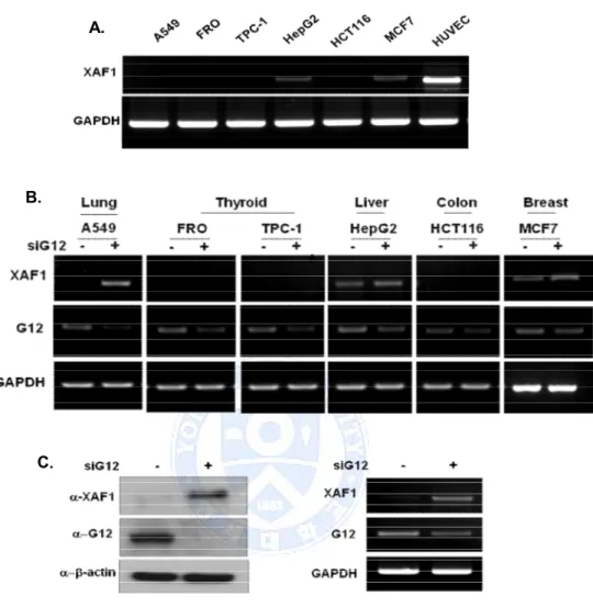
We screened the expression of the XAF1 in various cancer cell lines. To measure the endogenous expression level of XAF1, RT-PCR analyses was performed on various cancer cell lines. The results indicate that XAF1 express in HepG2, MCF7 cells compared with HUVEC normal control cells, and not detectable in A549, FRO, TPC-1, and HCT116 cells (Figure 1.A). To determine whether Ga12 regulated XAF1 expression level, cancer cells were transient transfected with Ga12 siRNA. Knockdown of Ga12 increased the basal expression level of XAF1 in A549, HepG2, and MCF7 cells, but not changed XAF1 expression in other cell lines. Particularly, Western blot and RT-PCR for Ga12 in A549 cells were predominantly re-expressed XAF1 protein and mRNA level by Ga12 down regulation. (Figure 1.B, C). These results demonstrate that Ga12 adjusted XAF1 expression in A549 cells.
Figure 1. Expression level of XAF1 in various cancer cells and XAF1 expression was controlled by Ga12. (A) Endogenous XAF1 mRNA level was measured by RT-PCR in various cancer cell lines. (B) Effect of Ga12 on XAF1 expression level. Cancer cells were transient transfected with Ga12 siRNA for 48h. XAF1 mRNA level detected by RT-PCR (C) Western blot and RT-PCR for Ga12 in A549 cells reveal restoration of XAF1 protein and mRNA levels by Ga12 siRNA.
A.
B.
B. Restoration of XAF1 by Ga12 down-regulation was appeared in
NSCLCs
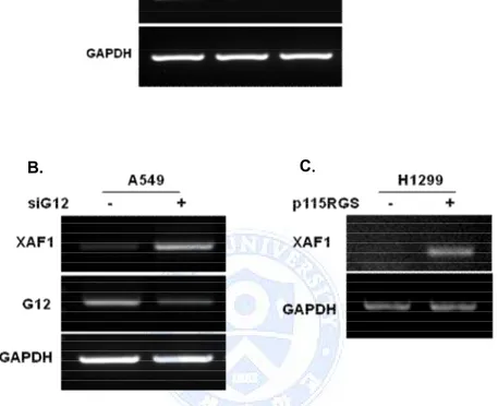
To describe the influence of Ga12 in XAF1 expression, we screened the expression of the XAF1 in other lung cancer cell lines. XAF1 basal mRNA level was very weakly detected in all NSCLC cells compare with normal cell line (Figure 2.A). However, XAF1 expression was markedly restored by blocking of Ga12 signal in A549 cells and H1299 cells (Figure 2.B).
Figure 2. Ga12 regulates expression level of XAF1 in human NSCLC cell lines. (A) Total RNA isolated from 2 NSCLC cell lines and HUVEC normal control cells. Ga12 and XAF1 mRNA basal level detected by RT-PCR (B, C) A549 cells were transfected with Ga12 siRNA for 48h. H1299 cells were transfected with p115RGS that inhibit downstream signals of Ga12. XAF1 expression level was measured by RT-PCR.
B. C.
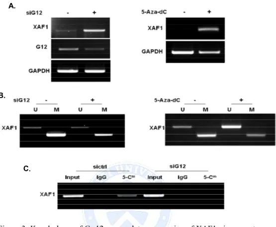
C. DNA methylation of XAF1 controlled by Ga12 in A549 cells To elucidate whether aberrant DNA methylation is associated with gene silencing of XAF1, A549 cell were treated the demethylating agent 5-Aza-2′deoxycytidine (5-Aza-dC). 5-Aza-dC treated in A549 cells was XAF1 re-expression that was shown same aspect increased XAF1 by Ga12 siRNA transfection (Figure 3.A). To determine whether Ga12 regulate methylation of XAF1 promoter region, we used MSP and Methylated DNA Immunoprecipitation (MeDIP) after siGa12 transfection. According to data from MSP, XAF1 was methylated in A549 cells. However, XAF1 promoter methylation was reduced by siGa12. The same effect was obtained when cells were treated 5-Aza-dC (Figure 3.B). Moreover, the data from MeDIP show that siGa12 lowers the frequency of 5-methylcytosine on the promoters of XAF1 (Figure 3.C)
Figure 3. Knock down of Ga12 up-regulates expression of XAF1 via promoter demethylation. (A) Effects of Ga12 and 5-Aza-dC on promoter methylation and expression of XAF1 in A549 cells. A549 cells were transfected with siGa12 RNA for 48h or treated 5-Aza-dC (10 µΜ) for 5days. XAF1 expression was evaluated by RT-PCR. (B) MSP analysis was performed to determine the promoter methylation of XAF1. 100 ng of bisulfite modified genomic DNA was subjected to PCR amplification of the XAF1 promoter sequences using unmethylation specific and methylation specific primer sets, respectively. M; methylated, U unmethylated. (C) Methylated DNA immunoprecipitation assay was performed to promoter methylation status. Immunoprecipitation of genomic DNA from A549 cells with antibody against 5-methylated-cytosine followed by RT- PCR analysis of MeDIP. Input as DNA control and IgG as negative control.
A.
B.
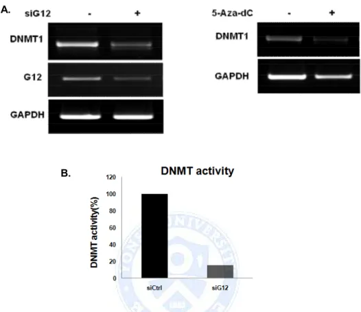
D. DNMT1 mediate DNA methylation change by Ga12 knock down As we confirmed that methylation in XAF1 was influenced by Ga12, we tried to find the mediating factors. First of all we checked the DNMT1 expression which is most common methyltransferase. In both siGa12 and 5-Aza-dC treatment, DNMT1 expression was decreased (Figure 4.A). In addition to complete loss of DNMT1 protein in the whole cell lysates and nuclear extracts, DNMT activity was severely reduced upon both siGa12 and 5-Aza-dC treatment (Figure 4.B).
From these results, we can show that Ga12 regulates XAF1 expression by epigenetic modification mechanism by inducing DNMT1 expression and activation.
Figure 4. Ga12 regulates both expression and activity of DNMT1. (A) A549 cells were transfected with siGa12 RNA and exposed to 5-Aza-dC for 48h and 5days, respectively. Expression of DNMT1 and Ga12 were analyzed by RT-PCR. Results were representative of three separated experiments. (B) A549 cells were transfected with Ga12 siRNA. DNMT activity was determined in the nuclear extracts, and the methylated DNA can be recognized with an anti-5-methylcytosine antibody. The ratio or amount of methylated DNA, which is proportional to enzyme activity, can then be measured using an ELISA-like reaction by reading the absorbance at 450 nm.
A.
E. Effect of the up-regulation of XAF1 expression by Ga12
knockdown on XIAP expression and localization
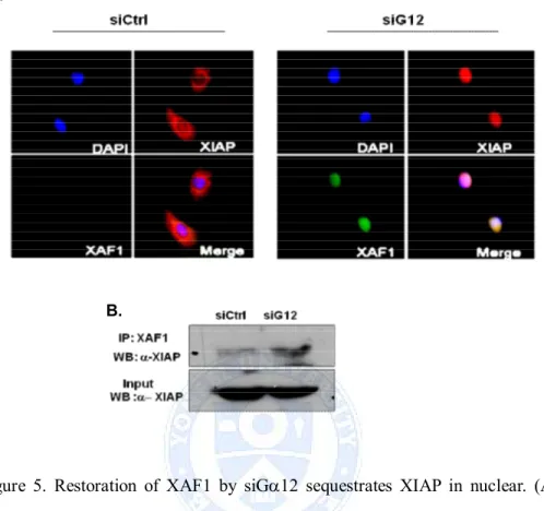
XAF1 was a nuclear protein to exert its proapoptotic effect by directly interacting with XIAP and inducing XIAP sequestration in nuclear inclusion. To investigate interaction between XAF1 and XIAP in siGa12 transfected A549 cells, we used fluorescence confocal microscopy to observe the localization of XAF1 and XIAP. XAF1 was not detected and endogenous XIAP was observed predominantly in the cytoplasm in control siRNA transfected cells. However, restoration of XAF1 expression by siGa12 triggered a edistribution of XIAP from the cytosol to the nucleus (Figure 5.A). The possibility of recovering XIAP by immunoprecipitation with an anti- XAF1 antibody was investigated. Coimmunoprecipitation analysis revealed the XIAP protein was precipitated in cellular nucleus and increased in Ga12 knockdown cells (Figure 5.B). As shown in these data, siGa12 increased XAF1 interaction with XIAP.
Figure 5. Restoration of XAF1 by siGa12 sequestrates XIAP in nuclear. (A) A549 cells were transfected with siGa12 for 48h. Confocal microscopy observed for the cellular localization of XAF1 with XIAP in siGa12 transfected A549 cells. Immunodetection was performed using specific antibodies linked to fluorescence performed using specific antibodies linked to fluorescence probes: Anti XIAP with cy3 (red) and anti XAF1 with FITC (green). Nuclei were stained with DAPI (blue). (B) Nuclear proteins were extracted from siGa12 transfected A549 cells to evaluate the coimmunoprecipitation of XIAP with anti-XAF1 antibodies. input as positive control.
A.
F. Effect of Ga12 knockdown on apoptosis
Next, to determined physiological roles of XAF1 in lung cancer, we performed functional studies. TUNEL assays revealed apoptotic DNA fragments after siGa12 transfection (Figure 6.A). I tried to investigate whether Ga12 enhance cisplatin action, because A549 is a cisplatin resistant cells. As shown in westernblot data, Ga12 knock down induced the cleaved caspase3 in A549 cells. Moreover, siGa12 transfected cells were increased apoptosis in the presence of cisplatin compared with only cisplatin (Figure 6.B). In flowcytometry analysis, there was a 3-fold increase in the percentage of apoptotic cells with siGa12 transfected cells + cisplatin when compare with siCtrl transfected cells+ cisplatin (Figure 6.C). Restoration of functional XAF1 by siGa12 could be effective in overcoming cisplatin resistance by sensitization of tumor cells to cisplatin induced apoptosis.
Figure 6. Effect of Ga12 knockdown on apoptosis. (A) Microscopic detection of apoptotic cells by TUNEL after siGa12 RNA transfection for 48h. (B) Casapse3 activation by Ga12 downregulation. Cells were transfected with
C. A.
immunoblotting assay using antibody specific for cleaved caspase3. The b-actin control using the same blot indicates equal protein loading. (C) The effect of siGa12 on either early or late stage of A549 apoptosis as detected by flowcytometry. A549 cells were transfected with Ga12 siRNA for 24h and then incubated without (control) or with 20 µM cisplatin. After 24 h, the cells were harvested, stained with Annexin V-FITC and PI, and analysed by flowcytometry. Diagrams of FITC-Annexin V/PI flowcytometry in a representative experiment are presented below the graphs. The lower right quadrants represent the cells in the early stage of apoptosis. The upper right quadrants contain the cells in the late stage of apoptosis or necrosis.
4. DISCUSSION
In this study, we characterized the role of Ga12 in the regulation of XAF1 function and demonstrate that downregulation of Ga12 with Ga12 siRNA mediates XAF1 induced apoptosis in A549 lung cancer cells.
Heterotrimeric G proteins regulate variety of cellular processes including proliferation, differentiation, junctional assembly, and apoptosis. The a-subunits of heterotrimeric G proteins can be divided into four families based on sequence homology: Gs, Gi, Gq, and G12.The last of the four families to be identified, the G12 family has been of particular interest to cancer researchers, since its members were found to promote the growth and oncogenic transformation of murine fibroblasts. 17 These findings led to the hypothesis that GPCRs may signal through the Ga12 proteins to promote tumorigenesis and tumor cell growth. Many studies have suggested a role for the members of the G12 family of heterotrimeric G proteins in oncogenesis and tumor cell growth. The Ga12 proteins are up-regulated in breast cancer and that Ga12 signaling promotes breast cancer metastasis by stimulating breast cancer cell invasion. Signaling by Ga12 proteins through the RhoA family of GTPases is a potent stimulator of prostate cancer cell invasion.16
In preliminary experiments, we used the Ga12 siRNA transfection system to the study on new roles of Ga12. We confirmed that expression of XIAP gene was controlled by siGa12 transfection in NSCLCs. XAF1 is one of the XIAP interacting partners and induce apoptosis. Therefore, this finding led to the hypothesis that Ga12 may adjust apoptosis of cancer cells via modulation of XAF1.
XAF1 was first identified as an interacting protein of XIAP. XAF1 antagonizes the anticaspase activity of XIAP. XAF1 is a nuclear protein that directly interacts with endogenous XIAP and results in XIAP sequestration in
compatible with the observed increase in the sensitivity of cancer cell lines to apoptotic triggers when XAF1 is overexpressed.18 Consistent with this, we observed that restoration of XAF1 expression by Ga12 knockdown reversed XIAP-mediated protection against apoptosis. Subcellular distribution studies revealed that XAF1 resides in the nucleus, and can effect a marked relocalization of endogenous XIAP protein from the cytoplasm to the nucleus. We also could detect the nuclear translocation of XIAP protein in tumor cells undergoing apoptosis by XAF1 restoration.
This study also demonstrated the mechanisms by which Ga12 regulates the apoptosis of lung cancer cells. XAF1 is a putative tumor suppressor that its mRNA is present ubiquitously in all normal tissues but is expressed at low or undetectable levels in various cancer cell lines. In many cancers, the CpG islands of selected genes are aberrantly methylated, resulting in repression of transcription of these genes.19Recent studies have suggested that loss of XAF1 expression may occur in different human cancers because of aberrant DNA methylation. 20,21 Zou et al found that loss of XAF1 expression is associated with tumor progression in human gastric and colon cancers. 21 Lee et al also discovered that downregulation of XAF1 expression is correlated with human urogenital malignancies.22
In this study, we found that XAF1 expression is upregulated in low expression A549 cells following Ga12 siRNA transfection and aberrant hypermethylation of the promoter is tightly associated with downregulation of XAF1 expression in A549 cells. And we show that XAF1 promoter methylation was reduced by siGa12. Thus this suggests that Ga12 regulates XAF1 expression by epigenetic modification mechanism. DNA methylation, catalyzed by DNA methyltransferase, involves the addition of a methyl group to the carbon 5’ position of the cytosine ring in the CpG dinucleotide to form methylcytosine. We found that the amount of DNMT1 expression and DNMT activity have correlation with the expression and methylation status of the
XAF1 gene. The eEF2 methylation is preceded by ras-raf-mitogen-activated protein kinase kinase (MEK)-extracellular signal-regulated kinase (ERK1/2)-p21Cip/WAF1 activation. eEF2, a key factor involved in protein translational elongation is symmetrically arginine-methylated in a reversible manner, being regulated by bFGF through MAPK signaling pathway.23 Soberanes, S. et al. observed that exposure to PM results in a mitochondrial-oxidant and JNK-mediated increase in the transcription and abundance of DNMT1 and increased methylation of the p16 promoter in the lung epithelium. Aberrant upregulation of DNMT1 could result in early and coordinated hypermethylation of key tumor suppressor genes in PM exposed patients, increasing the risk for the development of lung cancer. 24c-Jun NH(2)-terminal kinase (JNK) is a key downstream effector of G12 on this pathway. Expression of constitutively-active Ga12 or activation of G12 signaling by thrombin leads to increased JNK and c-Jun phosphorylation. These observations predict mechanism that G12 adjusts methylation of XAF1 promoter through MAPK pathway (e.g. ERK1/2 or JNK) mediated DNMT modulation.
Byun et al reported the importance of transcriptional regulation by 7 CpG dinucleotides located in the 5’ proximal region from -23 to -234 nucleotides in both gastric cancer cell lines and tumor tissues.25A cluster of methylated CpG sites instead of CpG islands located in the promoter area resulted in gene silencing of XAF1, and CpGs at -2nd, -1st, and +3rd positions are functionally more important in its transcriptional regulation.21 While we showed a correlation between aberrant methylation of XAF1 and Ga12 regulation and lung cancer progression, the exact promoter region could not be confirmed. Therefore, confirmation of XAF1 promoter methylation site in lung cancer cell may be need further study.
Some lung cancer cells are resistant against to cisplatin-induced tumor regression, leading to the conjecture that defective response to growth
context, it could be suspected that lung cancers with XAF1 inactivation might be more resistant to cisplatin drug therapy than cancers with normal XAF1 expression, and restoration of functional XAF1 could be effective in overcoming cisplatin resistance by sensitization of tumor cells to cisplatin induced apoptosis.
XAF1 induces cell cycle arrest during G2/M phase and mitotic catastrophe, and the restoration of XAF1 expression inhibits tumor growth in many types of human cancers. 26However, we could not observe that restoration of XAF1 expression inhibit tumor cell growth.
Several regulatory pathways of XAF1 expression have been reported. In addition to hypermethylation of XAF1 promoter, Wang, Jide et al reported that heat shock factor 1 negatively while interferon regulatory factor-1 and STAT1 positively modulated XAF1 transcription.27,28Inhibition of ERK1/2 stimulated XAF1 expression through indirect transcription regulation. Furthermore, XAF1 was an effector in ERK inhibition-induced cell apoptosis.29 The over-expression of XAF1 led to activation of wild-type p53 via post-translational modification in cells with or without DNA damage, which resulting in p53 nuclear accumulation and its increased transcriptional activity and enhancing p53-dependent apoptosis.30 However, we could not observation that Ga12–mediated mechanism for regulation of XAF1 expression and methylation. Therefore, other mechanisms of transcriptional regulation of the XAF1 gene may be present and need further research.
In conclusion, XAF1 undergoes epigenetic silencing in a considerable proportion of lung cancer cell lines by aberrant CpG sites hypermethylation of the gene promoter. In our data shown that siGa12 was enhanced demethylation of XAF1 promoter and was induced apoptosis. Therefore, our data presented here demonstrate that Ga12 was caused cancer cell progression and maintenance by controlling the methylation of XAF1.
Part Ⅱ
. Up-regulation of p16 by siG
a12 inhibits cellular
proliferation of HepG2 cells
1. INTRODUCTIONHepatocellular carcinoma (HCC) is one of the most common human malignancies worldwide, and is closely associated with infection of HBV and HCV and contamination of aflatoxin B1. Although the molecular mechanisms of hepatocarcinogenesis remain poorly understood, an increasing number of genetic abnormalities have been recognized.31
Cyclin-dependent kinase inhibitor 2A, (CDKN2A, p16Ink4A) also known as multiple tumor suppressor 1 (MTS-1), is a tumor suppressor protein, that in humans is encoded by the CDKN2A gene. P16 plays an important role in regulating the cell cycle, and mutations in p16 increase the risk of developing a variety of cancers.32
Inactivation of p16 by aberrant methylation of CpG islands is a frequent event in carcinomas and precancerous lesions of various organs, including the stomach. Gastric cancers with higher numbers of methylated genes have more distinct DNA methylation profiles than the originally defined CIMP-positive GCs. DNA methylation of tumor-related genes accumulates in conjunction with tumor progression. HepG2 cells treated with 5-Aza-cdR, showed demethylation of the p16 gene. The p16 mRNA and protein were all increased dramatically, cell cycle was arrested in G1, apoptotic rate increased and implanted tumor grew more slowly.33,34
Ga12 is reported to be involved in tumor cell invasion and progression. However, the precise mechanisms by which Ga12 regulates proliferation of cancer cells are poorly understood. Thus, we aimed to investigate the effect of
2. MATERIALS AND METHODS
A. Cell culture
A549, FRO, HepG2, HCT116, MCF7 cells were grown in RPMI1640 medium containing 10% FBS, 100 units/ml penicillin and 100 mg/ml. TPC-1, PLC/PRF5 cell lines were grown in DMEM medium supplemented with 10% FBS, 100 units/ml penicillin and 100 mg/ml. Cells were cultured in a 5% CO2 incubator at 37℃.
B. Small interfering RNA transfection
Ga12 was knocked down using ON-Target plus-SMARTpool pooled siRNAs. A nonspecific control pool was used for the negative control. Cells were transfected with siRNA using Lipofectamine 2000 reagents according to the manufacturer's guidelines. Briefly, 100 pmol of siRNA and Lipofectamine 2000 were added separately to OPTI MEM medium. After 5 min, the two solutions were mixed and incubated for 20 min at room temperature. The mixture was added to monolayer of cells seeded in 10 cm tissue culture plates. The media were added with complete cell culture medium after 6 h and whole-cell lysates were prepared 48 h later for detection of protein expression.
C. Western blot analysis
The whole cell lysates were prepared with lysis buffer (50 mM Tris-HCl, 150 mM Sodium chloride, 1% Triton X-100, 1% sodium deoxycholate, 0.1% SDS, pH 7.5, and 2 mM EDTA, 1 mM sodium orthovanadate, 0.1 mM phenylmethyl sulfonyl fluoride, 0.5% NP-40, protease inhibitor cocktail.) The protein concentration was determined by the bicinchoninic acid with bovine serum albumin as the standard. Equal aliquots of total cell lysates (50 ug) were solubilized in sample buffer and electrophoresed on denaturing
SDS-polyacrylamide gel (10% and 15% separating gel). The proteins were transferred to polyvinylidene difluoride membranes. The membranes were blocked with 5% nonfat dry milk in TBS containing 0.05% Tween 20 and incubated with primary antibodies overnight at 4℃ and then with horseradish peroxidase-conjugated secondary antibodies for 2h at RT. Antigen-antibody complexes were detected with WEST-SAVE Up luminol-based ECL reagent.
D. RNA isolation and RT-PCR
Total RNA from each cell was isolated using Isol-RNA Lysis Reagent according to the manufacturer's protocol. The RNA samples were treated with DNase I, quantified, and reverse-transcribed into cDNA using the ReverTra Ace-α First Strand cDNA Synthesis kit.
RT–PCR was performed using primers specific for p16 (sense primer: 5’-GGAAATTGGAAACTGGAAGC -3’, antisense primer: 5’- CTGCCCATCAT CATGACCTG-3’) and GAPDH (sense: 5’- CAAGGTCATCCATGACAACT -3’, antisense: 5’- TTCACCACCTTCTTGATGTC -3’). The cycling conditions were as follows: initial denaturation at 95℃ for 5min, followed 35cycles at 95℃ for 45s, 60℃ for 45s and 72℃ for 45s. Band intensities of the amplified DNAs were compared after visualization on an UV transilluminator.
E. Immunofluorescence.
HepG2 cells were grown on poly-D-lysine coated confocal dishs. Cells were transfected with Ga12 siRNA for 48h, washed in PBS, and fixed 10 min with 4% methanol. Cells were permeabilized 5 min with 0.1% Triton X-100, and blocked in 5% donkey serum. Cells were incubated with rabbit anti-p16 primary antibody at 1:50 in blocking buffer, followed by donkey anti-rabbit-FITC secondary antibody at 1:100. Antibody incubations were performed for 1 h at
F. Methylation-specific PCR (MSP)
Treatment of DNA with bisulfite converts cytosine residues to uracil, but leaves 5-methylcytosine residues unaffected. We used reagents supplied in Imprint DNA modification kit (Sigma, St. Louis, MO, USA) for bisulfite treatment, according to the one-step modification procedure recommended by the manufacturer. Briefly, 10 μl of DNA solution (100 ng) was combined with 110 μl of Imprint Bisulfite Modification Reagent, denatured at 99°C for 6 min and incubated at 65°C for 90 min. For purification of bisulfite-treated DNA, we used the Spin column in Imprint DNA modification kit. The modified DNA was then stored at −20°C until use. For MSP, the modified DNA was amplified with Taq polymerase. The methylated or unmethylated p16 primer sets were: unmethylated p16 sense, TTTGAGGGATAGGGTTGGAG -3', antisense, 5'-CTCCCCTTTTTCCAAAAAATCA -3'; methylated p16 sense, 5'-TTGAGGGA TAGGGTCGGAG-3', antisense, 5'- CCCTTTTTCCGAAAAA TCGAAA -3'.
G. Methylated DNA Immunoprecipitation (MeDIP)
1 mg of DNA extracted from cell lines. 5mC immunoprecipitation was carried out using the EpiQuik Methylated DNA Immunoprecipitation Kit according to the manufacturer's specifications. First, wells were washed once with Wash Buffer (WB; CP1) and then incubated at RT for 60 min in the presence of 100 ml of Antibody Buffer (AB; CP2) supplemented with 1 ml of 5mC antibody (or 1 ml of Normal Mouse IgG, as a negative control). DNA samples, diluted with ChIP Dilution Buffer (CP4), were added into the assay wells and incubated at RT for 90 min on an orbital shaker. The wells were washed six times with 1 WB, followed by the addition of TE Buffer. Afterwards, the DNA Release Buffer containing proteinase K were added to each well and samples were incubated at 65℃ for 15 min. Then, samples were incubated in Reverse Buffer at 65℃ for 30 min, Binding Buffer were subsequently added to
the wells, and the released samples, transferred to the F-Spin column, were centrifuged at 14 000 g for 20 s. Purified DNA was eluted in 15 ml of Elution Buffer. PCR was performed using immuno- precipitated DNA. PCR products were separated on 2% agarose gel and visualized by ethidium bromide staining.
H. DNMT Activity Assay
DNMT activity was determined in the nuclear extracts using the EpiQuik DNA Methyltransferase Activity Assay Kit (Epigentek Inc., Brooklyn, NY, USA) following the manufacturer’s protocol. Briefly, DNMT enzymes transfer a methyl group to cytosine from Adomet to methylate the DNA substrate, and the methylated DNA can be recognized with an anti-5-methylcytosine antibody. The ratio or amount of methylated DNA, which is proportional to enzyme activity, can then be measured using an ELISA-like reaction by reading the absorbance at 450 nm by the Tecan Infinite M2000 spectrophotometer. The DNMT activity was expressed as a percentage of the corresponding siRNA control.
I. Cell cycle analysis
The DNA contents of cell were measured by the PI (Sigma, St Louis, MO, USA) staining method. Cells (1 X 10⁶) were harvested by trypsinization and fixed with 70% ethanol for at least 2 h at -20℃. Fixed cells were rinsed twice with PBS, and re-suspended in PBS containing 50 µg/ml RNase A (Sigma, St Louis, MO, USA), then incubated in 37℃ for 30 min. PI was added to the cells suspension and incubated in the dark for 20 min. Stained cells were analyzed by a FACScan flow cytometry.
J. Cell Growth Assay
Cell growth was determined by cell count. A total of 1 × 10⁴cells were plated in a 6cm plate in complete medium. The following day, cells were transfected with Ga12 siRNA . Cell numbers were counted using a hemocytometer for 3days at 24h intervals.
K. Soft Agar Assay
1.5 × 10⁴HepG2 cells, expressing sictrl or siGa12, were mixed with 0.35% soft agar and plated on top of a 0.5% bottom agar in a 6-well plate. Cells were incubated at 37°C for 2 weeks to allow colony formation. Each cell line was plated in triplicate. Three random low-power view fields were chosen, and the total number of colonies was counted.
L. Statistical analysis
Results are expressed average ± S.D. Statistical analysis are as performed by student's t-test. Relationships were considered statistically significant when p-value was less than 0.05.
3. RESULTS
A. Effect of Ga12 knockdown on p16 expression in various cancer cell
lines
We screened the expression of the p16 gene in various cancer cell lines.
p16 basal mRNA level was low detected in HepG2, A549, MCF7 cell
lines compare with other cell lines. To determine whether G
a12 regulated
p16 expression, cancer cells were transiently transfected with G
a12
siRNA. However, down-regulation of G
a12 was restored p16 expression
in HepG2 cells.
Figure7. Effect of Ga12 knockdown on p16 expression in various cancer cell lines. Cancer cells were transient transfected with Ga12 siRNA for 48h. p16 mRNA level detected by RT-PCR.
B. Re-expression of p16 by siGa12 revealed in HepG2 cells
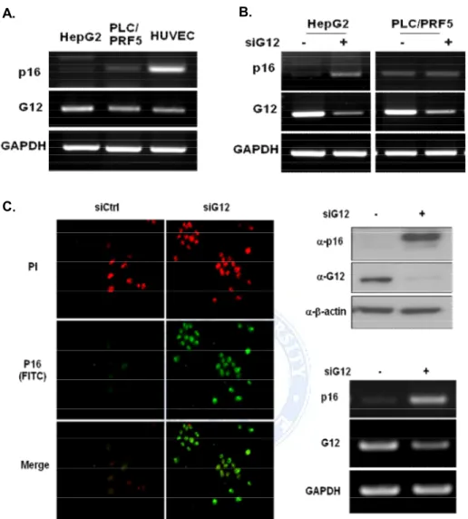
We screened the expression of the p16 in 2 hepatoma cancer cell lines. p16 basal mRNA level was very weakly detected in HepG2 cells and PLC/PRF5 cell lines (Figure 8.A). p16 expression was significantly enhanced in HepG2 cells after siGa12 transfection (Figure 8.B). We confirmed increased p16 expression level when blocked Ga12 signal from immunofluorescence study, western blot, and RT-PCR in HepG2 cells (Figure 8.C). Therefore all experiments were proceeding with HepG2 cells.
Figure 8. Restoration of p16 expression by siGa12 in HCCs. (A) Total RNA isolated from 2 HCCs and HUVEC normal control cells. Ga12 and p16 mRNA basal level detected by RT-PCR (B) HepG2 and PLC/PRF5 cells were transfected with Ga12 siRNA for 48h. p16 mRNA expression levels were measured by RT-PCR (C) Immunofluorscence, western blot and RT-PCR for Ga12 in HepG2 cells reveal restoration of p16 protein and mRNA levels by Ga12 siRNA.
A. B.
C. DNA methylation by Ga12 in HepG2 cells
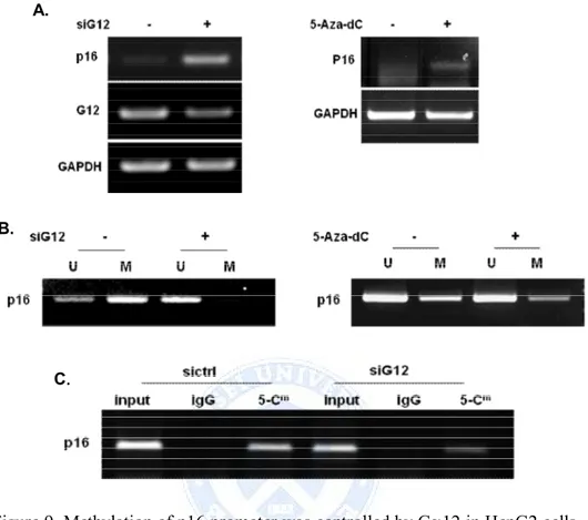
HepG2 cells were treated with 5-Aza-dC or transfected siGa12. 5-Aza-dC treated cells induced p16 expression and we obtained similar result by siGa12 (Figure 9.A). P16 is also frequently inactivated in several cancers via DNA methylation. Therefore, we examined whether Ga12 induces p16 promoter demethylation. Methylation of the p16 promoter CpG island was assessed by MeDIP analysis and MSP on genomic DNA isolated from HepG2 cells.
According to data from MSP, down regulation of Ga12 induced demethylation of p16, suggesting that expression of p16 is up-regulation by siGa12 via promoter demethylation. The same effect was obtained when cells were treated 5'-Aza-dC (Figure 9.B). MeDIP assay enabled the purification of enriched methylated-DNA by direct immunoprecipitation of the 5′-methylcytosine modification related to the CpG sites of the p16 promoter. As shown in here, siGa12 decreased 5’-methylcytosine on the promoters of p16 (Figure 9.C).
Figure 9. Methylation of p16 promoter was controlled by Ga12 in HepG2 cells. (A) Effects of Ga12 and 5-Aza-dC on promoter methylation and expression of p16 in HepG2 cells. HepG2 cells were transfected with siGa12 RNA for 48h or treated 5-Aza-dC (10 µΜ) for 5days. P16 expression was evaluated by RT-PCR. (B) Methylation of p16 promoter analyzed by MSP. MSP analysis of p16 promoter compared between siCtrl and siGa12 transfected cells (left). P16 methylation in response to exposure to 5-Aza-dC for 5 days (right). U; unmethylated, M; methylated DNA. (C) MeDIP assay was performed to promoter methylation status. Immunoprecipitation of genomic DNA from HepG2 cells with antibody against 5-Cm followed by RT-PCR analysis of MeDIP. Input as DNA control and IgG as negative control.
A.
B.
D. Down-regulation of Ga12 decreases DNMT1-mediated p16
methylation
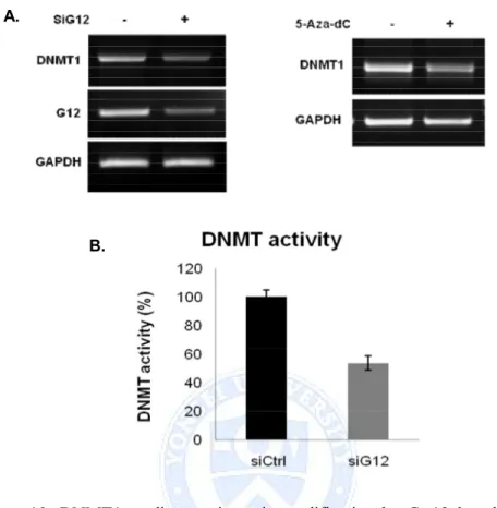
We next aimed to gain insights into the mechanisms that underlie hypermethylation in HepG2 cells. A crucial step in DNA methylation involves DNA methyltransferases that catalyze methylation of CpG dinucleotides in genomic DNA. To gain insights into the possible involvement of DNMTs, we performed RT-PCR on total RNA samples isolated from siGa12 transfected HepG2 cells. DNMT1 mRNA was decreased by Ga12 knockdown. The same effect was obtained when cells were treated 5'-Aza-dC (Figure 10.A). In addition, the DNMT activity in the nuclear protein extract of siGa12 cells decreased compared with sictrl (Figure 10.B), similar to the mRNA and protein pattern of DNMT1 by 5'-Aza-dC. From these results, we can show that Ga12 regulates p16 expression by epigenetic modification mechanism by inducing DNMT1 expression and activation.
Figure 10. DNMT1 mediates epigenetic modification by Ga12 knock down. (A) HepG2 cells were transfected with DNMT1 siRNA for 48h or treated 5-Aza-dC for 5days. DNMT1 and Ga12 mRNA levels were examined in G12-knocked down cells by RT–PCR. (B) Quantification of enzyme activity of DNMT1. HepG2 cells were transfected with siGa12 RNA. DNMT activity was determined in the nuclear extracts, and the methylated DNA can be recognized with an anti-5-methylcytosine antibody. The ratio or amount of methylated DNA, which is proportional to enzyme activity, can then be measured using an ELISA-like reaction by reading the absorbance at 450 nm.
A.
E. Regulation of Histone modification by Ga12
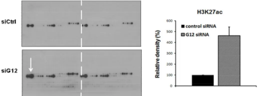
To investigate the histone modification by siGa12, we performed Modified Histone peptide array. Histone peptide array is experiment for the assessment of change in various histone modifications, such as methylation, acetylation, and phosphorylation modification. We obtained outstanding result that siGa12 increased Histone H3K27 acetylation. This result indicated that Ga12 alter chromatin structure by modifying histone tails.
Figure 11. Regulation of histone modification by Ga12. Protein extracts were prepared from cells 48 h after Ga12 siRNA transfection on the Modified Histone Peptide Array. P16 primary antibody was used at a 1:2000 dilution for overnight at 4℃. Anti-rabbit HRP secondary antibody was used at a 1:2500 dilution, followed by ECL detection. Active motif’s Array Analysis Software was used to analysis spot intensity.
F. Effect of Ga12 knock down on HepG2 cells proliferation
Next, to determined physiological roles of p16 in liver cancer, we performed functional studies. Flow cytometry was performed to observe the effects of Ga12 down regulation on cell cycle progression. The proportion of cells in the G0/G1 phase was significantly higher in the siGa12 transfected cells compared to the proportion of cells in the sictrl cells (Figure 12.A). And we were obtained result from cell counting assay that cell number was decreased in Ga12 knockdown (Figure 12.B). The levels of cell cycle regulatory proteins, such as phospho-Rb, E2F and CDK4, were also determined after siGa12 transfection. Cell cycle marker proteins were reduced by siGa12 (Figure 12.C). And we were obtained result from cell counting assay that cell number was decreased in Ga12 knockdown cells. We next examined the effect of Ga12 depletion in liver cancer cells on in vitro tumorigenicity using the soft agar colony formation assay. Consistent with the proliferation assays, siGa12 cells approximately decreased two fold in colony formation numbers. Colony size was reduced in Ga12 knockdown cells (Figure 12.D).
Figure 12. Ga12 promotes hepatoma carcinoma cell proliferation and tumorigenicity in HepG2 cells. (A) Cell cycle analysis of Ga12 knockdown HepG2 cell by flowcytometry. (B) Cell growth was determined by cell count assay. HepG2 cells were transfected with Ga12 siRNA. Cell numbers were counted using a hemocytometer for 3 days at 24h intervals. (C) Expression of cell cycle regulatory proteins E2F, Cdk4 and phospho-Rb in Ga12 knockdown cells. Western blots were performed on lysates from siGa12 cells. Comparable density of b-actin levels confirms equal loading of total protein. (D) Analysis of
in vitro tumorigenicity by soft agar assay. HepG2 cells transfected with siGa12
C.
A. B.
were seeded in soft agar and maintained for 2 weeks. Colonies were stained with crystal violet. Results are representative of at least three independent experiments performed in triplicates. Data are expressed as average ± SD *p<0.05;**p<0.01.
4. DISCUSSION
The G12 class includes Gα12 and Gα13 and is ubiquitously expressed in mammalian tissues and cells. The last of the four families to be identified, the G12 family has been of particular interest to cancer researchers, since its members were found to promote the growth and oncogenic transformation of murine fibroblasts. Many studies have suggested a role for the members of the Gα12 family of heterotrimeric G proteins in oncogenesis and tumor cell growth. 35,36
In breast and prostate cancer cells, previous studies showed a role of Gα12 and of Gα13 in invasiveness, but not in tumor cell proliferation.16,17In contrast, Grzelinski M et al. reported that in SCLC cells, the downregulation of either Gα12 or Gα13 leads to a clear inhibition of proliferation in vitro as well as in vivo.37Our observations indicate that in hepatoma cells, the downregulation of Gα12 leads to a clear inhibition of proliferation in vitro and the anchorage-independent colony formation. This finding further supports the concept that intact Gα12 signaling is a prerequisite for tumor growth in hepatoma cancer cells. Moreover, Gα12 siRNA restored the expression of p16Ink4 known to be silenced in many cancer cells and arrested the cell cycle progression of HepG2 cells.
Currently, p16 is considered a tumor suppressor protein because of its physiological role and downregulated expression in a large number of tumors. Inactivation of p16 by aberrant methylation of CpG islands is a frequent event in carcinomas and precancerous lesions of various organs.
Precancerous conditions with aberrant DNA methylation appear to generate HCCs rapidly. Liu, L. H. et al HepG2 cells treated with 5-Aza-dC, showed demethylation of the p16 gene. The p16 mRNA and protein were all increased dramatically, cell cycle was arrested in G1, apoptototic rate increased and implanted tumor grew more slowly.38We obtained similar result by siGα12. We
show that p16 hypermethylation is associated with HCC cell line and downregulation of Gα12 induces p16 promoter demethylation. According to data from MSP and MeDIP, we suggest that Gα12 expression is correlation with hepatocarcinogenesis through aberrant p16 hypermethylation.
Increased DNMT1 mRNA expression in a number of human cancers has been reported. Moreover, Sun et al. found that DNA methyltransferase mRNA levels were significantly higher in HCC and HCC cell lines. We confirmed that DNMT1 was increased in HepG2 cell, and DNMT1 mRNA was decreased by Gα12 knockdown. In addition, the DNMT activity in the nuclear protein extract of siGα12 cells decreased. From these results, we can show that Gα12 regulates p16 expression by epigenetic modification mechanism by inhibiting the activities and expression of DNMT. Sarkar et al reported that HDACi inhibited cell cycle progression, and reversed promoter methylation and silencing of three tumor suppressor genes: RARB2, p16 and p21. HDACi repressed MAP kinase I (ERK) activation and down-regulated DNMT1 levels. Recent studies suggest that DNA methylation in colon cancer cells and in NIH 3T3 cells may be regulated in some circumstances by ERK (MAP kinase1) activity.39 c-JUN is also involved in protecting the promoter region of the tumor suppressor p16(INK4a), which is consistently methylated over time in c-JUN deficient cells.40 The association of JNK1 with the epigenetic alternations, mainly the elevation of H3K4me3, H3K9me3, and the expression of EZH2 that is involved in both histone and DNA methylation indicates a pivotal role of JNK on the development of the human HCC.41 Mitsui, H. indicated that important roles of both Ras/MAPK and Ras/Rac/JNK cascades in activated Gα12-induced G1/S cell cycle progression.42 Ga12 stimulates cell proliferation and neoplastic transformation of NIH3T3 cells by attenuating p38MAPK-associated apoptotic responses, while activating the mitogenic responses through the stimulation of ERK- and JNK-mediated signaling pathway. These observations expected that