The Influence of Anti-Platelet Resistance on the Development
of Cerebral Ischemic Lesion after Carotid Artery Stenting
Tae-Jin Song,
1Sang Hyun Suh,
2,4Pil-Ki Min,
3,4Dong Joon Kim,
2Byung Moon Kim,
2Ji Hoe Heo,
1Young-Dae Kim,
1and Kyung-Yul Lee
1,4Departments of 1Neurology, 2Radiology, and 3Cardiology, Yonsei University College of Medicine, Seoul; 4Severance Institute for Vascular and Metabolic Research, Yonsei University, Seoul, Korea.
Received: February 7, 2012 Revised: May 18, 2012 Accepted: May 29, 2012
Corresponding author: Dr. Kyung-Yul Lee, Department of Neurology,
Gangnam Severance Hospital, Yonsei University College of Medicine, 211 Eonju-ro, Gangnam-gu, Seoul 135-720, Korea.
Tel: 82-2-2019-3325, Fax: 82-2-3462-5904 E-mail: [email protected]
∙ The authors have no financial conflicts of interest.
© Copyright:
Yonsei University College of Medicine 2013
This is an Open Access article distributed under the terms of the Creative Commons Attribution Non-Commercial License (http://creativecommons.org/ licenses/by-nc/3.0) which permits unrestricted non-commercial use, distribution, and reproduction in any medium, provided the original work is properly cited.
Purpose: Cerebral ischemic lesions are frequently observed after carotid artery stenting (CAS), and anti-platelet agents are used to prevent stent thrombosis and peri-procedural complications. However, despite the premedication, cerebral isch-emic lesions are observed, suggesting that they may rather be related to anti-plate-let resistance. We, therefore, investigated the effects of anti-plateanti-plate-let resistance on the development of cerebral ischemic lesions after CAS. Materials and Methods:
We retrospectively reviewed patients who received CAS and selected patients for whom brain MRI was performed within 24 hours after CAS and for whom anti-platelet resistance was checked. Anti-anti-platelet resistance was examined by the Veri-fyNow system. We analyzed the correlation between anti-platelet resistance and cerebral ischemic lesions detected on follow-up MRI. Results: Among 76 tients, 45 (59.2%) developed new ischemic lesions after CAS. Twelve (15.8%) pa-tients showed aspirin resistance and 50 (65.8%) papa-tients showed clopidogrel resis-tance. Patients with a new ischemic lesion demonstrated a significantly greater frequency of clopidogrel resistance than those who had no new ischemic lesion (82.2% versus 41.9%, p=0.001). The frequency of aspirin resistance was not sig-nificantly different between the groups of patients with and without new ischemic lesions (20.0% versus 9.7%, p=0.340). In multivariate analysis, clopidogrel resis-tance was a significant risk factor for post-procedural cerebral ischemia. Conclu-sion: Anti-platelet resistance can be used to predict new ischemic lesions after CAS. Anti-platelet resistance should be evaluated in all patients prior to CAS to prevent ischemic complications related to CAS.
Key Words: Cerebral infarction, aspirin resistance, clopidogrel resistance, carotid artery stent
INTRODUCTION
Cerebral ischemic lesions are observed by MRI in roughly 50% of patients after carotid artery stenting (CAS).1,2 Generally, anti-platelet agents are used as premed-ication to prevent stent thrombosis and peri-procedural complpremed-ications.3 Despite
pre-who took proton pump inhibitors. This study was approved by the Severance Hospital Institutional Review Board of the Yonsei University Health System in Seoul, Korea.
Carotid artery stent protocol
CAS was performed in patients with symptomatic (ischemic stroke or transient ischemic attack related to relevant artery) internal carotid artery (ICA) stenosis of 50% or more or as-ymptomatic stenosis of 70% or more according to the North American Symptomatic Carotid Endarterectomy Trial crite-ria on digital subtraction cerebral angiography. Four experi-enced neurointerventionist (S.H. Suh, D.J. Kim and B.M. Kim in radiology, P.K. Min in cardiology) managed all the procedures. We introduced a 90-cm-long 7 F or 8 F guiding catheter into the femoral sheath with a 120-cm-long 4 F or 5 F diagnostic catheter coaxially. Then, we removed the di-agnostic catheter after appropriate positioning of the guid-ing catheter proximal to the stenotic lesion.13 We did not perform aortogram separately. Pre-stenting balloon angio-plasty was performed in patients with severe stenosis. In cases of difficult penetration of the cerebral protection de-vice (CPD), balloon angioplasty was initially performed us-ing a 1.5-2 mm balloon catheter before placement of the CPD. After placing the CPD, a self-expandable stent [Pro-tege (ev3, Irvine, CA, USA), Precise (Cordis endovascular, Miami Lakes, FL, USA), or Wallstent (Boston Scientific, Natick, MA, USA)] was deployed in the proximal ICA or the distal common carotid artery, and post-stenting angio-plasty was done optionally when residual stenosis (more than 50%) was noted on angiography. The patients received a bolus injection of 3000-4000 IU heparin just before the start of the therapeutic procedure. The time intervals from pre-stent MRI to diagnostic cerebral angiography, pre-stent MRI to CAS, pre-stent MRI to post-stent MRI and diag-nostic cerebral angiography to post-stent MRI were deter-mined for patients who underwent CAS. If one-stage CAS was performed, the time interval from the first diagnostic angiography to CAS was considered as zero. At least 7 days before the procedure, all patients received 75 mg of clopido-grel and 100 mg of aspirin daily. Neurologic evaluation was performed before and at 24 hours after the procedure.
MRI protocol
All MRI examinations were performed on a 3.0T MRI sys-tem (Signa 3.0T, General Electric, Milwaukee, WI, USA; Achieva 3.0T, Philips Medical Systems, Best, the Nether-lands). Diffusion-weighted images (DWI) were obtained medication with aspirin and clopidogrel, however, cerebral
ischemic lesions are still frequently observed in patients upon brain diffusion-weighted MRI.1,2,4-6 Although the clini-cal implication of these cerebral ischemic lesions after CAS is not clear, they could potentially result in focal neurologic signs or cognitive dysfunction.1,2,4
Platelet function inhibition by aspirin or clopidogrel dif-fers from individual to individual, and some patients suffer recurrent cerebrovascular or cardiovascular events, regard-less of proper anti-platelet medication. In these cases, the patients may be clinically classified with anti-platelet resis-tance. A significant proportion of patients with coronary ar-tery occlusive disease show aspirin or clopidogrel resis-tance, which is related with major adverse coronary events after percutaneous coronary intervention and recurrent ath-erothrombotic events in patients with acute myocardial in-farction.7-9 In contrast to coronary artery occlusive disease, the role of anti-platelet resistance in carotid artery disease has not been well characterized. The reported prevalence of aspirin and clopidogrel resistance in cerebrovascular inter-vention ranges from 2 to 21% for aspirin and 43-52% for clopidogrel.10-12 However, these studies included only a few CAS patients (6.6%,10 16%,11 33.7%12) and did not provide any information about the clinical significance of anti-plate-let resistance in cerebrovascular stent placement. Therefore, we performed this study to determine the clinical signifi-cance of anti-platelet resistance in patients who underwent CAS, by investigating if there was any correlation between anti-platelet resistance and new cerebral ischemic lesions detected by 3.0T brain MRI after CAS.
MATERIALS AND METHODS
Study design
We retrospectively enrolled 76 patients who satisfied the following criteria from January 2007 to May 2011 in our registry: premedication of dual anti-platelet agents (aspirin and clopidogrel) at least 7 days before CAS, pre-stent brain MRI within 60 days, post-stent brain MRI within 24 hours, and aspirin and clopidogrel resistance test before CAS or within 2 days after CAS. Cases of emergent carotid stent insertion, those without pre or post-stent brain MRI, and those without anti-platelet resistance tests for both aspirin and clopidogrel were excluded. We obtained the past histo-ry, clinical features, and laboratory findings of patients by review of their medical records. We excluded the patients
age of the patients was 68.11±7.72 years, and 56 (73.7%) patients were males. In total, 39 patients (51.3%) underwent one-stage CAS without a previous diagnostic angiography procedure. Fifty seven (75.0%) patients had symptomatic carotid artery stenosis, 21 (36.8%) patients had a history of stroke confirmed by brain CT or MRI and 36 (63.2%) pa-tients had a transient history of ischemic attack related to the carotid stent side within 6 months before CAS. Pre-stent balloon angioplasty was performed in 61 (80.3%) patients, before placement of the CPD in 7 patients and after place-ment of the CPD in 54 patients. Seventy patients (92.1%) used the CPD. Post-stent balloon angioplasty was performed in 47 (61.8%) patients. The mean ARU and PRU values of all examined patients were 466.32±79.33 and 257.28±81.84, respectively. Twelve (15.8%) patients were resistant to aspi-rin, while 50 (65.8%) patients were resistant to clopidogrel, and 9 (11.8%) patients were resistant to both aspirin and clopidogrel. The frequency of aspirin resistance was similar to that of resistance to both drugs.
Among 76 patients, 45 (59.2%) patients had DWI-positive lesions; 40 patients were classified with asymptomatic stroke and 5 patients with symptomatic stroke. Eleven (24.4%) pa-tients had a single DWI-positive lesion, 16 (35.6%) papa-tients had 2 to 5 lesions, and 18 (40.0%) patients had more than 5 lesions. In 17 (37.8%) patients, lesions were located only on the ipsilateral side to stent placement. In contrast, lesions in 14 (31.1%) patients were detected in both the ipsilateral and non-ipsilateral vascular territory, and lesions in 14 (31.1%) patients were detected only in the non-ipsilateral vascular territory relative to the CAS. The DWI-positive lesions were frequently detected in the cerebral cortex and middle cere-bral artery (MCA) territory. The DWI-positive lesion size was less than 5 mm in 164 (72.8%), 5 mm to 10 mm in 35 (15.6%) and larger than 10 mm in 26 (11.6%).
All 5 (6.6%) symptomatic stroke cases after the proce-dure were from the symptomatic carotid artery stenosis group and had minor neurological deficits (National Insti-tutes of Health Stroke Scale score <4). These 5 patients were all DWI-positive and demonstrated aspirin and clopi-dogrel resistance. The location of the lesions according to CAS side was the ipsilateral MCA in 3 patients, the bilater-al MCA in one patient, and the ipsilaterbilater-al MCA and posteri-or inferiposteri-or cerebellar artery in one patient.
In patients with symptomatic carotid stenosis, the time in-terval between ischemic symptom onset and CAS was 19.69 days, and there was no time difference between DWI-posi-tive patients (22.67±20.04 days) and DWI-negaDWI-posi-tive patients using the following parameters: TR/TE 9000/81.4 ms,
ma-trix 128×128, FOV 240×240 mm, slice thickness 4 mm, b=1000 s/mm2. The resulting voxel volumes were 14 mm3. Acute probable ischemic lesions were defined as a hyperin-tense lesion on DWI or corresponding hypoinhyperin-tense lesion in apparent diffusion coefficient maps. A pre-stent MRI was performed within 60 days before the procedure. Post-stent brain DWI was carried out routinely within 24 hours after stent placement regardless of the presence of neurologic symptoms after CAS. When newly developed lesions were found after the procedure by comparing pre- and post-brain DWI, the case was defined as DWI-positive. If there were no newly developed lesions on follow-up DWI, the case was defined as DWI-negative. The number, size and loca-tion of the DWI-positive lesions according to the vascular territory and CAS side were recorded.14
Laboratory examination protocol
All patients received 75 mg of clopidogrel and 100 mg of aspirin for at least 7 days before undergoing aspirin and clopidogrel resistance tests. Aspirin resistance and clopido-grel resistance were determined by the VerifyNow system (Accumetrics, San Diego, CA, USA). The VerifyNow assay is a whole blood, light transmission-based optical detection assay that measures platelet aggregation in a cartridge con-taining fibrinogen-coated beads. An aspirin reaction unit (ARU) value ≥550 was defined as aspirin resistance.15 Clop-idogrel resistance was expressed in P2Y12 reaction units (PRU). A PRU value ≥240 was defined as clopidogrel re-sistance.16
Statistical analysis
Statistical analyses were performed using PASW Statistics 18 (IBM, Chicago, IL, USA). Continuous variables are pre-sented as means with standard deviations and categorical variables are presented as frequencies and percentages. Vari-ables were compared by independent t-test, chi-square test, Mann-Whitney U test and Fisher’s exact test. Univariate analysis was performed and factors with a p-value less of than 0.1 in univariate analysis were included in multivariate logistic regression analysis. A two-tailed value of p<0.05 was considered statistically significant.
RESULTS
DISCUSSION
Periprocedural microembolization is an important risk for pa-tients treated by angioplasty and stent placement of high-grade carotid stenosis. Atheromatous plaque and superim-posed thrombi are the main source of microemboli during CAS.17 Therefore, anti-platelet premedication and mainte-nance is very important for preventing peri-procedural isch-emic events. Recent investigations suggested that anti-platelet resistance is associated with cardiovascular or cerebrovascular events.9,18,19 Patients with aspirin resistance have been shown to have increased mortality and be at greater risk for acute coronary syndrome, vascular intervention failure, or new cerebrovascular events than aspirin-sensitive patients.18 Fur-thermore, clopidogrel resistant patients tend to have more recurrent cardiovascular events and a higher rate of sub-acute stent thrombosis after coronary stenting, compared (13.89±14.07 days) (p=0.063). For all patients, the duration
of aspirin and clopidogrel medication before CAS was a mean of 11.57 days, and there was also no time difference between positive patients (11.86±4.64 days) and DWI-negative patients (11.16±2.33 days) (p=0.439). In Spearman correlation analysis, there was no correlation between de-grees of carotid stenosis and ARU (p=0.985, rho=-0.002) or PRU (p=0.189, rho=0.152) values.
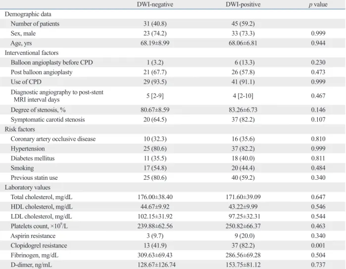
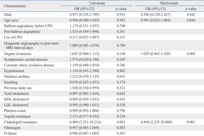
There was no difference in frequency of aspirin resistance between patients with and without DWI-positive lesions (p=0.340); however, clopidogrel resistance was detected more frequently in patients with DWI-positive lesions than patients without (82.2% versus 41.9%, p=0.001) (Table 1). After adjusting for age, gender, and degree of stenosis, clop-idogrel resistance was a significant predictor of DWI-posi-tive lesions after CAS upon multivariate analysis (odds ra-tio: 6.804; 95% confidence interval: 2.225-20.806; p=0.001) (Table 2).
Table 1. Comparison of Post Procedural Diffusion Weighted Image Findings
DWI-negative DWI-positive p value
Demographic data
Number of patients 31 (40.8) 45 (59.2)
Sex, male 23 (74.2) 33 (73.3) 0.999
Age, yrs 68.19±8.99 68.06±6.81 0.944
Interventional factors
Balloon angioplasty before CPD 1 (3.2) 6 (13.3) 0.230
Post balloon angioplasty 21 (67.7) 26 (57.8) 0.473
Use of CPD 29 (93.5) 41 (91.1) 0.999
Diagnostic angiography to post-stent
MRI interval days 5 [2-9] 4 [2-10] 0.467
Degree of stenosis, % 80.67±8.59 83.26±6.73 0.146
Symptomatic carotid stenosis 20 (64.5) 37 (82.2) 0.107
Risk factors
Coronary artery occlusive disease 10 (32.3) 16 (35.6) 0.810
Hypertension 25 (80.6) 37 (82.2) 0.999
Diabetes mellitus 11 (35.5) 18 (40.0) 0.811
Smoking 17 (54.8) 20 (44.4) 0.484
Previous statin use 25 (80.6) 40 (59.2) 0.340
Laboratory values Total cholesterol, mg/dL 176.00±38.40 171.60±39.09 0.647 HDL cholesterol, mg/dL 44.67±9.92 43.22±9.99 0.546 LDL cholesterol, mg/dL 102.15±31.92 97.25±32.31 0.544 Platelets count, ×109/L 239.88±62.56 250.82±66.37 0.463 Aspirin resistance 3 (9.7) 9 (20.0) 0.340 Clopidogrel resistance 13 (41.9) 37 (82.2) 0.001 Fibrinogen, mg/dL 309.63±69.43 286.56±69.28 0.504 D-dimer, ng/mL 128.67±126.74 153.75±81.12 0.737
DWI, diffusion weighted image; CPD, cerebral protection device; HDL, high density lipoprotein; LDL, low density lipoprotein. Values are number (%), mean±SD or median [interquartile range].
considered a very effective tool for detecting acute isch-emic brain lesions.1,2,4-6,14,23-25 Despite the use of a CPD to reduce embolic complications during CAS, post-procedural DWI-positive lesions were detected in 15 to 40% of cases in previous studies.2,26-29 Firstly, one possible reason for the high rate of new ischemic lesions detected after CAS is that new lesions may have developed before the stent place-ment. That is, we cannot exclude the possibility that new ischemic lesions developed spontaneously after the index stroke or by the time of the first diagnostic cerebral angio-graphic procedure. However, in our study, about half of the patients underwent one stage CAS and there was no differ-ence in the time interval from pre-stent MRI to diagnostic cerebral angiography, pre-stent MRI to CAS, pre-stent MRI to post-stent MRI and diagnostic cerebral angiography to post-stent MRI between the DWI-positive and negative groups. Therefore, the possibility of ischemic lesions not related to the CAS itself might not be that high.
Another possible reason for the high rate of new isch-emic lesions after CAS in our study may be the use of 3.0T MRI; previous studies used 1.5T MRI.1,2,14,27-29 Most impor-tant difference between 1.5T MRI and 3.0T MRI is the per-with clopidogrel sensitive patients.9,19
We investigated the relationship between anti-platelet re-sistance and the presence of new cerebral ischemic lesions that developed after CAS. In our study, 15.8% of patients were aspirin resistant and 65.8% were clopidogrel resistant, which was comparable to previous studies.10-12 Furthermore, the frequency of aspirin resistance was similar to that of re-sistance to both aspirin and clopidogrel in our study, consis-tent with a previous report.20 In aspirin resistance patients, platelets appear to have increased sensitivity to ADP-induced glycoprotein IIb/IIIa activation. These hyper-reactive plate-lets may be less sensitive to inhibition by clopidogrel.21,22
In our study, the frequency of clopidogrel resistance was significantly higher in positive patients than in DWI-negative patients. Therefore, medications which have phar-macological mechanisms different from that of clopidogrel should be considered, if clopidogrel resistance is detected in patients with CAS.
In this study, cerebral ischemic lesions were detected in 59.2% of patients after CAS, while a CPD was used in 92.1% of the patients. When performing CAS, cerebral ischemic lesions are frequently detected on DWI, which is
Table 2. Factors for Logistic Analysis for Post Procedural Diffusion Weighted Image-Positive Lesions
Characteristics Univariate Multivariate
OR (95% CI) p value OR (95% CI) p value
Male 0.957 (0.338-2.709) 0.933 0.586 (0.150-2.287) 0.442
Age (yrs) 0.998 (0.940-1.059) 0.943 0.993 (0.923-1.069) 0.844
Balloon angioplasty before CPD 1.179 (0.351-3.955) 0.790
Post balloon angioplasty 1.535 (0.589-3.998) 0.381
Use of CPD 0.217 (0.025-1.897) 0.167
Diagnostic angiography to post-stent
MRI interval days 1.009 (0.945-1.078) 0.788
Degree of stenosis 1.047 (0.984-1.115) 0.148 1.029 (0.962-1.102) 0.405
Symptomatic carotid stenosis 1.974 (0.630-6.186) 0.243
Coronary artery occlusive disease 1.159 (0.440-3.054) 0.766
Hypertension 1.110 (0.343-3.590) 0.862
Diabetes mellitus 1.212 (0.470-3.125) 0.691
Smoking 0.659 (0.263-1.653) 0.374
Previous statin use 1.920 (0.530-6.959) 0.321
Total cholesterol 0.997 (0.985-1.010) 0.642 HDL cholesterol 0.985 (0.939-1.033) 0.541 LDL cholesterol 0.995 (0.980-1.011) 0.538 Platelets count 0.999 (0.992-1.006) 0.796 Aspirin resistance 2.333 (0.577-9.432) 0.234 Clopidogrel resistance 6.404 (2.251-18.216) 0.001 6.804 (2.225-20.806) 0.001 Fibrinogen 0.997 (0.985-1.009) 0.583 D-dimer 0.996 (0.987-1.005) 0.367
Committee, Olsen TS, Langhorne P, Diener HC, Hennerici M, et al. European Stroke Initiative Recommendations for Stroke Man-agement-update 2003. Cerebrovasc Dis 2003;16:311-37. 4. Gossetti B, Gattuso R, Irace L, Faccenna F, Venosi S, Bozzao L, et
al. Embolism to the brain during carotid stenting and surgery. Acta Chir Belg 2007;107:151-4.
5. Schlüter M, Tübler T, Steffens JC, Mathey DG, Schofer J. Focal ischemia of the brain after neuroprotected carotid artery stenting. J Am Coll Cardiol 2003;42:1007-13.
6. Hauth EA, Jansen C, Drescher R, Schwartz M, Forsting M, Jaeger HJ, et al. MR and clinical follow-up of diffusion-weighted cere-bral lesions after carotid artery stenting. AJNR Am J Neuroradiol 2005;26:2336-41.
7. Angiolillo DJ, Fernandez-Ortiz A, Bernardo E, Ramírez C, Barre-ra-Ramirez C, Sabaté M, et al. Identification of low responders to a 300-mg clopidogrel loading dose in patients undergoing coro-nary stenting. Thromb Res 2005;115:101-8.
8. Marcucci R, Paniccia R, Antonucci E, Gori AM, Fedi S, Giglioli C, et al. Usefulness of aspirin resistance after percutaneous coro-nary intervention for acute myocardial infarction in predicting one-year major adverse coronary events. Am J Cardiol 2006;98: 1156-9.
9. Matetzky S, Shenkman B, Guetta V, Shechter M, Beinart R, Gold-enberg I, et al. Clopidogrel resistance is associated with increased risk of recurrent atherothrombotic events in patients with acute myocardial infarction. Circulation 2004;109:3171-5.
10. Prabhakaran S, Wells KR, Lee VH, Flaherty CA, Lopes DK. Prevalence and risk factors for aspirin and clopidogrel resistance in cerebrovascular stenting. AJNR Am J Neuroradiol 2008;29: 281-5.
11. Reavey-Cantwell JF, Fox WC, Reichwage BD, Fautheree GL, Ve-lat GJ, Whiting JH, et al. Factors associated with aspirin resistance in patients premedicated with aspirin and clopidogrel for endovas-cular neurosurgery. Neurosurgery 2009;64:890-5.
12. Lee DH, Arat A, Morsi H, Shaltoni H, Harris JR, Mawad ME. Dual antiplatelet therapy monitoring for neurointerventional pro-cedures using a point-of-care platelet function test: a single-center experience. AJNR Am J Neuroradiol 2008;29:1389-94.
13. Montorsi P, Galli S, Ravagnani P, Ruchin P, Lualdi A, Fabbiocchi F, et al. Randomized trial of predilation versus direct stenting for treatment of carotid artery stenosis. Int J Cardiol 2010;138:233-8. 14. Jaeger HJ, Mathias KD, Hauth E, Drescher R, Gissler HM, Hen-nigs S, et al. Cerebral ischemia detected with diffusion-weighted MR imaging after stent implantation in the carotid artery. AJNR Am J Neuroradiol 2002;23:200-7.
15. Patti G, Nusca A, Mangiacapra F, Gatto L, D’Ambrosio A, Di Sciascio G. Point-of-care measurement of clopidogrel responsive-ness predicts clinical outcome in patients undergoing percutaneous coronary intervention results of the ARMYDA-PRO (Antiplatelet therapy for Reduction of MYocardial Damage during Angioplas-ty-Platelet Reactivity Predicts Outcome) study. J Am Coll Cardiol 2008;52:1128-33.
16. Mangiacapra F, Patti G, Peace A, Gatto L, Vizzi V, Ricottini E, et al. Comparison of platelet reactivity and periprocedural outcomes in patients with versus without diabetes mellitus and treated with clopidogrel and percutaneous coronary intervention. Am J Cardiol 2010;106:619-23.
17. Piñero P, González A, Martínez E, Mayol A, Rafel E, González-Marcos JR, et al. Volume and composition of emboli in neuropro-tected stenting of the carotid artery. AJNR Am J Neuroradiol
formance of the gradient subsystem for controlling the quality of the DWI. The improved gradient subsystem in 3.0T MRI allows the voxel size and echo time of the DWI sequence to be decreased, thereby increasing resolution. This could make 3.0T MRI more accurate for pathologic lesion detection than 1.5T MRI, especially when perform-ing DWI.30
In our study, minor ischemic strokes occurred in five pa-tients within 24 hours after CAS. This result was compara-ble to previous studies, which reported 4.5-7.2% symptom-atic neurological complication after CAS.2,5,6,23 The fact that all of these patients in our study showed resistance to pre-given anti-platelet agents emphasizes the clinical signifi-cance of anti-platelet resistance in CAS.
Our study had several limitations. There was a possibility of selection bias, because patients who did not undergo post-stent MRI or ant-platelet resistance test were excluded. However, we routinely recommended both studies to all pa-tients with CAS, regardless of patient symptoms or signs af-ter CAS. In addition, we did not examine the long-af-term clin-ical significance of anti-platelet resistance after CAS. These limitations are due to retrospective design of the study.
In conclusion, the evaluation of anti-platelet resistance, es-pecially clopidogrel resistance, may be useful to predict the development of cerebral ischemia after CAS. We, therefore, recommend routine check-up of anti-platelet resistance. Long-term prospective studies using clinical outcome mea-surements are needed to clarify the significance of anti-plate-let resistance in CAS.
ACKNOWLEDGEMENTS
This work was supported by a Faculty Research Grant from Yonsei University College of Medicine (6-2010-0120).
REFERENCES
1. Poppert H, Wolf O, Resch M, Theiss W, Schmidt-Thieme T, Grae-fin von Einsiedel H, et al. Differences in number, size and location of intracranial microembolic lesions after surgical versus endovas-cular treatment without protection device of carotid artery steno-sis. J Neurol 2004;251:1198-203.
2. Hammer FD, Lacroix V, Duprez T, Grandin C, Verhelst R, Peeters A, et al. Cerebral microembolization after protected carotid artery stenting in surgical high-risk patients: results of a 2-year prospec-tive study. J Vasc Surg 2005;42:847-53.
J, et al. CT and diffusion-weighted MR imaging in randomized order: diffusion-weighted imaging results in higher accuracy and lower interrater variability in the diagnosis of hyperacute ischemic stroke. Stroke 2002;33:2206-10.
26. du Mesnil de Rochemont R, Schneider S, Yan B, Lehr A, Sitzer M, Berkefeld J. Diffusion-weighted MR imaging lesions after filter-protected stenting of high-grade symptomatic carotid artery steno-ses. AJNR Am J Neuroradiol 2006;27:1321-5.
27. Jaeger HJ, Mathias KD, Drescher R, Hauth E, Bockisch G, Demirel E, et al. Diffusion-weighted MR imaging after angioplas-ty or angioplasangioplas-ty plus stenting of arteries supplying the brain. AJNR Am J Neuroradiol 2001;22:1251-9.
28. Piñero P, González A, Mayol A, Martínez E, González-Marcos JR, Boza F, et al. Silent ischemia after neuroprotected percutane-ous carotid stenting: a diffusion-weighted MRI study. AJNR Am J Neuroradiol 2006;27:1338-45.
29. Palombo G, Faraglia V, Stella N, Giugni E, Bozzao A, Taurino M. Late evaluation of silent cerebral ischemia detected by diffusion-weighted MR imaging after filter-protected carotid artery stenting. AJNR Am J Neuroradiol 2008;29:1340-3.
30. Lee SY, Kim WJ, Suh SH, Oh SH, Lee KY. Higher lesion detec-tion by 3.0T MRI in patient with transient global amnesia. Yonsei Med J 2009;50:211-4.
2009;30:473-8.
18. Krasopoulos G, Brister SJ, Beattie WS, Buchanan MR. Aspirin “resistance” and risk of cardiovascular morbidity: systematic re-view and meta-analysis. BMJ 2008;336:195-8.
19. Müller I, Besta F, Schulz C, Massberg S, Schönig A, Gawaz M. Prevalence of clopidogrel non-responders among patients with stable angina pectoris scheduled for elective coronary stent place-ment. Thromb Haemost 2003;89:783-7.
20. Oqueli E, Hiscock M, Dick R. Clopidogrel resistance. Heart Lung Circ 2007;16 Suppl 3:S17-28.
21. Wang TH, Bhatt DL, Topol EJ. Aspirin and clopidogrel resistance: an emerging clinical entity. Eur Heart J 2006;27:647-54.
22. Lev EI, Patel RT, Maresh KJ, Guthikonda S, Granada J, DeLao T, et al. Aspirin and clopidogrel drug response in patients undergoing percutaneous coronary intervention: the role of dual drug resis-tance. J Am Coll Cardiol 2006;47:27-33.
23. McDonnell CO, Fearn SJ, Baker SR, Goodman MA, Price D, Lawrence-Brown MM. Value of diffusion-weighted MRI during carotid angioplasty and stenting. Eur J Vasc Endovasc Surg 2006; 32:46-50.
24. Warach S, Gaa J, Siewert B, Wielopolski P, Edelman RR. Acute human stroke studied by whole brain echo planar diffusion-weight-ed magnetic resonance imaging. Ann Neurol 1995;37:231-41. 25. Fiebach JB, Schellinger PD, Jansen O, Meyer M, Wilde P, Bender