Early Recurring Hepatocellular
Carcinoma after Partial Hepatic
Resection: Preoperative CT Findings
Objective: The purpose of this study was to determine the utility of preopera-tive CT in predicting early recurrence of hepatocellular carcinoma after partial hepatic resection.
Materials and Methods: Preoperative three-phase helical CT scans in 53 patients with hepatocellular carcinoma were retrospectively reviewed by two radi-ologists. In 27 patients (group I), HCC had recurred within six months, while 26 (group II) had remained disease free for at least two years. In each group, preop-erative CT findings were evaluated in each group for the tumor size and number, the presence or absence of capsule, distinctness of tumor margin, perinodular extension, and the presence or absence of portal vein thrombosis.
Results: In group I, a tumor capsule of tumor was seen in five of 27 patients (19%), and in group II, in 16 of 26 (62%) (p = .001). The tumor margin was dis-tinct in eight patients (30%) in group I and in 20 (77%) in group II (p = .001). Multiple tumors, perinodular extension, and portal vein thrombosis were more fre-quently seen in group I but the differences were not statistically significant (p > .05). Tumor size was similar in each group (p > .05).
Conclusion: Preoperative CT findings that may help predict the early recur-rence of hepatocellular carcinoma after surgical resection are an absence of cap-sule of tumors and an indistinct margin. Reference to these findings during preop-erative CT can guide clinicians in their choice of treatment.
f the remaining liver can maintain sufficient function to withstand resec-tion, limited resection - such as subsegmentectomy, segmentectomy or lobectomy - has been advocated as surgical treatment of hepatocellular carcinoma (HCC) (1, 2). The long-term results, however, are not yet satisfactory, due primarily to the high rate of recurrence or metastasis even after the complete resection of small tumors (3). Some patients show recurrence during the early postoperative pe-riod, with a recurrence rate of 56% within 1 year, 26% between 1 and 2 years, and 18% more than 2 years after surgery (4), and 80 100% 5 years after surgery (5, 6).
HCC can be treated nonsurgically by transarterial catheter embolization or percuta-neous ethanol injection therapy. Long-term survival has been reported (7, 8) and the results were comparable to those of surgery. If early recurrence of HCC can be pre-dicted preoperatively by imaging methods, unnecessary surgery can be avoided and such cases can be treated nonsurgically. Although potential clinical and pathologic prognostic factors have been investigated (2, 4, 9 13), no published report has fo-cused on the prediction of early recurrence, based on CT findings. In order to ascertain whether early recurrence could have been predicted preoperatively, the present study analyses the preoperative CT findings of HCC.
Jae Hoon Lim, MD1
Hyun-Jung Jang, MD1
Eung Yeop Kim, MD1
Cheol Keun Park, MD2
Jae-Won Joh, MD3 Yong Il Kim, MD3 Index words : Liver, CT Liver neoplasms, CT Korean J Radiol 2000 ; 1: 38-42 Received October 19, 1999; accepted after revision February 9, 2000.
Departments of 1Radiology, 2Diagnostic Pathology, and 3Surgery, Samsung Me-dical Center, Sungkyunkwan University School of Medicine.
This work was supported by the Sam-sung Medical Center Grant.
Address reprint requests to :
Jae Hoon Lim, MD, Department of Radiol-ogy, Samsung Medical Center, 50, Ilwon-Dong, Kangnam-Gu, Seoul 135-710, Korea.
Telephone: (822) 3410-2501 Fax: (822) 3410-2559
e-mail: jhlim@smc.samsung.co.kr
MATERIALS AND METHODS
The medical records of 239 consecutive patients with HCC who underwent surgery at our hospital between December 1994 and June 1999 were reviewed. From these we isolated 27 patients (group I) in whom HCC had recurred within 6 months in the remaining liver (n = 21), liver and lung (n = 2), or lung (n = 4), and 26 patients (group II) in whom HCC had not recurred within 2 years of surgery. We excluded 186 patients in whom HCC had recurred between 6 months and 2 years after surgery. Among the 53 remaining, 38 were men and 15 were wo-men, and they were aged between 30 and 68 (mean, 50) years. In group I, 16 patients had liver cirrhosis, seven had chronic hepatitis, and in four, the liver was normal. In group II, 17 had liver cirrhosis, four had chronic hepatitis, and five had a normal liver. All patients except one had good liver function (Child-Pugh Classification A). Fourteen of the 27 patients in group I and 17 of the 26 in group II had undergone preoperative transarterial chemoemboliza-tion treatment prior to surgery. The time interval between CT scans and surgery ranged from 3 to 145 (mean, 38) days 3 145 (mean, 37) days in group I, and 9 97 (mean, 40) days in group II. As preoperative diagnosis and staging, all patients had undergone three-phase helical CT, CT dur-ing arterial portography, and CT durdur-ing hepatic arteriogra-phy. Recurrence was based on the findings demonstrated by three-phase helical CT images of the liver, including the chest, and was defined as overt new growing mass in the
remaining liver, or new multiple lung nodules, as seen on CT scans. The recurrence-free interval after surgery ranged between 20 and 176 (mean, 97) days in group I, and be-tween 20 and 51 (mean, 33) months in group II. Among these 53 patients, the 27 in group I underwent hepatic lobectomy (n = 20), segmentectomy (n = 6), or wedge re-section (subsegmentectomy) (n = 1), while the 26 in group II underwent lobectomy (n = 18), segmentectomy (n = 7) or wedge resection (subsegmentectomy) (n = 1).
For all CT examinations, a helical CT scanner (HiSpeed Advantage; General Electric Medical Systems, Milwaukee, WI) was used. Images were obtained in the craniocaudal direction with 7 mm collimation and 7 mm/sec table speed during a single breath-hold helical acquisition of 25 30 sec, which varied according to liver size. Scans were ob-tained during the hepatic arterial, portal venous, and de-layed phase, 30, 60, and 180 sec, respectively, after the in-jection of 120 mL nonionic iodinated contrast material (Iopamiro 300; Bracco, Milano, Italy) via the antecubital vein at a rate of 3 ml/sec.
Three-phase dynamic CT findings were reviewed retro-spectively in random order by two experienced radiolo-gists (JHL, HJJ) who reached a consensus while unaware of the postoperative courses. The CT findings analyzed were (a) size and number of tumors, (b) presence of cap-sule, (c) distinctness of margin, (d) presence of portal vein thrombosis, and (e) perinodular extension. The capsule seen on CT scans was delineated by a clearly defined low attenuating rind, 1 3 mm in thickness at the margin of the tumor during arterial phase CT, and by a highly enhancing
A B
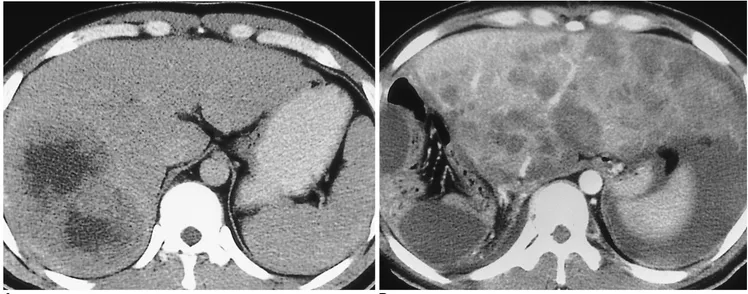
Fig. 1. A 31-year-old man with a large hepatocellular carcinoma in the right lobe who underwent right hepatic lobectomy (group I). A. CT scan obtained during the delayed phase shows ill-defined hepatocellular carcinoma without capsule.
B. CT scan obtained 2 months after surgery reveals that multiple nodular hepatocellular carcinoma in the entire liver had replaced about 70 80% of parenchyma.
rind of the same thickness during delayed phase CT. The distinctness of the tumor margin was visible on CT images obtained during the portal venous and delayed phases.
To study the relationship between the CT findings and recurrence, the Student t test, Fisher s exact test, and the Chi-Square test were used.
RESULTS
The CT findings of HCC in groups I and II are summa-rized in Table 1. Tumor size ranged between 2.1 and 18.0 (mean, 6.8) cm in group I, and between 1.0 and 10.0 (mean, 4.3) cm in group II. Forty-seven of the 53 patients had a single tumor, while among the remaining six, three in group I had massive-type HCC (a large single mass occu-pying a substantial portion of a hepatic lobe) and the other three, also in group I, had two tumors.
Among the 27 patients in group I, a tumor capsule was demonstrated in five (19%) (Fig. 1). In one patient in this group with two HCCs, one mass showed a capsule but the other did not; we therefore considered that the HCCs of this patient did not have a capsule. In contrast, among the 26 patients in group II, tumor capsules were seen in 16 (62%) (Fig. 2), and there was thus a statistically significant difference between the two groups (p = .001).
The tumor margin was distinct in eight of the 27 patients in group I (Fig. 3) and indistinct in the remaining 19, whereas it was distinct in 20 of the 26 patients in group II and indistinct in the remaining six. This difference was also statistically significant (p = .001).
Radiological evidences of perinodular extension into ad-jacent hepatic parenchyma was demonstrated in five pa-tients (19%) in group I and three (12%) in group II. Portal vein thrombosis was present in six patients (22%) in group I and one (4%) in group II (Fig. 4). These differences were not statistically significant ( p > .05).
DISCUSSION
Recent advances in imaging technologies have facilitated the detection of small HCCs, thus increasing the resectabil-ity rate. The postoperative long-term survival rate, howev-er, is far from satisfactory (3 5). To improve the surgical outcome, it is important to elucidate, before surgery, the factors which indicate risk of recurrent HCC. Many investi-gators have already reported that tumor size and number, the presence of capsule, satellite nodule, portal vein inva-sion, cirrhosis, and positive surgical margin may indicate risk of intrahepatic tumor recurrence (3, 5, 10, 11, 14, 15), but these factors were based on pathological findings. This study was designed to assess the usefulness of preoperative
Fig. 2. A 59-year-old man with nodular hepatocellular carcinoma in the right lobe who underwent right hepatic lobectomy 2 years and 6 months ago. Since this time he has been well, with no evi-dence of recurrence (group II). A CT scan obtained during the de-layed phase shows a well-defined, thin, enhancing capsule ar-ound the mass (arrowheads).
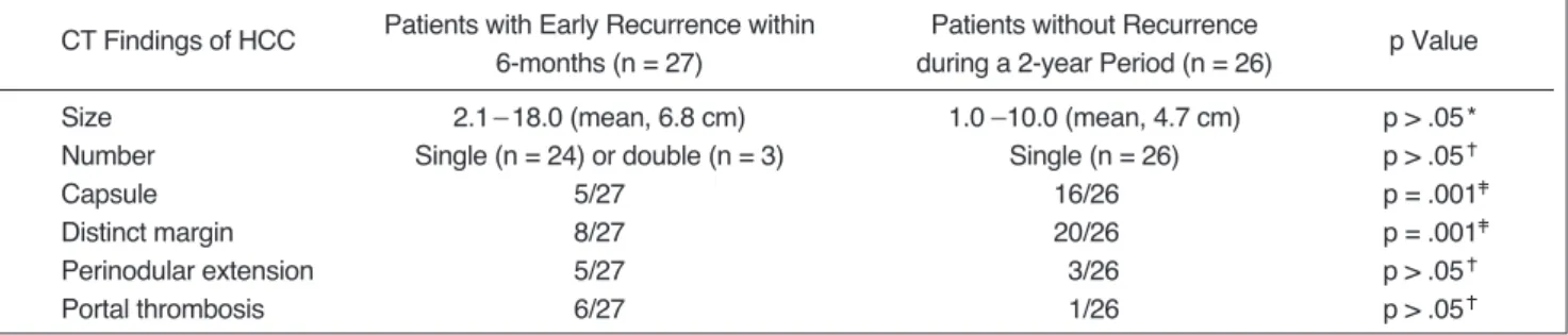
Table 1. CT Findings in Patients with Early Recurrence (within 6 months) and without Recurrence during a 2-year Period CT Findings of HCC Patients with Early Recurrence within Patients without Recurrence p Value
6-months (n = 27) during a 2-year Period (n = 26)
Size 2.1 18.0 (mean, 6.8 cm) 1.0 10.0 (mean, 4.7 cm) p > .05*
Number Single (n = 24) or double (n = 3) Single (n = 26) p > .05
Capsule 5/27 16/26 p = .001
Distinct margin 8/27 20/26 p = .001
Perinodular extension 5/27 3/26 p > .05
Portal thrombosis 6/27 1/26 p > .05
Note. Fisher s exact test
Chi-Square test * Student t test
CT findings in determining which patients may benefit from surgical versus nonsurgical therapies.
Shirabe et al. (10) reported that as tumor size increased, so did the rate of tumor spread. In our study, although mean tumor size was slightly greater in group I, there was no significant linkage between tumor size and early recur-rence. It has been reported that the number of tumors at the time of surgery is linked to early recurrence (11), but our study showed that there was slightly more chance of recurrence in patients with more than one tumor in the same lobe of the liver (not statistically significant).
It has been claimed that the presence or absence of fi-brous capsule surrounding an HCC is closely related to the rate of postoperative recurrence (10, 11, 16). By blocking the spread of tumor cells, the capsule may prevent tumor extension. Ueda et al. (17) confirmed radiologically and pathologically the vascular connection between tumor si-nusoid and very thin, slit-like vessels in the inner layer of fibrous capsule, with subsequent connection with the thick-er portal venules in the outthick-er laythick-er of fibrous capsule, and finally with the adjacent hepatic sinusoid. Blood from an HCC thus drains into adjacent liver parenchyma through the capsular portal vein. On the basis of this investigation it is believed that an intact capsule prevents tumor spread outside the confines of the tumor; the slit-like vascular space within the capsule prevents tumor propagation out-side of the capsule. When this is invaded, the tumor may spread through the capsular portal vein to the adjacent liv-er, archiving perinodular extension or distant metastasis through the portal and hepatic veins (10). An alternative
explanation is that HCC within a capsule is a histologically less aggressive lesion.
In this study, an indistinct HCC margin, as seen on CT, is thought to be closely correlated with a higher rate of recur-rence, and may represent permeative or replacement HCC growth outward to the hepatic parenchyma. In about 40 60 percent of cases, small HCCs (ranging in size from 2 to 4 cm) have a capsule. Although not proven pathologically, we hypothesize that HCC may become aggressive when the capsule is invaded and infiltrative growth occurs. Under such conditions, the tumor margin will subsequently be indistinct.
If a tumor is present in the portal vein at the time of sur-gery, this is known to be a significantly poor prognostic in-dicator (2, 9, 10, 18 20) and is considered to indicate risk of early recurrence. HCC quickly permeates the liver parenchyma through the portal venous system, spreading first within the liver itself and then into the hepatic vein and inferior vena cava and into the lung. The relationship between portal vein tumor thrombosis and early recur-rence may be explained in one of two ways: the presence of portal vein thrombosis, revealed by pathologic examina-tion, indicates possible hidden metastasis to a remote site of the hepatic parenchyma, though such evidence may not be visible on imaging studies; alternatively, manipulation of the liver at the time of surgery may facilitate the spread of tumor cells within the portal venous system (20). Nevertheless, our study did not reveal any statistically sig-nificant difference in the incidence of portal vein thrombo-sis between the two groups. This is because the number of Fig. 3. A 45-year-old man with a large hepatocellular carcinoma
in the right lobe who underwent right hepatic lobectomy 2 years and 6 months ago. Since this time he has been well, with no evi-dence of recurrence (group I). CT Scan obtained during the de-layed phase shows a low attenuating mass with an irregular but sharply circumscribed border. The central irregular area of low at-tenuation represents necrosis.
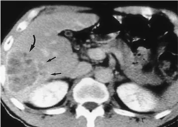
Fig. 4. A 60-year-old man with hepatocellular carcinoma in the right lobe who underwent posterior segmentectomy (group I). CT Scan obtained during the delayed phase shows an oval hepato-cellular carcinoma measuring 5 x 7 cm and two small perinodular extensions (straight arrows). Note disruption of the enhancing capsule (curved arrow).
cases was too small to be clinically relevant. Perinodular extension of HCC may be a sign of early recurrence, but in our study, also because of the small number of cases, this finding, too, was statistically insignificant.
In this study, sampling bias was inevitable. This was be-cause candidates for hepatic surgery were limited to pa-tients with good liver function (such as those with Child-Pugh classification A), patients with solitary or bitumoral nodular HCC, those with no portal vein thrombosis or ex-tensive perinodular tumor growth, and those with no evi-dence of extrahepatic metastasis. In our study, all but two HCCs were of the nodular type, and were clearly local-ized; the exceptions were cases with massive-type HCC. Patients in this study are thus not representative of those with HCC encountered in daily practice. Our reason for choosing patients with recurrence within six months and those without recurrence for two years was that we as-sumed that such intervals can guide clinicians in their choice of treatment options, namely surgery or nonsurgical methods such as transarterial chemoembolization and/or alcohol injection therapy.
In conclusion, the absence of tumor capsule and indis-tinct margin are the CT findings which most usefully pre-dict early recurrence after surgical resection of hepatocellu-lar carcinoma. Observation of these findings in conjunction with clinical data will be useful for the prediction of early recurrence, thus guiding clinician in his or her choice of treatment.
References
1. Kanematsu T, Takenaka K, Matsumata T, Furuta T, Sugimachi K, Inokuchi K. Limited hepatic resection effective for selected cirrhotic patients with primary liver cancer. Ann Surg 1984;199: 51-56
2. Izumi R, Shimizu K, Ii T, et al. Prognostic factors of hepatocellu-lar carcinoma in patients undergoing hepatic resection.
Gastroenterology 1994;106:720-727
3. Yamamoto M, Matsuda M, Iimuro Y, Fuji H, Nagahori K, Ainota K. Intrahepatic distant metastasis and metachronous multicentric occurrence in solitary hepatocellular carcinoma of less than five centimeters in diameter. Surg Today 1993;23:969-978
4. Nagao T, Inoue S, Yochimi F, et al. Postoperative recurrence of hepatocellular carcinoma. Ann Surg 1989;211:28-33
5. Chen M-F, Whang T-L, Jeng L-B, Wang C-S, Jan Y-Y, Chen
S-C. Postoperative recurrence of hepatocellular carcinoma: two hundred five consecutive patients who underwent hepatic resec-tion in 15 years. Arch Surg 1994;129:738-742
6. Belghiti J, Panis Y, Farges O, Benhamou JP, Fekete F. Intrahe-patic recurrence after resection of hepatocellular carcinoma complicating cirrhosis. Ann Surg 1991;214:114-118
7. Yamashita Y, Takahashi M, Koga Y, et al. Prognostic factors in the treatment of hepatocellular carcinoma with transcatheter ar-terial embolization and arar-terial infusion. Cancer 1991;67:385-391
8. Dusheiko GM, Hobbs KEF, Dick R, Burroughs AK. Treatment of small hepatocellular carcinoma. Lancet 1992;340:285-288 9. The Liver Cancer Study Group of Japan. Predictive factors for
long-term prognosis after partial hepatectomy for patients with hepatocellular carcinoma in Japan. Cancer 1994;74:2772-2780 10. Shirabe K, Kanematsu T, Matsumata T, et al. Factors linked to
early recurrence of small hepatocellular carcinoma after hepate-ctomy: univariate and multivariate analysis. Hepatology 1991; 14:802-805
11. Nagasue N, Uchida M, Makino Y, et al. Incidence and factors as-sociated with intrahepatic recurrence following resection of he-patocellular carcinoma. Gastroenterology 1993;105:488-494 12. Ouchi K, Matsubara S, Fukuhara K, Tominaga T, Matsuno S.
Recurrence of hepatocellular carcinoma in the liver remnant af-ter hepatic resection. Am J Surg 1993;166:270-273
13. Okada S, Shimada K, Yamamoto J, et al. Predictive factors for postoperative recurrence of hepatocellular carcinoma.
Gastro-enterology 1994;106:1618-1624
14. Franco D, Capussotti L, Smadja C, et al. Resection of hepatocel-lular carcinoma: results of 72 European patients with cirrhosis.
Gastroenterology 1990;98:733-738
15. Lai EC-S, Ng IO-L, Lok AS-F, et al. Long-term results of resec-tion for large hepatocellular carcinoma: a multivariate analysis of clinicopathological features. Hepatology 1990;11:815-818 16. Nagasue N. Liver resection for hepatocellular carcinoma:
indica-tions, techniques, complicaindica-tions, and prognostic factors.
Hepatobiliary Pancreat Surg 1998;5:7-13
17. Ueda K, Matsui O, Kawamori Y, et al. Hypervascular hepatocel-lular carcinoma: evaluation of hemodynamics with dynamic CT during hepatic arteriography. Radiology 1998;206:161-166 18. Nakashima T. Vascular changes and hemodynamics in
hepato-cellular carcinoma. In Okuda K, Peters RL. eds. Hepatohepato-cellular carcinoma. New York: Wiley Medical, 1987:169-203
19. Matsumata T, Kanematsu T, Tabenaka K, Yoshida Y, Nishizaki T, Sugimachi K. Patterns of intrahepatic recurrence after cura-tive resection of hepatocellular carcinoma. Hepatology 1989; 9:457-460
20. Okada S, Shimada K, Yamamoto J, et al. Predictive factors for postoperative recurrence of hepatocellular carcinoma.