Comparative Quantification of Contractile
Force of Cardiac Muscle Using a Micro-mechanical
Force Sensing System
Seokchang Ryu, Sukho Park, Deok-Ho Kim, and Byungkyu Kim*
Microsystem Research CenterKorea Institute of Science and Technology P.O.Box 131, Cheongryang, Seoul 103-650 Korea
{ seok, shpark, kim-dh, bkim}@kist.re.kr Abstract – To facilitate the cell based robot research, we
presented a micro-mechanical force measurement system for the biological muscle actuators, which utilize glucose as a power source for potential application in a human body or blood vessels. The system is composed of a micro-manipulator, a force transducer with a glass probe, a signal processor, an inverted microscope and video recoding system. Using this measurement system, the contractile force and frequency of the cardiac myocytes were measured in real time and the magnitude of the contractile force of each cardiac myocyte on a different condition was compared. From the quantitative experimental results, we estimated that the force of cardiac myocytes is about 20~40 µN, and showed that there is difference between the
control cells and the micro-patterned cells.
Index Terms – Cardiac myocytes, Cell force measurement, Micro-manipulation, Piezo resistive sensor.
I. INTRODUCTION
Over the last ten years, a novel concept of the Microrobot (or nanorobot) have been introduced [1][2][3]. Because of the size limitation of the robot with bio-mimetic actuation, however, the selection of the proper actuator is not easy and the realization of the robot is very difficult. In general, the electrostatic, electromagnetic, pneumatic, piezoelectric and thermal forces are mostly used as a micro actuator. These forces need external power source and have some limitations in the application of remote operation, such as locomotion in human digestive organ or blood vessels.
As an alternative, the cell based actuators are proposed and the miniature robot is also presented [4]. The cell based actuator is fuelled by a simple glucose nutrient in physiological fluids as an energy source and transforms the chemical energy into mechanical energy. In the presented micro-robots [4], as a structural backbone, an arch of silicon 50 micrometers wide is used and a cord of rat heart muscle fibers has been grown. Contraction and relaxation of the heart muscle makes the arch bend and stretch to produce a crawling motion.
In order to design the micro robot powered by living heart muscle, the estimation of the contractile force is very important. From the measured contraction force, the shear force which is generated by living and cultured cells is estimated and thus is utilized in the design of the microrobot
backbone. That is, by using the measured force, the motion of the microrobot structure can be simulated and thus the microrobot can be re-designed. The measurement methods of the cell’s contractile force are as follows.
First of all, there is a feasibility test of isometric force development in single cardiac myocytes from human ventricular muscle tissue obtained from small biopsies taken during open heart surgery [5]. This paper proposes the measurement method for the force of isolated cardiac myocytes with silicon glue to a sensitive force transducer and a piezoelectric motor. It was reported that the average isometric force at saturating calcium concentration obtained on 20 myocytes is about 51 kN/m2 and mechanical properties
of myocytes are correlated with the protein composition. Based on moving a magnetic bead, a single cardiac myocyte contractile force measurement technique is proposed [6]. A magnetic bead is attached on one end of the myocyte and adjusted magnetic field is applied on the magnetic bead. Using an inverted microscope with edge detection, the myocyte contractile force can be derived by the measuring of the maximal displacement of cell contraction and the magnetic field loading force on the bead. From this report, the estimated contraction force is about 10 µN.
The cell force measurement method based on microelectromechanical system (MEMS) technology is proposed in [7]. By using MEMS processes, two transducer beams with clamper and hinge are fabricated and one of two beams incorporates a strain gauge. The heart cell ends are attached to the transducer beams using clamps. Thus, when the cell is contracted, the strain is activated. The average measured maximal force was about 5.77 µN.
Elastic micro patterned substrates are used for cardiac myocyte force measurements in real time and in living cells [8][9]. The elastic micro posts are bent and the displacements of the posts are transformed to cell forces which can be calculated by a simple beam theory. From this method, the cell force is estimated about 600 nN.
In addition, a force measurements on living cells using atomic force microscopy (AFM) [10] and other methods [11] are presented. Although higher resolution can be achieved using AFM, a complex transmit-receive setup is required and in aqueous medium where cells survive, the reflection and
refraction of the transmitted light make the accuracy of cellular force measurement problematic [12].
In this paper, we propose the contractile force measurements of cardiac myocytes using a micro-manipulator. The measurements are carried in real time and the video images are also captured. Therefore, the experimental results show not only the magnitude of the cell force but also the frequency of the cell mobility. From the results of the cell force measurements, that is, the static characteristics (maximum displacement, tensile stress, etc) of the microrobot can be analyzed and the dynamic analyses (velocity, acceleration, friction, etc) are also possible.
We measured the contractile force of not only control cardiac myocytes but also micro-patterned cardiac myocytes. The motivation of this study is to improve the performance of such cell based actuators with regard to force and displacement by controlling beating activity of patterned cardiac myocytes. The micro-patterned cardiac myocytes unit can be also applied as a flexible platform for drug screening, cell-based sensors, and self-organizing cell motors.
The paper is organized as follows: In the following chapter, the proposed cell force measurement system and it’s subsystems are explained. Chapter III will present the experimental procedures and the results of the cardiac myocytes force measurements. From the experimental results, we can verify the proposed force measurement system and find the variations of the cell force signals due to the change of the culturing environments. Finally, concluding remarks will be drawn in Chapter IV.
II. CELL FORCE MEASUREMENT SYSTEM
A. Description of the overall system
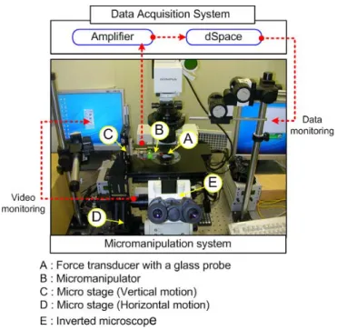
The overall schematic diagram of the force measurement system is illustrated in Fig. 1. The system consists of a micro-manipulator, a force transducer integrated with a glass probe, an inverted microscope, a signal amplifier, a force data acquisition system, and a video image recording system. By the micro-manipulator and the inverted microscope, when the end of glass probe approaches a cardiac cell, the beating force is transferred to probe and the force is sensed by the piezo-resistive force transducer. The force signal is amplified by the signal amp and the amplified cardiac beating force signal is A/D converted and recorded by dSPACE data acquisition system.
B. Micro-manipulation System & Force Transducer
The manipulation system consists of a micro-manipulator (Model: MM3A of Nanotek) controlled by its own controller and two micro stages operated manually. For the cell force measurement, a piezo-resistive type sensor (Model: AE801 of SensorOne) with a glass probe is used. With a precision load cell (Model: GSO-10 of Transducer technology Inc., Max. measurement range: 100mN, resolution: 10 µΝ) the calibration process was executed to convert the voltage signal of force transducer to realistic force
Fig. 1 Schematic diagram of cellular force measurement system. value. The calibration result, which is shown in Eq. 1, verifies that the transducer has a good linearity and the calibration factor is about 841 µΝ/V.
Force [µΝ] = 840.9 ⅹ Transducer Output [V] (1) C. Force Signal Processing
The force signal from the transducer is amplified by the signal amp (Model: 2301A of VISHAY) and the amplified force signal is A/D converted, filtered through a low-pass filter with cut-off frequency of 10Hz, and recorded by dSPACE data acquisition system. The ratio of amplification in the signal amp is about 10000, and the A/D converter of dSPACE has 16bit resolution and ±10 voltage range. And thus, the resolution of the force measurement system is about 0.257 µΝ. In general, it is reported that the contractile force level range of the cardiac cell is about 10~20 µΝ. Therefore, the resolution of the force measurement system is sufficiently small.
III. EXPERIMENTAL PROCEDURE AND RESULTS
A. Cell Preparation
Primary cardiac myocytes were isolated from postnatal day 1 Sprague-Dawley rat and cultured in Dulbecco’s modified Eagles’ medium (DMEM) (Gibco Invitrogen Co., Grand Island, NY, USA) supplemented with 10% Fetal Bovine Serum (Gibco Invitrogen) at 37°C in 5% CO2. The
isolated cardiac myocytes were directly seeded on the surface at a concentration of 1 x 10⁵ cells/cm2 and maintained for 7
days.
B. Cell Patterning into Microwells
We developed a simple method for patterning cardiac myocytes confined within microwells that contain a collagen-coated surface and a poly (ethylene glycol) (PEG) copolymer barrier.
Capillary lithography [13] was used to coat the PEG copolymer on glass substrate and to generate micro arrays for patterning cardiac myocytes. A few drops of 10% (w/v) PEG copolymer [poly(TMSMA-r-PEGMA)] [14] solution in ethanol were placed on a glass substrate. A thin film of the PEG polymer was achieved by spin coating (Model CB 15, Headaway Research, Inc., USA) at 1000 rpm for 10s. To make conformal contact, patterned PDMS molds were carefully placed onto the surface and then the molds were peeled off using a sharp tweezers after complete evaporation of the solvent for 30 min at room temperature. Prior to cell plating, collagen (Type I, Sigma Chemical Co., St. Louis, MO, USA) was coated for 3 h within the microwells on the PEG layer prepared on the glass substrate to enhance cell attachment to and survival in the microwell patterns.
By doing so, collagen was selectively absorbed on the exposed glass substrate to produce pre-patterned microwells. Cardiac myocytes suspended in medium were then plated onto the surfaces and the cell cultures were analyzed at intervals. After 20h, non-adhered cells present on the PEG layer were washed away with Hank’s balanced salt solution (HBSS, pH 7.4) (Gibco Invitrogen). Finally, cells were selectively attached on collagen-coated microwell surfaces due to an adhesion- resistant PEG surface.
C. Experimental Procedure
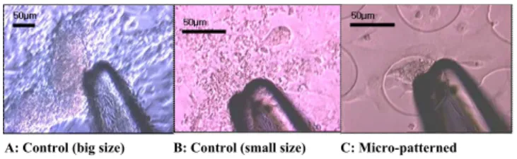
The experiments are executed for three cases: (a) big size control myocytes, (b) small size control myocytes, and (c) micro-patterned myocytes. First of all, among control cardiac myocytes groups, we choose the largest beating displacement group. In this group, many cardiac myocytes are aggregated and have a vivid beating feature. Then, the small size myocytes group is selected and its contractile force is also measured. Finally, by the proposed cell patterning method, the force measurements of micro-patterned cardiac myocytes are executed. To get the force signal from the cell, it is very important that the glass probe is aligned with the contraction direction of the target cell. If the glass probe is perpendicularly positioned with the direction of the cell’s contraction, the glass probe is just bent such as a simple beam and cannot transfer the cell force to the force transducer.
D. Experimental Results
As shown in Fig. 3, the glass tip is adjacent to the target cardiac myocytes. In addition, among three cases, (a) big size control myocytes and (c) micro-patterned myocytes aggregates and have some 3D structures. But, (b) small size control myocytes spread out and it seems like 2D planar
structure. Through the inverted microscope, we can know that each myocyte become synchronous and simultaneously beat.
A: Control (big size) B: Control (small size) C: Micro-patterned
Fig. 3 Photograph of cell contractile force measurement
The contractile force signals of the cardiac myocytes are shown in Fig. 4 and the statistics and the comparisons of the force magnitudes are presented in Table. 1. Firstly, (a) big size control myocytes have a contractile force about 35~40 µΝ and a beating frequency about 28~30 pulses/min. Secondly, in (b) small size control myocyte, a contractile force is about 15 µΝ and a beating frequency is about 40~42 pulses/min. This difference may be due to the strongly synchronized beating of three dimensionally aggregated large size control myocytes. Thirdly, the micro-patterned myocytes show a contractile force about 20 µΝ and a beating frequency is about 19~20 pulses/min. Asuming that the myocytes in Fig. 3 (c) are covered with a half of the well area, the force per unit area can be estimated as about 9.05 kN/m2, where the diameter of the
micro well is 75µm. Our quantitative experimental results indicates that the contractile force of cardiac cells is about 20-40 μN, and there are differences in the magnitude of the contractile force between on the control cells and the micro-patterned cells.
Condition Contractile Force
Control (big size) 15.4 ± 2.20 μ N Control (small size) 17.6 ± 0.87 μ N
Micro-patterned 36.9 ± 0.85 μ N
Table 1 Results of cell contractile force measurement
Fig. 4 Signal of contractile force of cardiac myocytes
E. Discussion
In these experiments, we could measure the contractile force of the some cell culture conditions (control and micro-patterned) and the differences of the force signals for each conditions. The results show the magnitude of the cell force measured in real time and can be used for the dynamic analysis of the cell mobility.
However, because the number of cells is not quantitative and the times of experiments are also lack, the comparisons of the results are impossible. Therefore, the repetitive experiments of the cell force measurements, the considerations of the cell quantities and the comparisons of the experimental results are necessary.
In the view of the quantitative analysis, we believe that the micro-patterned myocytes can be used as a proper tool for the contractile force measurements of cardiac myocytes. This is why the number of cells in the micro-well has small variations compared with a control myocytes group. Because the micro-patterned myocytes aggregates, for the same number of the myocytes, we also expect that the contractile force of the micro-patterned cells is larger than that of the control cells. Therefore, we are going to study for the characteristics of the micro-patterned cardiac myocytes.
In addition, if the micro patterns were not a well type but a line type (or a narrow beam type), the cardiac myocytes will be aligned along the longitudinal direction and the contractile force will be also amplified. These aligned myocytes will be applied to the design of the cell based microrobot. That is, the aligned cells will have lager force compared with general semi-sphere type control myocytes and thus it will be expected that the microrobot which is powered by the aligned cardiac myocytes can have large deformation and actuation forces.
IV. CONCLUSION
We proposed the micro-manipulation system for the contractile force measurements of cardiac myocytes. The force measurements system consists of a nano-manipulator, a force transducer, a glass probe, an inverted microscope, a signal amplifier, a force data acquisition system, and a video image recording system. By using this measuring system, the contractile force for control cardiac cells and micro-patterned cardiac myocytes can be measured in real time. From the experimental results, we can estimate that the contractile force of cardiac cells is about 20-40 µN and there are differences between the control cells and the micro-patterned cells.
ACKNOWLEDGMENT
This work was supported by the 21st Century's Frontier R&D Projects, under the contract number MS-02-324-01, sponsored by the Ministry of Science and Technology, Korea.
REFERENCES
[1] http://www.foresight.org, Foresight Institute, USA.
[2] L. Phee, D. Accoto, A. Menciassi, C. Stefanini, M.C. Carrozza, and P. Dario, “Analysis and development of locomotion devices for the gastrointestinal tract,” IEEE Trans. Biomedical Engineering , vol. 49, no. 6, pp. 613 – 616, 2002.
[3] J. Jung, B. Kim, Y. Tak, J. Park, “Undulatory tadpole robot (TadRob) using ionic polymer metal composite (IPMC) actuator,” Int. Conf.
Intelligent Robots and Systems, vol. 3, pp. 2133 – 2138, 2003.
[4] J. Xi, J. Schmidt, and C. Montemagno, “First self-assembled micro-robots powered by muscle,” SPIE Nanotechnology e-bulletin, 6, 2004.
[5] J. van der Velden, et al., “Force production in mechanically isolated cardiac myocytes from human ventricular muscle tissue,” Cardiovascular
Research, 38, pp. 414-423, 1998.
[6] S. Yin, X. Zhang, C. Zhan, J. Wu, and J. Cheung, “Measuring single cardiac myocyte contractile force via moving a magnetic bead,” Biophys
J. BioFAST, 2004.
[7] G. Lin, E. Palmer, K. Pister, and K. Roos, “Miniature Heart Cell Force Transducer System Implemented in MEMS Technology,” IEEE Trans.
Biomedical Engineering, vol. 48, no. 9, pp. 996-1006, 2001
[8] N. Balaban, et al., “Force and focal adhesion assembly: a close relationship studied using elastic micro patterned substrates,” Nature Cell
Biology, vol. 3, pp. 466-472, 2001.
[9] Y. Zhao, H. Yu, and X. Zhang, “Creating Polymer-based Microstructures with Various Aspect Ratios from a Single Template for Cellular Force Measurements,” Proceeding of the 18th IEEE International Conference
on Micro Electro Mechanical Systems (MEMS '05), Miami Beach, FL,
USA, January 30 - February 3, 2005, in press.
[10] E. Wojcikiewicz, X. Zhang, and V. Moy, “Force and Compliance Measurements on Living Cells Using Atomic Force Microscopy (AFM),”
Biological Procedures Online, vol. 6, no. 1, pp. 1-9, 2004
[11] J. van Vilet, G. Bao, and S. Suresh, “The biomechanics toolbox; experimental approaches for living cells and biomolecules,” Acta
Materialia, 51, pp. 5881-5905, 2003.
[12] Yu Sun, Bradley J. Nelson, “MEMS for cellular force measurements and molecular detection,” International Journal of Information Acquisition, vol. 1, no. 1, pp. 23-32, 2004.
[13] A. Khademhosseini, S. Jon, K. Y. Suh, T. T. Tran, G. Eng, J. Yeh, J. Seong, and R. Langer, “Direct patterning of protein- and cell-resistant polymeric monolayers and microstructures,” Advanced Materials, vol. 15, pp. 1995-2000, 2003.
[14] S. Jon, J. Seong, A. Khademhosseini, T. T. Tran, P. Laibinis, and R. Langer, “Construction of nonbiofouling surfaces by polymeric self-assembled monolayers,” Langmuir, vol. 19, pp. 9989-9993, 2003.