저작자표시-비영리-변경금지 2.0 대한민국 이용자는 아래의 조건을 따르는 경우에 한하여 자유롭게 l 이 저작물을 복제, 배포, 전송, 전시, 공연 및 방송할 수 있습니다. 다음과 같은 조건을 따라야 합니다: l 귀하는, 이 저작물의 재이용이나 배포의 경우, 이 저작물에 적용된 이용허락조건 을 명확하게 나타내어야 합니다. l 저작권자로부터 별도의 허가를 받으면 이러한 조건들은 적용되지 않습니다. 저작권법에 따른 이용자의 권리는 위의 내용에 의하여 영향을 받지 않습니다. 이것은 이용허락규약(Legal Code)을 이해하기 쉽게 요약한 것입니다. Disclaimer 저작자표시. 귀하는 원저작자를 표시하여야 합니다. 비영리. 귀하는 이 저작물을 영리 목적으로 이용할 수 없습니다. 변경금지. 귀하는 이 저작물을 개작, 변형 또는 가공할 수 없습니다.
Master's Thesis in Medicine
Adductor canal block versus femoral nerve block combined
with sciatic nerve block as an anesthetic technique for
hindfoot and ankle surgery:
A prospective, randomized non-inferiority trial
Ajou University Graduate School
Major in Medicine
Adductor canal block versus femoral nerve
block combined with sciatic nerve block as an
anesthetic technique for hindfoot and ankle
surgery:
A prospective, randomized non-inferiority trial
Young Uk Park, M.D., Ph.D., Advisor
I submit this thesis as the Master's thesis in Medicine.
February 2017
Ajou university Graduate School
Major in Medicine
The Master's thesis of Ji-Sang Yoon in Medicine is
hereby approved.
Thesis Defense Committee President
Young Uk Park
Member Jae Ho Cho
Member Doo Hyung Lee
Ajou University Graduate School
January 4th, 2017
i
ABSTRACT
Background:
A femoral nerve block (FNB) in combination with a sciatic nerve block (SNB) is
commonly used for anesthesia and analgesia in patients undergoing hindfoot and ankle surgery. The
effects of FNB on motor function, related fall risk, and rehabilitation are controversial. An adductor
canal block (ACB) potentially spares motor fibers in the femoral nerve, but the comparative effect
on hindfoot and ankle surgeries between the two approaches is not yet well defined. We
hypothesized that compared to FNB, ACB would cause less weakness in the quadriceps and produce
similar pain scores during and after the operation.
Methods:
Sixty patients scheduled for hindfoot and ankle surgeries (arthroscopy, Achilles tendon
surgery, or medial ankle surgery) were stratified randomized for each surgery to receive an FNB
(FNB group) or an ACB (ACB group) combined with an SNB. The primary outcome was the visual
analog scale (VAS) pain score at each stage. Secondary outcomes included quadriceps strength, time
profiles (duration of the block procedure, time to full anesthesia and time to full recovery), patients’
analgesic requirements, satisfaction, and complications related to peripheral nerve blocks such as
falls, neurologic symptoms, and local anesthetic systemic toxicity were evaluated. The primary
outcome was tested for the non-inferiority of ACB to FNB, and the other outcomes were tested for
the superiority of each variable between the groups.
Results:
31 patients received an ACB and 29 received a FNB. The VAS pain scores of the
ACB group were not inferior during and after the operation compared to those of the FNB
group. At 30 minutes and 2 hours after anesthesia, patients who received an ACB had
significantly higher average dynamometer readings than those who received a FNB
(34.2±20.4 and 30.4±23.7 vs. 1.7±3.7 and 2.3±7.4, respectively), and the results were
similar at 24 and 48 hours after anesthesia. There were no differences between the two
groups with regard to time profiles and patient satisfaction. No complications were noted.
Conclusions:
ACB preserved quadriceps muscle strength better than FNB, without a significant
difference in postoperative pain. Therefore, ACB may be a good alternative to FNB for reducing the
potential fall risk.
ii
TABLE
OF
CONTENTS
LIST OF TEXT
ABSTRACT i TABLE OF CONTENTS ii I. INTRODUCTION 1 II. METHODS 2 III. RESULTS 5 IV. DISCUSSION 8 V. CONCLUSION 10 REFERENCES 11 국문요약 14LIST
OF
TABLES
TABLE 1. 6Demographics
TABLE 2. 6VAS pain Scores over time
TABLE 3. 7Motor strength over time
TABLE 4. 7Block performance as time intervals
T . 9ent
- 1 -
I
NTRODUCTIONThe use of regional anesthesia has gained popularity because of its positive effects on patient comfort and safety.[1, 2] Most foot and ankle operations are performed on an outpatient basis, often under some form of regional anesthesia. The use of this type of anesthesia is supported by successful outcomes reported in the literature.[3-5] Ankle block and sciatic nerve block (SNB) are two of the most popular anesthetic techniques used in foot and ankle surgery. Both have been shown to be safe and effective for these surgical procedures.[6, 7]
Blockade of the saphenous nerve is essential for surgeries that involve the medial aspect of the foot or ankle, for which a regional technique is preferred.[8-10] Numerous approaches to saphenous nerve blockade have been described using landmarks, nerve stimulation, and ultrasound.[11-13] Although a saphenous nerve block is appropriate for anesthesia and analgesia for hindfoot and ankle surgery, a more proximal block for the medial side of the hindfoot and ankle is necessary when the tourniquet is applied to the distal thigh for surgery. Recently, a study showed that femoral nerve block (FNB) provides good surgical anesthesia and good postoperative pain control for patients with hindfoot and ankle conditions.[3] However, FNB leads to femoral quadriceps muscle weakness.[14, 15]
Consequently, quadriceps weakness results in functional impairment, and it is associated with an increased risk of postoperative falls.[16-18] Thus far, attempts to reduce quadriceps involvement after FNB without compromising analgesia have not been successful.[15, 19]
Partially because of the increase in the use of ultrasound guidance, a more targeted approach that only blocks sensory fibers of the femoral nerve that supply the operative site has been attempted; thus, the feasibility of adductor canal block (ACB) has been studied.[20, 21] In recent years, ACB has been successfully used for postoperative pain control after knee surgery.[22, 23]
However, no randomized, control study has compared ACB to FNB for hindfoot and ankle surgeries. We hypothesized that compared to FNB, ACB would demonstrate non-inferior pain scores during surgery and until 48 hours postoperatively, and cause less weakness in the quadriceps.
- 2 -
II.
M
ETHODSThis prospective, randomized non-inferiority trial was performed at Ajou University Hospital, Suwon, Republic of Korea between August 2015 and May 2016, and it was approved by our
institutional ethics committee (approval no.: AJIRB-MED-MDB-15-181). The study design was also prospectively registered (http://cris.nih.go.kr, KCT0001624).
After obtaining written informed consent, we enrolled 60 adult patients who were scheduled to undergo surgery on the hindfoot, medial side of the ankle, or both sides of the ankle (Achilles tendon surgery, the removal of an implanted device on the medial side, or ankle arthroscopy) under peripheral nerve blocks (PNBs); had an American Society of Anesthesiologists Physical status of I–II; and were aged 19-65 years. Exclusion criteria were an inability to cooperate; a history of alcohol or drug abuse; those with rheumatoid arthritis, coagulation disorders, peripheral neuropathy, and known allergies to local anesthetics; and the recent use of opioids, corticosteroids, or any other analgesics. If a patient complained of incomplete block before the end of surgery, we excluded the patient from the analysis.
Randomization was based on a computer-generated block randomization list (four numbers per block) in a 1:1 ratio. All patients underwent SNB, and ACB (ACB group) or FNB (FNB group) according to a randomized assignment.
Anesthesia and postoperative analgesia
All patients were anesthetized with PNBs for surgery. PNBs were performed in a block room at least 1 hour preoperatively. All PNBs were performed under ultrasound guidance (LOGIQ P6, GE
Healthcare, Chicago, IL, USA) using a high-frequency linear transducer (3.4–10.8 MHz). All blocks were performed after skin preparation with 2% chlorhexidine gluconate by one orthopedic surgeon (YUP) with vast experience in ultrasound-guided nerve block techniques. A 22-gauge Tuohy needle was introduced in-plane and 2 to 3 mL of saline was used to confirm the correct position of the needle tip near the target nerve. The study medication was administered through the needle as a bolus of 15 mL of a 1:1 mixed solution of ropivacaine 0.75% and lidocaine 1%.
For the SNB, the patient was placed in the supine position with the knee of the affected limb flexed at about 30°. SNB was performed immediately proximal to the bifurcation of the sciatic nerve into the tibial and common peroneal nerves. The needle was advanced until the needle tip was positioned at the anterior and posterior external surface of the sciatic nerve in the anterior-posterior plane and at the
- 3 -
midpoint of the sciatic nerve in the mediolateral plane. The needle-tip position was adjusted as necessary to ensure the circumferential spread of study medication around the sciatic nerve.
For the ACB, the patient was placed in the supine position with the extremity to be blocked slightly externally rotated. The ultrasound transducer was placed in a transverse cross-sectional view at the midpoint of the inguinal crease and the medial femoral condyle on the medial thigh. The femoral artery and the saphenous nerve just lateral to the artery were identified underneath the sartorius muscle. After the needle tip was placed just lateral to the artery and saphenous nerve, the study medication was injected.
For the FNB, the femoral nerve was identified lateral to the femoral artery at the inguinal crease in the transverse section. The study medication was injected anterior and posterior to the nerve. The actual or estimated needle insertion sites of ACB and FNB were covered with a dressing bandage in all patients to blind those assessing the outcome of the procedure to the treatment. Intravenous ketorolac tromethamine (30 mg, maximum dose 90 mg/d) was given as a rescue analgesic during the first 48 hours postoperatively when a patient reported a VAS score of ≥5 or if the patient requested pain relief.
Assessment
The primary (non-inferiority) outcome was the visual analog scale (VAS) pain score at each stage. All patients were educated preoperatively regarding the use of a 10-cm VAS pain score (0 cm, no pain; 10 cm, worst pain). VAS pain scores were evaluated during the anesthetic procedure, during surgery, immediately postoperatively and at 2, 12, 24, and 48 hours postoperatively. Tourniquet pain during the operation was evaluated separately.
The secondary (superiority) comparative outcomes included motor strength of the quadriceps muscle, time profiles (i.e., the duration of the procedure, time to full anesthesia, recovery time, and operative time), postoperative analgesic requirements, patient satisfaction, and the incidence of complications.
The quadriceps strength was assessed as the maximum voluntary isometric contraction at 30 minutes, 2 hours, 24 hours, and 48 hours postoperatively compared to baseline (preoperatively). We used a handheld dynamometer (HHD; microFET2, Hoggan Industries, Salt Lake City, UT, USA) to measure muscle strength. The HHD is a reliable and valid instrument, and we used standardized, recommended procedures to obtain valid measurements.14,21 To evaluate the quadriceps muscle strength, we placed the patient in a seated position with the knees flexed 60°. We used a non-elastic strap with Velcro closures to fix the HHD to the leg. We attached the Velcro strap to a chair and around the patient’s ankle, perpendicular to the lower leg. The HHD was placed under the Velcro strap on the
- 4 -
anterior surface of the tibia (5 cm above the transmalleolar axis). We instructed the patient to take 3 seconds to reach and maintain maximum effort, and then relax. For each assessment, the patient performed three consecutive contractions, separated by a 30-second pause between each trial. We used the mean value at each time point to calculate muscle strength.
The duration of the procedure was defined as the time interval from needle insertion to pull out. Time to full anesthesia was defined when a patient verbally responded “no” when asked if cutaneous pressure or a pinprick was being applied to the area in question. The recovery time (time elapsed between the onset of anesthesia and full motor and sensory recovery) was also documented.
Additional data collected included the following: 1) the numbers and proportions of patients requiring a rescue analgesic during the first 48 hours; 2) patient satisfaction (each patient was interviewed using a scale of 1–5 (5 being the most satisfied) at 48 hours postoperatively); and 4) the incidence of complications related to a PNB (if any), including falls, neurologic symptoms, and local anesthetic toxicity..
Statistical analyses
The sample size determination was based on a recently completed trial[3] at our institution evaluating anesthetic and analgesic effects of sciatic and femoral nerve blocks in the hindfoot and ankle surgeries involving the medial side of ankle. We chose a non-inferior design to demonstrate that ACB was not inferior to FNB as an anesthetic and analgesic technique, while demonstrating the superiority of preservation of quadriceps motor strength after ACB compared to FNB. The sample size was determined by assuming a standard deviation of 1.47 cm and an upper end confidence limit of 1 cm. We calculated a sample size of 27 patients in each group with a predicted 10% dropout rate, totaling 60 patients. This would permit a type-1 error rate of 0.05, with a type-2 error rate of 0.20.
Data were analyzed using SPSS version 13.0 (SPSS Inc., Chicago, IL). Data are presented as the mean±standard deviation, median (interquartile range) or the number (percentage) of patients. For the primary outcome variable, the non-inferiority of ACB versus FNB was demonstrated if the upper 95% confidence interval limit of the difference between the groups excluded the defined non-inferiority margin of 1 cm. We compared secondary outcomes using a t-test for parametric variables, a Mann-Whitney U test for non-parametric variables, and either a chi-square or Fisher exact test for categorical data. A P value <0.05 was considered statistically significant.
- 5 -
III.
R
ESULTSWe enrolled 60 patients during a 12-month period beginning in June 2015. Patients were randomized to receive either ACB (n=31) or FNB (n=29) (Table 1). The PNBs used in this study were the sole anesthetic techniques used for hindfoot and ankle surgery in 59 patients. One patient in the FNB group complained of incomplete block; therefore, this patient received general anesthesia. Surgical
procedures performed in this study were arthroscopy (n=32), Achilles tendon surgery (n=9), and medial ankle surgery (medial malleolar old fracture, n=19).
The VAS pain scores were comparable between the two groups (Table 2). The upper confidence limits of VAS pain scores were less than the δ at every stage, except the block procedure. Therefore, the ACB was not inferior to the FNB with regard to the pain score during and after hindfoot and ankle surgery.
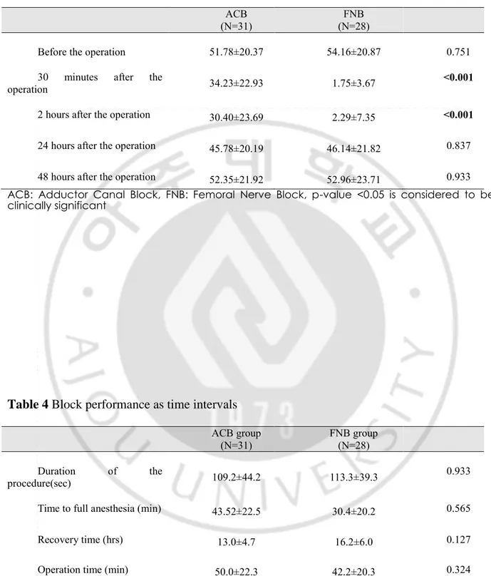
Table 3 presents data for the motor strength of the quadriceps muscle over time. We compared dynamometer readings between the groups at each time point. At 30 minutes and 2 hours
postoperatively, the mean strength during extension of the knee was significantly higher in the ACB group than in the FNB group (ACB/FNB: 34.23±22.93 vs. 1.75±3.67 at 30 minutes postoperatively and 30.40±23.69 vs. 2.29±7.35, respectively (P<.001). At 24 and 48 hours, there was no significant difference between the ACB and FNB groups, P values=.84 and .93, respectively.
The time profiles (duration of the procedure, time to full anesthesia, operative time, and recovery time) were not different between the two groups (Table 4).
Eleven patients were very satisfied, 41 were satisfied, and 6 were fairly satisfied, and no patients were unsatisfied. Patient satisfaction was similar for both types of nerve blocks (P=.80). Eight patients complained of temporary numbness in their operated leg 24 hours postoperatively (ACB: 3, FNB: 5, P=.46) However, their symptoms completely recovered without specific management.
- 6 -
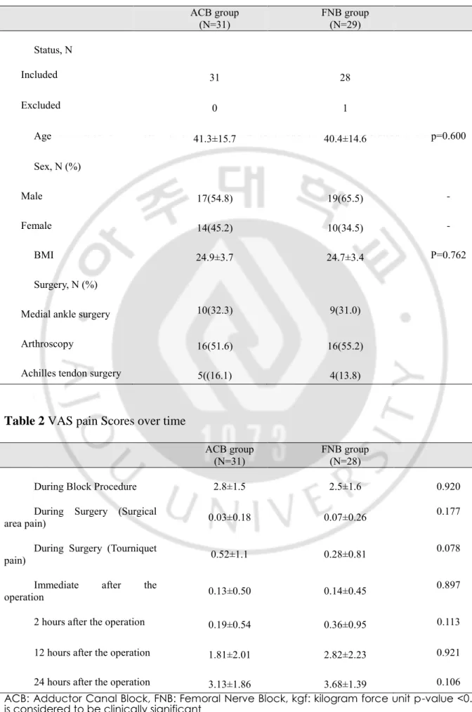
Table 1 Demographics
ACB group (N=31) FNB group (N=29) Status, N Included 31 28 Excluded 0 1 Age 41.3±15.7 40.4±14.6 p=0.600 Sex, N (%) Male 17(54.8) 19(65.5) - Female 14(45.2) 10(34.5) - BMI 24.9±3.7 24.7±3.4 P=0.762 Surgery, N (%)Medial ankle surgery 10(32.3) 9(31.0)
Arthroscopy 16(51.6) 16(55.2)
Achilles tendon surgery 5((16.1) 4(13.8)
Table 2 VAS pain Scores over time
ACB group
(N=31) FNB group (N=28)
During Block Procedure 2.8±1.5 2.5±1.6 0.920
During Surgery (Surgical
area pain) 0.03±0.18 0.07±0.26 0.177
During Surgery (Tourniquet
pain) 0.52±1.1 0.28±0.81 0.078
Immediate after the
operation 0.13±0.50 0.14±0.45 0.897
2 hours after the operation 0.19±0.54 0.36±0.95 0.113
12 hours after the operation 1.81±2.01 2.82±2.23 0.921
24 hours after the operation 3.13±1.86 3.68±1.39 0.106 ACB: Adductor Canal Block, FNB: Femoral Nerve Block, kgf: kilogram force unit p-value <0.05 is considered to be clinically significant
- 7 -
Table 3 Motor strength over time
ACB
(N=31) (N=28) FNB
Before the operation 51.78±20.37 54.16±20.87 0.751
30 minutes after the
operation 34.23±22.93 1.75±3.67 <0.001
2 hours after the operation 30.40±23.69 2.29±7.35 <0.001
24 hours after the operation 45.78±20.19 46.14±21.82 0.837
48 hours after the operation 52.35±21.92 52.96±23.71 0.933 ACB: Adductor Canal Block, FNB: Femoral Nerve Block, p-value <0.05 is considered to be clinically significant
Table 4 Block performance as time intervals
ACB group (N=31) FNB group (N=28) Duration of the procedure(sec) 109.2±44.2 113.3±39.3 0.933
Time to full anesthesia (min) 43.52±22.5 30.4±20.2 0.565
Recovery time (hrs) 13.0±4.7 16.2±6.0 0.127
Operation time (min) 50.0±22.3 42.2±20.3 0.324
ACB: Adductor Canal Block, FNB: Femoral Nerve Block, sec: seconds, min: minutes, hrs: hours, p-value <0.05 is considered to be clinically significant
- 8 -
IV.
D
ISCUSSIONThis prospective study demonstrated that ACB is an effective alternative to FNB for patients undergoing hindfoot and ankle surgery. The most important finding of this study was that ACB
significantly spared quadriceps strength at 30 minutes to 2 hours post-surgery, and it was not inferior to FNB in terms of pain scores, analgesic requirements, and time profiles.
For intraoperative anesthesia or postoperative pain control after hindfoot and ankle surgery, a
popliteal SNB is an effective method when the surgery does not involve the medial side of the hindfoot and ankle.[6, 7, 9] When the surgery involves the medial side of the hindfoot and ankle, the saphenous nerve should be blocked to ensure proper anesthesia or analgesia in combination with an SNB.
Although FNB can be used for the saphenous nerve block,[3, 24] ACB or a saphenous nerve block instead of an FNB is sufficient anesthesia or analgesia for hindfoot and ankle surgery. Several studies have already validated ACB as an effective analgesic method compared to an FNB, as it spares quadriceps muscle strength, but most studies have investigated arthroscopic knee surgery or total knee replacement.[25-27] This advantage of ACB over FNB was also demonstrated beyond knee surgery in our study, which is the first study to directly compare multiple outcomes of ACB versus FNB in patients undergoing hindfoot and ankle surgery.
The non-inferiority of VAS pain scores after hindfoot and ankle surgery with ACB compared to FNB is not surprising, because an ACB and FNB can effectively block the saphenous nerve, which
innervates the medial side of the hindfoot and ankle. Although the saphenous nerve block instead of an ACB can provide sufficient anesthesia and analgesia for hindfoot and ankle surgery, we performed an ACB because we applied a tourniquet on the lower thigh during the surgery. To reduce tourniquet-related discomfort, we think that an ACB rather than the saphenous nerve block is appropriate, because an ACB performed at the level of the mid-thigh involves the saphenous nerve and several other sensory nerves that innervate the medial, lateral, and anterior aspects of the knee, encompassing the superior pole of the patella to the proximal tibia.[26] In the current study, all patients in both groups tolerated tourniquet-related discomfort well, and there was no difference in tourniquet-related pain between the two groups.
Our results showed that the mean strength during extension of the knee was significantly higher for the ACB compared to the FNB group at 30 minutes and 2 hours postoperatively, and the strength became similar between the two groups at 24 and 48 hours. This result is similar to that reported in other studies.[15, 26] Jaeger et al. reported that motor strength was reduced by a mean of 49% with FNB (vs. 8% with ACB) compared to baseline.[15] Kim et al. reported a steeper decrease in motor strength after FNB, 81.5% after FNB and 53.2% after ACB compared to baseline.[26] In the present
- 9 -
study, the mean reduction of quadriceps strength from baseline was 95.8% 2 hours after FNB, and 41.3% 2 hours after ACB. The relatively low motor strength in our findings compared to those of other studies is most likely due to the concentration and dose of local anesthetic solution used.
No postoperative falls were noted in this study. However, given the small sample size (n=59), we cannot draw conclusions about a risk reduction of falls. Other secondary outcomes such as patient satisfaction, time profiles, and complications related to PNB were not significantly different between the groups. This can be attributed to the ACB providing analgesia that is not different from that of an FNB.
Our study has several limitations. First, patients and the investigator who performed the nerve blocks were not blinded to the treatment. We did not find it appropriate to perform two invasive procedures in each patient. Instead, a dressing covered the actual or estimated needle insertion site of ACB and FNB for all patients, with the purpose of blinding those assessing the outcome to the procedure. Second, our observation period was limited to 48 hours postoperatively, and we could not account for conclusions and potential complications detected thereafter. However, we are not aware of any untoward events associated with the use of either type of block in any of our patients. Finally, our data should be interpreted in the context of three kinds of hindfoot and ankle surgeries (arthroscopy, Achilles tendon surgery, and medial ankle surgery) involving the medial side of the ankle, and the use of our techniques may be different depending on the kind of surgery.
In conclusion, we found that the use of ACB compared to FNB for hindfoot and ankle surgery yielded similar results in pain scores, time profiles, and patient satisfaction with preserving quadriceps muscle strength better than FNB at 30 minutes to 2 hours postoperatively. Thus, ACB may represent a good alternative anesthetic and analgesic technique to FNB for reducing the potential fall risk in hindfoot and ankle surgery.
- 10 -
V.
C
ONCLUSIONIn conclusion, adductor canal block
preserved quadriceps muscle strength better than femoral
nerve block, without a significant difference in postoperative pain. Therefore, adductor canal
block may be a good alternative to femoral nerve block for reducing the potential fall risk.
- 11 -
R
EFERENCES1. Feely NM, Popat MT, Rutter SV. Regional anaesthesia for limb surgery: a review of anaesthetists' beliefs and practice in the Oxford region. Anaesthesia. 2008;63(6):621-625.
2. Pearce CJ, Hamilton PD. Current concepts review: regional anesthesia for foot and ankle surgery. Foot Ankle Int. 2010;31(8):732-739.
3. Lee KT, Park YU, Jegal H, Roh YT, Kim JS, Yoon JS. Femoral and sciatic nerve block for hindfoot and ankle surgery. J Orthop Sci. 2014;19(4):546-551.
4. Myerson MS, Ruland CM, Allon SM. Regional anesthesia for foot and ankle surgery. Foot Ankle. 1992;13:282-288.
5. Sarrafian SK, Ibrahim IN, Breihan JH. Ankle-foot peripheral nerve block for mid and forefoot surgery. Foot Ankle. 1983;4:86-90.
6. Monso A, Santaliestra J, Barbal F, Fito F, Riudeubas J. Sciatic nerve block at the popliteal fossa for foot surgery. Rev Esp Anestesiol Reanim. 1996;43(1):27-29.
7. Rongstad K, Mann RA, Prieskorn D, Nichelson S, Horton G. Popliteal sciatic nerve block for postoperative analgesia. Foot Ankle Int. 1996;17(7):378-382.
8. Blumenthal S, Borgeat A, Neudorfer C, Bertolini R, Espinosa N, Aguirre J. Additional femoral catheter in combination with popliteal catheter for analgesia after major ankle surgery. Br J Anaesth. 2011;106(3):387-393.
9. Hansen E, Eshelman MR, Cracchiolo A 3rd. Popliteal fossa neural blockade as the sole anesthetic technique for outpatient foot and ankle surgery. Foot Ankle Int. 2000;21(1):38-44.
10. Chen J, Lesser J, Hadzic A, Resta-Flarer F. The importance of the proximal saphenous nerve block for foot and ankle surgery. Reg Anesth Pain Med. 2013;38(4):372.
11. De Mey JC, Deruyck LJ, Cammu G, De Baerdemaeker LE, Mortier EP. A paravenous approach for the saphenous nerve block. Reg Anesth Pain Med. 2001;26(6):504-506.
12. Comfort VK, Lang SA, Yip SW. Saphenous nerve anaesthesia—a nerve stimulator technique. Can J Anaesth. 1996;43(8):852-857.
13. Gray AT, Collins AB. Ultrasound‐guided saphenous nerve block. Reg Anesth Pain Med. 2003;28(2):148.
- 12 -
14. Charous MT, Madison SJ, Suresh PJ, et al. Continuous femoral nerve blocks varying local anesthetic delivery method (bolus versus basal) to minimize quadriceps motor block while maintaining sensory block. Anesthesiology. 2011;115(4):774-781.
15. Jaeger P, Nielsen ZJ, Henningsen MH, Hilsted KL, Mathiesen O, Dahl JB. Adductor canal block versus femoral nerve block and quadriceps strength: a randomized, double-blind, placebo-controlled, crossover study in healthy volunteers. Anesthesiology. 2013;118:409-415.
16. Ilfeld BM, Duke KB, Donohue MC. The association between lower extremity continuous peripheral nerve blocks and patient falls after knee and hip arthroplasty. Anesth Analg.
2010;111(6):1552-1554.
17. Johnson R, Kopp SL, Hebl JR, Erwin PJ, Mantilla CB. Falls and major orthopaedic surgery with peripheral nerve blockade: a systematic review and meta-analysis. Br J Anaesth. 2013:110(4):518-528.
18. Muraskin SI, Conrad B, Zheng N, Morey TE, Enneking FK. Falls associated with lower-extremity–nerve blocks: a pilot investigation of mechanisms. Reg Anesth Pain Med. 2007;32(1):67-72. 19. Charous MT, Madison SJ, Suresh PJ, et al. Continuous femoral nerve blocks: varying local anesthetic delivery method (bolus versus basal) to minimize quadriceps motor block while maintaining sensory block. Anesthesiology. 2011;115:774-781.
20. Horn JL, , Pitsch T, Salinas F, Benninger B. Anatomic basis to the ultrasound-guided approach for saphenous nerve blockade. Reg Anesth Pain Med. 2009;34(5):486-489.
21. Manickam B, , Perlas A, Duggan E, Brull R, Chan VW, Ramlogan R. Feasibility and efficacy of ultrasound-guided block of the saphenous nerve in the adductor canal. Reg Anesth Pain Med. 2009;34(6):578-580.
22. Jaeger P, Grevstad U, Henningsen MH, Gottschau B, Mathiesen O, Dahl JB. Effect of adductor‐canal‐blockade on established, severe post‐operative pain after total knee arthroplasty: a randomised study. Acta Anaesthesiol Scand. 2012;56:1013-1019.
23. Jenstrup MT, Jæger P, Lund J, et al. Effects of adductor-canal-blockade on pain and ambulation after total knee arthroplasty: a randomized study. Acta Anaesthesiol Scand. 2012;56(3):357-364.
- 13 -
25. Grevstad U, Mathiesen O, Valentiner LS, Jaeger P, Hilsted KL, Dahl JB. Effect of adductor canal block versus femoral nerve block on quadriceps strength, mobilization, and pain after total knee arthroplasty: a randomized, blinded study. Reg Anesth Pain Med. 2015;40:3-10.
26. Kim DH, Lin Y, Goytizolo EA, et al. Adductor canal block versus femoral nerve block for total knee arthroplasty: a prospective, randomized, controlled trial. Anesthesiology. 2014;120(3):540-550.
27. Memtsoudis SG, Yoo D, Stundner O, et al. Subsartorial adductor canal vs femoral nerve block for analgesia after total knee replacement. Int Orthop. 2015;39(4):673-680.
- 14 -
- 국문요약-
Adductor canal block versus femoral nerve block combined with sciatic
nerve block as an anesthetic technique for hindfoot and ankle surgery: A
prospective, randomized non-inferiority trial
아주대학교 대학원의학과 윤지상 (지도교수: 박영욱) 대퇴신경 차단술과 좌골신경 차단술의 병합은 후족부 및 족부족관절 수술을 위한 부분마취 로 흔히 사용되는 술기이다. 하지만 대퇴신경 차단술로 인하여 운동기능의 저하로 인단 낙상 의 위험성 증가 및 재활의 어려움은 논란의 여지가 있다. 내전근관 차단술은 대퇴신경의 운동 신경 분지를 제외한 채 감각신경분지만 마취할 수 있다는 장점이 있으나, 족근관절 및 후족부 수술에 있어서 두 마취방법간의 비교연구는 현재까지 이뤄지지 않았다. 본 연구에서는 두 마 취 방법간의 전향적 비교연구를 시행하였다. 60 명의 환자를 대상으로 하여 무작위 선정을 통해 대퇴신경 차단술을 적용한 군과 내전근 관 차단술을 시행한 군으로 나누어 골절 및 연부조직 재건술, 절골술을 시행하였고 시술에 소 모된 시간과, 수술 중 및 끝난 뒤의 통증 정도, 수술 후의 운동 능력을 평가하여 통계적으로 비교하였다. 그 결과 수술 중 통증은 두 군간에서 통계적으로 유의미한 차이를 보이지 않았으며, 운동능 력에 있어서는 수술 후 30 분과 2 시간 뒤에 측정한 결과값에서 내전근관 차단술 군이 우월한 결과를 보였다. 마취 시간과 수술 후 만족도 면에서는 두 군간 차이를 보이지 않았다. 결론적으로, 내전근관 차단술은 대퇴신경 차단술 군에 비해 통증의 증가 없이 대퇴사두근의 운동능력을 더 크게 보존하는 결과를 보였다. 따라서 내전근관 차단술은 후족부 및 족근관절 수술에 있어서 대퇴신경 차단술을 대체할 수 있는 술기라고 판단 할 수 있다.