저작자표시-비영리-변경금지 2.0 대한민국 이용자는 아래의 조건을 따르는 경우에 한하여 자유롭게 l 이 저작물을 복제, 배포, 전송, 전시, 공연 및 방송할 수 있습니다. 다음과 같은 조건을 따라야 합니다: l 귀하는, 이 저작물의 재이용이나 배포의 경우, 이 저작물에 적용된 이용허락조건 을 명확하게 나타내어야 합니다. l 저작권자로부터 별도의 허가를 받으면 이러한 조건들은 적용되지 않습니다. 저작권법에 따른 이용자의 권리는 위의 내용에 의하여 영향을 받지 않습니다. 이것은 이용허락규약(Legal Code)을 이해하기 쉽게 요약한 것입니다. Disclaimer 저작자표시. 귀하는 원저작자를 표시하여야 합니다. 비영리. 귀하는 이 저작물을 영리 목적으로 이용할 수 없습니다. 변경금지. 귀하는 이 저작물을 개작, 변형 또는 가공할 수 없습니다.
A Dissertation
for the Degree of Master of Science
Studies on isolation methods of
Sox9 positive cells from chicken
femoral region.
닭의 장골로부터의 효과적인
Sox9 발현 세포 추출 방법에 관한 연구
August, 2019
By
Ji yoon Kim
Biomodulation Major
ABSTRACT
Mesenchymal stromal cells (MSCs) which have multipotent and immune privilege are one of the main resource for cell-to-tissue regeneration and experiment modelling for differentiation. Bone marrow-derived cells are considered as a main source for the mesenchymal stromal cell. For chicken, some methods are suggested for isolating bone marrow-derived stromal cell from long bone, but the most efficient method for obtaining bone marrow-derived stromal cell is unknown. In this study, I aimed to figure out more efficient method for isolating bone marrow-derived stromal cell from 4-day-old white leghorn’s long bone.
First, I investigated which part of white leghorn’s long bone was high in oste-chondro progenitor cell. I used a method of flushing cell from long bone’s epiphysis, metaphysis and diaphysis and compared expression of osteo-chondro progenitor cell marker Sox9 and ColⅡ. In this study, I found that the area of epiphysis and metaphysis is more made up with Sox9 and Col Ⅱ positive cell.
compared retrieved cell numbers, morphology of the cell, RNA level by RT-PCR, attachment ability and immunofluorescence. In this study, I found the retrieved cell number of enzymatically isolated cell is larger and the attachment ability of that is also better. But isolated by flushing method cells show more cuboidal morphology and higher level of Sox9 and ColⅡ in RT-PCR and IF.
The third study was for investigating differentiation ability of flushing method and enzyme treatment method in Epiphysis-Metaphysis area. I induced two types of cell and made a conclusion that flushing cell can differentiate into osteocyte and adipocyte but enzymatically isolated cells not only failed to differentiate in osteocyte or adipocyte but also were observed cell death.
In conclusion, these studies will contribute to choose flushing method as more efficient method for isolating Sox9 positive cells from white leghorn's long bone. It may help to study osteogenesis, adipogenesis of white leghorn more efficiently. Keywords: Osteogenesis, Adipogenesis, Sox9, Bone marrow derived cells, Chicken, White leghorn, isolation methods, Mesenchymal stromal cell.
CONTENTS
ABSTRACT ... i
CONTENTS ... vii
LIST OF FIGURES ... x
LIST OF ABBREVIATIONS ... xiii
CHAPTER 1 : General Introduction ... 1
CHAPTER 2 : Literature Review ... 7
1. Chicken, experimental animal ... 8
2. Ossification ... 9
3. bone marrow derived cell ... 10
3.1. chicken cell culture medium ... 13
3.2. Culture conditions of osteogenic differentiation ... 15
4. bone formation ... 18
5. bone marrow derived cell ... 19
5.1. Mesoderm differentiation ... 20
5.2. Ectoderm differentiation ... 21
CHAPTER 3 : General material and methods ... 26
CHAPTER 4 : THE EFFICIENT AREA OF WHITE LEGHORN’S LONG BONE TO ISOLATE SOX9 POSITIVE CELL ... 31
1. Introduction ... 32
2. Materials and Methods ... 34
3. Results ... 38
4. Discussion ... 48
CHAPTER 5 : Characterization of isolated cells by mechanical methods or enzymatic methods ... 50
1. Introduction ... 51
2. Materials and Methods ... 53
3. Results ... 59
4. Discussion ... 73
CHAPTER 6 : DIFFERENTIATION ABILITY OF ISOLATED CELLS BY MECHANICAL METHODS OR ENZYMATIC METHODSCharacterization of isolated cells by ... 75
1. Introduction ... 76
2. Materials and Methods ... 78
3. Results ... 83
CHAPTER 7 : General Discussion and Conclusion ... 90 REFERENCES ... 95 SUMMARY IN KOREAN ... 110
LIST OF FIGURES
Figure1 Sox9 expression of epiephysis-metaphysis area and diaphysis area in 12 hours culture passage 0. ... 40
Figure 2 Sox9 expression of epiephysis-metaphysis area and diaphysis area in 7 days culture passage 0 and passage2 with DMEM/F12 medium ... 41
Figure 3 Sox9 expression of epiephysis-metaphysis area and diaphysis area in 7 days culture passage 0 and passage2 with Low glucose DMEM medium. ... 43
Figure 4 Col Ⅱ expression of epiephysis-metaphysis area and diaphysis area in 7 days culture passage 0 and passage2 with DMEM/F12 medium. ... 44
Figure 5 Col Ⅱ expression of epiephysis-metaphysis area and diaphysis area in 7 days culture passage 0 and passage2 with Low glucose DMEM medium ... 46
Figure 6 General experimental procedures ... 60 Figure 7 Retrieved cell numbers and doubling time of the cells that isolated by mechanical methods and enzymatic methods ... 62
Figure 8 Morphology of the cell from enzymatic isolated cells and mechanical isolated cells ... 64
Figure 9 Colony forming ability of enzymatic isolated cells and mechanically isolated cells ... 67
Figure 10 Level of gene that related to Osteocyte, chondrocyte and adipocyte. ... 70
Figure 11 Cell surface marker levels of Sox9, Col I and Col II. ... 71
Figure 12 Adipogenic and osteogenic differentiation ability of mechanically isolated cells ... 85
Figure 13 Adipogenic and osteogenic differentiation ability of enzymatic isolated cells. ... 87
LIST OF ABBREVIATIONS
ARS : alizarin red s BM : bone marrow
BMC : bone marrow derived-adherent cell CFU-F : Computed tomography
CO2
:
carbon dioxide:
DMEM
: dulbecco’s modified Eagle
’s medium
DPBS
: dubecco’s phosphate buffer saline
FBS
: fetal bovine serum
HBSS
: Hanks' Balanced Salt Solution
IF
: immunofluorescence
MSC
: mesenchymal stem cell
PBS
: phosphate buffer saline
PCR
: polymerase chain reaction
RBC
: Red blood cell
RT-PCR : Reverse transcriptase polymerase chain
reaction
CHAPTER 1
Diversity cells of bone marrow induced cells have been used in stem cell engineering and clinical treatment as characterization and clinical application of human, bone marrow tumor cells, and as characterization of vascular endocardial stimulating cells in chicken marrow. However, in chicken bone marrow cells, basic information and conditions for extracts and culture are unknown.
MSCs, have differentiation potential to chondrocytes, adipocytes and osteocytes(E. J. Kim, Kim, & Cho, 2013). MSCs also called multipotent stromal cells or mesenchymal progenitor cells(Dominici et al., 2006).These multipotent progenitor cells are usually isolated from bone marrow(Pittenger et al., 1999). Multipotency of MSCs, secretion factor of MSCs and migration ability make MSCs potential medical sources.(J. R. Choi, Yong, & Wan Safwani, 2017; N. Kim & Cho, 2013). It is expected therapeutic potential in musculoskeletal injury, articular cartilage (Wilke, Nydam, & Nixon, 2007) and tendon.(Del Bue et al., 2008) (Smith, Korda, Blunn, & Goodship, 2003) (Nixon, Dahlgren, Haupt, Yeager, & Ward, 2008; Schnabel et al., 2009) (Violini, Ramelli, Pisani, Gorni, & Mariani, 2009). Until now diverse
endochondral ossification is responsible for bone formation.(Provot & Schipani, 2005; Reddi, 1981). Bone morphogenesis is almost completing in newborn objects in mammals. Cell compound and distribution following the development stage is also well known in mammals. But in chicken the morphogenesis is differ from mammals and the step is not clearly confirmed. The cell distribution in each stage is also unknown and standard protocol for manipulation and cellular properties has not been reported. Standard protocols should be confirmed for using chicken bone marrow derived cells to clinical applications and preclinical model research.
There are two big category of isolating MSCs, mechanical methods and enzymatic methods. First, non-enzyme and mechanical method is usually collect tibiae and hydrostatically expelled and disaggregation bone marrow plug, bone marrow flushing methods (Schrepfer, Deuse, Reichenspurner, et al., 2007). Additionally, centrifugal methods such as ficoll and fercoll is exist and effort to increase the purity of isolated cell, FACS and MACS are existed. Second, enzymatic isolating method is using collagenase type 1,2 and trypsin. (Kern, Eichler, Stoeve, Kluter, & Bieback, 2006),(R. L. Williams et al., 1988) (Yu, Williams, Camp, Olson, & Goodman, 2010), (van de Lavoir et al., 2006).
The most common isolation method for obtaining mesenchymal stem cell is to use enzymatic digestion in various tissue. Usually, enzymes such as trypsin, collagenase type 1~4, neutral protease or dispase are used to digest tissue.(Banitalebi Dehkordi et al., 2016; Dragoo & Chang, 2017; Houlihan et al., 2012; S. H. Jin et al., 2015; Ledesma-Martinez, Mendoza-Nunez, & Santiago-Osorio, 2016; Olivier, Rybicki, & Bouhassira, 2006; Sakaguchi et al., 2004). The isolation methods to obtain MSC is various, but certain parts of them have a common procedure. The first common procedure is washing steps for inactivation enzyme. DPBS contains FBS or culture media contains FBS. The Second common procedure is centrifugation to collect the pure cell from the fluid but some differences in condition can be exsists. Finally erythrocyte lysis Steps and filtration steps commonly exist in several isolation methods. The most representative enzyme used for enzymatic digestion is collagenase. Collagenase is is an enzyme derived from the fermentation of Clostridium histolyticum. The type of collagenase depends on the tissue. Collagenase type 1 contains average amounts of assayed activities (collagenase, caseinase, clostripain, and tryptic
used for mammary cells and fetal cell. Finaly collagenase type 4 contains low tryptic activity is commonly used for islets. As explained previously, experimenter can choose specific collagenase for their target tissues. Also collagenase is used in combination with other enzymes, usually mixed with trypsin and neutral protease (dispase). Dispase is found in some bacteria and it is suitable for the gentle dissociation of a wide variety of tissues. Dispase caused minimal cell damaged. Since trypsin selectively degrades extracellular proteins, trypsin acts ineffectively in tissue dissociation for itself. Neutral protease(dispase) is also used as a secondary enzyme for tissue dissociation and many primary cell isolation.
The most common isolation method for obtaining mesenchymal stem cell is to use enzymatic digestion in various tissue. Usually, enzymes such as trypsin, collagenase type 1~4, neutral protease or dispase are used to digest tissue. (Banitalebi Dehkordi et al., 2016; Dragoo & Chang, 2017; Houlihan et al., 2012; S. H. Jin et al., 2015; Ledesma-Martinez et al., 2016; Pacini et al., 2007) (Raposio & Bertozzi, 2017; Sakaguchi et al., 2004; Sanap et al., 2017). The most representative enzyme used for enzymatic digestion is collagenase. Collagenase is an enzyme derived from the fermentation of Clostridium histolytic. The type of collagenase depends on the tissue. Collagenase type 1 contains average amounts of assayed activities, collagenase,
caseinase, clostripain, and tryptic activities. It usually recommended for liver, lung, epithelial and fat. Collagenase type 2 is usually used for bone, cartilage, heart, muscle, and thyroid because it contains greater clostripain activity. Collagenase type 3 contains low proteolytic activity used for mammary cells and fetal cell. Finally, collagenase type 4 contains low tryptic activity is commonly used for islets. As explained previously, experimenter can choose specific collagenase for their target tissues. Also collagenase is used in combination with other enzymes, usually mixed with trypsin and neutral protease. Usually, enzymes such as trypsin, collagenase type 1~4, neutral protease or dispase are used to digest tissue (Banitalebi Dehkordi et al., 2016; Dragoo & Chang, 2017; Houlihan et al., 2012; S. H. Jin et al., 2015; Ledesma-Martinez et al., 2016; Olivier & Bouhassira, 2011; Raposio & Bertozzi, 2017; Sakaguchi et al., 2004; Sanap et al., 2017).
CHAPTER 2
1. Chicken, experimental animal
In medical and medical studies these days, chickens are used as experimental animal models. Immunotherapy also uses chicken as an experimental model. The reasons for using chicken as an experimental model are short reproductive time, manageability, high productivity of eggs and simplicity of in vitro research and development research. (Farzaneh, Hassani, Mozdziak, & Baharvand, 2017).
With these advantages, chicken and egg is used in various bio and therapeutic area. Because of egg’s relatively easiness and simplicity to control the system, it is easy to produce vaccines (Cloney & Franz-Odendaal, 2015; Farzaneh et al., 2017).
In recent years, transgenic animals that are made with chicken and experimental model in medical field’ study is increased.
2. Ossification
To complete bone morphogenesis, cranial endocrinization and endocrinization are important, and are further distinguished by bone cells and condrosis pedigree. (Shapiro, 2008). Long bones, such as the tibia and femur, are formed on the basis of endothelial misprints. The formation of cartilage is the first stage for the development of length and the formation of long bones, and the repair of damage consists of subsequent ovarianization activities.
In mammals, almost mislabeling occurs before birth, except that the length extension continues through puberty. This means that the raw cells for conundrum disappear as the endoclastic miscalculation is completed in the new-born object. However, in the case of chickens, the defibrillation of the endothelial pancreas continues even after birth, and primitive cells for ovarianization of early chickens are preserved.
Even after endocrine endocrine endocrinosis, the iliac bone becomes a major source of bone-in cells, because the iliac growth of bones and calcium deposition occurs in the iliac region.
3. bone marrow derived cell
Made up of various types of bone marrow cells and was hired, stem cell engineering and pharmaceutical to medical research consists of. (Bai et al., 2013; Bai et al., 2012; Haynesworth, Goshima, Goldberg, & Caplan, 1992; Krebsbach, Kuznetsov, Bianco, & Robey, 1999). Various types of bone marrow cells have been established to date. (Bianco, Riminucci, Gronthos, & Robey, 2001; Jiang et al., 2002; Schwartz et al., 2002) and in chickens, the body chimera comes from a patient who develops eggs from the transplantation of bone marrow cells. (Heo, Lee, Kim, Kim, & Lee, 2012; Heo, Lee, Yang, Kim, & Lee, 2011. Bone marrow is sponge tissue and consists multi different types of population like single nucleus cells that include monocytes, lymphocytes, stem related cells and progenitor cells{Alvarez-Viejo, 2013 #889) . Single nuclei derived from bone marrow are very useful in tissue engineering, such as gene and regenerative disease research. (Henrich et al., 2015a, 2015b) . Bone marrow is a soft, flexible tissue found inside a
and platelets. Hemorrhagic cells mature and move into signusoid and enter a cycle when ae is formed. (Lang et al., 1992) . According to age, red bone marrow is found in flat bones such as pelvic girdles and sternum at the adult stage. But in children, it is found in the hollow of long bones, such as the femur. (Gurevitch, Slavin, & Feldman, 2007; Moore & Dawson, 1990). Yellow manorows are also known to produce fat, cartilage, and bone in deformed cells. (Gurevitch et al., 2007; Zakaria & Shafrir, 1967). Almost all bone marrow is yellow bone marrow by the time it gets older. Its main function is to store fat cells that can be a source of energy. (Tavassoli, Houchin, & Jacobs, 1977; Velasco et al., 2010). Bone marrow is the transfer iron of hemoglobin-producing fat cells, bone cells, bone cells and endocytes that form the synosoid, and belittles the multicellular form that includes fibrous and macrophages. (Gordon, Pluddemann, & Martinez Estrada, 2014; Mansour et al., 2012; Yin & Li, 2006). They originate from endothelial stem cells that also exist in bone marrow..
Mesenchymal is embryonic connective tissue that is derived from the mesoderm and that differentiates in to hematopoietic and connective tissue, whereas MSCs do not differentiate in to hematopoietic cells (Phinney & Prockop, 2007). The mesenchymal stem cells are multifunctional stem cells,
which have various differentiation capabilities in a polyline. (Pittenger et al., 1999) such as including osteoblast (Heino & Hentunen, 2008) adipocyted, chondrocytes (Mackay et al., 1998), myocytes (Xie, Wang, Cao, & Zhang, 2006). Also it can be isolated using cell surface marer. Mesenchymal stem cells are expressed CD44, CD73, CD90, CD105, while do not express CD11b, CD14,CD19,CD34 and CD45 sreface marker. (H. J. Jin et al., 2013; Kern et al., 2006).
Stem cells can be divided into two types depending on their ability to differentiate. Universal stem cells can make all kinds of organisms. (Verfaillie, 2009). Versatile stem cells, on the other hand, are limited to being more distinguished than cell types. Middle-brain stem cells have the mid-skin differentiation ability to produce cartilage, skeletal, parietal cells and fibrous connective tissue. (Pittenger et al., 1999). Mesenchymal stem cells also form layers of cells inside the bones. Bones can be divided into several stages of development, including mesenchymal stem cells, such as osteoporosis, prefrontal cells, and bone cells. (Aubin, 2001; Hofstetter et al., 1991).
(Camassola, de Macedo Braga, Chagastelles, & Nardi, 2012). MSCs are mainly found is bone marrow and also other specific tissue (Asgari et al., 2015; Gonzalez et al., 2015; Zhu et al., 2015). Mesenchymal-derived multipotent cells have become valuable resources for experimental modeling for intercellular regeneration, differentiation and reprogramming (Anbari et al., 2014). These cells are also critical to the development of new biotechnology for stem cells. It is considered a basic substance of clinical application from various characteristics of bone marrow cells to injury and degenerative diseases. (Krebsbach et al., 1999). These cells are also critical to the development of new biotechnology for stem cells. It is considered a basic substance of clinical application from various characteristics of bone marrow cells to injury and degenerative diseases.
3.1 chicken cell culture medium
Several incubation systems have been developed to effectively maintain chicken bone marrow cells. Low-capacity glucose has been used in most chicken bone marrow cell culture systems (Bai et al., 2013; Khatri, O'Brien, & Sharma, 2009). In response to recent calls for optimizing the stem cell manipulation system, the establishment of a standard cultural system and the
development of a simple and optimized medium have become part of the main research objective. Basically Dulbecco’s modified Eagle’s medium (DMEM) is in general use of supporting the growth of chicken stem cells, but its high osmolality may have an adverse effect in the middle of cultivation on the cells(van de Lavoir et al., 2006). In chicken stem cell culture, knockout Dulbesco’s modified eagle’s medium (Knockout DMEM or KO/DMEM) is a basic culture medium which can support the growth ability in vitro. Although DMEM based culture medium can support the growth of various types of stem cells, the composition of KO/DMEM is suitable for all kinds of mammalian and non-mammalian stem cells and optimized culture environment of their growth rate (Amit et al., 2000; van de Lavoir et al., 2006). Traditionally, the presence of fetal bovine serum(FBS) in KO/DMEM has been commonly used in cell proliferation due to the prescene of growth factors. The fetal bovine serum that is routinely used in the cultivation of mouse embryonic stem cells, may not be optimized for the chicken cell culture environment; This is because the fetal environment from which FBS is derived is rich in growth factors. (Horiuchi,
unknown compounds. Because they contain elements of self-replication and differentiation. (Skottman & Hovatta, 2006).
3.2 Culture conditions of osteogenic differentiation
The standard procedures for the osteogenic differentiation of mesenchymal stem cells is addition with dexamethasone (Dex), ascorbic acid (Asc) and β-glycerophosphate (β-Gly). Osteogenic differentiation protocols using these three chemicals are often used in many experimental studies, including the differentiation detection or tissue engineering of stem cell cultures affected by dex, β-gly, dex, dex, and β-gly during the differentiation process and does not start with an initial differentiation that if any insufficient. In some reports, Dex prevents apoptosis of BMSCs in confluent cultures(Song, Caplan, & Dennis, 2009) and promotes mesenchymal stem cell proliferation (Wang et al., 2012). In osteogenic differentiation, dexamethasone acts as a key molecule that induces bone marrow generation through the osteosin differentiation of BMSC and dexamethasone as an FHL2 equation upward regulation (Hamidouche et al., 2008). Than FHL2 induced the upregulation of the key transcription factor Runx2. Furthermore, Dexametasone induces upregulation of
TAZ, which binds to Runx2 (Hong et al., 2009). Additionally, Dexametasone further induces osteogenuc differentiation is the modulation of Runx2 phosphorylation through MKP-1 (Langenbach & Handschel, 2013). Ascorbic acid facilitates osteogenic differentiation by increasing the secretion of Collagen type Ⅰ, as the binding increasing the secretion of Collagen type Ⅰ, as the binding increased integrin α2β1 to ColⅠ(Kishimoto et al., 2013). This signal path increases the phosphorylation of ERK1.2 and PERK1/2 moved to the nucleus in the MAPK signal path, where they are joined to Runx2 to increase the lower signal associated with osteoplastic, and then the osteoplastic protein is expressed. (Chaudhary & Avioli, 2000). β-Glycerophosphate increased osteogenic differentiation by the phosphorylation of EFR1/2 (Langenbach & Handschel, 2013).
Induction of differentiation
To confirm differentiation ability of chicken bone derived cells isolated with mechanical methods and enzymatic methods in vitro, osteogenic and adipogenic differentiation differentiations
Korea) suplplemented with 100 µM Dexametasone 10 mM β- glycerophosphate and 50 µM ascorbate-2-phosphate. In passage 2, after the cell confluency reach 80-90% culture medium changed from F-12 DMEM medium to osteogenic culuture medium. After washed with DPBS (Welgene Inc., Daegu korea), ostegenic medium changed every three days for 2 weeks. After 2 weeks, the cells were fixed with 4% (v/v) paraformaldehyde for 30 minutes and stained with Alizarin Red S staining (ARS). For adipogenic differentiation, alpha DMEM medium with antibiotic-antimycotic solution (Gipco Invitrogen, Grand Island, NY) and 2% (v/v) fatal bovine serum (FBS: Welgene Inc., Daegu, Korea) and supplemented with 0.5 mM isobutyl-methylxanthine, 200 µM indometacin, 1 µM dexametasone and 10 µg /ml bovine insulin. . In passage 2, after the cell confluency reach 70-80%, culture medium changed from F-12 DMEM medium to adipogenic culuture medium. After washed with DPBS (Welgene Inc., Daegu korea), adipogenic medium changed every three days for 2 weeks. After 2 weeks, the cells were fixed with 4% (v/v) paraformaldehyde for 30 minutes and stained with Oil Red O.
4. Bone formation.
There are two processes for bone formation. The ossification of the inner bone and inner bone plays a pivotal role in inducing the formation of the shape of bone differentiated by bone cells and cuds. (Shapiro, 2008; Zhang et al., 2011). Endocondral oshikhwa is responsible for the formation of long bones, such as femur and tibia. The cartilage is formed before ovality, and subsequent ovaries acquire activities such as basic long bone formation, extension of length and natural healing against various damages. Myocardial misprinting induces bone formation without cartilage development. While both processes complete bone formation before birth, the extension of bone length continues until puberty. In mammals, endocondral ossification was completed by birth, and primitive cells for ossification usually disappeared from offspring. The long bones are the sources of bone marrow cells, of which endochondral ossification following hypertrophic proliferation and calcium deposition occurs.
5. Mesenchymal stem cells differentiation pathways.
Bone marrow derived mesenchymal stem cells can differentiate into multi lineages of mesodermal, ectodermal and endodermal. (Woodbury, Reynolds, & Black, 2002). These differentiation results can be bone, fat, chondrocyte, muscle, neuron and liver cells under the conditions of differentiation culture in vitro. (Campagnoli et al., 2001; Pittenger et al., 1999). Differentiation is controlled by specific genetic and signal-regulatory paths that can be induced into a particular line of descent. In addition, induced chemicals and growth factors are accelerating appropriate diffusion and differentiation (Indrawattana et al., 2004; Li et al., 2007) {Kim, 2005 #1235}.
5.1 Mesoderm differentiation
In general, central mother cells are prone to differentiation into central mother cells. The reason is that they have the same lineage. Osteogenic, chondrogenic and adipogenic is the different main lineage of mesenchymal differentiation. In osteogenic differentiation, differentiation medium was made with dexamethasone, β-glycerophosphate, ascorbic-2-phosphate (Eslaminejad et al, 2013). And Alizarin red S (ARS) was used to demonstrating calcium deposition. Adipogecic differentiation,
differentiation medium consisting of isobutymethlxanthine. Indomethacin, dexamethasone and insulin.(J. H. Choi, Bellas, Vunjak-Novakovic, & Kaplan, 2011; Fu et al., 2014). And it can be demonstrated by formation of lipid droplet using Oil Red O stating solution (Ramirez, Anderson, Herp, & Raff, 1992). Chondrigenic differentiation, insulin-transferrin-selenium x . bovine serum album, linoleic, ascorbic acid-2-phosphate, TGF-β1 made chondrogenic medium(Day, Guo, Garrett-Beal, & Yang, 2005; Joyce, Roberts, Sporn, & Bolander, 1990; Kolambkar, Peister, Soker, Atala, & Guldberg, 2007; Roark & Greer, 1994). And it can be demonstrated by the formation of proteoglycan using alcian blue staining solution (Akiyama, Chaboissier, Martin, Schedl, & de Crombrugghe, 2002; Day et al., 2005). Gene regulation of PPAR-γ and C/EBP are involved in adipogenesis an dSOX9 induce chondrogenesis (Hu, Tontonoz, & Spiegelman, 1995; Z. Wu, Xie, Bucher, & Farmer, 1995). And also PPAR-γ and Cbfa-1 gene have relationship. Cbfa-1 gene was repressed by overexpression of PPAR-γ in osteogenic cells (Heim et al., 2004; Lian et al., 2004).
5.2 Ectoderm differentiation
Neuron is an example of endometriosis stem cells being differentiated into the isoderm lineage. Notch-1 and protein kinase A are known as a key in regulate lineage repression (H. Wu, Peisley, Graef, & Crabtree, 2007). In neuronnalgenic differentiation, differentiation medium consisting of culture medial supplemented with β-mercaptoethanol, Dimethyl sulfoxide-DMSO, butylated hydroxyanisole-BHA, Kel, Vaporic acid, forskolin, hydrocortisone, insulin (Patterson & Nawa, 1993).
6. Mesenchymal stem cells isolation
MSC is one of the adult stem cells, and is easy to isolate and use in vitro culture tools. (Camassola et al., 2012). MSCs’s main sources are bone and fluid, cord blood and chorion and plavanta tissue(Asgari et al., 2015; Gonzalez et al., 2015; Zhu et al., 2015). Because mesenchymal stem cells surface marker is not clear, it is difficult to identificate mesenchymal stem cells. The suspension of the culture of the whole bone marrow, culture medium, and the proper number plating of culture plates are necessary for purified isolation of heavy
nuclear stem cells. Non adherent cells are removed until 24hours by changing culture medium. Remained cells are attached on the culture dish plate after 3 days. (Bennaceur-Griscelli, Tourino, Izac, Vainchenker, & Coulombel, 1999; Bernardo et al., 2007; Ohgushi et al., 1996). The cells are defined as bone marrow tumor cells. Changing the badge every three days and removing non-identical cells refer to the central mother cells, which are morphologically homogenous and characteristically similar forms.
Mesenchymal stem cells can choose various culture medium depending on organ and species and organ of origin. Generally used media is Dulbesco’s modified Eale’s medium (DMEM) and α-minimum essential medium (Beyer et al., 2006). For expansion of MSCs the fetal bovine serum is used (Bieback et al., 2009). In the culture of central hair cells, adding some kinds of growth factors also leads to the final characteristic of central hair cells. Leukemia inhibitory factor (LIF) and using fibronectin-coated surface influence on growth of murine bone marrow derived multipotent adult progenitor cells (Dowsing, Hayes, Bennett, Morrison, & Messina, 2000). In addition, the
6.1 Isolation methods of Mesenchymal stem cells
Some protocols are proposed that establish central mother cells derived from bone marrow (Soleimani & Nadri, 2009). The most popular method for the separation of medial stem cells from bone marrow is bone marrow adhesion analysis. (Dobson, Reading, Haberey, Marine, & Scutt, 1999). The whole marrow from the tibia was sprayed on a culture plate. This method is to cut the edges of the bones and use syringes to push the whole bone marrow away. After sowing seeds in a culture dish, 24 hours later, the non-sophilic cells are removed and adhering cells, such as mesotheliary substrate cells, are homogenous and rapidly proliferated.
Cartilage and fat can be the sources for mesenchymal stem cells(Pittenger, Mosca, & McIntosh, 2000). Enzyme treatment tests such as collagenase are also needed to separate mesenchymal stromal stem cells from tissues. Enzyme can obtain purified and homogeneous mesenchymal stem cells compared to the whole marrow flushing methods (Sakaguchi et al., 2004). Also enzymatically isolated cells show higher cell metabolic activity.
The last way to separate medium-secret stem cells is to use density gradients in bone marrow (Tondreau et al., 2004). This protocol includes high density with low osmosis viscosity to separate monoclonal cell parts of the entire bone marrow. Density gradation has created layers, and it helps to get central hair cells from certain areas. To develop cell surface binding techniques to prevent hematopoietic cell contamination. Analyze MACS and flow cell measurements, for example. (Chang, Hsieh, & Chao, 2009; Insausti et al., 2012; Xia, Zuo, Wang, & Wang, 2013). MACS used magnetic beads that binding on cell surface labelled with antibody-coated magnetic beads (Amiri et al., 2015). And flow cytometry can categorize cells using the fluorescence intensity released while passing through the detector's nozzle.
7. Sox9 positive cells
A variety of undifferentiated protozoans formed chicken bone marrow-inducing cells. And these cells can provide an experimental environment for cell differentiation information
osteochondral progenitor cells like mesenchymal stem cells in bone. Osteoblast, chondrocyte, osteocytes, osteochondro progenitor cell, preosteoblast, proliferative chondrocyte and hypertrophic chondrocyte (Aubin, 2001; Hofstetter et al., 1991). Osteochondral progenitor cells are potential candidates for new osteoblasts or chondroblast. And also these cells can be differentiated in osteoblstic, chondroblastic lineages. (Friedenstein, Piatetzky, & Petrakova, 1966; Morikawa et al., 2009; Orlic et al., 2001; Toma, Pittenger, Cahill, Byrne, & Kessler, 2002). For making bone and cartilage osteochondro progenitor cells are very important.
For these reasons, more homogeneous osteogenic and chondrogenic progenitor cells are needed to treat bone or cartilage more effectively.
Sox9 is a transcriptional mediator, transforming growth factor β(TGF-β) and bone morphogenetic protein (BMP) sigmalling play a important role in initiating expression of cartilaginous ECM such as aggrecan, collagen, fibronectin and tenascin as shown in in vitro murine micro mass cultures (Chimal-Monroy & Diaz de Leon, 1999; Hatakeyama, Tuan, & Shum, 2004). Easily isolated, plentiful and having a less risk to patients is important in clinical applications.
CHAPTER 3
GENERAL MATERIAL
AND METHODS
Experimental animals and ceollection of bone marrow cells. Four-year-old white leghorn (WL, Gallus gallus) were selected as bone marrow cell donors. The animals were cared at the University Animal Farm, Seoul National University, Korea, using our standard management program. Every procedure all around for animal management euthanasia and breeding were performed base on the standard protocols of Seoul National University and institutional animal care and all relevant process of our experiments had approbation of unit committee (SNU-140912-4). And also all procedure used for this animal experiment was followed the standard operating procedure of our laboratory.
Immunofluorescence analysis
Cells were washed with PBS, fixed with 4% paraformaldehyde for 20min at room temperature and permeabilized with 0.1% of Triton X-100 (Sigma-Aldrich, St. Louis, MO). After further washing, Cells were exposed to primary antibody overnight at 4°C and secondary antibodies for 2 hour at room temperature. Washed coverslips were then mounted onto microscope slides
with a DAPI-impregnated mounting media (Vector Laboratories, Burlingame, CA). Images were captured with a confocal microscope (LSM 700 confocal microscope; Carl Zeiss, Thornwood, NY, USA)
Statistical analysis
all experiments were replicated more than three times. a generalized linear model (PROC-GLM) created using Statistical Analysis System(SAS) software version 9,4** (SAS Inst., Cary, NC) was used to analyze the data. Comparison between groups were subsequently conducted the Duncan methodods when a significant model effent was detected. A P valude of less than 0.005 determined as a significant difference.
Isolation and culture of bone marrow-derived privary cells.
The tibia and femurs extracted from both legs of euthanized 4-day-old chicken were washed with 1% (v/v)
bone fragment was wiped out and the spongious part of the bone was cut out so bone cavity was exposed. After cut off the diaphysis area in mechanical methods, bone marrow cells were retrieved by the syringe flushing with 2% (v/v) FBS containing DPBS. In enzymatic methods, after cut off the diaphysis area the epiphysis and metaphysis area was minced enough and incubate with 0.25% trypsin-EDTA (Welgene Inc., Daegu korea) in 37 ℃, 5% CO2 in humidified atmosphere. and than the fragments were incubated with 0.2% (v/v) collagenase Ⅱ in Hanks' Balanced Salt Solution, HBSS(Gipco Invitrogen, Grand Island, NY) in 37 ℃, 5% CO2 in humidified atmosphere for 1 hour. These cells, whole bone marrow-derived cells include red bood cells, were spread on the culture dished
Culture of bone marrow-derived cells.
one million bone marrow cells were seeded monolayer in 100mm tissue culture dishes and cultured in F-12 (DMEM/F-12 ; Welgene Inc., Daegu korea) supplemented with 10% (v/v) FBS(Welgene Inc., Daegu, Korea) and 1% (v/v) antibiotic-antimycotic solution(Gipco Invitrogen, Grand Island, NY) in 37 ℃, 5% CO2 in humidified atmosphere. 12 hours after culture, red blood cells and non-adherent cells were removed and medium was changed every three days interval. When bone maroow
drived cells reached to 80-90% cell confluence, the cells were dissociated by 0.25% trypsin-EDTA (Welgene Inc., Daegu korea) and counted cell number with hematocymeter. And before the subculture, morpholgy of the cultured bone marrow derived cells were observed under inverted microscope (TS-100-F, Nikon, Tokyo, Japan)..\
Analyzing gene levels using real-time PCR.
Chicken bone marrow derived cells Isolated by different two methods cultured for passage 0 and passage2. Two different passages’s cells assessed their gene expression level by real-time PCR. After extracted RNA at each passage by the RNeasyTM Mini Kit the samples were stored at –80 ℃. With M-MLV Reverse Transcriptase cDNA of each samples were extracted. The primer sequences are listed in table 1.
CHAPTER 4
THE EFFICIENT AREA OF WHITE
LEGHORN’S LONG BONE
TO ISOLATE SOX9 POSITIVE CELL
1. Introduction
Variety cells from bone marrow derived cells has been used for stem cell engineering and clinical therapy (characterization of cells with osteogenic potential from human, bone marrow stromal cells characterization and clinical application, characterization of vascular endothelial progenitor cells from chicken bone marrow). Bone marrow-derived cells has an enormous value for the research on mesenchymal stem cells and cell transformation (Pal & Das, 2017). But in chicken bone marrow cells, basic information and condition for extract and culture is unknown.
Long bone, such as femur and tibias, have been estimated as a major source for bone marrow derived cells in which endochondral ossification is responsible for bone formation. (Provot & Schipani, 2005; Reddi, 1981). Bone morphogenesis is almost completing in newborn objects in mammals. Cell compound and distribution following the development stage is also well known in mammals. But in chicken the morphogenesis is differ from mammals and the step is not clearly confirmed. The
chicken bone marrow derived cells to clinical applications and preclinical model research.
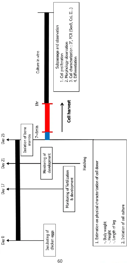
2. Materials and methods Experimental Design
In this chapter, histological section ‘s experiment was conducted for the femurs each parts, metaphysis and epiphysis area and diaphysis area from 4-day-old chicks. The cell retrieved from each part was cultured for 12 hours (passage 0), 7 days (passage 0) and passage 2 with Low glucose DMEM and F/12 DMEM. In these experiments, Sox9 and Col 2 expression was monitored in passage 0 (12hour, 7days) and passage 2 with immunofluorescence.
Experimental animals.
Four-year-old white leghorn (Gallus gallus) were selected as bone marrow cell donors. The animals were cared at the University Animal Farm, Seoul National University, Korea, using our standard management program. Every procedure all
140912-4). And also all procedure used for this animal experiment was followed the standard operating procedure of our laboratory.
Isolation and culture of bone marrow- derived primary cells. The tibia and femurs extracted from both legs of euthanized 4-day-old chicken were washed with 1% (v/v) antibiotic-antimycotic solution (Gipco Invitrogen, Grand Island, NY) and 2% (v/v) fatal bovine serum (FBS: Welgene Inc., Daegu, Korea) containing Dubecco’s phosphated buffer saline (DPBS ; Welgene Inc., Daegu, Korea). Muscle tissues attached to the bone fragment was wiped out and the spongious part of the bone was cut out so bone cavity was exposed. Bone marrow cells were retrieved by the syringe flushing with 2% (v/v) FBS containing DPBS. These cells, whole bone marrow-derived cells include red bood cells, were spread on the culture dished.
Culture of bone marrow-derived cells.
one million bone marrow cells were seeded in 100mm tissue culture dishes and cultured in two medium conditioned. First one is F-12 (DMEM/F-12; Welgene Inc., Daegu korea) supplemented with 10% (v/v) FBS(Welgene Inc., Daegu, Korea) and 1% (v/v) antibiotic-antimitotic solution(Gipco Invitrogen, Grand Island, NY) and the second one is Low/DMEM (Welgene Inc., Daegu korea) supplemented with 10% (v/v) FBS(Welgene Inc., Daegu, Korea) and 1% (v/v) antibiotic-antimitotic solution(Gipco Invitrogen, Grand Island, NY). These two conditioned cells were cultured in 37 ℃, 5% CO2 in humidified atmosphere. 12 hours after culture, red blood cells and non-adherent cells were removed and medium was changed every three days’ interval. When bone marrow drived cells reached to 80-90% cell confluence, the cells were dissociated by 0.25% trypsin-EDTA (Welgene Inc., Daegu korea) and counted cell number with hematocymeter.
(v/v) of Triton X-100. The washed cells were blocked with 10% (v/v) normal goat serum and treated with Sox 9 and Collagen type Ⅱ antibodies during over night at 4 ℃. Secondary antibody were treated for 2 hours at room temperature with dark condition. After three times washing, treats DAPI staining solution for nuclear visualization. Then image captured with fluorescent microscopy using NIS elements, Inc ‘s software.
Statistical analysis
all experiments were replicated more than three times. a generalized linear model (PROC-GLM) created using Statistical Analysis System(SAS) software version 9,4** (SAS Inst., Cary, NC) was used to analyze the data. Comparison between groups were subsequently conducted the Duncan methods when a significant model effect was detected. A P valued of less than 0.005 determined as a significant difference.
3. Results
Which part of the 4-day-old chicken’s legs is more positive
for Sox9?
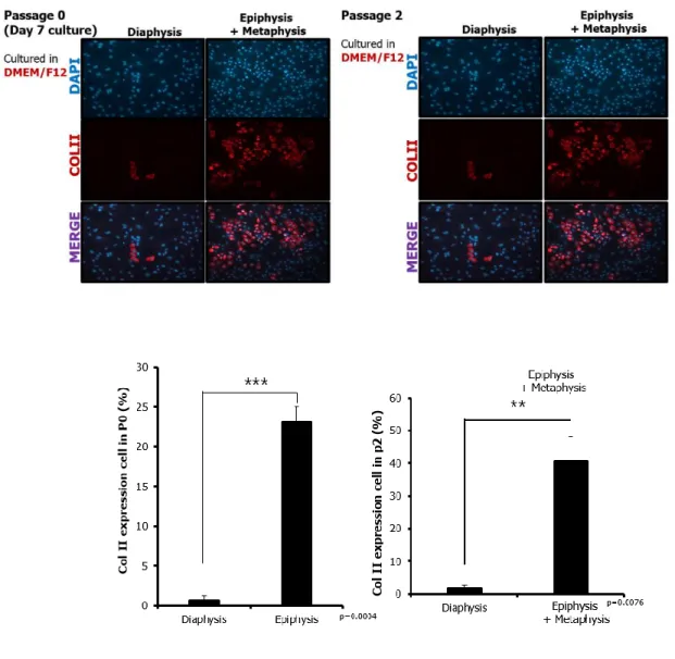
In 12 hours passage 0 culture in low glucose DMEM and DMEM/F12 medium seems to be no significant differences between two parts, epiphysis-metaphysis area and diaphysis area with Sox9 and Col Ⅱ immunofluorescence results there (Fig.1). But in further culture, there are significant differences between epiphysis-metaphysis area and diaphysis area’s cells. Level of Sox9 positive cells in DMEM/F12 medium culture, passage 0 with 7 days’ culture from seeding show a significant model effect (p=0.0072) between epiphysis-metaphysis area and diaphysis area. And also in passage 2, Sox9 expressed significantly high (p=0.126) in epiphysis-metaphysis area. In low glucose DMEM culture same tendency was observed. In 7 days’ culture passage 0, epiphysis-metaphysis area show higher Sox9 protein expression level (p=0.0002). Experiment in passage 2 also confirmed cells isolated from
epiphysis-marker. In 7 days’ passage 0 and passage2 cultured with DMEM/F12 medium, Col Ⅱ expressed significantly high in epiphysis-metaphysis area (p=0.0004, p=0.0076). And condition of low glucose DMEM culture, Col Ⅱ expressed significantly high in epiphysis-metaphysis area in both days passage 0 (p<0.0001). In passage2, expression level of Col Ⅱ shows tendency that higher in epiphysis and metaphysis area but model effect is not occurred (p=0.3395).
Figure 1. Sox9 expression of epiephysis-metaphysis area and diaphysis area in 12 hours’ culture at passage 0.
Immunofluorescence analysis of Sox9 expression of epiphysis-metaphysis area and diaphysis area in 12 hours’ culture
passage 0. There are no significant differences at Sox9 expression level between epiphysis-metaphysis area and diaphysis area.
Figure 2. Sox9 expression of epiephysis-metaphysis area and diaphysis area in 7 days’ culture passage 0 and passage2 with DMEM/F12 medium. Immunofluorescence analysis of Sox9 expression of epiphysis-metaphysis area and diaphysis area in 7 days’ culture passage 0 and passage 2. Sox9 expression level is significantly high in epiphysis-metaphysis area both in 7
days’ passage 0 culture and passage 2 culture. (p=0.0072, p=0.0126)
Figure 3. Sox9 expression of epiephysis-metaphysis area and diaphysis area in 7 days’ culture passage 0 and passage2 with
Low glucose DMEM medium. Immunofluorescence analysis of Sox9 expression of epiphysis-metaphysis area and diaphysis area in 7 days’ culture passage 0 and passage 2. Sox9
expression level is significantly high in epiphysis-metaphysis area both in 7 days’ passage 0 culture and passage 2 culture. (p=0.0002, p=0.0011)
medium. ColⅡ expression level is significantly high in
epiphysis-metaphysis area both in 7 days’ passage 0 culture and passage 2 culture. (p=0.0004, p=0.0076)
Figure 5. Col Ⅱexpression of epiphysis-metaphysis area and diaphysis area in 7 days culture passage 0 and passage2 with Low glucose DMEM medium.
epiphysis-metaphysis area both in 7 days’ passage 0 culture and merely high in passage 2 culture. (p<0.0001, p=0.3395)
4. Discussion
Femur and tibia from long bone in major sources for the bone marrow derived cells, that is used in a clinical and medical experiment area. To be more specific, sectioned the long bone and compare the surface Sox9 expression rate by the immunofluorescence, metaphysis-epiphysis area and diaphysis area.
After isolate the cells with mechanical methods, flushing by the syringe, the cells were seeded in different 100mm tissue culture dish. We made a different environment with different medium, DMEM/F12 and low glucose DMEM, and different culture time.
I compare the dishes in time point of 12hours, 7days and passage 2. As a result, no significant differences is found in 12 hours culture but in 7day culture and passage2 there is significantly high Sox9 surface expression level in epiphysis-metaphysis area than diaphysis in DMEM/F12 culture. (p-0.0072m p=0.0126)
After Sox9, Col Ⅱ is selected as a second surface marker. In same protocol in 7day culture and passage2 there is significantly high Col Ⅱ surface expression level in epiphysis-metaphysis area than diaphysis in DMEM/F12 culture. (p=0.0004, p=0.0076)
Also in low glucose DMEM culture, in 7day culture and passage2 there is significantly high Col Ⅱ surface expression level in epiphysis-metaphysis area than diaphysis in DMEM/F12 culture. (p<0.0001, p=0.3395)
CHAPTER 5
Characterization of isolated cells
by mechanical methods or enzymatic
1. Introduction
MSCs, have differentiation potential to chondrocytes, adipocytes and osteocytes(E. J. Kim et al., 2013). MSCs also called multipotent stromal cells or mesenchymal progenitor cells (Dominici et al., 2006). hese multipotent progenitor cells are usually isolated from bone marrow (Pittenger et al., 1999). Multipotency of MSCs, secretion factor of MSCs and migration ability make MSCs potential medical sources.(J. R. Choi et al., 2017; N. Kim & Cho, 2013). It is expected therapeutic potential in muscoloseletal injury, articular cartliage(Wilke et al., 2007) and tendon(Del Bue et al., 2008; Guest, Smith, & Allen, 2008; Nixon et al., 2008; Schnabel et al., 2009; Smith et al., 2003; Violini et al., 2009). Until now diverse methods of isolating MSCs exists. But more high purity and equivallent quality of MSCs is required for medical uses. There are two big category of isolating msc, physical methods and enzymatic methods. First, non-enzyme method is usually collect tibiae and hydrostatically expelled and disaggragation bone marrow plug, bone marrow flushing methods. (Schrepfer, Deuse, Lange, et al., 2007). Additionally, centrifugal methods such as ficoll and fercoll is exist and effort to increase the purity of isolated cell, FACS and MACS are existed. Second, enzyme method is using collagenase type 1,2 and trypsin. (Kern et al., 2006; K. J.
Williams et al., 2008; Xu, De Becker, Van Camp, Vanderkerken, & Van Riet, 2010).
2. Materials and methods Experimental Design
General experimental procedure was described on Fig.1. Two different methods were conducted to isolate Sox9 positive cells from epiphysis-metaphysis area of four-day-old chicken’s long bone. First one is mechanical methods, flushing and the second is enzymatic methods with collagenase Ⅱ. Several experiment was conducted to evaluated the character of two types of bone marrow derived cells. Physical profile of the donor – body weight, height, chest circumference and the length of the leg -, number of retrieved cells and cell morphology comparison was conducted. And the capacity for cell expansion and colony forming ability was examined. After that steps, collected bone marrow cell’s protein and RNA level of the osteo, chondro, adipo lineage was examined.
Experimental animals
Four-year-old white leghorn (Gallus gallus) were selected as bone marrow cell donors. The animals were cared at the University Animal Farm, Seoul National University, Korea,
using our standard management program. Every procedure all around for animal management euthanasia and breeding were performed base on the standard protocols of Seoul National University and institutional animal care and all relevant process of our experiments had approbation of unit committee (SNU-140912-4).
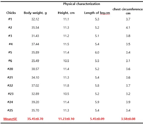
Measurement of physical characteristics.
Measurement of height, length of leg, chest circumference and body weight was conducted before euthanasia. The length from the top of the head to bottom feet was estimated, the length between wings was estimated to know the chest circumference and the length from the pelvic arch to the epicondyle of the tibia was estimated for the length of the leg.
Isolation and culture of bone marrow-derived privary cells. The tibia and femurs extracted from both legs of euthanized 4-day-old chicken were washed with 1% (v/v)
bone fragment was wiped out and the spongious part of the bone was cut out so bone cavity was exposed. After cut off the diaphysis area in mechanical methods, bone marrow cells were retrieved by the syringe flushing with 2% (v/v) FBS containing DPBS. In enzymatic methods, after cut off the diaphysis area the epiphysis and metaphysis area was minced enough and incubate with 0.25% trypsin-EDTA (Welgene Inc., Daegu korea) in 37 ℃, 5% CO2 in humidified atmosphere. and then the fragments were incubated with 0.2% (v/v) collagenase Ⅱ in HBSS in 37 ℃, 5% CO2 in humidified atmosphere for 1 hour. These cells, whole bone marrow-derived cells include red bood cells, were spread on the culture dished
Culture of bone marrow-derived cells.
one million bone marrow cells were seeded monolayer in 100mm tissue culture dishes and cultured in F-12 (DMEM/F-12; Welgene Inc., Daegu korea) supplemented with 10% (v/v) FBS(Welgene Inc., Daegu, Korea) and 1% (v/v) antibiotic-antimycotic solution(Gipco Invitrogen, Grand Island, NY) in 37 ℃, 5% CO2 in humidified atmosphere. 12 hours after culture, red blood cells and non-adherent cells were removed and medium was changed every three days’ interval. When bone marrow derived cells reached to 80-90% cell confluence, the cells were
dissociated by 0.25% trypsin-EDTA (Welgene Inc., Daegu korea) and counted cell number with hematocymeter. And before the subculture, morphology of the cultured bone marrow derived cells were observed under inverted microscope (TS-100-F, Nikon, Tokyo, Japan)..\
Examined proliferation ability during in vitro culture of the bone marrow derived cells
The number of the cells seeded for each passage and the number of cultured cells at the end of the passage was counted cell number with hematocymeter. The number of cells collected during the whole passage, at the end of each passage and doubling time were measured for checking out cell proliferation ability during in vitro culture. Doubling time of bone marrow derived cells calculated equation was tlog2/(logNt-logN0). t is time to confluence, Nt is the number of cells at the end of each passage and N0 is the number of initially seeded cells.
every 3 days after non-adherent cells and red blood cells were washed away. after ** days culture, cells were washed with DPBS (Welgene Inc., Daegu korea), fixed with 4% (v/v) paraformaldehyde for 30 minutesf at room temperature. Then fixed cells were stained with 1% crystal violet in 4% (v/v) paraformaldehyde for 5minutes in room temperature. After washing with DPBS (Welgene Inc., Daegu korea) more than 3 times, colony that cells gathered more than 55 was counted under light microscope at low magnification. Results were expressed as total number of colonies on 100mm tissue culture dish.
Analyzing gene levels using real-time PCR.
Chicken bone marrow derived cells Isolated by different two methods cultured for passage 0 and passage2. Two different passages’s cells assessed their gene expression level by real-time PCR. After extracted RNA at each passage by the RNeasyTM Mini Kit the samples were stored at –80 ℃. With M-MLV Reverse Transcriptase cDNA of each samples were extracted. The primer sequences are listed in table 1.
Immunochemistry
The bone marrow derived cells were washed with DPBS (Welgene Inc., Daegu korea), fixed with 4% (v/v) paraformaldehyde and subsequently permeablized with 0.1% (v/v) of Triton X-100. The washed cells were blocked with 10% (v/v) normal goat serum and treated with Sox 9 Collagen Type Ⅰ and Collagen type Ⅱ antibodies during over night at 4 ℃. Secondary antibody was treated for 2 hours at room temperature with dark condition. After three times washing, treats DAPI staining solution for nuclear visualization. Then image captured with fluorescent microscopy using NIS elements, Inc ‘s software.
Statistical analysis
All experiments were replicated more than three times. a generalized linear model (PROC-GLM) created using Statistical Analysis System(SAS) software version 9,4** (SAS Inst., Cary, NC) was used to analyze the data. Comparison between groups were subsequently conducted the Duncan
3. Results
Isolation and proliferation of bone marrow derived cells.
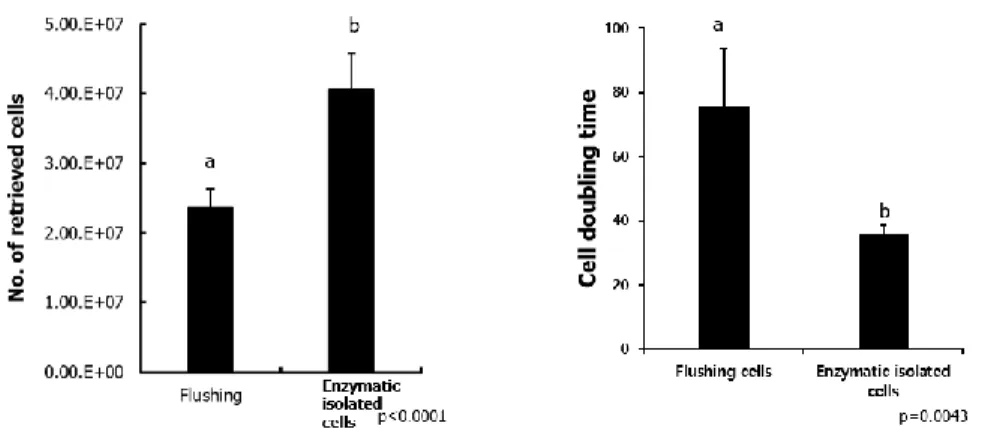
In total, 11 chickens were euthanized to collect bone marrow derived cells from epiphysis-metahphysis area in this experiments. The physical profile average of the cell donors was as follows: body weight, 35.45±0.70g; height, 11.23±0.10cm; length of leg, 5.45±0.09 cm; chest circumference, 3.58± 0.08. (table.1) The whole isolated cell number was counted before seeding, retrieved cell number is meaningly higher in enzymatic isolated cells. (p=0.0043) (table2, figure 1 A) After isolated cells from epiphysis-metaphysis area with mechanical and enzymatic methods, one million cells were seeded into the 100mm tissue and cell culture dish. In every subculture, cell doubling time was calculated by the tlog2/ (logNt=log N0). Cell doubling time shows enzymatic isolated cells have better proliferation ability. (p<0.0001) (figure 1 B)
As shown in figure2 cuboidal cell-dominant population is observed in mechanical methods isolated cells. On the other hand, stromal like cell-dominant population is observed in enzymatic isolated cells(figure 2).
Table 2. Physical characterization of white leghorn chicks employed as the donor of bone marrow-retrieved cells.
Figure 7. Retrieved cell numbers and doubling time of the cells that isolated by mechanical methods and enzymatic methods. Retrieved Cell number is higher in Enzymatic isolated cells (p<0.0001) (figure 7). And doubling time calculation confirmed proliferation speed is much higher in enzymatic isolated cells (p=0.043) (Figure 7).
Figure 8. Morphology of the cell from enzymatic isolated cells and mechanical isolated cells. The cuboidal cells that is one of the prominent shaped of menchymal Stromal Cells and osteo-chondro lineage primary cells is more observed in mechanically isolated cells. On the other hand, stromal like shaped cells observed in enzymatic isolated cells dominantly.
Comparisons of colony forming ability of bone marrow-derived cells in different methods of isolation
Isolated bone marrow derived cells which is mixed population morphology. To confirm chicken bone marrow derived primary cells attachment and colony forming ability colony forming unit-fibroblast analysis were conducted. One million cells obtained from chicken epiphysis- metaphysis area with mechanical or enzymatic methods are seeded in 100mm tissue culture dish. With crystal violet staining, single cell derived clones during passage 0, 7 days’ culture was detected. Contained more than 50 single positive cells was defined as colony. It shows isolated with mechanical methods cells didn’t forming colony in 7 days’ culture. In enzymatic isolated cells, colony was observed significantly. (p=0.0248)(figure 3)
Figure 9. Colony forming ability of enzymatic isolated cells and mechanically isolated cells. 24 hours’ culture after one million of cells are seeded in 100mm tissue culture dish, CFU-F staining is executed for confirming colony forming ability. In mechanically isolated cells, colony that more than 50 cells gathered was not found. But in enzymatic isolated cells, 62 colonies were observed in average. So Colony forming ability is much higher in enzymatically isolated cells(p=0.0248).
Analysis gene expression on chicken bone marrow-derived cells from epiphysis-metaphysis area with different isolated methods.
Gene expression of osteogenic, chondrogenic and adipogenic cell markers in general were analysis at passage 0 and passage 2 with real-time PCR. (figure 4) Sox9 gene expression seems to be higher in mechanical isolated methods both passage 0 and passage 2 results. And Collagen Ⅱ gene expression is also higher in passage 0 in mechanical methods isolated cells.
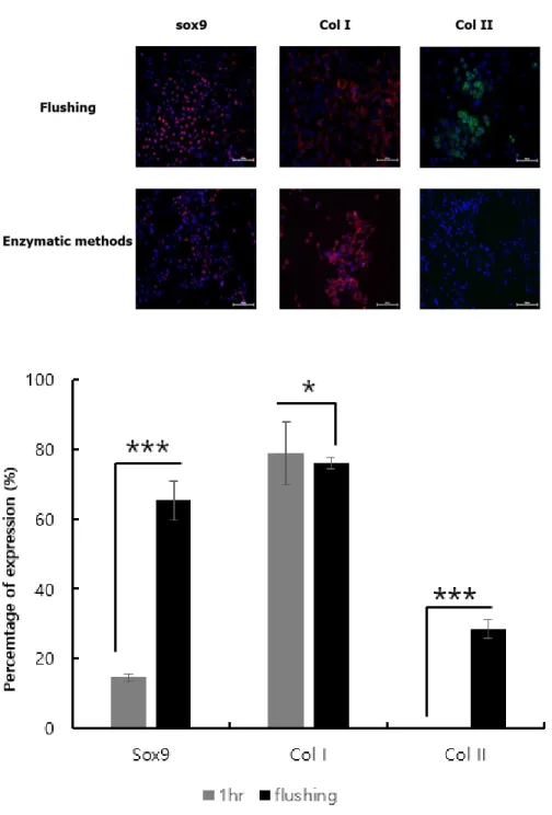
Comparison of cell surface marker expression of chicken bone marrow-derived cells isolated by different methods.
To detect expression of cell surface marker of the chicken bone marrow-derived cells immunocytochemistry at passage 2 is performed. Sox9, Collagen Ⅰ and Collagen Ⅱ surface marker that related to osteo-chondro lineage were detected by immunofluorescence. Except collagen Ⅰ ,which enzymatic isolated cells show slightly high level, Sox9 and Collagen Ⅱ cell surface
Figure 10. Level of gene that related to Osteocyte, chondrocyte and adipocyte. (A) At passage 0, Sox9 gene expression level is merely higher in mechanically isolated cells. Also ColⅡ gene expression level is much higher in mechanically isolated cells. (B) At passage 2, Sox9 gene expression level is higher in mechanically isolated cells. But Runx 2 gene expression level is higher in enzymatic isolated cells.
Figure 11. Cell surface marker levels of Sox9, Col I and Col II. Sox9 and ColⅡ surface marker expression level is much higher in mechanically isolated cells of passage 2. (p<0.0001). Also it
was confirmed that ColⅡ wasn’t expressed in enzymatic isolated cells. But in ColⅠ surface marker, expression level is merely higher in enzymatic isolated cells.
4. Discussion
In basic character of two cells, mechanically isolated cells and enzymatic isolated cells, proliferation speed is significantly faster in enzymatic isolated cells(p=0.0043). And also in primitive cell’s stage, retrieved cell number is also higher in enzymatic isolated cells. But in morphological aspects, cuboidal cells that is known as major morphology of osteo-chondro progenitor cells are observed in mechanically isolated cells.
In colony forming ability, in 24 hours culturing, mechanically isolated cells showed no colony but in enzymatic isolated cells about 60 colonies in average is formed. (p=0.0248) Gene expression level and cell surface marker expression level related to osteo-chondro lineage, it seems both gene and surface marker expressed higher in mechanically isolated cells.
As a result, this chapter experiments consists that enzymatic isolated cells have better ability in proliferation but shows low level of relevance to osteo-chondro related factor in gene, surface marker and morphology. In Mechanically isolated cells, these show low level of retrieved primary cell and proliferation speed. But mechanically isolated cells show much higher level of osteo- chondro related factor. It shows cuboidal
morphology, higher expression level of Sox9 and ColⅡ in gene and cell surface marker.
CHAPTER 6
DIFFERENTIATION ABILITY OF
ISOLATED CELLS
BY MECHANICAL METHODS OR
ENZYMATIC METHODS
1. Introduction
Variety cells from bone marrow derived cells has been used for stem cell engineering and clinical therapy (characterization of cells with osteogenic potential from human, bone marrow stromal cells characterization and clinical application, characterization of vascular endothelial progenitor cells from chicken bone marrow). Bone marrow-derived cells has an enormous value for the research on mesenchymal stem cells and cell transformation (Pal & Das, 2017). But in chicken bone marrow cells, basic information and condition for extract and culture is unknown.
Long bone, such as femur and tibias, have been estimated as a major source for bone marrow derived cells in which endochondral ossification is responsible for bone formation. (Provot & Schipani, 2005; Reddi, 1981). Bone morphogenesis is almost completing in newborn objects in mammals. Cell compound and distribution following the development stage is also well known in mammals. But in chicken the morphogenesis is differ from mammals and the step is not clearly confirmed. The
chicken bone marrow derived cells to clinical applications and preclinical model research.
2. Materials and methods Experimental Design
This study was designed to know which cells have differentiation ability to osteocyte lineage and adipocyte lineage. In both enzymatic isolated cells and mechanical isolated cells, differentiation induced with chemical conditioned. After two weeks’ culture, degree of differentiation is assessed by each staining assay protocol.
Experimental animals.
Four-year-old white leghorn (Gallus gallus) were selected as bone marrow cell donors. The animals were cared at the University Animal Farm, Seoul National University, Korea, using our standard management program. Every procedure all around for animal management euthanasia and breeding were performed base on the standard protocols of Seoul National
Isolation and culture of bone marrow-derived primary cells The tibia and femurs extracted from both legs of euthanized 4-day-old chicken were washed with 1% (v/v) antibiotic-antimitotic solution (Gipco Invitrogen, Grand Island, NY) and 2% (v/v) fatal bovine serum (FBS: Welgene Inc., Daegu, Korea) ontaining Dubecco’s phosphated buffer saline (DPBS ; Welgene Inc., Daegu, Korea). Muscle tissues attached to the bone fragment was wiped out and the spongious part of the bone was cut out so bone cavity was exposed. After cut off the diaphysis area in mechanical methods, bone marrow cells were retrieved by the syringe flushing with 2% (v/v) FBS containing DPBS. In enzymatic methods, after cut off the diaphysis area the epiphysis and metaphysis area was minced enough and incubate with 0.25% trypsin-EDTA (Welgene Inc., Daegu korea) in 37 ℃, 5% CO2 in humidified atmosphere. and then the fragments were incubated with 0.2% (v/v) collagenase Ⅱ in Hanks' Balanced Salt Solution, HBSS(Gipco Invitrogen, Grand Island, NY) in 37 ℃, 5% CO2 in humidified atmosphere for 1 hour. These cells, whole bone marrow-derived cells include red blood cells, were spread on the culture dished.