저작자표시-비영리-변경금지 2.0 대한민국 이용자는 아래의 조건을 따르는 경우에 한하여 자유롭게 l 이 저작물을 복제, 배포, 전송, 전시, 공연 및 방송할 수 있습니다. 다음과 같은 조건을 따라야 합니다: l 귀하는, 이 저작물의 재이용이나 배포의 경우, 이 저작물에 적용된 이용허락조건 을 명확하게 나타내어야 합니다. l 저작권자로부터 별도의 허가를 받으면 이러한 조건들은 적용되지 않습니다. 저작권법에 따른 이용자의 권리는 위의 내용에 의하여 영향을 받지 않습니다. 이것은 이용허락규약(Legal Code)을 이해하기 쉽게 요약한 것입니다. Disclaimer 저작자표시. 귀하는 원저작자를 표시하여야 합니다. 비영리. 귀하는 이 저작물을 영리 목적으로 이용할 수 없습니다. 변경금지. 귀하는 이 저작물을 개작, 변형 또는 가공할 수 없습니다.
A Thesis for the Degree of Doctor of Philosophy
Signaling Mechanisms Modulating
Synergistic Activation of Natural Killer Cells
자연살해세포의 시너지 활성을 조절하는
신호전달 기전
August, 2016
Hyung-Joon Kwon
The Interdisciplinary Program in Agricultural Biotechnology
College of Agriculture and Life Sciences
Signaling Mechanisms Modulating
Synergistic Activation of Natural Killer Cells
자연살해세포의 시너지 활성을 조절하는
신호전달 기전
지도교수 유 상 렬
이 논문을 박사학위논문으로 제출함
2016년 8월
서울대학교 대학원
농업생명과학대학 협동과정 농업생물공학 전공
권 형 준
권형준의 박사학위논문을 인준함
2016년 8월
위 원 장 윤 철 희 (인)
부위원장 유 상 렬 (인)
위 원 하 남 출 (인)
위 원 김 헌 식 (인)
위 원 서 상 욱 (인)
i
Signaling Mechanisms Modulating
Synergistic Activation of Natural Killer Cells
Abstract
Hyung-Joon Kwon
The Interdisciplinary Program in Agricultural Biotechnology
College of Agriculture and Life Sciences
Seoul National University
Natural Killer (NK) cells are discriminated among innate lymphoid cells (ILCs) with their cytolytic activity that has a key role in the control of viral infection and tumorigenesis. NK cells not only kill virus-infected or transformed cells including cancer stem cells by releasing cytotoxic granules, but also contribute to modulate diverse immune responses by secretion of cytokines (e.g., IFN-γ and TNF-α) and chemokines (e.g., MIP-1α and MIP-1β). Therefore, the malfunction of NK cells is linked various diseases, and the manipulation of NK cells function have hold promise in therapeutic applications. However, the limited information for the mechanisms regulating activation of NK cells has hampered the progression toward therapeutic strategies. Accordingly, understanding accurate activating and inhibitory receptors signaling mechanisms controlling NK cells function can augment successful NK cell-based therapies.
The triggering of NK cell effector functions relies on the engagement of activating or inhibitory receptors for ligands on their target cells. The balanced
ii
signals from activating and inhibitory receptors are known to dictate NK cells’ activity. However, even in the case of inhibitory signal is absent, activating signal from single coactivation receptor is insufficient to induce enough activation of resting NK cells. Signals from multiple activating receptors, not a single receptor, are required for NK cell functions. And only specific combinations among them can induce additive or synergistic enhancement of NK cells’ activity. For this reason, the study of signaling mechanisms modulating synergistic activation of NK cells is critical for harnessing the ‘arm’ of NK cells for clinical purposes. In this study, two critical signaling molecules for immune cell functions, glycogen synthase kinase (GSK)-3β and NF-κB are examined for regulation of synergistic activation of NK cells, and their interaction mechanisms with other signaling pathways are investigated.
In a synergistic activation model of NK cells that combines NKG2D and 2B4 activating receptors, representative non-ITAM-associated activating receptors, GSK-3β negatively regulated the synergistic activation of both cytotoxicity and cytokine secretion. The individual or combinational stimulation of NKG2D and 2B4 induced inhibitory phosphorylation of GSK-3, and the extent of phosphorylation in accordance with the level of NK cell activity. The inhibition of GSK-3β by siRNA knockdown or pharmacological inhibition increased NK cell function, but GSK-3α knockdown had no effect. This negative role of GSK-3β was dependent on its kinase activity. The regulation of NK cell function by GSK-3β was a common mechanism for both NKL cell line and primary NK cells. And NK cell activations by either ITAM-associated or non-ITAM activating receptors were regulated by GSK-3β.
iii
immune responses. Nevertheless, its role in NK cells activated by multiple activating receptors during target cell recognition is not defined. Synergistic combination of NKG2D, 2B4, or DNAM-1 induced synergistic and sufficient activation of NF-κB pathway that is linked to synergistic NK cell activity, whereas single receptor stimulation was insufficient. Receptors cooperation for NF-κB activation was regulated at the level of Vav1 phosphorylation, the 1st checkpoint, and this
Vav1-dependent synergistic signal also cooperates with separate PI3K-Akt signal for synergistic p65 phosphorylation, the 2nd checkpoint, in a stepwise manner. As
confirmed with a X-linked lymphoproliferative disease (XLP) NK cells model that is defective for 2B4 receptor signaling because of mutations of SAP adaptor, stepwise checkpoint signaling mechanism for NF-κB activation was suggested.
These newly uncovered signaling mechanisms during multiple receptor stimulation provides new insights of receptor cooperation to determine specificity and magnitude of NK cell activation and bases for establishing NK cell-based therapeutic strategies.
Key words: Natural Killer cells, Synergistic activation, Receptor signaling,
GSK-3β, NF-κB, XLP
iv
Contents
Abstract …...……….………...… i
Contents ……….……….………... iv
List of Figures ……….………... x
List of Tables ………...………. xvi
List of Abbreviations ……….………... xvii
Chapter I. General Introduction……….…………... 1
I-1. Research background... 2
I-1-1. Cancer therapies... 2
I-1-2. NK cell-based cancer therapy... 3
I-2. Innate lymphoid cells... 5
I-2-1. Classification of innate lymphoid cells... 5
I-2-2. Differences of NK cells from other ILCs... 6
I-3. Functional characteristics of NK cells... 7
I-3-1. NK cells... 7
v
I-3-3. Anti-viral activity of NK cells... 9
I-3-4. Crosstalk of NK cells with other immune cells... 9
I-4. Inhibitory and activating receptors of NK cells... 11
I-4-1. Inhibitory receptors and signaling... 11
I-4-2. Activating receptors and signaling... 12
I-5. Signaling for synergistic activation of NK cells... 15
I-6. X-linked lymphoproliferative disease... 17
I-7. Purpose of study and rationale for study design... 18
I-8. References... 19
Chapter II. NK cell function triggered by multiple activating
receptors is negatively regulated by glycogen synthase
kinase-3β... 32
II-1. Abstract... 33
II-2. Introduction... 35
II-3. Materials and Methods... 38
II-3-1. Cell lines and reagents... 38
II-3-2. NK cell expansion... 38
vi
II-3-4. Flow cytometric analysis of NK cell activation... 40
II-3-5. Cell stimulation... 40
II-3-6. Measurement of cytokine secretion... 40
II-3-7. Immunoblot analysis... 41
II-3-8. RNA interference... 41
II-3-9. RT-PCR... 42
II-3-10. Cytotoxicity assay... 43
II-3-11. Flow cytometric analysis of Ca
2+mobilization... 43
II-3-12. Retroviral transduction... 44
II-3-13. Statistical analysis... 45
II-4. Results... 46
II-4-1. Effector functions of NK cells are enhanced by
pharmacological inhibition of GSK-3... 46
II-4-2. GSK-3 phosphorylation correlates with the activation
status of NK cells... 52
II-4-3. GSK-3 phosphorylation by PI3K- and
MAPK-dependent Pathways... 60
II-4-4. GSK-3β-dependent regulation of NK cell effector
functions... 63
vii
II-4-5. GSK-3β regulates calcium-dependent signaling for
NK cell Activation... 68
II-4-6. Regulation of NK cell function by GSK-3β is
dependent on its kinase activity... 72
II-4-7. Role of GSK-3β in primary NK cell activation... 79
II-5. Discussion... 85
II-6. References... 91
Chapter III. Stepwise phosphorylation of p65 promotes NF-κB
activation and NK cell responses during target cell
recognition... 100
III-1. Abstract... 101
III-2. Introduction... 102
III-3. Materials and Methods... 106
III-3-1. Cells and reagents... 106
III-3-2. Patient samples... 107
III-3-3. Abs... 107
III-3-4. Cellular assays... 108
III-3-5.
Receptor
crosslinking
and
cell
mixing
viii
experiments... 110
III-3-6. Assessment of NF-κB activation by EMSA... 111
III-3-7. NF-κB reporter assay... 111
III-3-8. RNA interference... 112
III-3-9. Ex vivo expansion of NK cells... 113
III-3-10. DNA constructs and transfections... 114
III-3-11. Immunoblotting... 114
III-3-12. Cell conjugation and immunofluorescence... 115
III-3-13. Real-time PCR... 116
III-3-14.
Flow
cytometric
analysis
of
p65
phosphorylation... 117
III-3-15. Statistical analyses... 117
III-3-16. Data availability... 117
III-4. Results... 118
III-4-1. Synergistic activation of NF-κB by NKG2D and
2B4... 118
III-4-2. NF-κB is required for NK cell functions by NKG2D
and 2B4... 127
III-4-3. Disparate signals converge on the NF-κB p65
ix
subunit... 142
III-4-4. Signal amplification is insufficient for NF-κB
activation... 157
III-4-5. Defective NF-κB activation in XLP1 NK cells by
NKG2D and 2B4... 162
III-4-6. Vav1 is required for the synergistic activation of
NF-κB... 179
III-5. Discussion... 184
III-6. References... 195
x
List of Figures
Fig. 1. GSK-3 regulates IFN-γ production by primary NK cells stimulated with
target cell... 47
Fig. 2. GSK-3 regulates degranulation of primary NK cells stimulated with target
cell... 49
Fig. 3. GSK-3 regulates IFN-γ production by primary NK cells stimulated with
synergistic activating receptor combination... 50
Fig. 4. GSK-3 regulates IFN-γ production by NKL cells stimulated with synergistic
activating receptor combination... 51
Fig. 5. The phosphorylation of GSK-3 after stimulation of NKG2D and/or 2B4 in
NKL cells... 53
Fig. 6. The phosphorylation of GSK-3 after stimulation of NKG2D and/or 2B4 in
primary NK cells... 54
Fig. 7. The phosphorylation of GSK-3 after stimulation of NKG2D and/or 2B4 is
increased by knockdown of c-Cbl... 55
Fig. 8. The phosphorylation of GSK-3 after stimulation of NKG2D and/or 2B4 is
inhibited by costimulation of CD94 inhibitory receptor... 57
Fig. 9. The phosphorylation of GSK-3 after stimulation of NKG2D and/or 2B4 is
inhibited by knockdown of Vav1... 58
Fig. 10. Small molecule GSK-3 inhibitors cause decrease in β-catenin
phosphorylation via an inhibitory phosphorylation of GSK-3α/β... 59
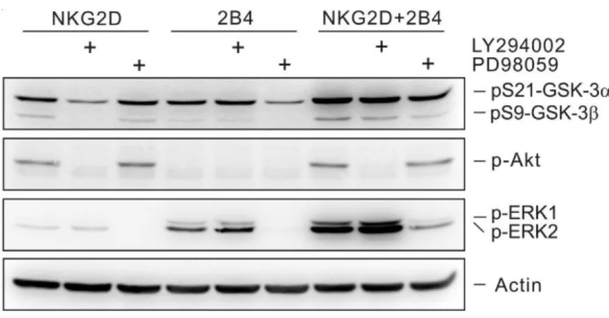
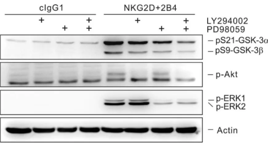
Fig. 11. PI3K and MAPK signaling pathways cooperate to promote GSK-3
phosphorylation... 61
Fig. 12. PI3K and MAPK signaling pathways cooperate to promote GSK-3
xi
Fig. 13. GSK-3 knockdown with siRNAs specific for each α or β-isoform... 64
Fig. 14. GSK-3β isoform selectively regulates NK cell cytokines transcription.... 65
Fig. 15. GSK-3β isoform selectively regulates NK cell cytokines secretion... 66
Fig. 16. GSK-3β isoform selectively regulates NK cell cytotoxicity... 67
Fig. 17. GSK-3β knockdown enhances calcium-dependent signaling... 69
Fig. 18. GSK-3β knockdown enhances PLC-γ2 phosphorylation and NFAT1 activity... 70
Fig. 19. Establishment of NKL cell line expressing GSK-3β mutants by retroviral transduction... 73
Fig. 20. GSK-3β activity regulates NK cell cytokines secretion... 74
Fig. 21. GSK-3β activity regulates NK cell cytotoxicity... 75
Fig. 22. GSK-3β activity regulates NK cell calcium signaling... 77
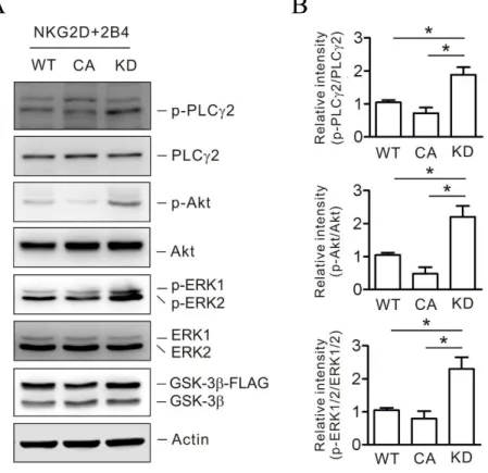
Fig. 23. GSK-3β activity regulates phosphorylation of PLC-γ2, Akt, and ERK by combination of NKG2D and 2B4 in NK cell... 78
Fig. 24. GSK-3 knockdown with β-isoform-specific siRNAs in primary NK cells... 80
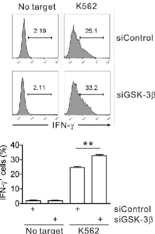
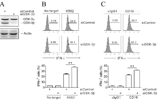
Fig. 25. GSK-3β knockdown enhances IFN-γ expression of primary NK cells induced by K562 target cells... 81
Fig. 26. The phosphorylation of GSK-3 after stimulation of both ITAM- and non-ITAM-dependent receptors in primary NK cells... 82
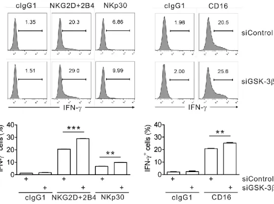
Fig. 27. GSK-3β knockdown enhances IFN-γ expression of primary NK cells induced by both ITAM- and non-ITAM-dependent receptors... 83
Fig. 28. Enhanced IFN-γ production by CD16.NK92 cells after GSK-3β knockdown... 84
co-xii
engagement... 119
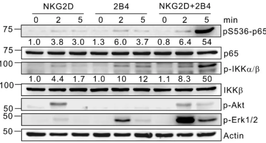
Fig. 30. Kinetics of phosphorylation of NF-κB signaling pathway by NKG2D and
2B4 co-engagement... 120
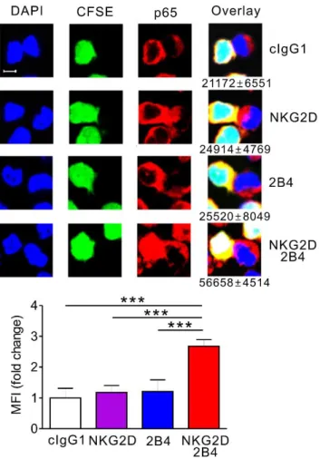
Fig. 31. Immunofluorescence for nuclear translocation of p65 by NKG2D and 2B4
co-engagement... 122
Fig. 32. Subcellular fractionation for nuclear translocation of p65 by NKG2D and
2B4 co-engagement... 123
Fig. 33. DNA binding nuclear p65 after NKG2D and 2B4 co-engagement... 124 Fig. 34. GFP expression in NKL-κB-GFP cells is NF-κB-dependent... 125 Fig. 35. Transcriptional activity of NF-κB after NKG2D and 2B4
co-engagement... 126
Fig. 36. Profile of gene expression related to NK cell function during
coactivation... 128
Fig. 37. BAY11-7082 inhibits cytokine and chemokine secretion of NKL cells by
NKG2D and 2B4 coactivation... 129
Fig. 38. Knockdown of p65 in NKL cells with p65-specific siRNA... 130 Fig. 39. p65-specific siRNA inhibits cytokine, chemokine, granzyme B, and IκBα
mRNA expression of NKL cells by NKG2D and 2B4 coactivation... 131
Fig. 40. p65-specific siRNA inhibits cytokine and chemokine secretion of NKL cells
by NKG2D and 2B4 coactivation... 133
Fig. 41. IFN-γ and MIP-1α production by NKG2D and 2B4 ligands is
NF-κB-dependent... 134
Fig. 42. p65-specific siRNA inhibits Granzyme B secretion of NKL cells by NKG2D and 2B4 coactivation... 135
Fig. 43. The proportion of cells showing p65 phosphorylation is synergistically
xiii
Fig. 44. Phosphorylation of p65 in primary NK cells by NKG2D and 2B4
coactivation... 137
Fig. 45. The proportion of cells showing cytokine production by NKG2D and 2B4
coactivation is synergistically increased... 138
Fig. 46. The proportion of cells showing cytokine production by NKG2D and 2B4
coactivation is inhibited by BAY11- 7082... 139
Fig. 47. Effects of BAY11-7082 on the viability of NK cells... 141 Fig. 48. NKG2D and 2B4 coengagement induces synergistic phosphorylation of p65
in NKL cells at serine 536 and 276... 143
Fig. 49. Synergistic phosphorylation of p65 by NKG2D and 2B4 ligands... 144 Fig. 50. Phosphorylation of p65 at serine 536 or 276 in NKL cells depends on
separate Akt or Erk pathway... 145
Fig. 51. Akt and Erk pathway are required for NK cells cytokines secretion... 147 Fig. 52. Requirement of Akt1 and Erk2 for optimal p65 phosphorylation and IFN-γ
and MIP-1α production following coactivation... 148
Fig. 53. NKG2D and 2B4 coengagement induces synergistic phosphorylation of p65
in primary NK cells at serine 536 and 276... 150
Fig. 54. Phosphorylation of p65 at serine 536 or 276 in primary NK cells depends
on separate Akt or Erk pathway... 151
Fig. 55. Requirement of Akt and Erk pathways for IFN-γ and TNF-α production
following NKG2D and 2B4 coactivation... 152
Fig. 56. Requirement of Akt and Erk pathways for transcriptional activity of NF-κB
following NKG2D and 2B4 coactivation... 153
Fig. 57. DNAM-1 synergizes with 2B4 to induce competent p65 phosphorylation
and NF-κB activation... 155
xiv
phosphorylation... 158
Fig. 59. c-Cbl depletion augments but is not sufficient for transcriptional activity of
NF-κB... 160
Fig. 60. c-Cbl depletion augments but is not sufficient for cytokines secretion by
NKL cells... 161
Fig. 61. SAP-specific knockdown in NKL cells... 163 Fig. 62. SAP is required for Ca2+ mobilization by NKG2D and 2B4
coactivation... 164
Fig. 63. SAP is required for NF-κB transcriptional activity by NKG2D and 2B4
coactivation... 165
Fig. 64. SAP is required for cytokines secretion by NKG2D and 2B4
coactivation... 166
Fig. 65. SAP is required for p65 phosphorylation and IFN-γ expression in primary
NK cells via NKG2D and 2B4 coactivation... 167
Fig. 66. SAP deficiency in XLP1 NK cells... 169 Fig. 67. XLP1 NK cells have defects in degranulation in response to 221 target
cells... 171
Fig. 68. XLP1 NK cells have defects in phosphorylation of p65 at serine 536 and
276 and Erk by NKG2D and 2B4 coactivation... 172
Fig. 69. XLP1 NK cells have defects in p65 nuclear translocation in response to
NKG2D and 2B4 coactivation... 173
Fig. 70. XLP1 NK cells have defects in IFN-γ production following NKG2D and
2B4 coactivation... 174
Fig. 71. XLP1 NK cells have defects in degranulation following NKG2D and 2B4
coactivation... 175
xv
cells... 177
Fig. 73. XLP1 NK cells have defects in Vav1 phosphorylation following NKG2D
and 2B4 coactivation... 178
Fig. 74. Synergistic phosphorylation of p65 is impaired by co-crosslinking
CD94-NKG2A... 180
Fig. 75. Synergistic increase of NF-κB transcriptional activity is impaired by
co-crosslinking CD94-NKG2A... 181
Fig. 76. Synergistic phosphorylation of p65 is impaired by Vav1 knockdown... 182 Fig. 77. Synergistic increase of NF-κB transcriptional activity is impaired by Vav1
knockdown... 183
Fig. 78. Proposed mechanism of NF-κB activation via coactivation receptors on NK
cells... 185
Fig. 79. Preactivation with IL-2 enhances IFN-γ expression and NF-κB
activation... 191
Fig. 80. Preactivation with IL-2 enhances the responsiveness of NK cells for IFN-γ
expression and NF-κB activation... 192
xvi
List of Tables
Table 1. List of primers used for real-time PCR analyses of the indicated
xvii
List of Abbreviations
ADCC – antibody-dependent cell-mediated cytotoxicity AML – acute myeloid leukemia
CA – constitutively active DC – dendritic cell
DNAM-1 – DNAX accessory molecule-1 EBV – Epstein-Barr virus
ELISA – enzyme-linked immunosorbent assay ERK – extracellular signal-regulated kinase FACS – Fluorescence-activated cell sorting GSK-3 – glycogen synthase kinase-3 GVHD – graft-versus-host disease HLA – human leukocyte antigen IFN - interferon
IKK – IκB kinase
ITAM – immunoreceptor tyrosine-based activation motif ITIM - immunoreceptor tyrosine-based inhibition motif ITSM – immunoreceptor tyrosine-based switch motif KD – kinase dead
KIR – killer-cell immunoglobulin-like receptor LAT – linker for activation of T cell
MAPK – mitogen-activated protein kinase MCMV – mouse cytomegalovirus
MEK – MAPK/ERK kinase
MHC – major histocompatibility complex MIC – MHC class I polypeptide-related sequence MIP – macrophage inflammatory protein
xviii
NCR – natural cytotoxicity triggering receptor NFAT – nuclear factor of activated T cell NF-κB – nuclear factor κB
NK – natural killer
NTAL – non-T-cell activation linker
PBMC – peripheral blood mononuclear cell
PI3K – phosphatidylinositol-4,5-bisphosphate 3-kinase PKC – protein kinase C
PLC – phospholipase C
PLGF – placental growth factor SAP – SLAM-associated protein
SHP – Src homology region 2 domain-containing phosphatase siRNA – small interfering RNA
SLAM – signaling lymphocytic activation molecule
SLP-76 – SH2 domain-containing leukocyte phosphoprotein of 76 kD TDZD - thiadiazolidinone
TNF – tumor necrosis factor
TRE – transcription response element ULBP – UL16 binding protein
VEGF – vascular endothelial growth factor WT – wild type
XLP – X-linked lymphoproliferative disease YINM – tyrosine-based signaling motif
1
Chapter I.
2
I-1. Research background
I-1-1. Cancer therapies
It is well-recognized that cancer is an old-age disease because cancer incidence increases with an extended life expectancy caused by progress of health care technology. Most primary tumors are now being treated by combinational therapy including surgery, chemotherapy, and radiation. But these conventional therapies can cause immunosuppressive side effect and tumor relapse by micrometastasis of residual cancer stem cells (1, 2). For this reason, targeted cancer therapy, interfering specific target molecules required for tumor cell cycle, is expected to be more safe and effective than conventional therapies. However, targeted therapy also has limitation that most cancer cells’ phenotype is resulted from multiple genes’ modification while current targeted therapies are focusing on a single target (3). And it is effective only before tumor is resistant to targeted therapeutic agent. Moreover, tumor progression is not only dependent on cell-autonomous gene alterations, but also dependent on microenvironment including cytokines and immune responses against tumor cells (4). The strategy exploiting the host’s immune system for cancer treatment depends on the insight that immune system can surveil initial transformation and eradicate residual cancer stem cells after therapies (5). Cancer immunotherapy, ranging from passive to active, encompasses cancer vaccines, adoptive cellular immunotherapy, immune checkpoint blockade, and
3
oncolytic viruses (6). Increasing clinical studies have combined these conventional therapy and immunotherapy and suggest that combinations can create a synergy (7).
I-1-2. NK cell-based cancer therapy
Allogeneic hematopoietic stem cell transplantation (HSCT) is an established strategy for cancer immunotherapy, especially for hematological malignancies (8). The patients are treated by irradiation and/or chemotherapy prior to transplantation to suppress the recipient’s immune rejection and are through the introduction of competent donor immune cells. This donor immune system can trigger graft-versus-tumor (GVT) effect. The patients with acute myeloid leukemia (AML) undergoing HSCT, in which a KIR-ligand mismatch exists, showed improved survival rate and reduced relapse rate (9, 10). After transplantation, donor T cells may become activated by recipient antigen-presenting cells (APC) and kill residual tumor cells. In the meantime, these T cells may also cause tissue damage referred to as ‘graft-versus-host disease’ (GVHD). NK cells are also known to mediate GVT effect, but not to involve GVHD. They rather decrease GVHD inducing inefficient priming of donor T cells by killing recipient’s APCs (11). Several strategies were suggested to take advantage of this strong point of NK cells, such as depleting T or B cells and ex vivo expansion of NK cells (12, 13). In addition, several strategies enhancing transferring NK cells’ activity, by means of cytokines or modulation of activating or inhibitory receptors, are suggested. But the establishment of NK cell-based therapy remained difficult because of the lack of
4
information about mechanisms for NK cell activation and requires for detailed investigations.
5
I-2. Innate lymphoid cells
I-2-1. Classification of innate lymphoid cells
NK cells belong to innate lymphoid cells (ILCs), a recently expanding family of innate immunity (14). Unlike T or B cells, ILCs are a group of lymphocytes that do not have rearranged antigen-specific receptors (15). ILCs are classified on the basis of transcriptional and functional profile. Similar to T cells classification, ILCs are also grouped into cytotoxic- and helper-ILC, and NK cells belong to cytotoxic-ILCs (16). Helper-ILCs further divided into ILC1, ILC2, and ILC3. ILC1 development depends on T-bet transcription factor and secrete IFN-γ. ILC2 depends on GATA3 transcription factor and secrete type 2 cytokines, such as IL-5 and IL-13 (17). ILC3 express RORγt and secrete mainly IL-17 and IL-22 (18). But this information for ILC development is mostly from mice studies, and less is characterized in humans (19). In the meantime, a recent study found common lymphoid progenitor population in secondary lymphoid tissues that express RORγt and generate all known ILC subsets, implying different system in humans (20). With diverse cytokine profile and function, ILC subsets contribute to defenses against a variety of pathogens (21, 22). Their role for host defense is crucial in immune-compromised mice (23), while it seems to be marginal in the presence of a functional T cells. In humans, the role of ILCs including NK cells may become more important in case of T cell-depleted HSCT recipient to prevent opportunistic infections.
6
Therefore, establishment of strategies for NK cell-based cancer therapy has to consider the role of whole ILC population including both cytotoxic- and helper-ILCs.
I-2-2. Differences of NK cells from other ILCs
Conventional NK cells were initially classified in group 1 ILCs as they express transcription factor T-bet and secrete IFN-γ. However, NK cells are developmentally dependent on Eomesodermin (Eomes), not on T-bet. In addition, NK cells express cytotoxic molecules, such as perforin and granzyme, that mediate their cytotoxic activity. This cytotoxic function of NK cells discriminates themselves from other ILCs as an innate counterpart of CD8+ cytotoxic T cells (24). NK cells
are also different from other ILCs for the independency on 7. NK cells are IL-7Rα negative while other helper-ILCs are IL-IL-7Rα positive.
NK cells hold their specialized position as critical innate effectors for anti-tumor and anti-viral immunity in spite of characterization and expansion of ILC family.
7
I-3. Functional characteristics of NK cells
I-3-1. NK cells
NK cells had been named as large granular lymphocytes with ‘natural’ cytotoxic activity to target cells without priming or inhibition. And now they are categorized as separate lymphoid lineage with both cytotoxicity and cytokine secretion activity (25-28). Following studies have proposed that NK cells recognize and kill target cells that are lack of self MHC class I molecules (“Missing self” hypothesis) (29, 30) and then found inhibitory receptors discriminating non-self from self cells. Thereafter, discovery of diverse activating NK cell receptors changed the activation of NK cells against ‘missing self’ target cells from nonspecific response to highly organized process with ‘dynamic balance’ (31). Although NK cells are lack of dominant antigen receptors, expressed by T or B cells, they express various array of germ-line encoded receptors. And their multiple engagement during target cell recognition result in the outcome of NK cell activity (32, 33). Activating receptors of NK cells sense ligands on stressed, transformed, or virus-infected cells, such as ULBP and MIC molecules for NKG2D receptor (34). Activation of NK cells depends not only on ligands of target cells, but also on cytokine microenvironment and interaction with other immune cells (35). NK cells can be activated by type I IFN, IL-12, IL-18, and IL-15 (36), and CD4+ T cell-derived IL-2 enhances NK cell
functions (37). NK cells also can be regulated by transforming growth factor (TGF)-β produced by regulatory T cells (38, 39). Cytokines, such as IFN-γ or TNF-α, secreted by NK cells also shape other immune cells responses (40, 41).
8
NK cells, with these sophisticated regulation mechanisms, interpret their environment and perform specialized tasks in anti-tumor or anti-viral immunity and immune cells cross-talk.
I-3-2. Anti-tumor activity of NK cells
Many in vivo and in vitro studies advocates the anti-tumor activity of NK cells (25). NK cells in mouse show in vivo rejection of several transplanted tumors in a NK receptor-target cell ligand-dependent manner (42). In some experiments, anti-tumor activity of NK cells induces generation of specific T cell responses against the same tumor cells (43) that could be a strong point of NK cell-based therapeutic strategy. In addition, immunosurveillance for spontaneous or induced tumors by NK cells was reported (44). In humans, although the characterization of NK cell function in vivo is difficult because of the lack of NK cell-specific deficiency (45), stacked epidemiologic survey suggests that the level of NK cell activity in peripheral blood is correlated with cancer risk (46). And allogeneic hematopoietic stem cell transplantation into recipient tumor patient, such as acute myeloid leukemia (AML), lacking donor KIR ligands leads to the development of donor NK cells and better post-transplant survival without graft-versus-host disease (GVHD) (47). In other mouse in vivo experiment, the blocking of NK cell inhibitory receptor specific for MHC class I molecules increased anti-tumor activity of NK cells (48) and this approach is in clinical trial in human AML. Understanding molecular mechanisms for NK cell activation is expected to strengthen this potential of NK cells as a tool for cell-based anti-tumor therapy.
9
I-3-3. Anti-viral activity of NK cells
Important clues about anti-viral activity of NK cells were obtained by NK cell depletion or adoptive transfer studies with mice. Deficiency of NK cell activities including cytotoxicity or cytokine production increased the susceptibility of mice to the herpesvirus MCMV infection (49, 50). NK cells recognize cell surface ligands, such as m157, encoded by CMV, on MCMV-infected cells with NK cell activating receptor Ly49H (51, 52). The struggling of MCMV with genes that play a role in evasion of NK cell recognition also emphasizes the critical role of NK cells in virus infections. Among these genes are involved in downregulation of ligands for NKG2D or in expression of MHC class I homologs that stimulate inhibitory receptors on NK cells (53).
Another emerging aspect in anti-viral activity of NK cells is regulation of immunopathology (54). NK cell depletion in mice leads to accelerated the encephalitis by Theiler’s virus or myocarditis by coxsackie B3 virus (55, 56). NK cells promote the trigger of CD8+ T cell responses and so control type I IFN-induced
immunopathology by virus dissemination (57). NK cells also suppress over-activated macrophages with their cytotoxic activity (58).
Therefore, NK cells perform dual mechanism during virus infection by eliminating both virus-infected cells and over-activated pathologic immune cells.
I-3-4. Crosstalk of NK cells with other immune cells
10
by means of both cytotoxicity and cytokine secretion. NK cells interact and influence DCs in two different ways (59, 60). NK cells can maintain DC homeostasis by eliminating immature DCs (61). On the other hand, NK cells promote DC maturation by secretion of IFN-γ and TNF, and also are activated by IL-12 secreted by DCs (62). In the interaction with T cells, NK cells can help the priming of type I helper T cells by secretion of IFN-γ (63). NK cells also kill activated T cells that are without enough MHC class I molecules (64). Therefore, blocking inhibitory receptors can enhance the cytotoxic activity of NK cells against CD4+ T cells, that is applicable to
the therapy for T cell-mediated autoimmunity. NK cells are also known to suppress autoreactive B cells (65). Distinct subset of NK cells in uterine can interact with endothelial cells by promoting angiogenesis in case of pregnancy (66). These NK cells secrete vascular endothelial growth factor (VEGF) and placental growth factor (PLGF) (67). Accordingly, effector functions of NK cells are not limited to anti-cancer or anti-viral activity, but also to broad immune-modulatory activity.
11
I-4. Inhibitory and activating receptors of NK cells
I-4-1. Inhibitory receptors and signaling
The discovery of immunoreceptor tyrosine-based inhibition motif (ITIM) as a site with a sequence motif for binding of tyrosine phosphatase SHP-1 to inhibitory killer cell Ig-like receptors (KIR) in human (68) triggered the finding of many other ITIM-associated receptors (69). The KIR family in human binds classical MHC class I molecules, such as HLA-B and HLA-C. The Ly49 family in mice binds H-2 class I molecules. The CD94-NKG2A, that is conserved in human and mice, binds nonclassical MHC molecule HLA-E in huaman and Qa1 in mice. In spite of the structural diversity between KIR and Ly49 and CD94-NKG2A, their signaling through ITIM is remarkably conserved.
Signaling by inhibitory receptors starts with phosphorylation of their ITIM motif. Phosphorylated ITIM recruits the tyrosine phosphatases SHP-1 and SHP-2. KIR is also known to bind additional molecule, arrestin 2 (70). The lack of β-arrestin 2 downregulates the inhibition by KIR because β-β-arrestin 2 facilitates SHP-1 and SHP-2 recruitment to KIR. The inhibition by ITIM-bound SHP is at very proximal signaling step, which is through dephosphorylation of Vav1 by SHP-1. Given the critical role of Vav1 for actin cytoskeleton rearrangement, synapse formation, and receptors clustering, this proximal inhibitory process suppresses the tyrosine phosphorylation of activating receptors even before it begins (71, 72). The inhibitory signaling after binding to MHC class I ligand also can be through tyrosine phosphorylation of small adaptor Crk that forms a complex with Abl, a process
12 dissociating Crk from scaffold protein Cbl (73).
Inhibitory signaling also play a role for NK cell education process, which is determined by MHC class I repertoire (74). NK cells that have detected more MHC molecules by their inhibitory receptors become more responsive. Conversely, NK cells with no contact for MHC become hyporesponsive. Two education mechanisms are suggested, that is anergy induced by continuous stimulation without enough inhibition (75) or acquisition of responsiveness by instructive signal through inhibitory receptors (76).
I-4-2. Activating receptors and signaling
Instead of a dominant antigen-specific receptor for activation, such as TCR or BCR, NK cells express diverse receptors which can activate effector functions in synergy (77). These activating receptors recognize ligand expressed on virus-infected, stressed, or transformed cells (78, 79). The resting human NK cells can be activated only by combination of activating receptors, which is called coactivation receptors, not by single receptor stimulation (80). Whereas, an exceptional receptor is the low-affinity Fc receptor FcγRIIIa (CD16), which recognize Fc region of antibody and is sufficient alone to activate resting NK cells for antibody-dependent cellular cytotoxicity (ADCC). In contrast to inhibitory receptors, which has varied expression in distinct peripheral blood NK cell subsets, activating receptors are expressed mostly by all NK cells. Moreover, activating receptors induce diverse signaling pathways, while inhibitory receptors use common mechanism for inhibition.
13
motif (ITAM) or associated with adaptors carrying ITAM includes CD16, natural cytotoxicity receptors (NCRs), such as NKp30, NKp44, and NKp46, and activating KIRs, such as KIR2DS and KIR3DS, and CD94/NKG2C heterodimer (77). These receptors, upon ligand engagement, are tyrosine-phosphorylated by Src family kinases and recruit tyrosine kinases, such as Syk and ZAP-70. Syk and ZAP-70 kinases phosphorylate linker for activation of T cells (LAT) and non-T-cell activation linker (NTAL) followed by activation of PI3K, PLC-γ1, 2, and Vav2, 3. (81, 82).
Among non-ITAM receptors are NKG2D, CD2, 2B4, CRACC, NTB-A, and DNAM-1. NKG2D associates with DAP10 adaptor protein which is containing a tyrosine-based signaling motif (YINM). The YINM is phosphorylated by Src family kinases and recruits p85 subunit of PI3K or Grb2. These interaction activates Vav1 and PLC-γ2 that lead to the activation of Ca2+ mobilization and cytotoxicity
(83). 2B4, NTB-A, and CRACC that belong to SLAM family receptors contain immunoreceptor tyrosine-based switch motif (ITSM) (84) which recruits, after phosphorylation, SAP, EAT-2, and ERT adaptor proteins (85). SAP binds Src family kinase Fyn, and this interaction leads to NK cell activation through phosphorylation of Vav1 and PLC-γ2 (86, 87).
NK cells are equipped with diverse kinases, adaptors, and signaling modules depending on different activating receptors. Whereas, inhibition of signals from different activating receptors by a common ITIM-mediated mechanism suggests that there are critical signaling molecule as a convergent point. The redundancies of signaling molecules at each steps are different. Src family kinases, such as Lck, Fyn, Src, Lyn, Yes, and Fgr, are important molecules for most of activating receptors. But, no single kinase is indispensable as they are redundant for
14
NK cell activation (88). On the other hand, PLC-γ and Vav proteins are non-redundant for NK cell activation and play as a critical point for NK cell activation (82, 89). Therefore, the identification of signaling components play as critical checkpoints for NK cell activation can facilitate the understanding of cooperative mechanisms between coactivation receptors.
15
I-5. Signaling for synergistic activation of NK cells
NK cell activation is strictly regulated by the requirement for multiple engagement of coactivation receptors during target cells recognition (77, 90). The signals from specific combinations of coactivation receptors, such as NKG2D and 2B4, synergize to induce effective killing of target cells (77, 91). But, other combinations can make different effects, such as no increase over another activating signal or simple additive effect. Study of mechanisms how signals from disparate activating signals are integrated to produce potent effector functions of NK cells is a very difficult task because diverse activating receptors signals through different modules. Although NK cells are stimulated by synergistic combination of receptors, a single ITIM-containing inhibitory receptor stimulation is dominant over the synergy. This implies there are central checkpoints where signals from multiple receptors converge but inhibitory signal intersect.
Studies using NKG2D and 2B4 synergy model identified several checkpoints and molecular mechanisms integrating signals from multiple receptors (87, 92). The Vav1 as a central convergent point is advocated by the mechanism that Vav1 is an essential target of SHP-1 recruited to inhibitory receptors (93). Coengagement of NKG2D and 2B4 induces Vav1 phosphorylation and synergistic phosphorylation of PLC-γ2, Ca2+ mobilization, and degranulation, while single
receptor stimulation also induces Vav1 phosphorylation, but not the following events including effector functions of NK cells. This is explained by cell-intrinsic inhibition mechanism mediated by E3 ubiquitin ligase c-Cbl (87). The threshold inhibiting
16
Vav1-dependent downstream signal, that is formed by c-Cbl, is not overcome by a signal from single activating receptor, but by synergistic signal from certain combination of coactivation receptors.
Following study of NKG2D and 2B4 synergy unveiled another mechanism for synergistic activation (92). The extent of Vav1 phosphorylation by NKG2D and 2B4 combination is equivalent to the sum of phosphorylations induced by individual receptors. But, certain combinations of receptors do not enhance the phosphorylation of Vav1 over the extent induced by single receptor, even though each receptor stimulation independently induced phosphorylation of Vav1. This suggests a regulation mechanism that is upstream of Vav1. SLP-76 and LAT adaptor molecules are known as essential molecules for Vav1 signaling and signaling complexes formation in T cells (94). In NK cells, synergistic activation by NKG2D and 2B4 is largely dependent on SLP-76, but not LAT. Signals from each NKG2D and 2B4 induced phosphorylation of SLP-76 at disparate tyrosine residues, Tyr128 and Tyr113 respectively. Both phosphorylations at Tyr128 and Tyr113 are required for potent activation of Vav1 and subsequent synergistic NK cell activation. This mechanism appoints SLP-76 as an another checkpoint for permissive activation during synergy between activating receptors.
17
I-6. X-linked lymphoproliferative disease
X-linked lymphoproliferative disease (XLP) is a rare disease that has an incidence of 1~2 per million males (95). Key features of XLP are severe EBV-induced fulminant infectious mononucleosis (FIM), hemophagocytic lymphohistiocytosis (HLH), B-cell lymphoma, and dysgammaglobulinemia (96). Similar to other primary immunodeficiencies, HSCT is considered to be the only therapy to treat XLP (97). In 1998, SH2D1A gene encoding SAP was found as a molecular cause of the disease, and clues to how mutations of SH2D1A, which is associated with SLAM family receptors, contribute to disease (98). SAP associates with conserved ITSM motif in the cytoplasmic domain of SLAM via its SH2 domain. SAP competes with another SH2 containing protein, SHP-2 phosphatase, for binding to SLAM limiting its recruitment. SAP-deficient NK cells from XLP were found to have impaired cytotoxicity (99). Therefore, SAP-deficiency is known to render SLAM receptors inhibitory through the recruitment of phosphatases (100). But it was also reported that the inhibitory role of SAP-deficiency is not the case of NKG2D or DNAM-1 stimulation (101). Therefore, in NKG2D and 2B4 combination model, only activating role of 2B4 is to be considered. And NK cells from XLP patient can be a primary cell model to confirm mechanisms found in NKG2D and 2B4 costimulation of NK cell line.
18
I-7. Purpose of study and rationale for study design
The purpose of this study is to understand activating receptor signaling mechanisms of NK cells and provide theoretical basis in the establishment of activity-modulating strategies for NK cell-based therapy.
For this purpose,
1. Human NK cell line and primary NK cells, not mouse NK cells, were used for investigation.
2. NK cells were activated not only with single activating receptor stimulation but also with multiple activating receptor stimulation to mimic target cell recognition in physiological condition.
3. Specific activating receptors combination, that is NKG2D and 2B4, that induces synergistic pattern of NK cell activation was selected for activity-enhancing strategy.
4. Relatively less-known non-ITAM-associated activating receptors, not ITAM-associated receptors, were selected for investigation.
5. Critical checkpoint molecules of signal transduction were searched. : Intrinsic inhibition point – target for activity-enhancing strategy
: Signal convergence point – criteria for selection of receptor combination 6. NK cells from XLP1 patient which is deficient in 2B4 receptor signaling
were used to confirm newly found mechanism.
7. Mechanisms discovered from non-ITAM receptor model were verified in ITAM receptor model to confirm whether they are common mechanisms of NK cells.
8. Role of each signal from separate receptors at convergence point were investigated to explain how separate signals are integrated.
9. The level of changes between signal activation and following activity of NK cells were compared and checked for their coincidence.
19
I-8. References
1. Weinblatt, M. E. et al. Efficacy of low-dose methotrexate in rheumatoid arthritis.
N. Eng. J. Med. 312, 818-822 (1985).
2. Mumprecht, S., Matter, M., Pavelic, V. & Ochsenbein, A. F. Imatinib mesylate selectively impairs expansion of memory cytotoxic T cells without affecting the control of primary viral infections. Blood 108, 3406-3413 (2006).
3. Anand, P., Sundaram, C., Jhurani, S., Kunnumakkara, A. B. & Aggarwal, B. B. Curcumin and cancer: An “old-age” disease with an “age-old” solution. Cancer
Lett. 267, 133-164 (2008).
4. Zitvogel, L., Tesniere, A. & Kroemer, G. Cancer despite immunosurveillance: immunoselection and immunosubversion. Nat. Rev. Immunol. 6, 715-727 (2006).
5. Sharma, P., Wagner, K., Wolchok, J. D. & Allison, J. P. Novel cancer immunotherapy agents with survival benefit: recent successes and next steps.
Nat. Rev. Cancer 11, 805-812 (2011).
6. Farkona, S., Diamandis, E. P. & Blasutig, I. M. Cancer immunotherapy: the beginning of the end of cancer? BMC Med. 14, 73 (2016).
7. Zitvogel, L., Apetoh, L., Ghiringhelli, F. & Kroemer, G. Immunological aspects of cancer chemotherapy. Nat. Rev. Immunol. 8, 59-73 (2008).
20
immunotherapy of human cancer. Nat. Rev. Immunol. 7, 329-339 (2007). 9. Ruggeri, L. et al. Effectiveness of donor natural killer cell alloreactivity in
mismatched hematopoietic transplants. Science 295, 2097-2100 (2002).
10. Ruggeri, L. et al. Role of natural killer cell alloreactivity in HLA-mismatched hematopoietic stem cell transplantation. Blood 94, 333-339 (1999).
11. Shlomchik, W. D. et al. Prevention of graft versus host disease by inactivation of host antigen-presenting cells. Science 285, 412-415 (1999).
12. Miller, J. S. et al. Successful adoptive transfer and in vivo expansion of human haploidentical NK cells in patients with cancer. Blood 105, 3051-3057 (2005). 13. Klingemann, H. G. & Martinson, J. Ex vivo expansion of natural killer cells for
clinical applications. Cytotherapy 6, 15-22 (2004).
14. Vacca, P. et al. NK cells and other innate lymphoid cells in hematopoietic stem cell transplantation. Front. Immunol. 7, 188 (2016).
15. Bernink, J. H. et al. Human type 1 innate lymphoid cells accumulate in inflamed mucosal tissues. Nat. Immunol. 14, 221-229 (2013).
16. Seillet, C., Belz, G. T. & Huntington, N. D. Development, homeostasis, and heterogeneity of NK cells and ILC1. Curr. Top. Microbiol. Immunol. 395, 37-61 (2015).
17. Halim, T. Y. Group 2 innate lymphoid cells in disease. Int. Immunol. 28, 13-22 (2016).
21
(ILC3s): origin, differentiation, and plasticity in humans and mice. Eur. J.
Immunol. 45, 2171-2182 (2015).
19. Montaldo, E. et al. Human innate lymphoid cells. Immunol. Lett. (2016). 20. Scoville, S. D. et al. A progenitor cell expressing transcription factor RORγt
generates all human innate lymphoid cell subsets. Immunity 44, 1140-1150 (2016).
21. McKenzie, A. N., Spits, H. & Eberl, G. Innate lymphoid cells in inflammation and immunity. Immunity 41, 366-374 (2014).
22. Sonnenberg, G. F. & Artis, D. Innate lymphoid cells in the initiation, regulation and resolution of inflammation. Nat. Med. 21, 698-708 (2015).
23. Klose, C. S. et al. Differentiation of type 1 ILCs from a common progenitor to all helper-like innate lymphoid cell lineages. Cell 157, 340-356 (2014).
24. Fuchs, A. et al. Intraepithelial type 1 innate lymphoid cells are a unique subset of IL-12- and IL-15-responsive IFN-γ-producing cells. Immunity 38, 769-781 (2013).
25. Trinchieri, G. Biology of natural killer cells. Adv. Immunol. 47, 187-376 (1989). 26. Herberman, R.B., Nunn, M.E., Holden, H.T., Lavrin, D.H. Natural cytotoxic reactivity of mouse lymphoid cells against syngeneic and allogeneic tumors, II: characterization of effector cells. Int. J. Cancer. 16, 230-239 (1975).
27. Kiessling, R., Klein, E., Pross, H., Wigzell, H. “Natural” killer cells in the mouse, II: cytotoxic cells with specificity for mouse Moloney leukemia cells:
22
characteristics of the killer cell. Eur. J. Immunol. 5, 117-121 (1975).
28. Lanier, L.L., Phillips, J.H., Hackett, J.Jr., Tutt, M., Kumar, V. Natural killer cells: definition of a cell type rather than a function. J. Immunol. 137, 2735-2739 (1986).
29. Ljunggren, H.G., Karre, K. In search of the ‘missing self’: MHC molecules and NK cell recognition. Immunol. Today. 11, 237-244 (1990).
30. Karre, K., Ljunggren, H.G., Piontek, G. & Kiessling, R. Selective rejection of H-2-deficient lymphoma variants suggests alternative immune defense strategy.
Nature 319, 675-678 (1986).
31. Vivier, E., Nunes, J.A. & Vely, F. Natural killer cell signaling pathways. Science
306, 1517-1519 (2004).
32. Tomasello, E., Blery, M., Vely, F. & Vivier, E. Signaling pathways engaged by NK cell receptors: double concerto for activating receptors, inhibitory receptors and NK cells. Semin. Immunol. 12, 139-147 (2000).
33. Lanier, L.L. Natural killer cell receptor signaling. Curr. Opin. Immunol. 15, 308-314 (2003).
34. Lanier, L.L. NK cell recognition. Annu. Rev. Immunol. 23, 225-274 (2005). 35. Long, E.O. Ready for prime time: NK cell priming by dendritic cells. Immunity
26, 385-387 (2007).
36. Walzer, T., Dalod, M., Robbins, S.H., Zitvogel, L. & Vivier, E. Natural-killer cells and dendritic cells: “I’ union fait la force.” Blood 106, 2252-2258 (2005).
23
37. Fehniger, T.A. et al. CD56bright natural killer cells are present in human lymph
nodes and are activated by T cell-derived IL-2: a potential new link between adaptive and innate immunity. Blood 101, 3052-3057 (2003).
38. Laouar, Y., Sutterwala, F.S., Gorelik, L. & Flavell, R.A. Transforming growth factor-β controls T helper type 1 cell development through regulation of natural killer cell interferon-γ. Nat. Immunol. 6, 600-607 (2005).
39. Ghiringhelli, F. et al. CD4+CD25+ T regulatory cells inhibit natural killer cell functions in a transforming growth factor-β-dependent manner. J. Exp. Med. 202, 1075-1085 (2005).
40. Moretta, L. et al. Effector and regulatory events during natural killer-dendritic cell interactions. Immunol. Rev. 214, 219-228 (2006).
41. Martin-Fontecha, A. et al. Induced recruitment of NK cells to lymph nodes provides IFN-γ for TH1 priming. Nat. Immunol. 5, 1260-1265 (2004).
42. Stewart, C.A. & Vivier, E. Strategies of NK cell recognition and their roles in tumor immunosurveillance. in How the Immune System Recognizes Self and
Nonself: Immunoreceptors and Their Signaling (ed. Kitamura, D.) 37-81
(Springer, Tokyo, 2007).
43. Diefenbach, A., Jensen, E.R., Jamieson, A.M. & Raulet, D.H. Rae1 and H60 ligands of the NKG2D receptor stimulate tumor immunity. Nature 413, 165-171 (2001).
44. Smyth, M.J. et al. NKG2D function protects the host from tumor initiation. J.
24
45. Orange, J.S. Human natural killer cell deficiencies. Curr. Opin. Allergy Clin.
Immunol. 6, 399-409 (2006).
46. Imai, K., Matsuyama, S., Miyake, S., Suga, K. & Nakachi, K. Natural cytotoxic activity of pheripheral-blood lymphocytes and cancer incidence: an 11-year follow-up study of a general population. Lancet 356, 1795-1799 (2000). 47. Ruggeri, L., Aversa, F., Martelli, M.F. & Velardi, A. Allogeneic hematopoietic
transplantation and natural killer cell recognition of missing self. Immunol. Rev.
214, 202-218 (2006).
48. Koh, C.Y. et al. Augmentation of antitumor effects NK cell inhibitory receptor blockade in vitro and in vivo. Blood 97, 3132-3137 (2001).
49. Lee, S.H., Miyagi, T. & Biron, C.A. Keeping NK cells in highly regulated antiviral warfare. Trends Immunol. 28, 252–259 (2007).
50. Scalzo, A.A., Corbett, A.J., Rawlinson, W.D., Scott, G.M. & Degli-Esposti, M.A. The interplay between host and viral factors in shaping the outcome of cytomegalovirus infection. Immunol. Cell Biol. 85, 46–54 (2007).
51. Smith, H.R. et al. Recognition of a virus-encoded ligand by a natural killer cell activation receptor. Proc. Natl. Acad. Sci. USA 99, 8826–8831 (2002).
52. Arase, H., Mocarski, E.S., Campbell, A.E., Hill, A.B. & Lanier, L.L. Direct recognition of cytomegalovirus by activating and inhibitory NK cell receptors.
25
53. Vidal, S.M. & Lanier, L.L. NK cell recognition of mouse cytomegalovirus-infected cells. Curr. Top. Microbiol. Immunol. 298, 183–206 (2006).
54. Raulet, D.H. Interplay of natural killer cells and their receptors with the adaptive immune response. Nat. Immunol. 5, 996–1002 (2004).
55. Paya, C.V., Patick, A.K., Leibson, P.J. & Rodriguez, M. Role of natural killer cells as immune effectors in encephalitis and demyelination induced by Theiler’s virus. J. Immunol. 143, 95–102 (1989).
56. Fairweather, D., Kaya, Z., Shellam, G.R., Lawson, C.M. & Rose, N.R. From infection to autoimmunity. J. Autoimmun. 16, 175–186 (2001).
57. Robbins, S.H. et al. Natural killer cells promote early CD8 T cell responses against cytomegalovirus. PLoS Pathog. 3, e123 (2007).
58. van Dommelen, S.L.H. et al. Perforin and granzymes have distinct roles in defensive immunity and immunopathology. Immunity 25, 835–848 (2006). 59. Moretta, L. et al. Effector and regulatory events during natural killer-dendritic
cell interactions. Immunol. Rev. 214, 219–228 (2006).
60. Piccioli, D., Sbrana, S., Melandri, E. & Valiante, N.M. Contact-dependent stimulation and inhibition of dendritic cells by natural killer cells. J. Exp. Med.
26
61. Hayakawa, Y. et al. NK cell TRAIL eliminates immature dendritic cells in vivo and limits dendritic cell vaccination efficacy. J. Immunol. 172, 123–129 (2004). 62. Degli-Esposti, M.A. & Smyth, M.J. Close encounters of different kinds: dendritic
cells and NK cells take centre stage. Nat. Rev. Immunol. 5, 112–124 (2005). 63. Martin-Fontecha, A. et al. Induced recruitment of NK cells to lymph nodes
provides IFN-γ for TH1 priming. Nat. Immunol. 5, 1260–1265 (2004).
64. Lu, L. et al. Regulation of activated CD4+ T cells by NK cells via the Qa-1– NKG2A inhibitory pathway. Immunity 26, 593–604 (2007).
65. Takeda, K. & Dennert, G. The development of autoimmunity in C57BL/6 lpr mice correlates with the disappearance of natural killer type 1-positive cells: evidence for their suppressive action on bone marrow stem cell proliferation, B cell immunoglobulin secretion, and autoimmune symptoms. J. Exp. Med. 177, 155–164 (1993).
66. Anne Croy, B., van den Heuvel, M.J., Borzychowski, A.M. & Tayade, C. Uterine natural killer cells: a specialized differentiation regulated by ovarian hormones.
Immunol. Rev. 214, 161–185 (2006).
67. Hanna, J. et al. Decidual NK cells regulate key developmental processes at the human fetal-maternal interface. Nat. Med. 12, 1065–1074 (2006).
27
68. Burshtyn, D.N. et al. Recruitment of tyrosine phosphatase HCP (SHP-1) by the killer cell inhibitor receptor. Immunity 4, 77-85 (1996).
69. Long, E.O. Negative signaling by inhibitory receptors: The NK cell paradigm.
Immunol. Rev. 224, 70-84 (2008).
70. Yu, M.C. et al. An essential function for beta-arrestin 2 in the inhibitory signaling of natural killer cells. Nat. Immunol. 9, 898-907 (2008).
71. Endt, J. et al. Inhibitory receptor signals suppress ligation-induced recruitment of NKG2D to GM1-rich membrane domains at the human NK cell immune synapse. J. Immunol. 178, 5606-5611 (2007).
72. Watzl, C. & Long, E.O. Natural killer cell inhibitory receptors block actin cytoskeleton-dependent recruitment of 2B4 (CD244) to lipid rafts. J. Exp. Med.
197, 77-85 (2003).
73. Peterson, M.E. & Long, E.O. Inhibitory receptor signaling via tyrosine phosphorylation of the adaptor Crk. Immunity 29, 578-588 (2008).
74. Anfossi, N. et al. Human NK cell education by inhibitory receptors for MHC class I. Immunity 25, 331-342 (2006).
75. Gasser, S. & Raulet, D.H. Activation and self-tolerance of natural killer cells.
Immunol. Rev. 214, 130-142 (2006).
76. Yokoyama, W.M. & Kim, S. Licensing of natural killer cells by self-major histocompatibility complex class I. Immunol. Rev. 214, 143-154 (2006). 77. Bryceson, Y.T., March, M.E., Ljunggren, H.G. & Long, E.O. Activation,
28
coactivation, and costimulation of resting human natural killer cells. Immunol.
Rev. 214, 73-91 (2006).
78. Bottino, C., Castriconi, R., Moretta, L. & Moretta, A. Cellular ligands of activating NK receptors. Trends Immunol. 26, 221-226 (2005).
79. Gasser, S. & Raulet, D.H. Activation and self-tolerance of natural killer cells.
Immunol. Rev. 214, 130-142 (2006).
80. Bryceson, Y.T., March, M.E., Ljunggren, H.G. & Long, E.O. Synergy among receptors on resting NK cells for the activation of natural cytotoxicity and cytokine secretion. Blood 107, 159-166 (2006).
81. Ting, A.T., Karnitz, T.L., Schoon, R.A., Abraham, R.T. & Leibson, P.J. Fc gamma receptor activation induces the tyrosine phosphorylation of both phospholipase C (PLC)-gamma 1 and PLC-gamma 2 in natural killer cells. J.
Exp. Med. 176, 1751-1755 (1992).
82. Cella, M. et al. Defferential requirements for Vav proteins in DAP10- and ITAM-mediated NK cell cytotoxicity. J. Exp. Med. 200, 817-823 (2004).
83. Upshaw, J.L. et al. NKG2D-mediated signaling requires a DAP10-bound Grb2-Vav1 intermediate and phosphatidylinositol-3-kinase in human natural killer cells. Nat. Immunol. 7, 524-532 (2006).
84. Claus, M., Meinke, S., Bhat, R. & Watzl, C. Regulation of NK cell activity by 2B4, NTB-A and CRACC. Front. Biosci. 13, 956-965 (2008).
85. Sayos, J. et al. The X-linked lymphoproliferative-disease gene product SAP regulates signals induced through the co-receptor SLAM. Nature 395, 462-469
29 (1998).
86. Latour, S. et al. Binding of SAP SH2 domain to FynT SH3 domain reveals a novel mechanism of receptor signalling in immune regulation. Nat. Cell Biol. 5, 149-154 (2003).
87. Kim, H.S., Das, A., Gross, C.C., Bryceson, Y.T. & Long, E.O. Synergistic signals for natural cytotoxicity are required to overcome inhibition by c-Cbl ubiquitin ligase. Immunity 32, 175-186 (2010).
88. van Oers, N.S., Lowin-Kropf, B., Finlay, D., Connolly, K. & Weiss, A. alpha beta T cell development is abolished in mice lacking both Lck and Fyn protein tyrosine kinases. Immunity 5, 429-436 (1996).
89. Caraux, A. et al. Phospholipase C-gamma2 is essential for NK cell cytotoxicity and innate immunity to malignant and virally infected cells. Blood 107, 994-1002 (2006).
90. Bryceson, Y.T., Ljunggren, H.G. & Long E.O. Minimal requirement for induction of natural cytotoxicity and intersection of activation signals by inhibitory receptors. Blood 114, 2657-2666 (2009).
91. Lanier, L.L., Corliss, B. & Phillips, J.H. Arousal and inhibition of human NK cells. Immunol. Rev. 155, 145-154 (1997).
92. Kim, H.S. & Long, E.O. Complementary phosphorylation sites in the adaptor protein SLP-76 promote synergistic activation of natural killer cells. Sci. Signal.
5, ra49 (2012).
30
as a mechanism for inhibition of cellular cytotoxicity. Mol. Cell. Biol. 23, 6291-6299 (2003).
94. Koretzky, G.A., Abtahian, F. & Silverman, M.A. SLP76 and SLP65: complex regulation of signalling in lymphocytes and beyond. Nat. Rev. Immunol. 6, 67-78 (2006).
95. Tangye, S. G. XLP: Clinical features and Molecular Etiology due to Mutations in SH2D1A encoding SAP. J. Clin. Immunol. 34, 772-779 (2014).
96. Booth, C. et al. X-linked lymphoproliferative disease due to SAP/SH2D1A deficiency: a multicenter study on the manifestations, management and outcome of the disease. Blood 117, 53-62 (2011).
97. Lankester, A. C. et al. Allogeneic stem cell transplantation in X-linked lymphoproliferative disease: two cases in one family and review of the literature.
Bone Marrow Transplant. 36, 99-105 (2005).
98. Sayos, J. et al. The X-linked lymphoproliferative-disease gene product SAP regulates signals induced through the co-receptor SLAM. Nature 395, 462-469 (1998).
99. Bottino, C. et al. NTB-A, a novel SH2D1A-associated surface molecule contributing to the inability of natural killer cells to kill Epstein-Barr virus-infected B cells in X-linked lymphoproliferative disease. J. Exp. Med. 194, 235-246 (2001).
100. Dong, Z. et al. The adaptor SAP controls NK cell activation by regulating the enzymes Vav-1 and SHIP-1 and by enhancing conjugates with target cells.
31
Immunity 36, 974-985 (2012).
101. Meazza, R. et al. XLP1 inhibitory effect by 2B4 does not affect DNAM-1 and NKG2D activating pathways in NK cells. Eur. J. Immunol. 44, 1526-1534 (2014).
32
Chapter II.
NK cell function triggered
by multiple activating receptors
is negatively regulated
by glycogen synthase kinase-3β
Published in
33
II-1. Abstract
Activation of NK cells is triggered by combined signals from
multiple activating receptors that belong to different families. Several NK cell
activating receptors have been identified, but their role in the regulation of
effector functions is primarily understood in the context of their individual
engagement. Therefore, little is known about the signaling pathways broadly
implicated by the multiple NK cell activation cues. Here we provide evidence
pointing to glycogen synthase kinase (GSK)-3β as a negative regulator of
multiple NK cell activating signals. Using an activation model that combines
NKG2D and 2B4 and tests different signaling molecules, we found that
GSK-3 undergoes inhibitory phosphorylation at regulatory serine residues by the
engagement of NKG2D and 2B4, either individually or in combination. The
extent of such phosphorylation was closely correlated with the degree of NK
cell activation. NK cell functions, such as cytokine production and
cytotoxicity, were consistently enhanced by the knockdown of GSK-3β or its
inhibition with different pharmacological inhibitors, whereas inhibition of the
GSK-3α isoform had no effect. In addition, NK cell function was augmented
by the overexpression of a catalytically inactive form of GSK-3β. Importantly,
the regulation of NK cell function by GSK-3β was common to diverse
activating receptors that signal through both ITAM and non-ITAM pathways.
Thus, our results suggest that GSK-3β negatively regulates NK cell activation
34