저작자표시-비영리-변경금지 2.0 대한민국 이용자는 아래의 조건을 따르는 경우에 한하여 자유롭게 l 이 저작물을 복제, 배포, 전송, 전시, 공연 및 방송할 수 있습니다. 다음과 같은 조건을 따라야 합니다: l 귀하는, 이 저작물의 재이용이나 배포의 경우, 이 저작물에 적용된 이용허락조건 을 명확하게 나타내어야 합니다. l 저작권자로부터 별도의 허가를 받으면 이러한 조건들은 적용되지 않습니다. 저작권법에 따른 이용자의 권리는 위의 내용에 의하여 영향을 받지 않습니다. 이것은 이용허락규약(Legal Code)을 이해하기 쉽게 요약한 것입니다. Disclaimer 저작자표시. 귀하는 원저작자를 표시하여야 합니다. 비영리. 귀하는 이 저작물을 영리 목적으로 이용할 수 없습니다. 변경금지. 귀하는 이 저작물을 개작, 변형 또는 가공할 수 없습니다.
A Thesis
for the Degree of Master of Science
In vitro generation of human CD141
+dendritic
cells from CD14
+monocytes
CD14
+단핵구로부터 CD141
+수지상 세포의
생체 외 생성
August 2017
By
Soo Ji Kim
School of Agricultural Biotechnology
Graduate School, Seoul National University
농 학 석 사 학 위 논 문
In vitro generation of human CD141
+dendritic
cells from CD14
+monocytes
CD14
+단핵구로부터 CD141
+수지상 세포의
생체 외 생성
지도교수 윤철희
이 논문을 농학석사 학위논문으로 제출함
2017년 08월
서울대학교 대학원
농생명공학부
김 수 지
김수지의 석사학위논문을 인준함
2017년 08월
위 원 장 한 승 현 (인)
부위원장 윤 철 희 (인)
위 원 박 병 철 (인)
Summary
Human CD141+ (BDCA-3+) dendritic cells (DCs) are specialized to cross-presentation and, thus extensively studied for developing DC-based therapy against cancer. A series of attempts was made to generate CD141+ DCs from cord blood CD34+hematopoietic progenitors to overcome a practical limitation,
in vivo rareness. However, in vitro differentiation of CD141+ DC needs to be
further investigated. In the present study, I identified CD141 expression in the well-known culture system that generates DCs from CD14+ monocytes in presence of GM-CSF and IL-4. After 8-days of the culture, CD141 was detected only on the cells which adhered to the bottom of the culture plate. The attached cells exhibited a typical feature of immature monocyte-derived DCs (moDCs), except for higher CD86 expression, more dendrites, and higher granularity compared to those which did not attach. Additional 3 days of culture further increased CD141 expression in the cells retaining adhesion, which partially expressed CELC9A. Indeed, they exhibited relatively effective uptake of dead cells. Interestingly, the attached moDCs differently responded to polyinosinic:polycytidylic acid (poly I:C) stimulation as well as the mixed lymphocyte reaction. Collectively, in vitro generation of human CD141+ DCs from CD14+ monocytes will contribute to further investigation into yielding
Contents
Summary... I Contents ... III List of Figures ... IV List of Abbreviations ...V I. Review of Literature ...1 1. Human monocyte-derived DC ... 1 1.1 Monocyte ... 11.2 In vitro derived moDCs ... 2
2. Human CD141+ dendritic cell ... 3
2.1 Introduction ... 3
2.2 Characterization of CD141+DCs ... 5
2.3 The role of CD141+DCs in T cell responses ... 6
2.4 Cross-presentation of CD141+DCs ... 7
2.5 Development of CD141+ DCs ... 8
II. Introduction ...9
III. Materials and Methods ...12
1) Ethical statement ... 12
2) Isolation of human monocytes ... 12
3) CD14+monocyte culture ... 13
4) Wright–Giemsa Staining ... 13
5) Flow cytometry ... 14
6) PolyI:C stimulation ... 14
7) Mixed lymphocyte reaction... 15
8) Dead cell uptake ... 15
9) Enzyme-linked immunosorbent assay (ELISA) ... 15
IV. Results ...17
1) Three different forms of moDCs after the differentiation of peripheral blood CD14+monocytes ... 20
2) Attached cells after CD14+ monocyte culture are immature DCs with relatively high expression of CD141 and CD86 ... 23
3) Non-attached cells are not important to CD141 expression of the attached cells in GM-CSF/IL-4-driven culture system ... 26
4) Adhesive capacity is partially involved in CD141 expression of moDCs .... 28
5) Dead cells uptake and TLR3 response of Attached CD141+moDCs ... 31
6) Allogeneic mixed lymphocyte reaction by attached CD141+moDCs ... 34
V. Supplementary results...35
1) The absolute number of nine different subsets after additional 3-day culture35 2) Surface molecules of nine different subpopulations after additional 3-day culture ... 36
VI. Discussion ...37
VII. Literature Cited ...40
List of Figures
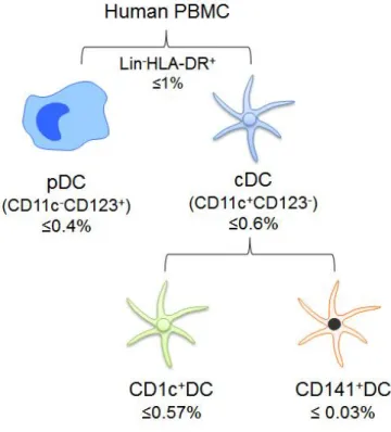
Figure 1. The subsets of DCs in human PBMC
Figure 2. Three different forms of moDCs after the differentiation of peripheral blood
CD14+monocytes
Figure 3. Attached cells in monocyte culture are immature DCs which relatively express high CD141 and CD86
Figure 4. Non-attached cells are not important to CD141 expression on the attached cells in GM-CSF/IL-4-driven culture system
Figure 5. Adhesive capacity is partially involved in CD141 expression of moDCs
Figure 6. Dead cell uptake and TLR3 response of attached CD141+moDCs
Figure 7. Allogeneic MLR by attached CD141+moDCs
Supplementary Figure 1. The absolute number of nine different subsets after additional 3-day culture
Supplementary Figure 2. Surface molecules of nine different subpopulations after additional 3-day culture
List of Abbreviations
Abs Antibodies
Ags Antigens
BDCA Blood dendritic cellsantigen
BMDCs Bone marrow derived dendritic cells
cDC Conventional dendritic cells
CTL Cytotoxic T lymphocyte
DC Dendritic cell
ELISA Enzyme-linked immunosorbent assay
FBS Fetal bovine serum
GM-CSF Granulocyte-macrophage colony-stimulating factor
HIV Human immunodeficiency virus
IFN Interferon
IL Interleukin
MFI Mean fluorescence intensity
MHC Major Histocompatibility Complex
MLR Mixed lymphocyte reaction
moDCs Monocyte derived dendritic cells
PBMC Peripheral blood mononuclear cell
PBS Phosphate buffered saline
PolyI:C Polyionsinic:polycytidylic acid
I. Review of Literature
1. Human monocyte-derived DC
1.1 Monocyte
Monocytes are a group of immune cells that originate in bone marrow and are released into peripheral blood, where they circulate for several days [1]. Monocytes represent 5-10% of peripheral leucocytes and are probably best known for serving as a systemic reservoir of myeloid precursors that are needed for the renewal of tissue macrophages and dendritic cells [2]. In humans, monocytes have been divided into three subtypes based on relative surface expression of LPS co-receptor CD14 and FCγ III receptor CD16 [3] (Fig. 2). Classical monocytes (CD14++CD16-) are the major population of human monocytes (~80%). the remaining population (~20%) of human monocytes are further subdivided into two subtypes. The more abundant non-classical subsets (CD14+CD16++) are characterized by very low expression of surface CD14 and high levels of CD16, whereas intermediate subset (CD14++CD16+) express high levels of CD14 molecules [4].
1.2 In vitro-derived moDCs
Human DC function has been obtained from DC derived from monocytes differentiated in vitro by culture with GM-CSF and IL-4 over the past two decades [1, 5]. In vitro culture system provides a versatile environment for skewing DC with potent cytotoxic T lymphocyte response (CTL) [6], Th1 [7], 2
[8], 17[9] or regulatory functions [10] by adding and substituting cytokines, growth factors, and other stimuli to culture. These in vitro derived moDCs have been extensively used in the clinic, mostly as vaccines to induce anti-tumor immune responses in cancer patients [11]. Although the physiological relevance of moDCs is unclear, the similarity of these cells with DCs found in human inflamed tissues suggests them to be most closely related to inflammatory DCs [12]. However, in vitro cultured moDCs are heterogeneous. Some of them attached on the bottom of culture plate were observed but not reported about their characteristics although suspended one is generally recognized as immature moDCs [1, 5, 13]. Therefore, in vitro cultured moDCs are still in need for further investigation.
2. Human CD141
+dendritic cell
2.1 Introduction
Human CD141 (BDCA3)+ DCs are found in various tissues such as blood, tonsil, lymph nodes, spleen, skin, liver, lung, and intestine [14-16]. CD1c (BDCA1)+ DCs, the other subset of conventional dendritic cells (cDCs),
represent by far the most predominant cDC subset in human blood, whereas CD141+ DCs consist of a minor blood population (Fig. 1). They exist approximately 0.03% in peripheral blood mononuclear cell (PBMC) [3, 16, 17]. Although this rareness was limitation for detailed functional analysis of this subset, transcriptional analysis suggests high correlation of function between human CD141+DCs and their mouse CD8+/CD103+counterparts [18, 19].
2.2 Characterization of CD141+ DCs
CD141 is a cell surface-expressed transmembrane glycoprotein, known as thrombomodulin or BDCA3. Together with BDCA1 (CD1c), BDCA2 (CD303) and BDCA4 (CD304), CD141 is generally used to classify human DC. However, CD141 expression is not selectively expressed by the CD141+ DC subset, since intermediate expression of CD141 has also been found on monocytes and other DC subsets, such as blood plasmacytoid DCs (pDCs), skin DCs [18, 20, 21]. Although the typical CD141+ DC subsets express higher levels of CD141 than other DCs, it is important to use CD141 in combination with
distinct markers that are unique to CD141hi DC subsets to assure selection of CD141+ DCs. CD141+ DCs uniquely express the CLEC9A (c-type lectin domain family 9A), known as DNGR-1, which is damage-associated molecular pattern (DAMP) receptor that senses necrotic cells [22, 23]. In addition, they express chemokine receptor, XCR1 and preferentially induce optimal cytotoxic T lymphocyte (CTL) responses through the interaction of XCR1 with XCL1 selectively expressed on CD8+ T cells [24-26]. DEC205, endocytotic receptor, also expressed high levels on CD141+ DCs recognizes dead cells [16, 27]. CD141+ DCs specifically express cell adhesion molecule 1 (CADM1), also
called as nectin-like protein 2 (Necl2) [28]. Although CADM1 is expressed on various cell types, its expression among blood leukocytes is restricted to CD141+ DCs [29]. Furthermore, CD141+ DCs express significantly high level of toll like receptor (TLR) 3 [16]. Needless to say that TLR3 ligation by polyionsinic:polycytidylic acid (polyI:C) is commonly used for in vitro stimulation of CD141+DCs.
2.3 The role of CD141+ DCs in T cell responses
One of the first studies on the function of CD141+ DCs demonstrated that blood CD141+DCs are superior to produce IL-12p70 in response to stimulation with a cytokine cocktail of polyI:C, interferon (IFN)γ, tumor necrosis factor (TNF)a, IFNα, interleukin (IL)-1β compared to CD1c+ DCs [16]. In addition,
blood CD141+ DCs were reported to produce IFNα, IFNβ, IL-6, IL-8, TNFα, and CXCL10 in response to polyI:C [16, 30]. The production of these cytokines
by CD141+ DCs upon stimulation suggests that these cells could induce Th1 responses, which play a key role in anti-viral immune responses. Co-culture of CD141+ DCs with allogeneic naïve CD4+ T cells induced high production of the Th1 cytokines IFNγ and IL-2 [16, 17]. Furthermore, CD141+ DCs induced high proliferation of allogeneic naïve CD4+ T cells. However, many studies showed it is not clear the plasticity of CD141+ DCs in terms of Th2-skewing [31] and tolerogenic functions [20, 32] in addition to their Th1-inducing ability.
2.4 Cross-presentation of CD141+ DCs
DCs are specialized in capture antigen and present antigenic molecule to T cells. The majority of antigens derived from endogenously synthesized proteins presented by DCs in MHC class I, however, DCs have the capacity to cross-present exogenous-derived antigen in MHC class I molecules. This pathway actives CD8+T cells which play a key role in the immune control of tumors and viral infections [33]. CD141+ DCs initially reported that they exhibit a
specialized cross-presenting capacity because they were likely to be more efficient at cross-presentation of soluble and cell associated antigens than other DC subsets [17, 25]. However, recent studies showed that other DCs are able to cross-present soluble antigens as efficient as CD141+ DCs [34-36]. Although controversial results were reported about the efficiency of CD141+ DCs at
cross-presentation of soluble antigens, CD141+ DCs exhibit a specialized
mechanism to transfer antigens from late endosomes and lysosomes to the cytosol [37] and superiority at the uptake and cross-presentation of necrotic
cell-associated antigens [16, 17, 25, 36]. CLEC9A and TLR3 were shown to mediate and enhance cross-presentation of dead cell-associated antigens [22, 38, 39]. In addition, XCR1 may contribute to activate CD8+ T cells upon presentation of exogenous antigens on MHC class I by CD141+DCs, which can
enhance cellular interaction with CD8+ T cells by the XCR1-XCL1 axis [24, 25].
2.5 Development of CD141+ DCs
The development of CD141+ DCs is poorly defined because of their low
abundance and difficult isolation. Nevertheless, possibility to generate CD141+
DCs in vitro has recently activated research into the development of this subset [17, 28, 40]. In vitro systems have been described that generate CD141+ DCs from HPC [17, 28]. In the system human lineage-negative cord blood cells, including HPC, were cultured under the stem cell factor (SCF), Flt3L, IL-3, and IL-6 and then differentiated with Flt3L, GM-CSF, and IL-4 which induced the small population of CD141+ DCs [17]. The protocol described by other group involved thrombopoietin (TPO), SCF, Flt3L, IL-6 and yielded much higher numbers of CD141+ DCs [28]. Although these culture systems involve Flt3L, the requirement of Flt3L in CD141+ DC development remain to be elucidated. In addition, GM-CSF, a cytokine involved in development of peripheral DCs, may contribute to development of CD141+ DCs [41, 42]. Furthermore, the
nature of precursors of CD141+DCs in human needs to be further characterized. According to CD141+ DCs in vivo, it is likely that lymphoid tissue and
peripheral tissue CD141+ DCs derived from blood CD141+ DCs because blood CD141+ DCs not fully differentiated until reside in peripheral tissue like skin [18, 34]. On the other hand, recent studies describe that the heterogeneous precursors in blood are pre-committed to become either CD1c+or CD141+DCs
[43, 44]. Therefore, roles for cytokines and precursors in development of CD141+DCs remain to be determined.
II. Introduction
Dendritic cells (DC), as one of the professional antigen-presenting cells, process antigenic materials and present epitopes to T lymphocytes [45, 46], bridging between the innate and the adaptive immune system [47, 48]. As well studied, exogenous antigens, such as extracellular pathogens and tumor, engulfed by DC are enzymatically processed into peptide fragments fitting onto major histocompatibility complex (MHC) II which interact with T-cell receptor of CD4+ helper T cells [49]. However, in addition to this conventional processing, DCs are able to cross-prime CD8+ T cells by alternatively loading the endocytosed antigens on MHC I, eliciting the clearance of the damaged cells including tumor cells [49]. This cross-priming ability allowed DC as a prime candidate that deserves intense investigation for the development of anti-cancer vaccine [50]. In practice, clinical trials of DC-based immunotherapy promised a long-term survival of melanoma, prostate cancer, primary brain tumors (glioma), and renal cell cancer patients [11, 51, 52].
Less than two decades ago, researchers found that human CD141+ DCs are counterpart of mouse CD8α+ lymphoid DC or CD103+ non-lymphoid tissue equivalent, both of which are specialized in cross-presentation [17-19]. In particular, human CD141+ DCs highly express CLEC9A, XCR1, and TLR3, which play unique function in cross-priming [16, 17, 25]. They are prone to induce cytotoxic T lymphocyte (CTL) responses through interaction of XCR1 with XCL1 selectively expressed on CD8+ T cells [24-26], as well as to sense
and present necrotic cell bodies via CLEC9A, c-type lectin receptor [16, 17]. Moreover, CD141+ DCs could be strongly activated by polyI:C, toll like receptor 3 (TLR3) agonist, as assessed by expression of costimulatory molecules such as CD80 and CD40 and production of inflammatory cytokines such as IL-6 [16, 18, 53]. However, there are discrepancies between the initial reports characterizing human CD141+DCs and still, many questions about their ontogeny and distribution need to be answered.
A major obstacle to investigating CD141+ DC biology and for further applying them to the therapeutic is their paucity, ‘in vivo rareness’. [54, 55]. There are a series of attempts to overcome this problem by in vitro generating CD141+ DCs from cord blood (CB) progenitors [17, 28]. According to the published protocol, CD141+ DCs could be differentiated into CD34+ cells isolated from CB, then cultured with five different cytokines for 7 days, followed by additional culture for 13 days with some of the cytokines replaced [17]. It was recently reported that a modification of this protocol advanced in yielding the more cell numbers [28]. However, fundamental limitation of CB as resources strongly demands further investigation for in vitro generating CD141+ DCs.
In the present study, I found that CD141+ DCs could be generated from adult peripheral blood CD14+monocytes, cultured with GM-CSF and IL-4 for 8 days.
[1, 13, 46] Among these monocyte-derived dendritic cells (moDC), the cells attached on the bottom of culture plate [5] expressed high CD141 than compared to other cells that loosely attached or suspended. The attached
CD141+ moDCs partially expressed CLEC9A and performed dead-cell uptake effectively compared to the others. They exhibited increase of costimulatory molecules and cytokines such as IL-6 in response to polyI:C treatment. In addition, they did not activate allogeneic naïve T cells in mixed lymphocyte reaction. These data suggested that CD14+ monocytes in adult peripheral blood could be an alternative source to obtain in vitro generation of CD141+ DCs for clinical application.
III. Materials and methods
Ethical statement
Normal adult blood samples were anonymously provided by the Blood Center of Korean Red Cross, Seoul under the approval of the Institutional Review Board of Korean Red Cross and the agreement for research purpose. The written informed consent from blood donors with respect to taking blood samples for research purposes was obtained and approved by the Ethics Committee of Korean Red Cross. All experimental procedures using human blood were performed under the approval of the Institutional Review Board at the Seoul National University (IRB no. E1702/001-001). Data were all analyzed anonymously.
Isolation of human monocytes
Peripheral blood mononuclear cells (PBMC) were purified from normal adult human blood by density gradient centrifugation using Ficoll-Paque Plus™ (GE Healthcare, Life Sciences). CD14+ monocytes were isolated from the PBMC using the BD IMagTM anti-human CD14 antibody, a magnetic bead-based positive selection kit (BD Biosciences). As soon as isolated, the cells were washed with RPMI 1640 more than two times and used immediately for further experiments. CD14+ monocytes are routinely obtained with above 95% purity, verified by flow cytometry.
CD14+monocyte culture
CD14+ monocytes were cultured in RPMI 1640 containing heat-inactivated 10% (vol/vol) FBS and 1% (vol/vol) antibiotics/antimycotic solution (all from Invitrogen) at 37°C under 5% CO2. For generation of moDCs, the cells were
incubated at 1 ~ 2 × 10⁶cells/ml for 8 days with 500 U/ml rhIL-4 and 800 U/ml rhGM-CSF (all from JW CreaGene Inc.). Fresh media containing rhIL-4 and rhGM-CSF were added by half of the initial volume every 3 days. Unless indicated otherwise, the cells were split into three different phases (suspended, semi-attached, and attached cells) after 8 days of the culture and then directly used for analysis or further cultured separately for 3 days. In our experimental setting, suspended cells (Sus) were obtained by directly collecting the culture media, and then semi-attached cells (Sem), by adding and gently washing with PBS. Finally, attached cells (Att) were obtained after incubation with cell dissociation buffer (Invitrogen) for 6 min.
Wright–Giemsa staining
CD14+ monocyte culture was performed in the cell culture slide (SPL) for moDC generation, as above mentioned. The cells except for the attached cells were diluted at 2×10⁵ cells/ml in 4% paraformaldehyde and used for the cytospin at 1100 rpm for 10 min. The slides were air-dried for 15 min, fixed and stained with Differential Quik Stain Kit (Polysciences, Inc.). Images were taken with an OLYMPUS CKX58 microscope.
Flow cytometry
The cells were incubated with antibodies at 4 °C for 20 min in staining buffer (1 × PBS containing 0.1% BSA and 0.1% sodium azide). For intracellular staining, the cells were fixed, permeabilized with an intracellular fixation and permeabilization buffer set (eBioscience) according to the manufacturer’s instructions, and stained for TLR3. Samples were acquired on Canto II (BD Biosciences) and data were analyzed with FlowJo software (TreeStar). Antibodies used for flow cytometric analyses were fluorochrome-labeled mAbs against human CD86 (IT2.2), CD207 (10E2), CD1c (L161), HLA-DR (L243), CD16 (3G8), CD1a (HI149), CD64 (10.1), CD209 (9E9A8), CD326 (9C4), CD141 (M80), CD11b (ICRF44), (67A4), CD15 (W6D3), CD163 (GHI/61), CD80 (2D10), LAP (TW4-2F8), CD273 (24F.10C12), CD274 (29E. 2A3), CD284 (HTA125), CD282 (TL2.1), CD40 (5C3), CD83 (HB15e), Siglec-8 (7C9), CD25 (M-A251), TLR3 (TLR3.7), CLEC9A (683409), XCR1 (1097A) (from BD Biosciences, eBioscience or R&D systems).
Poly I:C stimulation
poly I:C (InvivoGen) were added at 10μg/ml on 11 days of the CD14+ monocyte culture (as mentioned above). After 16h of culture, cells were analyzed by flow cytometry, or after 1 day of culture, the supernatants were collected to detect cytokines.
Mixed lymphocyte reaction
CD4+ T or CD8+ T cells were isolated from PBMC with BD IMagTM anti-human CD4 antibody or anti-anti-human CD8 antibody, respectively. Isolated T cells were labeled with CTV (2.5μM) and then co-cultured for 6 days with moDCs pre-incubated in the presence or absence of 10μg/ml poly I:C for 16 h. The ratio of moDCs to T cells was 1:10. Allogeneic T cell proliferation and activation was assessed by flow cytometry.
Dead cell uptake
Dead cell uptake was performed as described previously with modification [17]. Cryopreserved autologous PBMC were thawed and heat killed (90ºC, 30min) then labeled with CFSE. Dead cells were added to moDCs at 1:1 ratio for 2 h at 4°C or 37°C. For flow cytometric analysis, cells were stained for HLA-DR and the percentage of CFSE+HLA-DR+ cells was calculated by subtracting the frequency of positive events at 4°C (binding) from the frequency at 37°C (binding + uptake).
Enzyme-linked immunosorbent assay (ELISA)
Human IL-6, TNF-α, IL-2, IFN-γ, IL-17 in the culture supernatants were measured by using DuoSet ELISA kits (R&D Systems, USA) according to the manufacturer’s instruction. Optical density was measured by VersaMax ELISA Microplate Reader (Molecular Devices, USA) at wavelength 450 nm.
Statistical analysis
The mean value ± standard deviation was determined on the basis of at least 3 different blood samples. All the experiments were performed at least three times with different blood samples. All the data are representative or independent experiments yielding similar results. For comparison of means between two groups, the data were analyzed using two-tailed paired student’s t-test and considered statistically significant when p-value was less than 0.05. For multiple group comparison, one-way ANOVA followed by a Friedman test and the results were corrected by Dunn’s multiple comparison test. A P-value of less than 0.05 was considered significant. All statistical analyses were performed using GraphPad Prism 5 version 5.01 (GraphPad Software, USA).
IV. Results
1) Three different forms of moDCs after the differentiation of peripheral blood CD14+monocytes
moDCs obtained from monocyte culture in the presence of GM-CSF and IL-4 have been widely used in human DC studies [5, 13]. These moDCs were observed to take attached, semi-attached, or suspended forms in the culture condition [5], among which the last is generally recognized as an immature human moDCs [5, 56]. However, the attached or semi-attached cells are not well investigated, with a superficial understanding of heterogeneity of moDCs. Thus, I distinguished the three forms of moDCs, based on their adhesive capacity (Fig. 2A) as explained thoroughly in the Materials and Methods. This protocol allowed us to gain consistently app. 0.56 × 10⁶of attached cells (Att) and 0.42 × 10⁶of semi-attached cells (Sem) from 1 × 10⁷of CD14+ monocytes
(Fig. 2B). The attached and semi-attached cells consisted of app. 20 % and 12 % of the total moDCs (Fig. 2C), respectively. As previously reported [5, 56], suspended cells were the major population. I stained cells with Wright–Giemsa solution and found that the three forms of cells were similar in the cell-size, but the attached cells showed irregular shapes with dendrites elongated (Fig. 2D). FSC-A value of the attached cells was relatively higher than that of the others (Fig. 2E), which is seemingly attributed by their dendrites (Fig. 2D). Their SSC-A value was also slightly greater than the others (Fig. 2E), indicating that
the attached cells exhibited somewhat higher internal-complexity. Collectively, these data suggested that CD14+monocyte treated with GM-CSF and IL-4 for 8 days yielded the attached cells having relatively larger with elongated dendrites.
Figure 2. Three different forms of moDCs after the differentiation of peripheral blood CD14+ monocytes. CD14+ monocytes (1 x 10⁷) were cultured with GM-CSF and IL-4 for 8 days. (A) To analyze each subset
supernatant from moDC culture was harvested for suspended cells (Sus) and PBS washing for collecting semi-attached cells (Sem). Then, attached cells (Att) on the bottom of plate were collected by using cell dissociation buffer. (B) Absolute numbers of three subsets of moDCs are shown. Results from twelve independent cultures for each DC subset are shown as individual line. To determine the significance, one-way ANOVA followed by a Friedman test corrected by Dunn’s multiple comparison test was performed on twelve independent cultures from twelve different donors. *p<0.05, **p<0.01, ***p<0.001. (C) The proportion of three subsets of moDCs from twelve independent cultures. (D) Microscopy analysis of the morphology of Giemsa stained moDC subsets separated at the end of culture. Bar = 10 µm. (E) FSC-A and SSC-A level of three moDC subsets. One representative result of at least nine independent cultures is shown.
2) Attached cells in monocyte culture are immature DCs which relatively express high CD141 and CD86
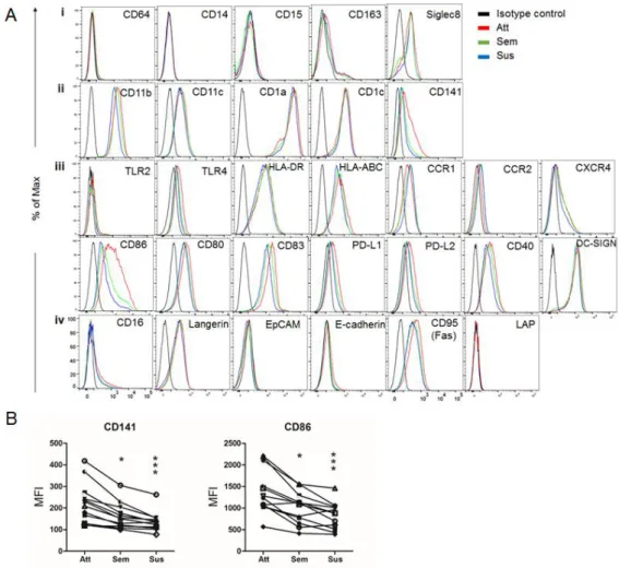
Next, to determine phenotypic traits of each type of cells obtained by our protocol, I examined the expression of surface molecules involved in T cell-stimulatory/inhibitory, epithelia-interacting, bacteria-sensing, and migratory features of DC, as well as cell-identifying markers (Fig. 3A). CD64, CD14, CD15, CD163 and Siglec 8 were not expressed on the attached one (Fig. 3Ai), ruling out the possibility that they are monocytes, macrophages and granulocytes [3, 57-59]. Instead, the attached cells expressed typical molecules defining human myeloid DC subsets, such as CD11b, CD11c, CD1a and CD1c, at similar level with that of the semi-attached or suspended one (Fig. 3Aii) [60]. The expressions of almost all the functional molecules, which I analyzed, including TLRs, MHC classes, chemokine receptors and B7 families except CD86 were similar among the three forms, whereas, interestingly, CD141 and CD86 were considerably higher in the attached cells. Indeed, the mean fluorescence intensity (MFI) of CD141 and CD86 was significantly greater in the attached cells relative to those of the others (Fig. 3B), suggesting that the attached cells in the 8-day culture of monocytes in the presence of GM-CSF and IL-4 are immature moDCs selectively expressing relatively higher CD141 and CD86.
Figure 3. Attached cells in monocyte culture are immature DCs which relatively express high CD141 and CD86. CD14+ monocytes were cultured with GM-CSF and IL-4 for 8 days. (A) A variety of cell surface markers of three subsets (Att, attached; Sem, semi-attached; Sus, suspended) of moDCs were analyzed by using flow cytometry. (i, phenotype markers of macrophages, monocytes, and granulocytes; ii, specific molecules for DCs; iii, TLRs, MHC molecules, chemokine receptors and B7 families; iv, additional markers
histogram. One representative result of at least four independent experiments in shown. (B) MFI of CD141 and CD86 on each subset. Results from twelve independent cultures for each DC subset are shown as individual line. To determine the significance, one-way ANOVA followed by a Friedman test corrected by Dunn’s multiple comparison test was performed on twelve different donors. *p<0.05, **p<0.01, ***p<0.001.
3) Non-attached cells are not important to CD141 expression of the attached cells in GM-CSF/IL-4-driven culture system
Next, I sought to gain an insight into factors contribute to the generation of the attached cells and their CD141 expression. Of note, I could obtain the attached cells only after 1 day of the culture and, absolute number of the attached cells was gradually decreased (Fig. 4A). However, the preferential expression of CD141 and CD86 on attached cells was observed at the late phase of the culture (Fig. 4A). To determine whether the cells attached at 1-day culture could express CD141 and CD86 without subsequent influences by non-attached cells, I discarded the cells floating in the supernatant of 1-day or 6-day culture and then supernatant was added back to the cells after washing with PBS (Fig. 4B). Interestingly, neither CD141 nor CD86 on the attached cells were not affected by discarding the non-attached cells, which suggested direct or indirect factors by non-attached cells are negligible to selective expression of CD141 and CD86 on the attached moDCs (Fig. 4C).
Figure 4. Non-attached cells did not have an impact on CD141 expression on the attached cells in GM-CSF/IL-4-driven culture system. CD14+
monocytes were treated with GM-CSF and IL-4. (A) The cells were harvested and analyzed for the absolute number and the MFI of CD141 and CD86 on three types of moDCs at indicated days. Results from at least four independent cultures for each DC subset are shown as dot with bar overlays indicating SEM. To determine the significance, one-way ANOVA followed by a Friedman test corrected by Dunn’s multiple comparison test was performed on three different donors. *p<0.05, **p<0.01, ***p<0.001. (B) To exclude influence of non-attached cells including suspended (Sus, blue) and semi-non-attached cells (Sem, green) ① supernatant containing Sus of 1-day or 6-day culture for moDCs was collected. ② The plate was washed with PBS and harvested, ③ which were centrifuged at 1200 rpm for 5min. ④ Then, the supernatant excluding Suswas added back to the culture left with only attached cells (red). (C). Attached moDCs from control group (con) and the groups that non-attached cells discarded were harvested analyzed at 1- (1d) and 6-day (6d). MFI of CD141 and CD86 were normalized with that of control group. Results with at least five independent experiments for each DC subset.
4) Adhesive capacity is partially involved in CD141 expression of moDCs
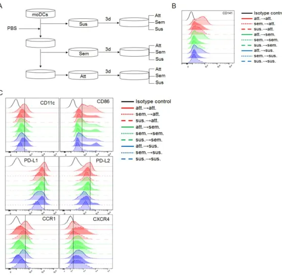
To determine whether attached property affects the expression of CD141, I separated subsets at 8 days after the culture and then cultured for additional 3 days (Fig. 5A). It was evidence that the Att were mostly remained attached but some became suspended. Similarly, suspended and semi-attached groups were mostly semi-attached but some attached after the additional culture (Supplementary Fig. 1). Then, I checked expression of CD141 on the nine different subsets after the additional culture. The expression of CD141 was increased on the moDCs retaining adhesion to the plate bottom (att→att) (Fig. 5B). The attached moDCs showed higher expression of surface CD141 than those of the others, attached cells derived from suspended (sus→att) and semi-attached moDCs (sem→att) or non-semi-attached cells derived from semi-attached moDCs (att→sem, att→sus). It suggested that attached property could affect expression of CD141. In addition, unlike other molecules (Supplementary Fig. 2), CD11c, CD86, PD-L1, PD-L2, and CCR1 were increased on attached moDCs (Fig. 5C). Especially, the expression of PD-L1, PD-L2, and CXCR4 are expressed on all the subsets which were not expressed at 8 days. These results suggested that all subsets after additional 3-day culture have potential of heterogeneity.
Figure 5. Adhesive capacity is partially involved in CD141 expression of
moDCs. CD14+monocytes were treated with GM-CSF and IL-4 for 8 days. (A) Three types of moDCs, based on the attachment, were cultured separately for additional 3 days with media containing GM-CSF and IL-4. Then, nine different subpopulations were analyzed for (B) the expression of CD141 (C) CD11c, CD86, PD-L1, PD-L2, CCR1, and CXCR4 expression. Isotype control is shown as gray on each histogram. One representative result of at least three
5) Dead cell uptake and TLR3 response of attached CD141+moDCs
To further characterize these moDCs, I examined the expression of CLEC9A [5, 26], XCR1 [13, 14], TLR3 [3, 53], and CADM1 [5, 17] since they are highly expressed on CD141+ DCs as previously reported. As shown in Figure 6A,
attached CD141+moDCs partially expressed CLEC9A, XCR1, and TLR3 more than other subsets. Additionally, none of the subsets expressed CADM1.
It has been suggested that human CD141+ DCs expressed high levels of CLEC9A which specialized in antigen derived from dead cell uptake [12, 28]. It has been shown to mediate and enhance cross-presentation of dead cell-associated antigens from viral infected of tumor cells [10, 26, 27]. Therefore, I examined CFSE labeled-heat killed autologous PBMCs uptake by attached CD141+ moDCs. I observed that the percentage of attached CD141+ moDCs expressed CFSE was higher than other subsets (Fig. 6B). Therefore, attached CD141+moDCs partially expressing CLEC9A (Fig. 6A) were efficiently uptake dead cells.
Human CD141+ DCs specifically express high level of TLR3 and are triggered by synthetic ligand, poly I:C enhancing cross-presentation [13, 61, 62]. Next, the attached CD141+ moDCs were treated with polyI:C for 16 hrs. The results demonstrated that attached CD141+ moDCs matured phenotypically in response to poly I:C stimulation compared to the other subsets (Fig. 6C). In addition, IL-6 and IL-1β production by attached CD141+ moDCs treated with
poly I:C was higher than other subsets (Fig. 6D). However, TNFα, 8, and IL-10 secretion were not upregulated in attached CD141+moDCs.
Figure 6. Dead cell uptake and TLR3 response of attached CD141+moDCs.
CD14+monocytes were treated with GM-CSF and IL-4 for 8 days. Three types of moDCs, based on the attachment, were cultured separately for additional 3 days with media containing GM-CSF and IL-4. (A) MFI of CLEC9A, XCR1, and TLR3 on each of the cell subsets. Results from three independent cultures for each DC subset are shown as individual line.
(B) The moDCs were incubated with CFSE-labeled heat killed (90℃, 30 min) autologous PBMCs at 1:1 ratio as described in the Materials and Methods. Uptake of dead cells by moDCs was quantified by flow cytometry. (C) Fold change of MFI of costimulatory molecules CD80, CD83, CD86, and CD40 by the subsets after 16hr culture in media alone or in the presence of poly I:C. The fold change of MFI for at least three donors is shown as individual lines. To determine significance, one-way ANOVA followed by a Friedman test corrected by Dunn’s multiple comparison test was performed on twelve independent cultures from twelve different donors. *p<0.05, **p<0.01, ***p<0.001. (D) moDC subsets were stimulated with polyI:C (PIC). Culture supernatants were examined by using ELISA. Results from three independent cultures for each DC subset are shown as mean ± SEM.
6) Allogeneic mixed lymphocyte reaction by attached CD141+moDCs
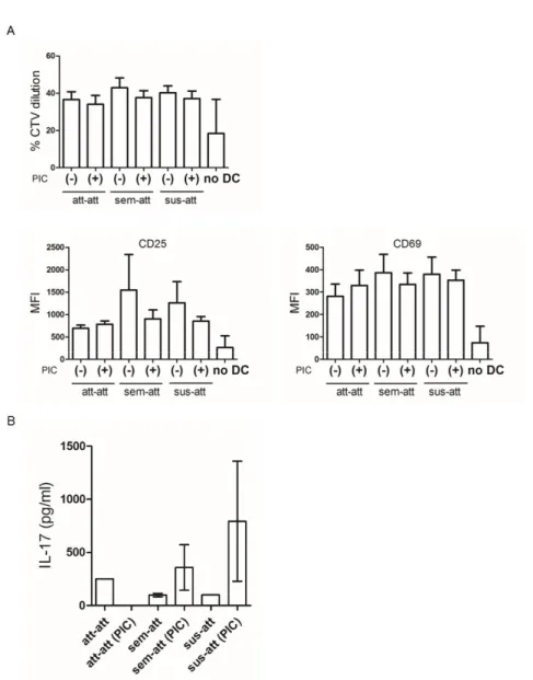
One of important features of DCs is the ability to induce the proliferation of allogeneic T cells in a mixed lymphocyte reaction (MLR). In addition, CD141+ DCs are known as strong inducers of Th1 responses particularly after poly I:C stimulation [3, 16]. Interestingly, the results showed that the attached CD141+ moDCs induced neither differentiation nor activation (Fig. 7A) of allogeneic CD4+T cells in the MLR. In addition, IL-2 and IFN-γ were not detected by all the subsets while IL-17 was observed exclusively in each group (Fig. 7B).
Figure 7. Allogeneic MLR by attached CD141+ moDCs. Bead-sorted
allogeneic CD4+ T cells, labeled with CTV, were co-cultured with unstimulated (-) or poly I:C (PIC) activated (+) moDCs, or in the absence of DC (no DC) for 6 days. (A) CD4+ T cell proliferation by CTV dilution, and MFI of CD25 and
CD69 was measured using flow cytometry. Results from at least five different samples are shown as mean ± SEM. (B) The supernatants were harvested and
IL-2, IFN-γ, and IL-17 production was examined by using ELISA. Results from three independent cultures for each DC subset are shown as mean ± SEM.
V. Supplementary results
Supplementary Fig. 1. The absolute number of nine different subsets after additional 3-day culture. CD14+ monocytes (1 x 10⁷) were treated with GM-CSF and IL-4 for 8 days. Three types of moDCs (1 x 10⁶), based on the
attachment, were cultured separately for additional 3 days with media containing GM-CSF and IL-4. Then, nine different subpopulations were analyzed for absolute numbers were examined. Results from at least ten different samples are shown as mean ± SEM.
Supplementary Fig. 2. Surface molecules of nine different subpopulations after additional 3-day culture Nine different moDC subsets were harvested
and analyzed for the expression of surface molecules. Isotype control is shown in gray on each histogram. One representative result of at least three independent experiments is shown.
VI. Discussion
CD141+ DCs are known to enhance cytotoxic T lymphocyte responses and,
therefore, has been studied for the potential use of therapeutic vaccines against viruses [15, 63] and potentially cancers. However, they are rare in vivo consisting less than 0.03% in periphery in human [48, 64]. Thus, CD141+DCs differentiated in vitro has a high value to investigate. The current study demonstrated that immature CD141+ DCs could be generated in vitro from
CD14+ monocytes in human peripheral blood mononuclear cells. It has been
reported that in vitro derived CD141+ DCs obtained from human CD34+ hematopoietic progenitor of cord blood [5, 16]. However, CD141+ DCs from peripheral blood are better candidate for a therapeutic vaccine than those from cord blood because the former could be obtained consistently as needed in fresh condition.
After the culture peripheral CD14+ monocytes for the generation of DCs, three different types of cells are observed depending on the ability to adhere to the culture plate. It has been demonstrated that suspended, loosely attached, and attached mouse bone marrow derived dendritic cells (BMDCs) cells exist after the culture [65, 66]. However, the characteristics of these subsets from human
in vitro cultured moDCs are unknown. Since the loosely attached mouse
BMDCs are reported as monocyte-derived macrophages expressed CD11c and MHC class II [65] and the attached BMDCs are divided into two populations,
MHCIIloF4/80hi macrophages and MHCIIhiF4/80lo DCs [66], I postulated that the attached or semi-attached moDCs can be macrophages or mixed cells containing macrophages and DCs. However, I found that all the subsets are not macrophages but immature moDCs and the attached moDCs expressed surface CD141 higher than those of the other subsets. This study is the first, at the best of our knowledge, describing the characteristics of subsets based on the attachment after the differentiation into moDCs.
Although the relationship between attachment and CD141 expression is unclear from the previous studies, I postulate that the attached property may affect the expression of CD141. At this end, I purified attached moDCs and other subsets and then cultured for additional 3 days. It was noting that the attached moDCs at 8 days are mostly remained the attached but some of them became suspended. In addition, moDCs maintained attached on the bottom of culture plate after additional 3-day culture expressed high level of CD141. It suggested that the attached property could affect the expression of CD141.
Additionally, I found that increase of CD141 on attached moDCs after 3-day culture was not affected by GM-CSF and IL-4 [67]. CD141 expression on attached moDCs after 3-day culture without these cytokines in media was not different from them cultured with the cytokines (data not shown). It suggested that it is not GM-CSF and IL-4 but other factors to increase CD141 on attached moDCs after 3-day culture.
On the other hand, the fact that human monocytes are divided into three types of subsets based on CD14 and CD16 could important to understand generation
of attached CD141+ moDCs [4, 34, 35]. According to previous studies, the subsets of monocytes could differentiate different types of DCs. For example, DCs in vitro generated from CD16+ monocytes expressed high level of CD86 [68, 69]. In present study, there are approximately 6% of CD14+CD16+ monocytes in total CD14+ monocyte from PBMC (data not shown). Therefore, It needs to be further investigated that these monocytes subsets could associate with generation of attached CD141+ moDCs [34, 70]. Additionally, the recent studies show CD141+ DCs could differentiated from pre-conventional DCs (cDCs) in PBMC [31, 32]. It has been reported that human pre-cDCs are a mixture of cells committed to + moDCs either the CD141+ lineage (CD172a−
pre-cDCs) or the CD1c+ lineage (CD172a+ pre-cDCs) [31]. In addition,
CD100+CD34int blood precursors gave rise to CD1c+ and CD141+cDCs [32, 71]. These studies can help to understand whether some pre-committed progenitors could be related with generation of attached CD141+ moDCs. It would be also interesting to determine whether some factors in culture environment including cell to cell interaction, cytokine and specific adhesion molecules from each cell influence the generation of attached CD141+moDCs.
In conclusion, I suggested that immature CD141+DCs could be generated in
vitro from CD14+monocytes in human peripheral blood, which could be further developed into the strategy to yield cross-priming human DCs in vitro.
VII. Literature Cited
1. Lindstedt, M., K. Lundberg, and C.A.K. Borrebaeck, Gene Family Clustering Identifies Functionally Associated Subsets of Human In Vivo Blood and Tonsillar Dendritic Cells. The Journal of Immunology, 2005. 175(8): p. 4839-4846.
2. Bamboat, Z.M., et al., Human liver dendritic cells promote T cell hyporesponsiveness. J Immunol, 2009. 182(4): p. 1901-11.
3. Jongbloed, S.L., et al., Human CD141+ (BDCA-3)+ dendritic cells (DCs) represent a unique myeloid DC subset that cross-presents necrotic cell antigens. J Exp Med, 2010. 207(6): p. 1247-60.
4. Ziegler-Heitbrock, L., et al., Nomenclature of monocytes and dendritic cells in blood. Blood, 2010. 116(16): p. e74-80.
5. Poulin, L.F., et al., Characterization of human DNGR-1+ BDCA3+ leukocytes as putative equivalents of mouse CD8alpha+ dendritic cells. J Exp Med, 2010. 207(6): p. 1261-71.
6. Haniffa, M., et al., Human tissues contain CD141hi cross-presenting dendritic cells with functional homology to mouse CD103+ nonlymphoid dendritic cells. Immunity, 2012. 37(1): p. 60-73.
7. Robbins, S.H., Novel insights into the relationships between dendritic cell subsets. Genome Biology, 2008. 9(1).
8. Chu, C.C., et al., Resident CD141 (BDCA3)+ dendritic cells in human skin produce IL-10 and induce regulatory T cells that suppress skin inflammation. J Exp Med, 2012. 209(5): p. 935-45.
9. Andrzej Dzionek, Y.I., Katsuya Okawa, Jun Nagafune, Ju¨ rgen Ro¨ ck, Yoshiaki Sohma, Gregor Winkels, Monika Zysk, Yasunori Yamaguchi, and Ju¨ rgen Schmitz, Plasmacytoid: Dendritic Cells From Specific Surface Markers to Specific Cellular Functions. human immunology, 2002.
sensing of necrosis to immunity. Nature, 2009. 458(7240): p. 899-903.
11. Zhang, J.G., et al., The dendritic cell receptor Clec9A binds damaged cells via exposed actin filaments. Immunity, 2012. 36(4): p. 646-57.
12. Bachem, A., et al., Superior antigen cross-presentation and XCR1 expression define human CD11c+CD141+ cells as homologues of mouse CD8+ dendritic cells. J Exp Med, 2010. 207(6): p. 1273-81.
13. Crozat, K., et al., The XC chemokine receptor 1 is a conserved selective marker of mammalian cells homologous to mouse CD8alpha+ dendritic cells. J Exp Med, 2010. 207(6): p. 1283-92.
14. Hartung, E., et al., Induction of potent CD8 T cell cytotoxicity by specific targeting of antigen to cross-presenting dendritic cells in vivo via murine or human XCR1. J Immunol, 2015. 194(3): p. 1069-79.
15. Tullett, K.M., et al., Targeting CLEC9A delivers antigen to human CD141+ DC for CD4+ and CD8+T cell recognition. JCI Insight, 2016. 1(7): p. e87102. 16. Balan, S., et al., Human XCR1+ dendritic cells derived in vitro from CD34+
progenitors closely resemble blood dendritic cells, including their adjuvant responsiveness, contrary to monocyte-derived dendritic cells. J Immunol, 2014. 193(4): p. 1622-35.
17. Galibert, L., et al., Nectin-like protein 2 defines a subset of T-cell zone dendritic cells and is a ligand for class-I-restricted T-cell-associated molecule. J Biol Chem, 2005. 280(23): p. 21955-64.
18. Meixlsperger, S., et al., CD141+ dendritic cells produce prominent amounts of IFN-alpha after dsRNA recognition and can be targeted via DEC-205 in humanized mice. Blood, 2013. 121(25): p. 5034-44.
19. Yerkovich, S.T., et al., Allergen-enhanced thrombomodulin (blood dendritic cell antigen 3, CD141) expression on dendritic cells is associated with a TH2-skewed immune response. J Allergy Clin Immunol, 2009. 123(1): p. 209-216 e4.
20. Florian W. Velten, K.D., Johannes Bohlender, Patraporn Metharomand Sergij Goerdt, A gene signature of inhibitory MHC receptorsidentifi es a BDCA3+subset of IL-10-induceddendri tic cells with reduced allostimulatory
capacityin vitro. Eur. J. Immunol. , 2004.
21. Joffre, O.P., et al., Cross-presentation by dendritic cells. Nat Rev Immunol, 2012. 12(8): p. 557-69.
22. Segura, E., et al., Characterization of resident and migratory dendritic cells in human lymph nodes. J Exp Med, 2012. 209(4): p. 653-60.
23. Nizzoli, G., et al., Human CD1c+ dendritic cells secrete high levels of IL-12 and potently prime cytotoxic T-cell responses. Blood, 2013. 122(6): p. 932-42.
24. Segura, E., M. Durand, and S. Amigorena, Similar antigen cross-presentation capacity and phagocytic functions in all freshly isolated human lymphoid organ-resident dendritic cells. J Exp Med, 2013. 210(5): p. 1035-47.
25. Cohn, L., et al., Antigen delivery to early endosomes eliminates the superiority of human blood BDCA3+ dendritic cells at cross presentation. J Exp Med, 2013. 210(5): p. 1049-63.
26. Zelenay, S., et al., The dendritic cell receptor DNGR-1 controls endocytic handling of necrotic cell antigens to favor cross-priming of CTLs in virus-infected mice. J Clin Invest, 2012. 122(5): p. 1615-27.
27. Dalod, M., Professional cross-presenting CD8alpha-type CD141(hi) dendritic cells: we have got you in our skin! Immunity, 2012. 37(1): p. 3-5.
28. Poulin, L.F., et al., DNGR-1 is a specific and universal marker of mouse and human Batf3-dependent dendritic cells in lymphoid and nonlymphoid tissues. Blood, 2012. 119(25): p. 6052-62.
29. Greter, M., et al., GM-CSF controls nonlymphoid tissue dendritic cell homeostasis but is dispensable for the differentiation of inflammatory dendritic cells. Immunity, 2012. 36(6): p. 1031-46.
30. van der Aa, E., et al., BDCA3 expression is associated with high IFN-lambda production by CD34(+)-derived dendritic cells generated in the presence of GM-CSF, IL-4, and/or TGF-beta. Eur J Immunol, 2015. 45(5): p. 1471-81. 31. Breton, G., et al., Human dendritic cells (DCs) are derived from distinct
circulating precursors that are precommitted to become CD1c+ or CD141+ DCs. J Exp Med, 2016. 213(13): p. 2861-2870.
32. Villani, A.C., et al., Single-cell RNA-seq reveals new types of human blood dendritic cells, monocytes, and progenitors. Science, 2017. 356(6335).
33. Winfried F. Pickl, O.M., Petra Kohl, Johannes Stockl, Elisabeth Riedl, Clemens Scheinecker,t Concha Bello-Fernandez,t and Walter Knapp, Molecular and Functional Characteristics of Dendritic Cells Generated from Highly Purified CD14+ Peripheral Blood Monocytes. journal of immunology, 1996. 157: p. 3850-3859.
34. Patel, A.A., et al., The fate and lifespan of human monocyte subsets in steady state and systemic inflammation. J Exp Med, 2017.
35. Wong, K.L., et al., Gene expression profiling reveals the defining features of the classical, intermediate, and nonclassical human monocyte subsets. Blood, 2011. 118(5): p. e16-31.
36. Nikolaus Romani , D.R., Marion Heuer, Susanne Ebner, Brigitte Eibl, Dietger Niederwieser, Gerold Schuler, Generation of mature dendritic cells from human blood An improved method with special regard to clinical applicability. Journal of Immunological Methods 1996. 196: p. 137-151. 37. Thomas Tu¨ting, C.C.W., Dina M. Martin* Yvette L. Kasamon, Jennifer Rowles,,
C.L.S. Debora I. Ma, Jr. Stephan N. Wagner, Pierre van der Bruggen,, and M.T.L. Joseph Baar, and Walter J. Storkus, Autologous Human Monocyte-Derived Dendritic Cells. JI, 1998.
38. BERND HILDENBRAND1, D.L., BARBARA SAUER, CHRISTIAN HERTKORN, MARINA A. FREUDENBERG, J. HINRICH PETERS, and C.U.a.M.A. THOMAS NESSELHUT, IFN-γ Enhances TH1 Polarisation of Monocyte-derived. Anticancer Research, 2008.
39. Hiroyuki Tanaka, C.E.D., Manuel Rubio, Guy Delespesse, and Marika Sarfati, Human Monocyte–derived Dendritic Cells Induce Naive. JEM, 2000.
40. Alonso, M.N., et al., T(H)1, T(H)2, and T(H)17 cells instruct monocytes to differentiate into specialized dendritic cell subsets. Blood, 2011. 118(12): p. 3311-20.
41. Raker, V.K., M.P. Domogalla, and K. Steinbrink, Tolerogenic Dendritic Cells for Regulatory T Cell Induction in Man. Front Immunol, 2015. 6: p. 569.
42. Anguille, S., et al., Clinical use of dendritic cells for cancer therapy. The Lancet Oncology, 2014. 15(7): p. e257-e267.
43. Segura, E., et al., Human inflammatory dendritic cells induce Th17 cell differentiation. Immunity, 2013. 38(2): p. 336-48.
44. Sallusto, F., Efficient Presentation of Soluble Antigen by Cultured Human Dendritic Cells Is Maintained by Granulocyte/Macrophage Colony-stimulating Factor Plus Iuterleukin 4 and Downregulated by Tumor Necrosis Factor α. J Exp Med, 1994. 179: p. 1109-1118.
45. Kambayashi, T. and T.M. Laufer, Atypical MHC class II-expressing antigen-presenting cells: can anything replace a dendritic cell? Nat Rev Immunol, 2014. 14(11): p. 719-30.
46. Haniffa, M., M. Collin, and F. Ginhoux, Ontogeny and functional specialization of dendritic cells in human and mouse. Adv Immunol, 2013.
120: p. 1-49.
47. Villadangos, J.A. and P. Schnorrer, Intrinsic and cooperative antigen-presenting functions of dendritic-cell subsets in vivo. Nat Rev Immunol, 2007. 7(7): p. 543-55.
48. Boltjes, A. and F. van Wijk, Human dendritic cell functional specialization in steady-state and inflammation. Front Immunol, 2014. 5: p. 131.
49. R.Carbone, W.R.H.a.F., CROSS-PRESENTATION IN VIRAL IMMUNITY AND SELF-TOLERANCE. Nat Rev Immunol, 2001.
50. Palucka, K. and J. Banchereau, Cancer immunotherapy via dendritic cells. Nat Rev Cancer, 2012. 12(4): p. 265-77.
51. Gross, S., et al., Twelve-year survival and immune correlates in dendritic cell-vaccinated melanoma patients. JCI Insight, 2017. 2(8).
52. Palucka, K. and J. Banchereau, Dendritic-cell-based therapeutic cancer vaccines. Immunity, 2013. 39(1): p. 38-48.
53. Colletti, N.J., et al., TLR3 Signaling Promotes the Induction of Unique Human BDCA-3 Dendritic Cell Populations. Front Immunol, 2016. 7: p. 88.
54. O'Keeffe, M., W.H. Mok, and K.J. Radford, Human dendritic cell subsets and function in health and disease. Cell Mol Life Sci, 2015. 72(22): p. 4309-25.
55. Geginat, J., et al., Immunity to Pathogens Taught by Specialized Human Dendritic Cell Subsets. Front Immunol, 2015. 6: p. 527.
56. Nikolaus Romani, S.G., Daniela Brang, and A.L. , Bettina Trockenbacher,Fritsch, Ralph M. Steinman,S and Gerold Schuler, Proliferating Dendritic Cell Progenitors in Human Blood. J Exp Med, 1994. 180: p. 83-93. 57. Lavin, Y. and M. Merad, Macrophages: gatekeepers of tissue integrity.
Cancer Immunol Res, 2013. 1(4): p. 201-9.
58. Nakayama, F., et al., CD15 expression in mature granulocytes is determined by alpha 1,3-fucosyltransferase IX, but in promyelocytes and monocytes by alpha 1,3-fucosyltransferase IV. J Biol Chem, 2001. 276(19): p. 16100-6. 59. Bochner, B.S., Siglec-8 on human eosinophils and mast cells, and Siglec-F
on murine eosinophils, are functionally related inhibitory receptors. Clin Exp Allergy, 2009. 39(3): p. 317-24.
60. Jacques Banchereau1, F.B., Christophe Caux, Jean Davoust, Serge Lebecque, Yong-Jun Liu, Bali Pulendran, and Karolina Palucka, IMMUNOBIOLOGY OF DENDRITIC CELLS. Annu. Rev. Immunol., 2000.
61. Lauterbach, H., et al., Mouse CD8alpha+ DCs and human BDCA3+ DCs are major producers of IFN-lambda in response to poly IC. J Exp Med, 2010.
207(12): p. 2703-17.
62. Crozat, K., E. Vivier, and M. Dalod., Crosstalk between components ofthe innate immune system:promoting anti-microbial defensesand avoiding immunopathologies. Immunological Reviews, 2009.
63. Aymeric Silvin, C.I.Y., Xavier Lahaye,, et al., Constitutive resistance to viral infection in human CD141+dendritic cells. science immunology, 2017.
64. Dzionek, A., et al., BDCA-2, BDCA-3, and BDCA-4: Three Markers for Distinct Subsets of Dendritic Cells in Human Peripheral Blood. The Journal of Immunology, 2000. 165(11): p. 6037-6046.
65. Helft, J., et al., GM-CSF Mouse Bone Marrow Cultures Comprise a Heterogeneous Population of CD11c(+)MHCII(+) Macrophages and Dendritic Cells. Immunity, 2015. 42(6): p. 1197-211.
Phenotypically Different Dendritic Cells and Macrophages. Mol Cells, 2016.
39(10): p. 734-741.
67. van de Laar, L., P.J. Coffer, and A.M. Woltman, Regulation of dendritic cell development by GM-CSF: molecular control and implications for immune homeostasis and therapy. Blood, 2012. 119(15): p. 3383-93.
68. CD16+ and CD16- human blood monocyte subsets differentiate in vitro to dendritic cells with different ailities to stimulate CD4+ T cells. international immunology, 2001.
69. Rivas-Carvalho, A., et al., CD16+ human monocyte-derived dendritic cells matured with different and unrelated stimuli promote similar allogeneic Th2 responses: regulation by pro- and anti-inflammatory cytokines. Int Immunol, 2004. 16(9): p. 1251-63.
70. Geissmann, F., Development of Monocytes, Macrophages, and Dendritic cells. science, 2010.
71. Luque, M.C., et al., CD100 and plexins B2 and B1 mediate monocyte-endothelial cell adhesion and might take part in atherogenesis. Mol Immunol, 2015. 67(2 Pt B): p. 559-67.
VIII. Summary in Korean
인간 CD141+ 수지상 세포는 항원 교차 제시에 특화된 세포로, 수지상 세포를 기반으로 하는 암 치료를 위하여 널리 연구되었다. 체내에 극히 적 게 존재하는 현실적 한계를 극복하기 위하여 제대혈 유래 CD34+ 조혈 전 구세포로부터 CD141+ 수지상 세포를 생체 외 분화, 생성시키려는 시도들 이 있었으나, 여전히 이 세포를 생체 외에서 분화시키는 연구는 부족한 실 정이다. 본 연구는 CD141+ 수지상 세포가 일반적으로 단핵구 유래 수지상 세포 를 분화시키기 위해 사용하는 보편적인 배양방법에 의해서 생성이 가능하다 는 것을 보여준다. CD14+ 단핵구를 GM-CSF, IL-4 과 함께 8일 간 배 양하여 얻은 단핵구 유래 수지상 세포 중 일부는 배양 접시 바닥에 붙어 있 으며 이들은 CD141 발현이 특이적으로 높았다. 이 붙어 있는 세포들은 다 른 군집들에 비해 크기가 크고 가지를 많이 뻗고 있으며 CD86가 높게 발 현한다는 점을 제외하고는 전형적인 미성숙 단핵구 유래 수지상 세포의 특 성을 지닌다. CD141의 높은 발현은 추가 3일 배양 이 후에도 여전히 붙어 있는 세포에서 명확히 나타났으며 일부 CLEC9A를 발현하였다. 무엇보다 도, 이들은 죽은 세포를 효과적으로 인지하여 받아들이는 기능이 있음을 확 인할 수 있었다. 또한 흥미롭게도, polyI:C 처리에 대한 반응성과 동종 이 계 CD4+ T 세포와의 혼합림프구 반응에서 다른 군집과 반응성이 다른 것을 확인하였다.
종합적으로, 본 연구는 인간 말초 혈액 CD14+ 단핵구로부터 생체 외 배
양을 통하여 CD141+ 수지상 세포를 얻기 위한 앞으로의 연구에 기여 할