저작자표시-비영리-변경금지 2.0 대한민국 이용자는 아래의 조건을 따르는 경우에 한하여 자유롭게 l 이 저작물을 복제, 배포, 전송, 전시, 공연 및 방송할 수 있습니다. 다음과 같은 조건을 따라야 합니다: l 귀하는, 이 저작물의 재이용이나 배포의 경우, 이 저작물에 적용된 이용허락조건 을 명확하게 나타내어야 합니다. l 저작권자로부터 별도의 허가를 받으면 이러한 조건들은 적용되지 않습니다. 저작권법에 따른 이용자의 권리는 위의 내용에 의하여 영향을 받지 않습니다. 이것은 이용허락규약(Legal Code)을 이해하기 쉽게 요약한 것입니다. Disclaimer 저작자표시. 귀하는 원저작자를 표시하여야 합니다. 비영리. 귀하는 이 저작물을 영리 목적으로 이용할 수 없습니다. 변경금지. 귀하는 이 저작물을 개작, 변형 또는 가공할 수 없습니다.
Master’s Thesis of Agriculture
Application of high temperature aging of beef with
low-dose electron beam
and X-ray irradiation
소고기 고온숙성 시
저선량 전자선 및 X선 조사의 적용
February, 2018
So Yeon Kim
Department of Agricultural Biotechnology
Graduate School
Application of high temperature aging
of beef with low-dose electron beam
and X-ray irradiation
Advisor: Prof. Cheorun Jo, Ph.D.
Submitting a Master’s Thesis of Agriculture
February, 2018
Department of Agricultural Biotechnology
Graduate School
Seoul National University
So Yeon Kim
Confirming the Master’s Thesis written by
So Yeon Kim
소고기 고온숙성 시
저선량 전자선 및 X선 조사의 적용
Application of high temperature aging
of beef with low-dose electron beam
and X-ray irradiation
지도교수 조 철 훈
이 논문을 농학석사학위논문으로 제출함
2018년 2월
서울대학교 대학원
농생명공학부 동물생명공학전공
김 소 연
김소연의 석사학위논문을 인준함
2018년 2월
위 원 장 (인)
부위원장 (인)
위 원 (인)
i
Abstract
Application of high temperature aging
of beef with low-dose electron beam
and X-ray irradiation
So Yeon Kim
Department of Agricultural Biotechnology
Program in Animal Science and Biotechnology
Graduate School of Seoul National University
The effects of irradiation source (electron beam [EB] and X-ray [XR]), aging temperature (4 °C and 14 °C), and aging time (0, 3, 7, and 14 days) were evaluated on microbial quality, physicochemical properties, and calpain-1 autolysis in beef M. semimembranosus. Regardless of irradiation source, irradiation prior to aging reduced the total number of aerobic bacteria in beef and this reduction was maintained during aging. Irradiation did not affect the pH, b* value, shear force, or myofibrillar fragmentation index of beef at day 0. Degradation of sarcoplasmic and myofibrillar proteins was greater in beef aged at 14 °C compared with beef aged at 4 °C. EB- or XR-irradiated samples showed slower autolysis of calpain-1; however, beef tenderness was not
ii
affected. Therefore, EB or XR irradiation can be applied to beef prior to aging to control microbial growth during high temperature (14 °C) aging, thus shortening the aging time without adversely affecting the physicochemical properties of beef.
Keywords: Beef, High temperature aging, Irradiation, Microbial quality,
Tenderness
iii
Contents
Abstract
··· iContents
··· iiiList of Tables
··· viList of Figures
··· viiList of Abbreviations
··· viiiChapter I. Literature review
1.1. Meat aging
1.1.1. Physicochemical changes during aging ··· 11.1.2. Endogenous proteolytic enzymes ··· 3
1.1.3. Dry vs. Wet aging ··· 4
1.1.4. High temperature aging ··· 5
1.2. Irradiation
1.2.1. Radiation ··· 71.2.2. Food irradiation··· 8
1.2.3. Mechanism of bacterial inactivation ··· 9
iv
Chapter II. Application of high temperature aging of beef with
low-dose electron beam and X-ray irradiation
2.1. Introduction
··· 122.2. Materials and methods
2.2.1. Sample preparation and irradiation processing ··· 152.2.2. Microbial analysis ··· 16
2.2.3. pH ··· 16
2.2.4. Instrumental color measurement ··· 16
2.2.5. Shear force measurement ··· 17
2.2.6. Myofibrillar fragmentation index (MFI) ··· 17
2.2.7. SDS-PAGE and Western blot 2.2.7.1. Preparation of meat extraction ··· 18
2.2.7.2. Sodium dodecyl sulfate-polyacrylamide gel electrophoresis (SDS-PAGE) ··· 19
2.2.7.3. Western blot ··· 20
2.2.8. Statistical analysis ··· 21
2.3. Results and discussion
2.3.1. Total aerobic bacteria ··· 222.3.2. pH ··· 25
2.3.3. Surface color ··· 27
2.3.4. Shear force ··· 30
v
2.3.6. Sodium dodecyl sulfate-polyacrylamide gel electrophoresis
(SDS-PAGE) ··· 35 2.3.7. Western blot ··· 38
2.4. Conclusion
··· 40References
··· 41Summary in Korean
··· 54Acknowledgement
··· 57vi
List of Tables
Chapter I.
Table 1. Characteristics of food irradiation sources ··· 8
Chapter II.
Table 1. Total aerobic bacterial counts (log CFU/g) of the EB- and XR-
irradiated beef samples aged at 4 °C or 14 °C during 14 days of aging ··· 24
Table 2. pH of the EB- and XR-irradiated beef samples aged at 4 °C or 14 °C
during 14 days of aging ··· 26
Table 3. Surface color of the EB- and XR-irradiated beef samples aged at
4 °C or 14 °C during 14 days of aging ··· 28
Table 4. Shear force values (N) of the EB- and XR-irradiated beef samples
aged at 4 °C or 14 °C during 14 days of aging ··· 32
Table 5.Myofibrillar fragmentation index (MFI) of the EB- and XR-
vii
List of Figures
Chapter I.
Fig. 1. The electromagnetic spectrum ··· 7
Chapter II.
Fig. 1. Sodium dodecyl sulfate-polyacrylamide gel electrophoresis of the EB
and XR irradiated beef samples aged for 0 and 14 days at 4 °C or 14 °C. (STD, standard molecular bands; Control, non-irradiated; EB, electron beam-irradiated; XR, X-ray-irradiated) ··· 37
Fig. 2. Western blot of calpain-1 in the sarcoplasmic fraction of the EB- and
XR-irradiated beef samples aged for 0 and 14 days at 4 °C or 14 °C. (Control, non-irradiated; EB, electron beam-irradiated; XR,
viii
List of Abbreviations
ATP : Adenosine-5-triphosphate
EB : Electron beam
MFI : Myofibrillar fragmentation index
SDS-PAGE : Sodium dodecyl sulfate-polyacrylamide
gel electrophoresis
STD : Standard molecular bands
WHC : Water holding capacity
1
Chapter I.
Literature review
1.1. Meat aging
1.1.1. Physicochemical changes during aging
Aging, also known as conditioning, is a process to convert animal muscles into meat (Khan et al., 2016). After death, mammalian muscles undergo the onset of rigor mortis and lose their extensibility. Anaerobic glycolysis during rigor mortis increases lactate and H+ ions in muscle, resulting pH decline (Coombs, 2017). At the completion of rigor, muscles become rigid and tough and have low water holding capacity (WHC). Therefore, beef aging is widely used in meat industry to improve the meat palatability, such as tenderness, juiciness, and flavor (Campbell et al., 2001). Several studies have been proposed that the degradation of cytoskeletal proteins could enable swelling of the myofibrils, which improves the WHC, resulting increase in juiciness of meat after aging period (Straadt et al., 2007; Huff-Lonergan & Lonergan, 2005; Kristensen & Purslow, 2001). In addition, adenosine-5-triphosphate (ATP) in muscles are degraded into inosine monophosphate, adenosine-5-diphosphate, and adenosine-5-monophosphate during aging and these nucleotide-related flavor compounds improve meat flavor (Yim et al. 2015). Meat tenderization is the most important goal of
2
aging because consumers consider tenderness as the most important organoleptic characteristic of meat (Miller et al., 2001). Multiple factors have been proposed to influence meat tenderness, including animal age and gender, rate of glycolysis, amount of intramuscular fat, connective tissue, sarcomere length, ionic strength, and degradation of myofibrillar proteins (Koohmaraie 1994; Belew et al., 1993; Wu & Smith, 1987). These factors make numerous physicochemical changes in skeletal muscle of carcasses during post-mortem aging, which result in improvement of meat tenderness. For example, ionic strength causes proteins solubilization from the thick and thin myofilaments, which can affect the actin/myosin bond and induce weakening of this interaction (Goll et al., 1995). Also, the amount of intramuscular fat is positively related to meat tenderness through the lubrication of muscle fibers (Savell & Cross, 1988). However, final meat tenderness largely depends on the extent of proteolysis of key proteins by endogenous proteolytic enzymes during aging process (Koohmaraie & Geesink, 2006; Taylor et al., 1995). Several myofibrillar and cytoskeletal proteins, such as actin, myosin, troponin-T, titin, nebulin and desmin are subjected to cleavage by proteolytic enzymes (Goll et al., 1992; Hopkins & Thompson, 2002; Lametsch et al, 2003). This results in the Z-disk degradation and fragmentation of myofibrils, which is translated into the loss of muscle integrity and the increase of meat tenderness.
3
1.1.2. Endogenous proteolytic enzymes
Proteolytic enzymes should have certain characteristics to be considered to be involved in the meat tenderization process. Firstly, the enzymes must be located in the skeletal muscle cells. Secondly, the enzymes must have access to the myofibrillar proteins. Lastly, it must be able to degrade the same proteins that are degraded during the post-mortem aging (Kemp et al., 2010; Koohmaraie, 1994).
The proteases involved in post-mortem protein degradation include the lysosomal cathepsins, the proteasomes or multicatalytic proteinase complex, and the calpains. Cathepsins are divided into cysteine (cathepsins B, H, L, and X), aspartic (cathepsins D and E), and serine (cathepsins G) peptidases. Many researches showed that cathepsins do not play an important role in proteolysis during aging. Actin and myosin degradation is not largely occurred during meat aging, while these are the primary substrates for cathepsins (Koohmaraie et al., 1991). Also, cathepsins are located in the lysosomes and therefore, they cannot access to myofibrillar proteins without some treatments, such as low pH or high temperature to disrupt the lysosomal membrane (Hopkins & Thompson, 2002; O’Halloran et al, 1997). The proteasome (or multicatalytic protease complex) is involved in the regulation of numerous cellular pathways, by their degradation of proteins in nucleus and cytosol (Coux et al., 1996).
Calpains are extensively researched protease and accepted that proteolytic calpain activity dose contribute to improvement of tenderness
4
during meat aging (Koohmaraie & Geesink, 2006). In skeletal muscle, the calpain system is divided into three proteases, μ-calpain (calpain 1), m-calpain (calpain 2), and p94 (calpain 3). μ- and m-Calpain are calcium-activated proteases, requiring micro- and millimolar calcium concentrations for enzyme activation, respectively. Calpains can degrade key myofibrillar proteins such as titin, nebulin, troponin-T, and desmin (Huff-Lornegan et al., 1996). μ-Calpain is activated within 3 days of slaughter and key myofibrillar proteins are degraded during this period (Taylor et al., 1995). m-Calpain is not activated in early post-mortem. According to Boehm et al. (1998), the calcium concentration in post-mortem muscle are lower than that required concentration of m-calpain activation.
1.1.3. Dry vs. Wet aging
Meat aging is defined as storing meat for a period of time to improve meat palatability, such as tenderness, flavor, and juiciness (Campbell et al., 2001). There are two commercial types of beef aging, which are dry aging and wet aging. In dry aging, the unpackaged meat is exposed to air in a refrigerated room with controlled relative humidity, whereas in wet aging, vacuum packaged meat is stored at refrigerated temperature (Smith et al., 2008). In both cases, aging time varied considerably according to the aging temperature and extended aging time can enhance the meat tenderness. Khan et al. (2016) reported that in refrigerated temperature (0-4 °C), 14-21 d of
5
aging time is optimal for dry aging and 7-10 d is needed for wet aging. Beefy and brown/roasted flavor is a unique characteristic of dry aged beef compared to wet aged or unaged beef, which have bloody and metallic flavor (Diles et al., 1994; Warren & Kastner, 1992). Meanwhile, a greater shrinkage and trimming loss occur during dry aging that result in weight loss about 10% when compared to wet aging (Parrish et al., 1991).
Controversial results exist about the effect of aging method on meat palatability. This is supported by research conducted by Parrish et al. (1991), which showed trained panel scores for juiciness, flavor intensity, and flavor desirability of dry- or wet-aged meat were not significantly different. Also, Dikeman et al. (2013) showed dry and wet aging resulted in similar beef steak palatability that sensory scores showed no differences in tenderness, juiciness or off flavor intensity. However, Smith et al. (2014) resulted wet aged beef M.
spinalis thoracis showed higher scores in overall acceptance and beef flavor
intensity compared to dry aged beef.
1.1.4. High temperature aging
Wet aging has several advantages such as minimal weight loss, reduced
bacterial growth, and prolonged shelf life compared to dry aging.Therefore,
wet aging is preferred aging method for most of the beef industry (Dikeman et al., 2013). Wet aging is a process that storing meat in refrigerated room about
6
2-3 weeks for improving the tenderness or flavor. However, wet aging process needs cost and energy for meat processors to store large quantities of meat in refrigerated room for extended periods (Crouse & Koohmaraie, 1990). High temperature aging is a process that holding the vaccum packed meat in high temperature around 10~20 °C. In high temperature, proteolytic enzyme activity is higher and can accelerate the proteolysis when compared to low temperature. According to Davey and Gilbert (1976), the aging process can be accelerated exponentially up to 40 °C. Lee et al. (1996) showed that aging pre-rigor beef at 30 °C for 2 days resulted in similar shear force as conventional wet aging at 2 °C for 7 or 14 days. Also, high temperatures around 10–15 °C result in the highest extent of meat tenderness, with lowest muscle shortening and maximum aging potential (Devine et al., 1999). High temperature aging can increase the rate of proteolysis and reduce the aging period. However, high temperature aging also can accelerate the microbial growth in meat. It is important to decontaminate the meat during aging process by irradiation or UV lamp.
7
1.2. Irradiation
1.2.1. Radiation
Radiation is defined as energy emitted in the form of electromagnetic waves, photons or subatomic particles which can travel through most materials (Grover and Kumar, 2002). Radiation can be classified according to wave frequency, such as radio wave, microwave, infrared, visible light, ultraviolet radiation, X-rays, and gamma-rays (Figure 1; Satin 1996). Also, the electromagnetic spectrum is divided into two types of radiation, non-ionizing and ionizing radiation. Near ultraviolet, visible light, infrared, microwave, radio waves, and low-frequency radio frequency (longwave) are all examples of non-ionizing radiation. Ionizing radiation spectrum is divided into particulate (alpha, beta, and neutron) and non-particulate (X-rays and gamma-rays; Shahbakhti et al., 2004). Ionizing radiation is produced by unstable atoms that have an excessive energy or mass and these atoms reach stable state by removing electrons from their orbit to emit the excess energy or mass (Calado et al., 2014).
8
1.2.2. Food irradiation
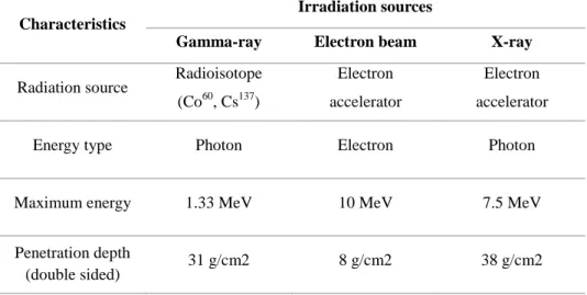
Food irradiation is a process exposing food to controlled doses of ionizing radiations such as gamma-rays emitted from the radioisotopes cobalt-60 and cesium-137, electron beams generated by electron beam accelerators (allowed up to 10 MeV), and X-rays generated by machine sources (allowed up to 5 MeV; Codex 2003). Only these sources can be used for food irradiation because energies emitted by these sources are much too low to induce radioactivity in any exposed material (Calado et al., 2014). Ionizing radiation in food processing can damage the DNA so that cells become inactivated, therefore microorganisms, insect gametes, and plant meristems are prevented from reproducing. At the same time, ionizing radiation induces minimal changes in chemical components in food (Thayer, 1990).
Table 1. Characteristics of food irradiation sources (adapted from Song, 2016).
Characteristics
Irradiation sources
Gamma-ray Electron beam X-ray Radiation source Radioisotope
(Co60, Cs137)
Electron accelerator
Electron accelerator
Energy type Photon Electron Photon
Maximum energy 1.33 MeV 10 MeV 7.5 MeV
Penetration depth
9
1.2.3. Mechanism of bacterial inactivation
The bacterial inactivation mechanism by ionizing radiation can be divided into direct and indirect effects. The direct mechanism of bacterial inactivation by ionizing radiation is the destruction of chemical bonds within the DNA, RNA, and other macromolecules that are essential for bacterial survival (Urbain, 1986). When a cell or an organism is exposed to radiation energy, direct damage to DNA can occur and destroy the cell components such as purine and pyrimidine bases, resulting in the breakage of double helix structure of DNA or its phosphodiester linkages (Zerial et al., 1978). This direct effect of ionizing radiation prevents the multiplying of microorganisms, therefore, achieving reproductive death (Black & Jaczynski, 2006). Also, direct effect is the dominant inactivation mechanism when spores of spore-forming bacteria are irradiated.
Indirect mechanism of bacterial inactivation by ionizing radiation is the damage caused by radicals from radiolysed water molecules in the bacteria. When water is subjected to radiation, it is radiolysed and can generate free radicals such as hydroxyl radical (OH·) (Farkas, 2006). Oxidative radicals are responsible for up to 90% of damage to DNA, therefore they are important factors affecting the radiation sensitivity of cell or organisms. Radicals can damage cell by destroying the structure and function of cell components such as carbohydrates, proteins, and fat. Such damages can result in chromosomal abnormalities, errors in cell division, and inactivation of endogenous enzymes.
10
Both direct and indirect bacterial inactivation occur simultaneously when foods are irradiated that direct effects account for 25% and indirect effects account for 75% of the bacterial inactivation (Moosekian et al., 2012).
Insects, fungi, vegetative bacteria, bacterial spores, and viruses have different radiation sensitivity because the molecular weight of DNA affects the radio-sensitivity. Insects are more sensitive to radiation energy because they have larger DNA in their cells compared to bacteria. Also, DNA structure can affect the radio-sensitivity that single-stranded DNA is much sensitive to radiation energy than double-stranded DNA.
Meanwhile, the bacterial inactivation effect of ionizing radiation is also affected by external environments when foods are irradiated. Irradiation temperature, oxygen concentration, and water activity of food are main factors influencing the radio-sensitivity of foods. Black and Jaczynski (2006) suggested that the physical state of water (frozen or unfrozen) is the major factor affecting the indirect effect of bacterial inactivation and irradiation temperature (4 and 22 °C) plays a minor role in the bacterial inactivation by indirect mechanism.
11
1.2.4. Electron beam and X-ray
Electron beam irradiation uses high energetic electrons which are particulate ionizing radiation (Tahergorabi et al., 2012). Electrons accelerated by a linear accelerator to the speed of light that these high energy electrons transferred to food by an electron beam gun, resulting in bacterial inactivation. Electron beam irradiation has lower penetration depth compared to gamma-ray or X-gamma-ray irradiation because electrons have a significantly larger diameter than photons that electron beam can penetrate only few centimeters (~ 4 cm) of food materials (Bhat et al., 2012).
X-rays are generated by colliding accelerated electrons with a target metal such as tungsten or tantalum (Miller, 2005). There are two factors influencing the efficiency of conversion from electron beam to X-ray power. These are energy of electrons and atomic number of target metal. Penetration depth of X-ray irradiation increases with higher electron energy, however, too much energy of electron beams that exceeds the threshold energy of target metal could induce radioactivity of metal by emission of neutrons (Grégoire et al., 2003). Threshold energies of tungsten, tantalum, and gold are 6.19, 7.58, and 8.07 MeV, respectively. Therefore, tantalum or gold is utilized for 7.5 MeV X-rays and tungsten is used for generation of 5 MeV X-ray (Cleland & Stichelbaut, 2013). The conversion efficiency from electron beam to X-ray is 8-9% at 5 MeV and 12-13% at 7.5 MeV when using tantalum (Meissner et al., 2000).
12
Chapter II.
Application of high temperature aging
of beef with low-dose electron beam and
X-ray irradiation
2.1. Introduction
In the meat industry, aging is widely used to improve meat tenderness, which can be impacted by complex changes in muscle metabolism after slaughter (Marino, Albenzio, Malva, Santillo, Loizzo, & Sevi, 2013). Aging is generally performed by storing meat for up to 3 weeks at refrigerated temperature (Lee, Sebranek, & Parrish, 1996). However, this conventional aging process has considerable refrigerated space requirements, operational costs, and energy usage (Dransfield, 1994). Higher aging temperatures around 10–15 °C result in the highest degree of meat tenderness, with lowest muscle shortening and maximum aging potential (Devine, Wahlgren, & Tornberg, 1999). The decreasing rates of shear force in beef M. longissimus thoracis et
lumborum and M. semitendinosus are greater at higher incubation
13
2004). This contributes to accelerated protein degradation, owing to the enhanced activity of proteolytic enzymes like calpain or cathepsin at higher aging temperatures (Hwang, Devine, & Hopkins, 2003).
Although high aging temperature can increase meat tenderness with reduced aging time, it may promote the proliferation of microorganisms in meat, which can lead to a significant reduction in shelf life (Zhu, Mendonca, & Ahn, 2004). For example, total viable bacterial counts increased 2 log
CFU/cm2 in beef stored at 10 °C for 72 h, compared with a 0.4 log CFU/cm2
increase in beef stored at 5 °C (Kinsella, Prendergast, McCann, Blair, McDowell, & Sheridan, 2009). Similarly, in beef samples packaged in polyethylene, Pseudomonas species showed faster growth rates with 0.345 log
(CFU/cm2)/day at 10 °C compared to 0.090 log (CFU/cm2)/day at 0 °C
(Giannuzzi, Pinotti, & Zaritzky, 1998). In this regard, a method to shorten beef aging time, while controlling microbial growth, is ideal for practical use to reduce the aging time and cost.
Irradiation technology, which is approved by the Food and Drug Administration (FDA) for the treatment of food, has been used for decades to ensure the microbial safety of meat without loss of nutritional quality (WHO, 1999). Compared to gamma irradiation, electron-beam irradiation (EB) and X-ray irradiation (XR) are more acceptable to consumers because they are free of radioisotopes (Kong et al., 2017). Park et al. (2010) found that EB up to 10 kGy showed reduced bacterial populations with no adverse effect on
14
quality and most sensory characteristics in beef sausage patties. XR is a relatively new technology for this application. Mahmoud, Chang, Wu, Nannapaneni, Sharma, & Coker (2015) reported that 2.0 kGy of XR reduced the population of Salmonella to below the detection limit (<1.0 log CFU/g) in chicken fillets.
Lee, Sebranek, & Parrish (1996) reported that aging of EB-irradiated (2 kGy) pre-rigor beef at 30 °C for 2 days resulted in similar shear values as conventional wet-aged beef at 2 °C for 7 or 14 days, without microbial spoilage. However, an irradiation dose of 6.4 kGy on beef muscle decreased the calpain activity by creating highly oxidizing conditions (Rowe, Maddock, Lonergan, & Huff-Lonergan, 2004a). Protein degradation and tenderness improvement during aging is highly associated with calpain-1 activation (Koohmaraie and Geesink, 2006). It can be hypothesized that a low-dose irradiation can be applied to minimize the impact on enzyme activity and meat tenderness, while preventing microbial spoilage. In addition, it is worth investigating the effects of XR irradiation because limited data are currently available. Therefore, the objective of this study was to evaluate the effect of low-dose EB and XR on microbial quality, physicochemical properties, and proteolytic calpain-1 autolysis of beef aged at 4 °C or 14 °C for 14 days.
15
2.2. Materials and methods
2.2.1. Sample preparation and irradiation processing
Beef M. semimembranosus (2 h post mortem) were obtained from a commercial slaughterhouse (Daejeon, Korea). Beef muscles were divided into three blocks (300 g each) for non-irradiated control, EB, and XR treatment samples. Each beef sample was vacuum-packaged in a sterilized polyethylene bag (20 cm × 30 cm; Sunkyung Co., Ltd, Seoul, Korea) then irradiated. Polyethylene bags were sterilized before use with an EB irradiation dose of 35 kGy.
EB or XR was performed within 4 h after slaughter, using a linear electron beam RF accelerator (ELV-8, 10 MeV, EB-Tech Co., Ltd., Daejeon, Korea) or the ELV-8 accelerator with an X-ray converter attached (7.5 MeV, EB tech Co., Ltd.), respectively. The beam current was 1 mA, and the dose rate was 2.95 kGy/s. The average absorbed dose was 5 kGy and was calculated using a cellulose triacetate dosimeter system (FTR-125, Fujifilm Co., Tokyo, Japan). All experiments were performed in triplicate, with three observations for each experiment. After irradiation, the beef samples were stored at refrigerated temperature (4 °C) or elevated temperature (14 °C) until further analysis. Samples were collected after 0, 3, 7, and 14 days of aging. An elevated temperature of 14 °C was selected because the highest tenderness and lowest cold shortening of beef has been shown at this temperature (Devine, Wahlgren, & Tornberg, 1999).
16
2.2.2. Microbial analysis
Five-gram beef samples were blended with 45 mL of 0.85% sterile saline solution for 2 min using a stomacher (BagMixer® 400, Interscience Ind., St. Nom, France). Samples for microbial testing were prepared in a series of decimal dilutions using sterile saline. Each diluent (0.1 mL) was spread on total plate count agar (Difco Laboratories, MI, USA) in triplicate, and the agar plates were incubated at 37 °C for 48 h. The number of colonies was counted and expressed as colony forming units per gram (log CFU/g).
2.2.3. pH
pH was measured by blending 1 g beef samples with 9 mL of distilled water (DW) for 30 s at 10,000 rpm using a homogenizer (T10 basic, Ika Works, Staufen, Germany). The homogenates were filtered by filter paper (No. 4, Whatman International Ltd., Kent, UK) after centrifugation at 2,265 ×g for 10 min (Continent 512R, Hanil Co., Ltd., Incheon, Korea). The pH of the filtrate was measured using a pH meter (SevenGo, Mettler-Toledo International Inc., Schwerzenbach, Switzerland).
2.2.4. Instrumental color measurement
17
were measured by a spectrophotometer (CM-5, Konica Minolta Censing Inc., Osaka, Japan) (Yong et al., 2017). The instrument was calibrated with a standard black and white plate before measurement. Measurements were taken in triplicate at different locations within each sample.
2.2.5. Shear force measurement
Beef samples were vacuum-packaged and cooked in a water bath at 85 °C for 30 min to achieve a core temperature of approximately 75 °C (Jayasena et al, 2015). Samples were then cooled at 4 °C and three core samples (1.0 × 1.5 × 3 cm) were taken in the longitudinal direction of muscle fibers. Each sample was cut at a speed of 120 mm/min at 20 N force using a Warner-Bratzler blade attached to a texture analyzer (LLOYD instruments, Ametek, Fareham, UK), with a maximum cell load of 10 kg and a target load of 10 g. The shear force value was calculated as the mean of the maximum force required to shear each set of core samples.
2.2.6. Myofibrillar fragmentation index (MFI)
MFI was determined by turbidity methods, as described by Hopkin et al. (2000), with some modifications. MFI is an indicator of measuring the extent of myofibrillar protein degradation of meat during aging (Olson et al., 1976). For each sample, 0.5 g of minced beef was homogenized with 30 mL of MFI
18
buffer containing 0.1 M KCl, 0.001 M EDTA, 0.001 M sodium azide (NaN3),
0.025 M potassium phosphate (0.007 M KH2PO4 and 0.018 M K2HPO4 giving
a pH 7.0 at 4 °C) at 10,000 rpm for 30 s. After homogenization, the mixture was left to rest for 30 s and then re-homogenized for 30 s. The resulting homogenate was filtered with a 1-mm mesh strainer to remove the connective tissues and washed with 10 mL of MFI buffer. The filtered homogenate was centrifuged at 10,000 ×g for 10 min (HM-150IV, Hanil Co. Ltd., Seoul, Korea) and then the supernatant was removed. The remaining pellet was mixed with 10 mL of MFI buffer and vortexed. This step was repeated five times. After removal of the supernatant, 10 mL of MFI buffer was added to the pellet and samples were vortexed. Aliquots of the resulting suspension were diluted with MFI buffer to 0.5 mg/mL of protein concentration, and the absorbance was measured at 540 nm using a spectrophotometer (X-ma 3100, Human Co. Ltd., Seoul, Korea). MFI values were calculated as absorbance units multiplied by 200.
2.2.7. SDS-PAGE and Western blot
2.2.7.1. Preparation of meat extraction
Beef samples were minced and 0.5 g of the minced sample was blended with 5 mL of Tris-EDTA buffer (0.05 M Tris and 0.01 M EDTA) at pH 8.3 using a homogenizer (T10 basic) for 1 min. The homogenized solution was centrifuged at 10,000 ×g for 20 min and the supernatant was collected and
19
mixed with an equal volume of 2× SDS sample buffer (0.125 M Tris-HCl buffer at pH 6.8, containing 20% glycerol, 2% SDS, 2% β-mercaptoethanol, and 0.02% bromophenol blue). Samples were boiled at 95 °C for 10 min and cooled at 4 °C for 2 min. The total protein concentration in meat extracts was 1 mg/mL, determined using the Lowry, Rosebrough, Farr, & Randall (1951) method. The prepared meat extracts were used for SDS-PAGE and western blot.
2.2.7.2. Sodium dodecyl sulfate-polyacrylamide gel electrophoresis
(SDS-PAGE)
The SDS-PAGE method of Laemmli (1970) was used with some modifications. The stacking gel and separating gel contained 4.5 % and 12.5 % polyacrylamide, respectively and 20 µL of meat extract was loaded onto the gel. Protein standards (Precision Plus Protein™ Unstained Standards, Bio-Rad, CA, USA) were included in each electrophoretic run to determine molecular size. Electrophoresis was performed using a Mini-slab Size Electrophoresis System AE-6531 (Atto Corporation, Tokyo, Japan) at 20 mA for 70 min. Gels were stained for 30 min in 0.1 % Coomassie Brilliant Blue R-250 solution, containing 30 % methanol and 10 % acetic acid. After staining, gels were destained for 90 min using a solution containing 30 % methanol and 10 % acetic acid.
20
2.2.7.3. Western blot
Autolysis of calpain-1 was studied by western blot according to the method of Towbin and Gordon (1979). After electrophoresis, gels were electroblotted onto polyvinylidene difluoride membranes (Bio-Rad, CA, USA) at 4 °C for 90 min at 90 V using a mini trans-blot cell (Bio-Rad, CA, USA). The transfer buffer (pH 8.3) consisted of 0.025 M Tris, 0.192 M glycine, and 20 % methanol. After transfer, membranes were incubated in blocking solution (5% skim milk in 0.05% Tween 20 [TBS-T]) at room temperature for 1 h on a shaker and washed in TBS-T. Membranes were then incubated overnight at 4 °C on a shaker with primary antibodies (Mu-calpain antibody, 9A4H8D3, Alexis Crop., CA, USA) diluted 1:5000 with 1% skim milk. After incubation, membranes were washed three times with TBS-T for 5 min each and incubated at room temperature for 1 h with HRP-conjugated secondary antibody (Goat Anti-mouse IgG-HRP, sc-2005, Santa Cruz Biotechnology Co. Ltd., CA, USA) diluted 1:5000 with TBS-T. Membranes were washed three times with TBS-T for 5 min each and then bound antibodies were detected using Clarity Western ECL Substrate (Bio-Rad, CA, USA). The signal intensity was determined using Image Lab Software Version 5.2.1 (Bio-Rad, CA, USA).
21
2.2.8. Statistical analysis
All experiments were performed in triplicate. Using SAS software (version 9.3, SAS Institute Inc., NC, USA), a multifactorial analysis of variance using the general linear model was applied to investigate the effect of irradiation (EB and XR), aging temperature (4 °C and 14 °C), and aging time (0, 3, 7, and 14 days). The differences among the mean values were identified using the Tukey’s multiple range test at a confidence level of P < 0.05. The mean values and standard errors of the means were recorded.
22
2.3. Results and discussion
2.3.1. Total aerobic bacteria
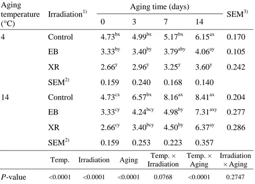
The initial number of total aerobic bacteria was reduced in irradiated samples when compared to non-irradiated controls (P < 0.05; Table 1). The efficiency between EB and XR irradiation on microbial inactivation was not different. Reductions were 1.40 and 2.07 log CFU/g for EB and XR, respectively, compared with 4.73 log CFU/g for non-irradiated samples. In most cases, irradiated samples showed significantly lower total aerobic bacterial counts than non-irradiated samples during 14 days of storage (P < 0.05). EB irradiation doses of 5 kGy resulted in approximately a 0.5 log CFU/g reduction in initial total aerobic bacterial counts in cooked beef patties (Park et al., 2010). Prendergast et al. (2009) also found that irradiation doses of 5 kGy gave a 2–3 log reduction in total aerobic bacterial counts in vacuum-packaged beef. Microbial inactivation by irradiation is mainly caused by the generation of free radicals such as OH·, H·, and e- resulting from water hydrolysis. Free radicals also generate hydrogen peroxide, which is an effective antimicrobial agent (Manas & Pagán, 2005). As a result, bacterial cell walls and DNA can be damaged and malfunction of microorganisms can occur.
Beef samples aged at 14 °C had higher total aerobic bacterial counts than samples aged at 4 °C, indicating that storage temperature had a significant effect on microbial growth rate (P < 0.0001). Similar results were reported by
23
Yim et al. (2016). In their study, EB irradiation resulted in a significant decrease in initial total aerobic bacterial counts, but bacteria proliferated rapidly in beef loins aged at 10 °C and 25 °C compared to those aged at 2 °C. In our results, the total aerobic bacterial counts of non-irradiated samples stored at 14 °C for 3 days were approximately 7 log CFU/g, which is considered the upper microbial limit for good quality fresh meat (ICMFS, 1986). However, the microbial counts of both irradiated samples aged at 14 °C for 7 days were below 7 log CFU/g. These results indicate that EB and XR can extend the shelf life of beef aged at high temperature, with a maximum shelf life of 7 days at 14 °C in this experiment.
24
Table 1. Total aerobic bacterial counts (log CFU/g) of the EB- and
XR-irradiated beef samples aged at 4 °C or 14 °C during 14 days of aging. Aging
temperature (°C)
Irradiation1)
Aging time (days)
SEM3) 0 3 7 14 4 Control 4.73bx 4.99bx 5.17bx 6.15ax 0.170 EB 3.33by 3.40by 3.79aby 4.06ay 0.105 XR 2.66y 2.96y 3.25y 3.60y 0.242 SEM2) 0.159 0.240 0.168 0.140 14 Control 4.73cx 6.57bx 8.16ax 8.41ax 0.204 EB 3.33cy 4.24bcy 4.98by 7.31axy 0.277 XR 2.66cy 3.40bcy 4.50by 6.37ay 0.286 SEM2) 0.159 0.253 0.223 0.357
Temp. Irradiation Aging Temp. × Irradiation Temp. × Aging Irradiation × Aging P-value <0.0001 <0.0001 <0.0001 0.0768 <0.0001 0.2747
1)Control, non-irradiated; EB, electron beam irradiation; XR, X-ray irradiation. 2)Standard error of the means (n=9), 3)(n=12).
a-cValues with different letters within the same row differ significantly (P < 0.05). x,y
25
2.3.2. pH
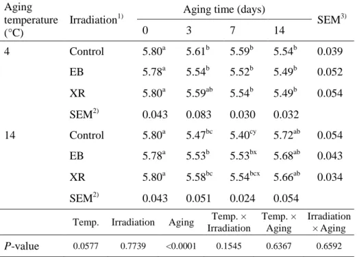
EB and XR did not change the initial pH values of beef samples, which had a mean value of 5.79 (Table 2). This result agrees with that reported by Lee et al. (1996) that the pH of beef steaks is not affected by EB up to 7 days, irrespective of storage temperature. However, in our study, pH values decreased at both temperatures during aging (P < 0.0001), except for a slight pH increase in samples aged at 14 °C for 7–14 days (P < 0.05). The decrease in pH at early storage times can be explained by the accumulation of inorganic phosphoric acid resulting from the depletion of muscle adenosine-5-triphosphate (ATP) (Scherer et al., 2005) and the production of lactic acid resulting from the degradation of glycogen (Kozioł et al., 2015). Meanwhile, the increase in pH of samples stored at 14 °C for 14 days may be attributed to the production of ammonia, amines, and other basic substances from the degradation of proteins by microorganisms and endogenous enzymes in beef (Muela et al., 2010). This would be expected, given the changes seen in aerobic bacterial count during storage (Table 1).
26
Table 2. pH of the EB- and XR-irradiated beef samples aged at 4 °C or 14 °C
during 14 days of aging. Aging
temperature (°C)
Irradiation1)
Aging time (days)
SEM3) 0 3 7 14 4 Control 5.80a 5.61b 5.59b 5.54b 0.039 EB 5.78a 5.54b 5.52b 5.49b 0.052 XR 5.80a 5.59ab 5.54b 5.49b 0.054 SEM2) 0.043 0.083 0.030 0.032 14 Control 5.80a 5.47bc 5.40cy 5.72ab 0.054 EB 5.78a 5.53b 5.53bx 5.68ab 0.043 XR 5.80a 5.58bc 5.54bcx 5.66ab 0.034 SEM2) 0.043 0.051 0.024 0.054
Temp. Irradiation Aging Temp. × Irradiation Temp. × Aging Irradiation × Aging P-value 0.0577 0.7739 <0.0001 0.1545 0.6367 0.6592
1)Control, non-irradiated; EB, electron beam irradiation; XR, X-ray irradiation. 2)Standard error of the means (n=9), 3)(n=12).
a-cValues with different letters within the same row differ significantly (P < 0.05). x,y
27
2.3.3. Surface color
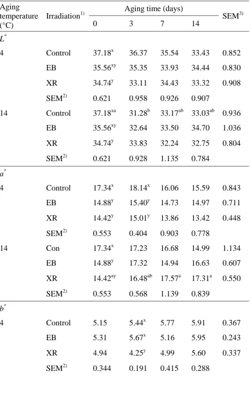
Meat color is an important visual factor that determines how consumers perceive product quality and it significantly influences the consumer’s purchasing decisions (Carpenter et al., 2001). XR significantly decreased the
initial L* value of beef when compared to the other groups, but there were no
differences in the L* value between control and irradiated samples after 3 days
of storage. The a* value of beef was initially lower in irradiated samples, but
was not different after aging for 14 days. The irradiation treatments and aging temperatures had no effect on the b* value of beef samples. Therefore, we conclude that EB and XR could affect the color of beef initially, but the difference may disappear with extended storage. Several studies have shown that irradiating fresh beef results in undesirable color changes (Brewer, 2004). Nam et al. (2003) reported that beef appears to be the meat most susceptible to irradiation-induced color changes, often changing to an unattractive greenish or brownish gray color (Kim et al., 2002). However, this study showed that 5 kGy of EB and XR had no effect on the overall color of beef after 3 days of storage, although a decrease in L* and a* values were detected at day 0 in irradiated samples when compared to non-irradiated samples. EB and XR irradiation treatments did not affect the surface color of beef samples at the end of the storage period (Table 3). Color changes in irradiated fresh meat occur because of the inherent susceptibility of the myoglobin molecule to energy input and alterations in the chemical environment, with heme iron being particularly susceptible (Brewer, 2004).
28
Table 3. Surface color of the EB- and XR-irradiated beef samples aged at
4 °C or 14 °C during 14 days of aging. Aging
temperature (°C)
Irradiation1)
Aging time (days)
SEM3) 0 3 7 14 L* 4 Control 37.18x 36.37 35.54 33.43 0.852 EB 35.56xy 35.35 33.93 34.44 0.830 XR 34.74y 33.11 34.43 33.32 0.908 SEM2) 0.621 0.958 0.926 0.907 14 Control 37.18xa 31.28b 33.17ab 33.03ab 0.936 EB 35.56xy 32.64 33.50 34.70 1.036 XR 34.74y 33.83 32.24 32.75 0.804 SEM2) 0.621 0.928 1.135 0.784 a* 4 Control 17.34x 18.14x 16.06 15.59 0.843 EB 14.88y 15.40y 14.73 14.97 0.711 XR 14.42y 15.01y 13.86 13.42 0.448 SEM2) 0.553 0.404 0.903 0.778 14 Con 17.34x 17.23 16.68 14.99 1.134 EB 14.88y 17.32 14.94 16.63 0.607 XR 14.42ay 16.48ab 17.57a 17.31a 0.550 SEM2) 0.553 0.568 1.139 0.839 b* 4 Control 5.15 5.44x 5.77 5.91 0.367 EB 5.31 5.67x 5.16 5.95 0.243 XR 4.94 4.25y 4.99 5.60 0.337 SEM2) 0.344 0.191 0.415 0.288
29
14 Control 5.15ab 5.10b 5.44ab 6.72a 0.358
EB 5.31 5.67 5.16 6.42 0.396
XR 4.94 4.58 5.99 6.29 0.400
SEM2) 0.344 0.371 0.513 0.269
Temp. Irradiation Aging Temp. × Irradiation Temp. × Aging Irradiation × Aging L* P-value 0.0046 0.0375 0.0002 0.2121 0.0705 0.2281 a* P-value 0.0016 0.0007 0.0466 0.0093 0.1922 0.0546 b* P-value 0.1432 0.2657 <0.0001 0.1047 0.3572 0.0606 1)
Control, non-irradiated; EB, electron beam irradiation; XR, X-ray irradiation.
2)Standard error of the means (n=9), 3)(n=12).
a,bValues with different letters within the same row differ significantly (P < 0.05). x,yValues with different letters within the same column differ significantly (P < 0.05).
30
2.3.4. Shear force
EB- and XR-irradiated beef showed no differences in shear force values compared with non-irradiated controls at all aging times (Table 4). Davis et al. (2004) reported that EB irradiation (4.4 kGy) had no effect on the shear force values of fresh pork loins. However, Rowe et al. (2004b) reported that irradiated (6.4 kGy) beef steaks showed significantly higher shear force values than non-irradiated steaks. Rowe et al. (2004b) also demonstrated that higher carbonyl content was associated with the higher shear force seen in irradiated beef. Irradiation of beef at an early postmortem stage induces oxidative conditions that increase protein oxidation. Oxidized amino acids cause denaturation and aggregation of myofibrillar protein and loss of proteolytic enzyme activity (Rowe et al., 2004a). Thus, application of low-dose irradiation on meat is important to avoid extensive oxidizing conditions that can cause meat toughness. Lee et al. (1996) showed that 2 kGy of electron beam irradiation did not affect shear force or myofibrillar fragmentation index in beef. The current study used a relatively low dose (5 kGy) of EB and XR irradiation compared with previous studies (6.4 kGy), and it had no adverse effect on beef tenderness. Our preliminary study indicated that 2 kGy of EB or XR could not induce microbial inactivation during storage at 14 °C (data not shown). Aging temperature had a significant effect on shear force values (Table 4). The decreasing rate of shear force was more evident in samples stored at 14 °C than in samples stored at 4 °C. The shear
31
force values of samples stored at 14 °C for 7 days are similar to the values of samples stored at 4 °C for 14 days. This may be attributed to the accelerated destruction of muscle structure and increased protease activity at high storage temperatures. It has been reported that rigor temperatures around 10–15 °C give the greatest improvement in tenderness by reducing muscle shortening and lessening the impact on enzyme activity (Devine et al., 1999).
32
Table 4. Shear force values (N) of the EB- and XR-irradiated beef samples
aged at 4 °C or 14 °C during 14 days of aging. Aging
temperature (°C)
Irradiation1)
Aging time (days)
SEM3) 0 3 7 14 4 Control 90.73a 89.92a 75.59b 69.18b 3.040 EB 92.15a 94.35a 79.28b 71.76b 1.825 XR 96.96a 94.82a 79.19b 71.15c 1.717 SEM2) 1.816 2.041 2.979 2.086 14 Control 90.73a 84.43a 65.98b 49.65c 2.396 EB 92.15a 85.64a 65.39b 53.86c 2.473 XR 96.96a 87.20b 66.16c 54.96d 1.555 SEM2) 1.816 1.848 2.925 1.940
Temp. Irradiation Aging Temp. × Irradiation Temp. × Aging Irradiation × Aging P-value <0.0001 0.0040 <0.0001 0.8032 <0.0001 0.7355
1)Control, non-irradiated; EB, electron beam irradiation; XR, X-ray irradiation. 2)Standard error of the means (n=9), 3)(n=12).
33
2.3.5. Myofibrillar fragmentation index (MFI)
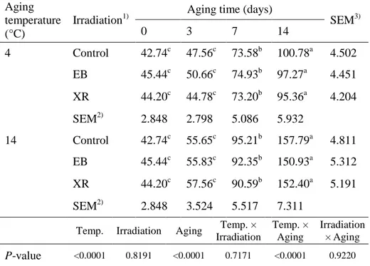
MFI is an indicator of the extent of myofibrillar protein degradation during the postmortem aging of meat (Li et al., 2012). It represents the degradation of structural proteins in the I-band of the myofibril and the weakening of myofibril linkages (Taylor et al., 1995). Irradiation treatment had no effect on MFI (Table 5). MFI values significantly increased over storage time (P < 0.0001) and they were significantly affected by aging temperature. Samples aged at 14 °C had higher MFI values compared to samples aged at 4 °C (P < 0.0001). Lee et al. (1996) reported that MFI increases in beef with increasing post-aging time, regardless of EB treatment. This may be attributed to Z-disk collapses in the beef intramuscular fibers as the post-mortem aging time increases. Li et al. (2012) reported that muscles kept at 14 °C showed increased MFI values and suggested that accelerated proteolysis is the reason for improved tenderness.
34
Table 5. Myofibrillar fragmentation index (MFI) of the EB- and
XR-irradiated beef samples aged at 4 °C or 14 °C during 14 days of aging.
Aging temperature (°C)
Irradiation1) Aging time (days) SEM3)
0 3 7 14 4 Control 42.74c 47.56c 73.58b 100.78a 4.502 EB 45.44c 50.66c 74.93b 97.27a 4.451 XR 44.20c 44.78c 73.20b 95.36a 4.204 SEM2) 2.848 2.798 5.086 5.932 14 Control 42.74c 55.65c 95.21b 157.79a 4.811 EB 45.44c 55.83c 92.35b 150.93a 5.312 XR 44.20c 57.56c 90.59b 152.40a 5.191 SEM2) 2.848 3.524 5.517 7.311
Temp. Irradiation Aging Temp. × Irradiation Temp. × Aging Irradiation × Aging P-value <0.0001 0.8191 <0.0001 0.7171 <0.0001 0.9220 1)
Control, non-irradiated; EB, electron beam irradiation; XR, X-ray irradiation.
2)Standard error of the means (n=9), 3)(n=12).
35
2.3.6. SDS-PAGE
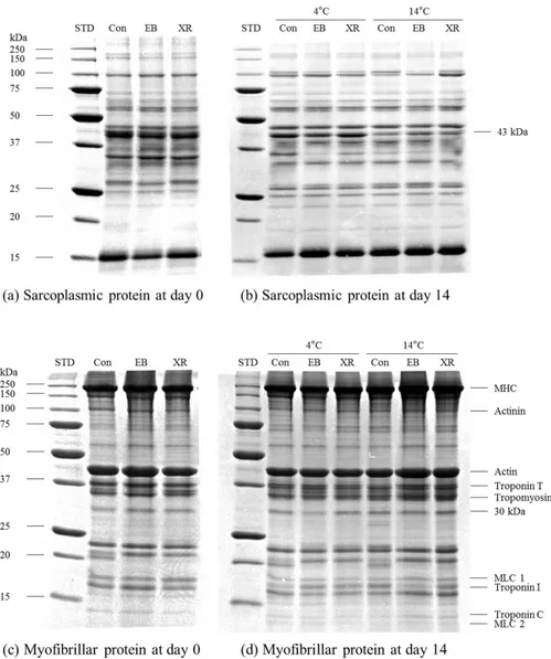
SDS-PAGE analysis showed no changes in the sarcoplasmic and myofibrillar protein patterns between non-irradiated and EB- or XR-irradiated samples at 0 and 14 days of aging (Fig. 1). Lee et al. (2000) also found no change in sarcoplasmic and myofibrillar proteins in gamma-irradiated (up to 10 kGy) beef round by SDS-PAGE. Similarly, no changes were detected in protein patterns between non-irradiated and gamma-irradiated beef, pork, and chicken meats below 10 kGy of irradiation dose (Yook et al., 1998). These results indicate that EB and XR irradiation at 5 kGy does not destroy or degrade protein structures in beef samples. The intensity of the 43-kDa band in sarcoplasmic protein patterns decreased in beef aged for 14 days at 14 °C, compared to beef aged for 14 days at 4 °C (Fig. 1). This result shows that high aging temperature enhances the degradation of this protein. Morzel et al. (2004) reported that meat tenderness during postmortem aging is positively correlated with the intensity of the 43-kDa sarcoplasmic protein band identified as creatine kinase. Similarly, in our results, lower shear force values were observed in beef aged for 14 days at 14 °C compared to beef aged at 4 °C. This was accompanied by decreasing intensity of the 43-kDa band in SDS-PAGE. This result suggests that sarcoplasmic protein degradation may reflect increased proteolysis due to high aging temperature and this could be used as a marker for beef tenderness (Bowker et al., 2008) Degradation of myofibrillar proteins, especially troponin T, myosin light chain 1, and troponin I, is observed during 14 days of aging. However, no differences were
36
found in the myofibrillar protein pattern between non-irradiated controls and irradiated beef samples. Using western blot analysis, Rowe et al. (2004b) reported that oxidative conditions caused by 6.4 kGy of irradiation have a negative effect on troponin T degradation, indicating a significant decrease in beef tenderness. This result can be explained by the increased sensitivity of western blot analysis to detect very low amounts of myofibril fragments. Therefore, samples were further analyzed by western blot.
37
Fig. 1. Sodium dodecyl sulfate-polyacrylamide gel electrophoresis of the EB
and XR irradiated beef samples aged for 0 and 14 days at 4 °C or 14 °C. (STD, standard molecular bands; Control, non-irradiated; EB, electron beam-irradiated; XR, X-ray-irradiated).
38
2.3.7. Western blot
EB and XR did not affect calpain-1 activity in sarcoplasmic extracts from beef samples at day 0 (Fig. 2). Calpain-1 is one of the proteolytic enzymes present in meat that acts on the cytoskeletal proteins in the myofibrils, resulting in a decrease in the binding force between the myofibrils, resulting in their collapse (Koohmaraie, & Geesink, 2006). Autolysis of calpain-1 is associated with enzyme activity in post-mortem muscle. Oxidation of the cysteine residue in the active site of calpain-1 results in loss of activity (Lametsch et al., 2008). Rowe et al. (2004b) found that strong oxidative conditions caused by irradiation decreased sarcoplasmic and myofibril-bound calpain-1 activity and reduced the degradation of nebulin, titin, desmin, and troponin-T in beef steaks. In addition, the beef steaks showed higher shear force compared to non-irradiated samples. Similarly, the present study showed a slower rate of sarcoplasmic calpain-1 autolysis in irradiated beef compared to non-irradiated controls at 14 days. Calpain-1 consists of 80, 78, and 76 kDa bands and degradation of the intact 80 kDa (inactive form) band to 76 kDa (active form) occurs during aging. This result indicates that calpain-1 autolysis was less extensive and there was lower enzyme activity in irradiated samples. However, the effect of 5 kGy of irradiation on calpain-1 activity may be negligible to meat tenderness, because there was no significant difference in shear force values or MFI between non-irradiated and irradiated samples.
39
Fig 2. Western blot of calpain-1 in the sarcoplasmic fraction of the EB- and
XR-irradiated beef samples aged for 0 and 14 days at 4 °C or 14 °C. (Control, non-irradiated; EB, electron beam-irradiated; XR, X-ray-irradiated).
40
2.4. Conclusion
These results suggest that low-dose EB and XR irradiation of beef prior to aging are effective in decreasing microbial growth during aging, with no undesirable changes in physicochemical properties. The low-dose EB and XR irradiation (5 kGy) of beef can be combined with high temperature (14 °C) aging to significantly decrease aging time. EB and XR irradiation may reduce calpain-1 autolysis during beef aging to some extent; however, these effects are not strong enough to reduce final meat tenderness. Therefore, the low-dose EB and XR irradiation of beef could be an effective technique for high temperature aging.
41
References
Bhat, R., Karim Alias, A., & Paliyath, G. (2012). Use of electron beams in food preservation. Progress in Food Preservation, 343-372.
Black, J. L., & Jaczynski, J. (2006). Temperature effect on inactivation kinetics of Escherichia coli O157: H7 by electron beam in ground beef, chicken breast meat, and trout fillets. Journal of Food Science, 71, 221-227.
Boehm, M. L., Kendall, T. L., Thompson, V. F., & Goll, D. E. (1998). Changes in the calpains and calpastatin during postmortem storage of bovine muscle. Journal of Animal Science, 76, 2415-2434.
Bowker, B. C., Fahrenholz, T. M., Paroczay, E. W., & Solomon, M. B. (2008). Effect of hydrodynamic pressure processing and aging on sarcoplasmic proteins of beef strip loins. Journal of Muscle Foods, 19, 175-193.
Brewer, S. (2004). Irradiation effects on meat color–a review. Meat Science,
68, 1-17.
Calado, T., Venâncio, A., & Abrunhosa, L. (2014). Irradiation for mold and mycotoxin control: a review. Comprehensive Reviews in Food Science
and Food Safety, 13, 1049-1061.
Campbell, R. E., Hunt, M. C., Levis, P., & Chambers, E. (2001). Dry‐aging effects on palatability of beef longissimus muscle. Journal of Food
42
Carpenter, C. E., Cornforth, D. P., & Whittier, D. (2001). Consumer preferences for beef color and packaging did not affect eating satisfaction.
Meat Science, 57, 359-363.
Cleland, M. R., & Stichelbaut, F. (2013). Radiation processing with high-energy X-rays. Radiation Physics and Chemistry, 84, 91-99.
Coombs, R. G. (2017). Tenderness, Consistency and cooking loss of beef loins and ground beef from two different genetic types of cattle (Doctoral dissertation).
Coux, O., Tanaka, K., & Goldberg, A. L. (1996). Structure and functions of the 20S and 26S proteasomes. Annual Review of Biochemistry, 65, 801-847.
Crouse, J. D., & Koohmaraie, M. (1990). Effect of freezing of beef on subsequent postmortem aging and shear force. Journal of Food Science,
55, 573-574.
Davey, C. L., & Gilbert, K. V. (1976). The temperature coefficient of beef ageing. Journal of the Science of Food and Agriculture, 27, 244-250. Davis, K. J., Sebranek, J. G., Huff-Lonergan, E., Ahn, D. U., & Lonergan, S.
M. (2004). The effects of irradiation on quality of injected fresh pork loins. Meat Science, 67, 395-401.
Devine, C. E., Wahlgren, N. M., & Tornberg, E. (1999). Effect of rigor temperature on muscle shortening and tenderisation of restrained and unrestrained beef M. longissimus thoracicus et lumborum. Meat Science,
43
Dikeman, M. E., Obuz, E., Gök, V., Akkaya, L., & Stroda, S. (2013). Effects of dry, vacuum, and special bag aging; USDA quality grade; and end-point temperature on yields and eating quality of beef Longissimus
lumborum steaks. Meat science, 94, 228-233.
Diles, J. J., Miller, M. F., & Owen, B. L. (1994). Calcium chloride concentration, injection time, and aging period effects on tenderness, sensory, and retail color attributes of loin steaks from mature cows.
Journal of Animal Science, 72, 2017-2021.
Dransfield, E. (1994). Optimisation of tenderisation, ageing and tenderness.
Meat Science, 36, 105-121.
Farkas, J. (2006). Irradiation for better foods. Trends in Food Science &
Technology, 17, 148-152.
Giannuzzi, L., Pinotti, A., & Zaritzky, N. (1998). Mathematical modelling of microbial growth in packaged refrigerated beef stored at different temperatures. International Journal of Food Microbiology, 39, 101-110. Goll, D. E., Geesink, G. H., Taylor, R. G., & Thompson, V. F. (1995). Does
proteolysis cause all postmortem tenderization, or are changes in the actin/myosin interaction involved?. In Annual International Congress of
Meat Science and Technology, 41, 537-544.
Goll, D. E., Thompson, V. F., Taylor, R. G., & Christiansen, J. A. (1992). Role of the calpain system in muscle growth. Biochimie, 74, 225-237.
Grégoire, O., Cleland, M. R., Mittendorfer, J., Dababneh, S., Ehlermann, D. A. E., Fan, X., ... & Stichelbaut, F. (2003). Radiological safety of food
44
irradiation with high energy X-rays: theoretical expectations and experimental evidence. Radiation Physics and Chemistry, 67, 169-183. Grover, S. B., & Kumar, J. (2002). A review of the current concepts of
radiation measurement and its biological effects. Indian Journal of
Radiology and Imaging, 12, 21.
Hopkins, D. L., Littlefield, P. J., & Thompson, J. M. (2000). A research note on factors affecting the determination of myofibrillar fragmentation. Meat
Science, 56, 19-22.
Hopkins, D. L., & Thompson, J. M. (2002). Factors contributing to proteolysis and disruption of myofibrillar proteins and the impact on tenderisation in beef and sheep meat. Australian Journal of Agricultural
Research, 53, 149-166.
Huff-Lonergan, E., Mitsuhashi, T., Beekman, D. D., Parrish, F. C., Olson, D. G., & Robson, R. M. (1996). Proteolysis of specific muscle structural proteins by mu-calpain at low pH and temperature is similar to degradation in postmortem bovine muscle. Journal of Animal Science, 74, 993-1008.
Huff-Lonergan, E., & Lonergan, S. M. (2005). Mechanisms of water-holding capacity of meat: The role of postmortem biochemical and structural changes. Meat science, 71, 194-204.
Hwang, I. H., Devine, C. E., & Hopkins, D. L. (2003). The biochemical and physical effects of electrical stimulation on beef and sheep meat tenderness. Meat Science, 65, 677-691.
45
Hwang, I. H., Park, B. Y., Cho, S. H., & Lee, J. M. (2004). Effects of muscle shortening and proteolysis on Warner–Bratzler shear force in beef
longissimus and semitendinosus. Meat Science, 68, 497-505.
ICMFS (1986). Microorganisms in Foods. 2. Sampling for microbiological analysis: Principles and specific applications (2nd ed.). Toronto: University of Toronto Press.
Jayasena, D. D., Nam, K. C., Kim, J. J., Ahn, H., & Jo, C. (2015). Association of carcass weight with quality and functional properties of beef from Hanwoo steers. Animal Production Science, 55, 680-690.
Joint FAO/WHO Codex Alimentarius Commission, Joint FAO/WHO Food Standards Programme, & World Health Organization. (2003). Codex
Alimentarius: Food hygiene, basic texts. Food & Agriculture Org..
Kemp, C. M., Sensky, P. L., Bardsley, R. G., Buttery, P. J., & Parr, T. (2010). Tenderness–An enzymatic view. Meat Science, 84, 248-256.
Khan, M. I., Jung, S., Nam, K. C., & Jo, C. (2016). Postmortem aging of beef with a special reference to the dry aging. Korean Journal for Food
Science of Animal Resources, 36, 159.
Kim, Y. H., Nam, K. C., & Ahn, D. U. (2002). Color, oxidation‐reduction potential, and gas production of irradiated meats from different animal species. Journal of Food Science, 67, 1692-1695.
Kinsella, K. J., Prendergast, D. M., McCann, M. S., Blair, I. S., McDowell, D. A., & Sheridan, J. J. (2009). The survival of Salmonella enterica serovar Typhimurium DT104 and total viable counts on beef surfaces at different
46
relative humidities and temperatures. Journal of Applied Microbiology,
106, 171-180.
Kong, Q., Yan, W., Yue, L., Chen, Z., Wang, H., Qi, W., & He, X. (2017). Volatile compounds and odor traits of dry-cured ham (Prosciutto crudo) irradiated by electron beam and gamma rays. Radiation Physics and
Chemistry, 130, 265-272.
Koohmaraie, M. (1994). Muscle proteinases and meat aging. Meat Science, 36, 93-104.
Koohmaraie, M., & Geesink, G. H. (2006). Contribution of postmortem muscle biochemistry to the delivery of consistent meat quality with particular focus on the calpain system. Meat Science, 74, 34-43.
Koohmaraie, M., Whipple, G., Kretchmar, D. H., Crouse, J. D., & Mersmann, H. J. (1991). Postmortem proteolysis in longissimus muscle from beef, lamb and pork carcasses. Journal of Animal Science, 69, 617-624.
Kozioł, K., Maj, D., & Bieniek, J. (2015). Changes in the colour and pH of rabbit meat in the aging process. Medycyna Weterynaryjna, 71, 104-108. Kristensen, L., & Purslow, P. P. (2001). The effect of ageing on the
water-holding capacity of pork: role of cytoskeletal proteins. Meat Science, 58, 17-23.
Laemmli, U. K. (1970). Cleavage of structural proteins during the assembly of the head of bacteriophage T4. Nature, 227, 680-685.
Lametsch, R., Karlsson, A., Rosenvold, K., Andersen, H. J., Roepstorff, P., & Bendixen, E. (2003). Postmortem proteome changes of porcine muscle
47
related to tenderness. Journal of Agricultural and Food Chemistry, 51, 6992-6997.
Lametsch, R., Lonergan, S., & Huff-Lonergan, E. (2008). Disulfide bond within µ-calpain active site inhibits activity and autolysis. Biochimica et
Biophysica Acta (BBA)-Proteins and Proteomics, 1784, 1215-1221.
Lee, M., Sebranek, J., & Parrish, F. C. (1996). Accelerated postmortem aging of beef utilizing electron-beam irradiation and modified atmosphere packaging. Journal of Food Science, 61, 133-136.
Lee, J. W., Yook, H. S., Lee, K. H., Kim, J. H., Kim, W. J., & Byun, M. W. (2000). Conformational changes of myosin by gamma irradiation.
Radiation Physics and Chemistry, 58, 271-277.
Li, K., Zhang, Y., Mao, Y., Cornforth, D., Dong, P., Wang, R., Zhu, H., Luo, X. (2012). Effect of very fast chilling and aging time on ultra-structure and meat quality characteristics of Chinese Yellow cattle M. Longissimus
lumborum. Meat Science, 92, 795-804.
Lowry, O. H., Rosebrough, N. J., Farr, A. L., & Randall, R. J. (1951). Protein measurement with the Folin phenol reagent. Journal of Biological
Chemistry, 193, 265-275.
Mahmoud, B. S., Chang, S., Wu, Y., Nannapaneni, R., Sharma, C. S., & Coker, R. (2015). Effect of X-ray treatments on Salmonella enterica and spoilage bacteria on skin-on chicken breast fillets and shell eggs. Food Control, 57, 110-114.
48
food preservation. Journal of Applied Microbiology, 98, 1387-1399. Marino, R., Albenzio, M., Della Malva, A., Santillo, A., Loizzo, P., & Sevi, A.
(2013). Proteolytic pattern of myofibrillar protein and meat tenderness as affected by breed and aging time. Meat Science, 95, 281-287.
Meissner, J. O. E. R. N., Abs, M., Cleland, M. R., Herer, A. S., Jongen, Y., Kuntz, F., & Strasser, A. (2000). X-ray treatment at 5 MeV and above.
Radiation Physics and Chemistry, 57, 647-651.
Miller, R. B. (2005). Food irradiation using X-rays. Electronic Irradiation of
Foods: An Introduction to the Technology, 75-121.
Miller, M. F., Carr, M. A., Ramsey, C. B., Crockett, K. L., & Hoover, L. C. (2001). Consumer thresholds for establishing the value of beef tenderness.
Journal of Animal Science, 79, 3062-3068.
Moosekian, S. R., Jeong, S., Marks, B. P., & Ryser, E. T. (2012). X-ray irradiation as a microbial intervention strategy for food. Annual Review of
Food Science and Technology, 3, 493-510.
Morzel, M., Chambon, C., Hamelin, M., Santé-Lhoutellier, V., Sayd, T., & Monin, G. (2004). Proteome changes during pork meat ageing following use of two different pre-slaughter handling procedures. Meat Science, 67, 689-696.
Muela, E., Sañudo, C., Campo, M. M., Medel, I., & Beltrán, J. A. (2010). Effect of freezing method and frozen storage duration on instrumental quality of lamb throughout display. Meat Science, 84, 662-669.

