INTRODUCTION
Birth defects are an important contributor to infant mor-tality among all racial/ethnic groups. Major congenital anoma-lies are defined as those that threaten life, require major surgery, or lead to a significant disability. Between 2% and 3% of all infants have a major congenital anomaly identified at birth and approximately 6% to 10% of such infants die within the first year of life (1). In more than 60% of such cases, the eti-ology of the congenital birth defect is unknown and primary prevention is impossible. In approximately 20% of congen-ital cases, the causes are monogeneous defects, 50% are caused by chromosome aberrations and 20% by virus infections, such as rubella, cytomegalovirus, and herpes virus (2, 3). Many environmental factors have been suspected to play an etiologic role in the formation of congenital anomalies. Chemical pol-lutants, dietary imbalances, ionizing radiation, pharmaceu-tical substances, and infections provide examples of known
or suspected agents (4). Unlike animal studies, molecular and biochemical studies in pregnant women are impossible. There-fore, epidemiological data about congenital malformations is of vital importance to scientific research on pathomorphogen-esis, aimed at prevention and public health education (3).
Currently, no nationwide birth defect monitoring system exists in Korea. Only a small number of reports on birth defect monitoring are available from general hospitals (5-7). Thus, our aim is to establish a multi-center birth defects monitoring system for the evaluation of the prevalence and serial occur-rence of birth defects in Korea.
MATERIALS AND METHODS
Study materials
The study materials were all recorded deliveries at 10 med-Jae-Hyug Yang, Yon-Ju Kim,
Jin-Hoon Chung, Moon-Young Kim, Hyun-Mee Ryu, Hyun-Kyong Ahn, Jung-Yul Han, Soon-Ha Yang*, Ahm Kim�
, Hyun-Se Kim� , Pyo-Jong Lee�
, Sung-Soo Kim‖, Young-Ju Kim¶, Kyung-Sim Koh**, Jong-Chul Shin��
, Yong-Kun Cho�� , Bo-Hyun Yoon��
Department of Obstetrics and Gynecology, Samsung Cheil Hospital, Samsung Medical Center*, Sungkyunkwan University; Asan Medical Center�
, University of Ulsan; Cheil Women’s Clinic�
; Ilsan Cheil Hospital�
; Bombit Women’s Hospital‖; Ewha Women’s University Mokdong Hospital¶; Institute for Occupation and Environmental Health**; Kangnam St. Mary’s Hospital��
, The Catholic University of Korea; Sanggye Baik Hospital��
, Inje University; Seoul National University Hospital��
, Seoul, Korea Received : 10 December 2003 Accepted : 6 April 2004 Address for correspondence Jae-Hyug Yang, M.D.
Department of Obstetrics and Gynecology, Samsung Cheil Hospital, 1-19 Mookjeung-dong, Choong-gu, Seoul 100-380, Korea
Tel : +82.2-2000-7273, Fax : +82.2-2000-7183 E-mail : [email protected]
*This work was supported by the Korea Food Drug Administration Grant. KFDA- ED 2002-21.
509
A Multi-center Study for Birth Defect Monitoring Systems in Korea
The aim of this study was to establish a multi-center birth defects monitoring system to evaluate the prevalence and the serial occurrence of birth defects in Korea. Ten medical centers participated in this program. A trained nurse collected relevant re
-cords from delivery units and pediatric clinics in participating hospitals on a monthly basis. We observed 1,537 cases of birth defects among 86,622 deliveries, which included live births and stillbirths. The prevalence of birth defects was 1.8%, and the sex distribution of the birth defect cases was 55.2% male and 41.6% female. The highest proportion of birth defects was in the cardiovascular system (17.5%), followed by birth defects involving in the genitourinary system (15.6%). Chromoso-mal anoChromoso-malies were detected 30.0 per 10,000 births. Of these chromosoChromoso-mal anoma-lies, Down syndrome was most frequently observed. This study led to an establish-ment of a multi-center active monitoring system for birth defects. To better under-stand the serial occurrence of birth defects in Korea, it is necessary to increase the number of participating hospitals and to launch on a nation-wide multi-center study.
ical centers in Korea, between May 1999 and October 2002. The ten medical center were: the Samsung Cheil Hospital, Samsung Medical Center, Asan Medical Center, Cheil Women’s Clinic, Ilsan Cheil Hospital, Bombit Women’s Hospital, Ewha Women’s University Mokdong Hospital, Kangnam St. Mary’s Hospital, Sanggye Baik Hospital, and Seoul National Univer-sity Hospital.
Active data collection
Records were obtained monthly, from delivery units and pediatric clinics by a visiting nurse. Trained staff from each center provided the medical records that included delivery files, stillborn files and newborn files. The entire medical record of each case was reviewed for followings: hospital stay, prenatal diagnostic test results, birth certificate work sheet, labor and delivery records, progress notes, pathology/autopsy findings, physical examination findings, and a discharge sum-mary. The collected database was checked by trained medi-cal doctor monthly.
Definition and coding of diagnoses
All live births, stillbirths, and spontaneous abortions after the 16th week of gestation were included. All neonates born
at one of the 10 centers were examined by a pediatrician with-in the first week of life. In addition to the clwith-inical examwith-ina- examina-tion, information from the prenatal and postnatal ultrasound examinations of the heart, brain and other organs were record-ed. Chromosome analysis was available for a number of birth defects that were coded according to the EUROCAT (Euro-pean Registration of Congenital Anomalies and Twins, an European Union Registry) (4) and the International Clearing-house for Birth Defects Monitoring System (ICBDMS) (8).
The nineteen groups of malformations as described by the ICBDMS (8) were 1) anencephaly, 2) spina bifida, 3) ence
-phalocele, 4) hydrocephaly, 5) microtia, 6) cleft palate, 7) total cleft lip, 8) esophageal atresia or stenosis, 9) anorectal atresia or stenosis, 10) hypospadias, 11) renal agenesis/dysgenesis, 12) limb reduction defects, 13) omphalocele, 14) gastroschi-sis, 15) abdominal wall defects, 16) diaphragmatic hernia, 17) transposition of the great vessels, 18) hypoplastic left heart syndrome, and 19) Down syndrome.
RESULTS
During the 4-yr period (May 1999-October 2002), we ob
-served 1,537 cases of birth defects among 86,622 births, in
-cluding live births and stillbirths, the prevalence rate being
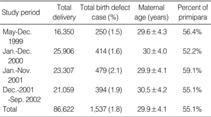
Study period Total delivery
Total birth defect case (%) Maternal age (years) Percent of primipara May-Dec. 16,350 250 (1.5) 29.6±4.3 56.4% 1999 Jan.-Dec. 25,906 414 (1.6) 30±4.0 52.2% 2000 Jan.-Nov. 23,307 479 (2.1) 29.9±4.1 59.1% 2001 Dec.-2001 21,059 394 (1.9) 30.5±4.2 55.1% -Sep. 2002 Total 86,622 1,537 (1.8) 29.9±4.1 55.1% Table 1.General characteristics of study populations
1999 2000 2001 2002 Total n=250 n=414 n=479 n=394 n=1,537 Cesarean section 65 104 144 121 438 (26%) (25.1%) (30.1%) (30.7%) (28.5%) Termination 80 136 159 110 484 (32%) (32.9%) (33.2%) (27.9%) (31.5%) Fetal sex male 138 209 278 219 848 (55.2%) (50.5%) (58%) (55.6%) (55.2%) female 103 186 187 164 639 (41.2%) (44.9%) (39%) (41.6%) (41.6%) ambiguous 1 (0.4%) 1 (0.2%) 2 (0.4%) 1 (0.3%) 5 (0.3%) unknown 13 (5.2%) 13 (3.1%) 12 (2.5%) 7 (1.8%) 45 (2.9%) Twin pregnancy 7 (2.8%) 17 (4.1%) 21 (4.4%) 24 (6.1%) 69 (4.5%) Table 2.General characteristics of infants with birth defects (n= 1,537) analysed by year USA: Atlanta Observed /10,000 China Observed /10,000 Japan Observed /10,000 Korea Observed /10,000 Congenital anomalies Anencephaly 3.3 1.9 5.4 1.8 Spina bifida 0.2 3.2 8.0 3.8 Encephalocele 1.6 0.9 2.5 1.1 Hydrocephalus 3.6 7.5 6.5 7.1 Microtia 2.7 1.6 - 0.9
Cleft palate, only 1.4 4.8 2.4 6.0
Cleft lip, and/or cleft palate 10.3 15.9 13.6 9.9 Esophageal atresia or 2.4 3.3 0.5 0.9 stenosis Anorectal atresia or 3.5 4.0 3.0 4.0 stenosis Hypospadias 1.2 3.5 3.1 7.7 Renal agenesis/ 7.6 4.3 0.9 5.3 dysgenesis
Limb reduction defect 1.3 3.8 5.3 4.9
Omphalocele 3.0 4.6 1.6 2.0
Gastroschisis 1.6 2.3 2.7 2.0
Abdominal wall defects 1.4 7.1 4.3 4.0
Diaphragmatic hernia 4.8 4.9 0.5 1.6
Transposition of great 2.1 2.1 - 4.2
vessels
Hypoplastic left heart 1.3 1.9 - 2.9
syndrome
Down syndrome 9.2 10.4 1.7 11.3
Table 3.Incidence comparison of 19 congenital anomalies by ICBDMS (1998) with other reports
1.8%. The mean maternal age was 29.9 (±4.1, 95% CI) yr, with a range 19-45 yr. Nineteen percent (291 cases) of the birth defects were associated with an elderly mother (≥35 yr). Primipara birth and multipara birth accounted for 55.1% and 44.9%, respectively. Table 1 shows the changes in the prevalence of birth defects over time.
The general characteristics of the birth defects are summa-rized in Table 2. The mean gestational age at delivery was 32.5±8.6 weeks, ranging from 11 weeks to 43 weeks. The mean birth weight was 2,198±1,319.5 g, with a range from 5 g to 5,060 g. Non-living births (termination or intra-uterine fetal death) accounted for 31.5% of deliveries, and 438 babies (28.5%) were born via Cesarean section. The sex distribution of the birth defect cases was 55.2% male and 41.6% female. Five cases had (0.3%) ambiguous genitalia, and in 45 cases (2.9%), the gender of the fetus could not be determined. Twin pregnancies accounted for 4.5% of cases.
Table 3 shows the comparison of incidences of 19 congen-ital anomalies in Korea as defined by ICBDMS (5) with those in Japan and China and with Atlanta, U.S.A. as determined by the Metropolitan Atlanta Congenital Defects Program
(MACDP) (9).
Among 1,537 birth defect cases, 260 cases (16.9%) had chromosomal abnormalities (Table 4). Among these, Down syndrome was most common (137 cases), followed by Edwards syndrome (57 cases).
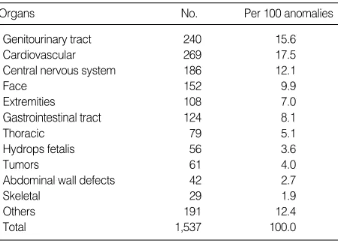
Table 5 shows the birth defects according to the involved organs. The highest proportion of birth defects (17.5%) in
-volved the cardiovascular system, followed by the genitouri-nary system (15.6%). Multiple anomalies accounted for 16% of all birth defect cases.
Thirty-four cases had polydactyly of the hand (4.0 per 10,000 fetuses) and 20 cases had polydactyly of the foot (2.4 per 10,000 fetuses). Syndactyly of the hand and foot occurred in 9 cases (1.2 per 10,000 fetuses) and 7 cases (0.8 per 10,000 fetuses), respectively.
DISCUSSION
The concept of congenital malformation is not strictly defined, and includes functional and metabolic disorders that, although present, may not necessarily be recognizable at birth (10, 11). Different classifications of congenital anomalies are used in different countries. The two most commonly used classification systems are: (1) the International Classification of Diseases system (12, 13), and (2) the International Clearing-house for Birth Defects Monitoring System (ICBDMS) (14). In our study, the classification of birth defects was performed according to the European Registration of Congenital Anoma-lies and Twins (EUROCAT) and the ICBDMS (1994) system. The EUROCAT program was initiated in 1979. This sur
-veillance system details 900,000 births per annum in 17 coun-tries. The International Clearing-house for Birth Defects Mon-itoring System (ICBDMS) is a WHO-related non-governmen-tal organization, and more than 25 countries including the United States, England, France, Australia, Japan, and China participate in the program. These monitoring systems are 1999 No. Per 100 Anomalies 2000 No. Per 100 Anomalies 2001 No. Per 100 Anomalies 2002 No. Per 100 Anomalies Total No. Per 100 Anomalies Per 10,000 Births Autosomal trisomy 21 8.4 45 10.9 38 7.9 98 24.9 202 13.1 23.3 Trisomy 21 15 6 21 5.1 21 4.4 80 20.3 137 5.0 15.8 Trisomy 18 6 2.4 21 5.1 14 3.4 16 4.1 57 3.6 6.5 Trisomy 13 0 0 3 0.7 3 0.6 2 0.5 8 0.5 0.9 Sex chromosomal 2 0.8 6 1.4 8 1.7 4 1.0 20 1.4 2.3 Turner syndrome 2 0.8 4 1.0 3 0.6 4 1.0 13 0.8 1.5 Klinefelter syndrome 0 0 2 0.5 2 0.4 0 0 4 0.3 0.5 Others 0 0 0 0 3 0.6 0 0 3 0.3 0.3 Structural abnormality 6 2.4 10 2.4 9 1.9 3 0.8 28 2.2 3.2 Others 3 1.2 1 0.2 5 1.0 1 0.3 10 0.8 1.2 Total 32 12.8 62 15.0 60 12.5 106 26.9 260 13.5 30.0
Table 4.Frequency estimates of chromosomal abnormalities
Organs No. Per 100 anomalies
Genitourinary tract 240 15.6
Cardiovascular 269 17.5
Central nervous system 186 12.1
Face 152 9.9 Extremities 108 7.0 Gastrointestinal tract 124 8.1 Thoracic 79 5.1 Hydrops fetalis 56 3.6 Tumors 61 4.0
Abdominal wall defects 42 2.7
Skeletal 29 1.9
Others 191 12.4
Total 1,537 100.0
based on registration. The Japanese Association of Obstetri-cians and Gynecologists (JAOG) program started in 1972. This system is a nationwide hospital-based monitoring system that covers about 10% of all births in Japan. In our study, the 10 medical centers (six tertiary centers, one secondary center, and three large local obstetric clinics) covered only about 3% of births in Korea.
During the 4-yr study period, the overall incidence of birth defects from the 10 medical centers in Korea was 1.8%. This is lower than the incidence of 2.4% provided by the EURO-CAT registration system, which tends to produce lower fig-ures than other passive registries (15). Moreover, it has been reported that active monitoring systems detect 50% more congenital malformations than passive monitoring systems (16). Therefore, the prevalence of birth defects in this study would be lower than the actual incidence.
The incidence of total chromosomal abnormalities was 0.3% and the incidence of the trisomy 21 was 9.2 per 10,000 births, which is slightly lower than the incidence in Glasgow (12.4 per 10,000 births) (17), in Atlanta, U.S.A. (11.3 per 10,000 births), and in Japan (10.4 per 10,000 births) (15).
In the present study, the most frequent abnormality was a cleft lip with or without a cleft palate with a prevalence of 10.3 per 10,000 births, which is higher than in the Atlanta (U.S.A.) study (9.9 per 10,000 births), but lower than in Japan (15.9 per 10,000 births) or China (13.6 per 10,000 births) (15). The incidence of cleft palate without cleft lip in Korea was found to be 1.4 per 10,000 births, which is lower than in other countries; Japan: 4.8 per 10,000 births, China: 2.4 per 10,000 births, and U.S.A.: 6.0 per 10,000 births. And the incidence of renal agenesis/dysgenesis in Korea was 7.6 per 10,000 births, which is higher than in Japan (4.3 per 10,000 births) and in the U.S.A. (5.3 per 10,000 births). The inci-dence of anencephaly and microtia in Korea (3.3 per 10,000 births and 2.7 per 10,000 births) were also higher than in Japan (1.9 per 10,000 births and 1.6 per 10,000 births) and in the U.S.A. (1.8 per 10,000 births and 0.9 per 10,000 births). The incidence of hypospadia (1.2 per 10,000 births) and spina bifida (0.2 per 10,000) in Korea were lower than in Japan (3.5/3.2 per 10,000 births) and in the U.S.A. (7.7/3.8 per 10,000 births). The incidences of hydrocephalus (3.6 per 10,000 births) and limb reduction defect (1.3 per 10,000 births) in Korea were lower than in Japan (7.5/3.8 per 10,000 births) and in the U.S.A. (7.1/4.9 per 10,000 births). Also, the incidence of gastroschisis (1.6 per 10,000 births) and abdominal wall defect (1.4 per 10,000 births) were lower than in Japan (2.3/7.1 per 10,000 births) and in the U.S.A. (2.0/4.0 per 10,000 births). The incidence of omphalocele (3.0 per 10,000 births) was lower than in Japan (4.6 per 10,000 births) but higher than in the U.S.A. (2.0 per 10,000 births).
The frequency of birth defects varies markedly between countries and depends on the observation time after birth, the types of malformations included, and on differences in the reporting and statistical procedures used (18). On the other
hand, this difference in the frequency of birth defects within countries would be affected by ethnicity, eating habits, and environmental factors, or combined. More studies are needed to elucidate the underlying causes for the different incidences. The highest proportion of birth defects involved the cardio-vascular system (17.5%) followed by the genitourinary system (15.6%). Recently, it was proposed that poor semen quality, cryptorchidism, hypospadias and testicular cancer are symp-toms of one underlying entity, testicular dysgenesis syndrome (TDS) (19). TDS may be caused by genetic or environmental factors, or both. Even though the clinical symptoms appear postnatally, TDS can cause an irreversible testicular dysgen-esis during early fetal development. In addition, there are many recent reports that environmental factors, especially endocrine disrupting chemicals (EDCs), can be the cause of congenital malformation (20). These EDCs may cause a vari-ety of defects in the endocrine and reproductive systems (21, 22). However, little is known about the underlying biochem-ical and molecular mechanisms, or the determinants of tera-tologic susceptibility, particularly in humans (20).
Possible explanations for the lower incidence of birth defects in the present study than in the EUROCAT program are: (1) we included neonates only within the first week of life, so that the defects recognizable thereafter might have been lost; (2) in stillbirth cases, autopsy is carried out less frequently than in the EUROCAT program. The lower frequency of autopsy in our study might have resulted in a lower derection rate of defects in the internal organs.
Our aim is to establish a multi-center birth defects moni-toring system to evaluate the prevalence and serial occurrence of birth defects in Korea. Birth defect registries that acquire data through active rather than passive reporting can provide additional important data on birth defects (21). Such a multi-center birth defects monitoring system can provide high-qual-ity information on birth defects in Korea. To check the serial occurrence of birth defects, it is necessary to increase the num-ber of participating hospitals and to launch a nation-wide multi-center study.
REFERENCES
1. Liu S, Joseph KS, Wen SW. Trends in fetal and infant deaths caused by congenital anomalies. Semin Perinatol 2002; 26: 268-76. 2. Kalter H, Warkany J. Medical progress. Congenital malformations:
etiologic factors and their role in prevention (first of two parts). N Engl J Med 1983; 308: 424-31.
3. Queisser-Luft A, Stolz G, Wiesel A, Schlaefer K, Spranger J. Mal-formations in newborn: results based on 30,940 infants and fetuses from the Mainz congenital birth defect monitoring system (1990-1998). Arch Gynecol Obstet 2002; 266: 163-7.
4. Lechat MF, Dolk H. Registries of congenital anomalies: EUROCAT. Environ Health Perspect 1993; 101 (Suppl 2): 153-7.
SP. The clinical study of congenital anomalies. Korean J Obstet Gynecol 1998; 41: 1698-703.
6. Park HK, Lee CH, Nam KH, Lee KH, Cho TH. The clinical epi-demiologic study of congenital anomalies in the newborn infants. Korean J Obstet Gynecol 1993; 36: 1383-90.
7. Park HK, Nam KH, Lee KH, Cho TH. The clinical epidemiologic study of congenital anomalies in the newborn infants. Korean J Peri-natol 1991; 2: 58-67.
8. International Clearinghouse for Birth Defects Monitoring Systems, Annual Report 1992. International Center for Birth Defects, Rome. 9. Metropolitan Atlanta Congenital Defect Program: Congenital
mal-formations surveillance. Atlanta: Centers for Disease Control, 1988. 10. Epstein CJ. Genetic disorders and birth defects. In: Rudolph AM, Hoffman JIE, Rudolph CD, eds. Rudolph’s pediatrics, 19th ed. Nor-walk, CT: Appleton & Lange, 1991; 265-269.
11. Shi LM, Chia SE, Chan OY, Chew SK, Foong BH. Prevalence of birth defects and parental work in Singapore live births from 1994 to 1998: a population-based study. Occup Med (Lond) 2002; 52: 325-31.
12. WHO. ICD-9. International Classification of Diseases, 9th revision. Geneva: WHO.
13. WHO. ICD-10. International Classification of Diseases, 10th revi-sion. Geneva: WHO.
14. BPA. Classification of Diseases Vols 1&2, 2nd ed. British Pediatric Association, 1987.
15. International Clearinghouse for Birth Defects Monitoring Systems, Annual Report 2000. International Center for Birth Defects, Rome. 16. Lindberg MC, Edmons LD. Surveillance of birth defects. In:
Halperin W, Baker EL (eds) Public health surveillance. Van Nos-trand Reinhold, New York, 1992; 12: 157-77.
17. Iliyasu Z, Gilmour WH, Stone D. Prevalence of Down syndrome in Glasgow, 1980-96; the growing impact of prenatal diagnosis on younger mothers. Health Bull (Edinb) 2002; 60: 20-6.
18. Taskinen HK. Effects of parental occupational exposures on spon-taneous abortion and congenital malformation. Scand J Work Envi-ron Health 1990; 16: 297-314.
19. Weber RF, Pierik FH, Dohle GR, Burdorf A. Environmental influ-ences on male reproduction. BJU Int 2002; 89: 143-8.
20. Wells PG, Winn LM. Biochemical toxicology of chemical teratoge-nesis. Crit Rev Biochem Mol Biol 1996; 31: 1-40.
21. Solomon GM, Schettler T. Environment and health: 6. Endocrine disruption and potential human health implications. CMAJ 2000; 163: 1471-6.
22. Wilson JG. Environmental and birth defects. New York: Academic Press; 1973.