Contents lists available atScienceDirect
Electrochemistry Communications
journal homepage:www.elsevier.com/locate/elecomFull communication
A one-step and label-free, electrochemical DNA detection using
metal ion-mediated molecular beacon probe
Songyi Baek
a,1, Jun Ki Ahn
a,1, Byoung Yeon Won
a, Ki Soo Park
b,⁎, Hyun Gyu Park
a,⁎ aDepartment of Chemical and Biomolecular Engineering (BK21+ Program), KAIST, 291 Daehak-ro, Yuseong-gu, Daejeon 305-701, Republic of Korea bDepartment of Biological Engineering, College of Engineering, Konkuk University, Seoul 05029, Republic of KoreaA R T I C L E I N F O Keywords: Molecular beacon G-quadruplex Lead ion Electrochemistry Biosensor A B S T R A C T
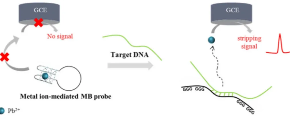
We developed a one-step and label-free, electrochemical DNA detection method using metal ion-mediated molecular beacon (MB) probe specially designed to have target-specific sequence in its loop and Pb2+-binding aptamer in its stem. In the absence of target DNA, MB probe, after the interaction with Pb2+, forms the in-tramolecular stem-loop hairpin structure, which limits Pb2+to freely diffuse onto the electrode surface, leading to the low electrochemical signal. In contrast, the presence of target DNA that forms the hybridization complex with MB probe, breaks down the intramolecular stem-loop structure of MB probe, and releases Pb2+that is freely diffused onto the electrode surface to generate the high electrochemical signal. By employing this method, the target DNA from Chlamydia trachomatis, one of the major pathogens causing sexually transmitted disease was successfully detected with the high selectivity.
1. Introduction
In recent years, intense interest has been aroused on the develop-ment of a simple and convenient DNA detection method that can be adopted into point-of-care-testing (POCT) and decentralized diagnostic testing [1,2]. Among many DNA detection approaches, the electro-chemical methods have received the special attention due to their sensitivity, portability, operational simplicity, cost effectiveness, and low power consumption [3–5]. The representative example in this type of sensing strategy relies on the molecular beacon (MB) probe whose one end is labeled with electrochemical reporter and the other end is immobilized onto electrode surface. The presence of target DNA com-plementary to MB probe controls the electrochemical signal by ad-justing the distance between the electrochemical reporter and the electrode surface [6–8]. In another study, Fang and co-worker sug-gested a similar strategy that utilizes MB probe labeled with carminic acid moieties as a key detection component [9]. Although these elec-trochemical methods developed to date provides the promising results, they involve the complicated and multi-step procedures including electrode modification, signaling molecule labeling, and washing [6,9–22]. These cumbersome, time-consuming procedures not only di-minish the overall detection reliability of the assay, but also increase the operational costs and inconveniences, which consequently limit their practical and widespread use in POCT [23,24].
To overcome these drawbacks, metal ions have been employed as the signaling molecules for electrochemical sensors. For example, metal nanoparticles such as CdS, PbS, and ZnS conjugated to DNA probe are first captured onto the electrode through its hybridization to target DNA and the captured metal nanoparticles are then dissolved by strong acid. As a result, the released metal ions generate the highly amplified electrochemical signals [25]. These methods allow for the sensitive DNA detection, but they still entail the afore-mentioned drawbacks in the previous electrochemical methods. Therefore, there is a great need for simple, convenient, electrochemical DNA detection strategies sui-table for POCT.
We herein developed a one-step and label-free, electrochemical DNA detection method using metal ion-mediated molecular beacon (MB) probe in which Pb2+-binding aptamer that forms G-quadruplex structure in the presence of Pb2+is rationally linked to target-specific sequence. In the absence of target DNA, Pb2+bound to MB probe is not freely diffused onto the electrode, generating the low electrochemical signal. In contrast, the presence of target DNA that forms the hy-bridization complex with MB probe, breaks down the intramolecular stem-loop structure of MB probe and releases Pb2+that generates the high electrochemical signal. With this novel strategy, the target DNA from Chlamydia trachomatis, one of the major pathogens causing sexu-ally transmitted disease, was analyzed with the selectivity even in the patient samples.
https://doi.org/10.1016/j.elecom.2019.01.023
Received 26 October 2018; Received in revised form 24 January 2019; Accepted 28 January 2019 ⁎Corresponding authors.
E-mail addresses:[email protected](K.S. Park),[email protected](H.G. Park). 1The authors equally contributed to this work.
Available online 30 January 2019
1388-2481/ © 2019 Published by Elsevier B.V. This is an open access article under the CC BY-NC-ND license (http://creativecommons.org/licenses/BY-NC-ND/4.0/).
2. Experimental
2.1. Materials
All the oligonucleotides were synthesized from Integrated DNA Technologies (IDT, Coralville, USA) and their sequences are listed in Table 1. The lead (II) nitrate, tris-acetate EDTA, sodium hydrogen phosphate, sodium dihydrogen phosphate, and sodium chloride were purchased from Sigma-Aldrich (St. Louis, MO, USA). All other chemi-cals were of analytical grade and used without further purification. Ultrapure DNase/RNase-free distilled water was purchased from In-vitrogen (Carlsbad, CA, USA).
2.2. Genomic DNA isolation
Samples (30 mL) of urine specimens, collected after prostatic mas-sage from patients infected by Chlamydia trachomatis and Trichomonas vaginalis, were centrifuged at 14,000 ×g for 2 min. The centrifuged pellets were washed twice with 1 mL of PBS (0.2 M phosphate, 1.5 M sodium chloride, pH 7.4) and resuspended in 400 μL of PBS. Genomic DNA was extracted from each pellet by using an Accuprep TM Genomic DNA Extraction Kit (Bioneer, Korea) according to the manufacturer's protocol and stored at −20 °C until use.
2.3. PCR amplification
Amplification of the specific site of Chlamydia trachomatis gene encoding virulence proteins was performed by using the PCR method on a DNA engine-Peltier thermo cycler (Bio-Rad, Hercules, CA) in a 50 μL solution containing DNA template (1 μL), 0.25 M each primer (Table 1), 10× PCR reaction buffer (500 mM Tris–HCl, 100 mM KCl, 50 mM (NH4)2SO4, 20 mM MgCl2), 0.2 mM dNTPs, and 1.25 U FastStart Taq DNA polymerase (Roche, Germany). PCR was programmed for 5 min at 95 °C, followed by 35 cycles of 30 s at 95 °C, 30 s at 55 °C, and 1 min at 72 °C, and 5 min at 72 °C. After the PCR amplification, the resulting PCR product (118 bp) was purified (NucleoSpin™, Macherey-Nagel, Duren, Germany) and used as the target DNA in this study. For a non-target DNA, the specific site of Trichomonas vaginalis gene was amplified and purified by following the same procedure used for the Chlamydia trachomatis except using the primer sets specific to Tricho-monas vaginalis gene (Table 1).
2.4. Electrochemical DNA detection
The as-prepared target DNAs were heat-denatured at 95 °C for 5 min and immediately cooled [26]. The 10 μL of the target DNA solution was mixed with 40 μL of 10 mM Tris-Acetate buffer (pH 8.0) solution con-taining the 1.65 μM molecular beacon (MB) probe and 0.41 μM Pb2+. The final solution (50 μL) was incubated for 1 h at room temperature. Synthetic target DNA (90)** ACA AGC TGC AAT CCC TTT TAA AAT AAC CCC GCA CGT
GCT TCG AGC AAC TTC GAG CAA
Synthetic target DNA (Mut-1)*** ACT ACA AGC TGC AAACCC TTT TAA AAT AAC
Synthetic target DNA (Mut-2)*** ACT ACA AGC TGC ACACCC TTT TAA AAT AAC
Synthetic target DNA (Mut-3)*** ACT ACA AGC TGC ACA TCC TTT TAA AAT AAC
Arbitrary DNA probe TAC AGT CTA GGA TTC GGC GTG GGT TAA GCT C. trachomatis
Forward primer AGG CGT TTG TAC TCC GTC AC C. trachomatis
Reverse primer TGG TGG GGT TAA GGC AAA TCG T. vaginalis
Forward primer CAC AAC ACC AAC ATA CGG CG T. vaginalis
Reverse primer TGA CAG CGA GCT TAC GAA GG
⁎The stem region that interacts with Pb2+is underlined, while the loop region that binds to the target DNA is indicated in red color.
⁎⁎The region complementary to loop region in MB probe is indicated in red color. ⁎⁎⁎The mutation site of the synthetic target DNA is indicated in blue color.
Then, square wave anodic stripping voltammetry (SWASV) was per-formed to measure the electrochemical signal of Pb2+ by using a GAMRY Reference 600 (Warminster, PA) with screen-printed carbon electrodes consisting of a carbon working (4 mm diameter), carbon auxiliary and silver pseudoreference electrodes (DropSens, Oviedo-As-turias, Spain). SWASV was measured under the following conditions: initial potential = −0.8 V, final potential = −0.2 V, pulse size poten-tial = 25 mV, step size = 0.05 V, frequency = 25 Hz, accumulation time = 120 s, and equilibrium time = 10 s [23,24]. For chron-oamperometric studies, the cell was left at +300 mV vs. Ag/AgCl re-ference for 20 s to reach the equilibrium state and then the current transient was measured for Pb2+, Pb2+ with MB probe, Pb2+ with arbitrary DNA probe (Table 1), Pb2+with MB probe and target DNA at different concentrations (100 pM and 10 nM) at −1 V (see Fig. S1 for details). The electrochemical signal differences for each case were as-sessed using a two-tailed t-test or one way analysis of variance (ANOVA) of the GraphPad Prism software, and P value smaller than 0.05 was considered significant.
3. Results and discussion
Fig. 1illustrates a one-pot, electrochemical DNA detection method which utilizes metal ion-mediated molecular beacon (MB) probe ra-tionally designed to contain target-specific sequence and Pb2+-binding aptamer. In the absence of target DNA, Pb2+-binding aptamer in MB probe interacts with Pb2+ to form G-quadruplex structure and thus Pb2+ bound to MB probe does not freely move onto the electrode, leading to the low electrochemical signal. On the other hand, the pre-sence of target DNA that forms the hybridization complex with MB probe breaks down the intramolecular stem-loop structure of MB probe, and releases Pb2+that is freely diffused onto the electrode surface to generate the high electrochemical signal. Based on the strong electro-chemical signal of Pb2+, the target DNA can be simply detected in a one-step without any modification and washing steps.
First, the ratio between MB probe and Pb2+that is essential for the efficient DNA detection was optimized by measuring the electro-chemical signal at ca. −0.48 V, the anodic peak potential of Pb2+. As shown in Fig. S2, the electrochemical signal decreased with increasing ratio of MB probe to Pb2+and became saturated at the ratio over 4, which indicates that most of Pb2+were captured by MB probe, limiting its access to the electrode. Thus, the optimal ratio of 4 was selected for further experiments.
Next, the detection feasibility of the developed method was verified with the synthetic target DNA (30-mer) specific to Chlamydia tracho-matis (C. trachotracho-matis). As envisioned in the schematic illustration (Fig. 1), the low electrochemical signal was obtained in the absence of target DNA, which is attributed to Pb2+bound to the stem region in MB probe. In contrast, the presence of target DNA breaks down the
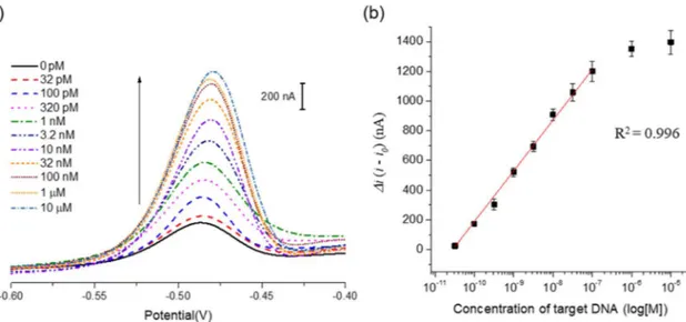
intramolecular stem-loop structure of MB probe and releases Pb2+that freely approaches the electrode, which was manifested by the high electrochemical signal (Fig. 2(a)). In addition, the peak current differ-ences (Δi) increased with increasing concentrations of target DNA (C. trachomatis) up to 100 nM, over which it reached a plateau. An ex-cellent linear relationship (R2= 0.996) existed in the range from 32 pM to 100 nM (Fig. 2(b)). For confirmation of performance on our system, in addition, we examined the electrochemical signaling effect on the length of target DNA (Fig. S3) and buffer condition (Fig. S4) and bio-logical interfering agent such as nucleic acid and proteins (Fig. S5).
We also investigated the ability of our system to detect DNA mu-tations. The results inFig. 3show that in the presence of target DNA containing three base pair mutations (Mut-3), the electrochemical signal from Pb2+was significantly decreased as compared to the one in the presence of complementary target DNA. In addition, the presence of target DNA with one or two base pair mutations (Mut-1 and 2) also decreases the electrochemical signal (P < 0.0139, unpaired two-tailed t-test). In addition, the effect of the non-complementary DNA on the hybridization of the MB probe with the target DNA was examined by measuring the electrochemical signal form Pb2+. As shown in the Fig. S6, the high electrochemical signals were observed, regardless of the presence of non-complementary target DNA (P = 0.3221, unpaired two-tailed t-test), which clearly confirms that non-complementary target DNA does not interfere with the hybridization of the molecular beacon probe with the target DNA.
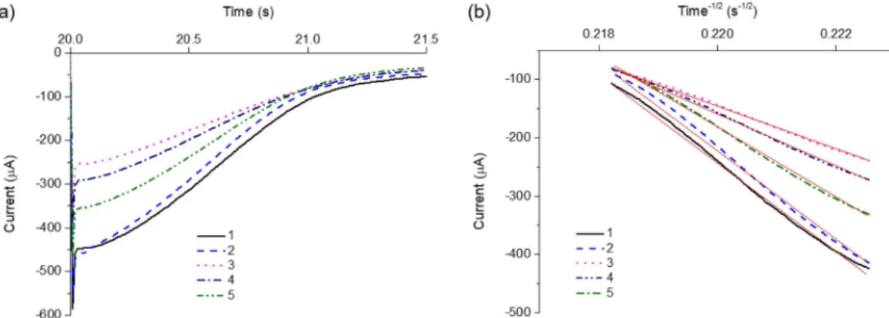
To further support the detection feasibility, the chronoampero-metric analyses of Pb2+, Pb2+with MB probe, and Pb2+with arbitrary DNA probe (Table 1), and Pb2+with MB probe and target DNA (100 pM and 10 nM) were performed. The results in Fig. 4(a) show that the current decreases as the time increases, which were replotted as a function of time−1/2to compare the slopes of linear trend line for Pb2+, Pb2+with MB probe, Pb2+with arbitrary DNA probe, and Pb2+with MB probe and target DNA (100 pM and 10 nM). According to Cottrell Eq.(1), where i is current, n is the number of electrons transferred per Pb2+, F is the Faraday constant, A is the electrode surface area, C is the total concentration of Pb2+, D is the diffusion coefficient of Pb2+, and t is time for which potential is applied (Fig. 4(b)) [27,28], the slope of the plot inFig. 4(b) is only proportional to D. The diffusion coefficients for Pb2+with MB probe and Pb2+with arbitrary DNA probe, and Pb2+ with MB probe and target DNA (100 pM and 10 nM) were found to be 0.23, 0.92, 0.58 and 0.78 relative to that for free Pb2+, respectively, which corresponds to 460.1, 1820.5, 635.1, and 1369.2 nA, respec-tively, in SWASV. This result indicates that Pb2+is selectively captured by Pb2+-binding aptamer in MB probe, leading to a significant reduc-tion of diffusion coefficient. These observareduc-tions clearly confirm that the developed strategy, which relies on metal ion-mediated MB probe, can be utilized for the one-step, label-free, and electrochemical detection of target DNA.
i nFACD t nFAC i t D 1/2 1/2 1/2 1/2 1/2 1/2 = = (1) Finally, the experiments to demonstrate the diagnostic capability of the developed method were carried out using the patient samples in-fected by C. trachomatis and Trichomonas vaginalis (T. vaginalis), major pathogens causing sexually transmitted diseases.
As shown inFig. 5, the peak current differences (Δi) increased as the concentration of target C. trachomatis DNA increased. An excellent linear relationship (R2= 0.998) existed in the range from 32 pM to 100 nM and the detection limit (3σ/slope) was determined to be 69.5 pM, a value that is comparable to those from other electrochemical strategies (Table S1) [6,9–22]. Importantly, no significant current dif-ference was observed when non-complementary DNA derived from T. vaginalis was tested, confirming the high specificity of the developed strategy. Overall, these results support that this method can be used for the reliable analysis of target DNA from clinical samples, verifying its practical applicability.
4. Conclusion
In summary, we have developed a one-step and label-free, electro-chemical DNA detection method that utilizes metal ion-mediated MB probe as a key component. By employing this strategy, the target DNAs derived from the patient samples infected with C. trachomatis and T. vaginalis were successfully analyzed with the high selectivity. In addi-tion, the diffusion coefficients of Pb2+ in different situations were compared to clearly support the proposed working principle. Importantly, this strategy does not involve multiple washing steps and expensive modifications required in the previous approaches, ensuring its widespread application in POCT. In addition, it has a huge potential for the multiplexed DNA detection and the detection of other biomo-lecules including small mobiomo-lecules, proteins, and cells, which could be accomplished by simply replacing Pb2+-binding aptamers with dif-ferent metal ion-binding aptamers such as mercury, silver, and copper etc. or substituting the target-specific sequence with other biomolecule-binding aptamers. Finally, we believe that this approach will pave the way for the development of new types of electrochemical sensors. Fig. 2. Quantitative analysis of synthetic target DNA specific to C. trachomatis. (a) Square wave anodic stripping voltammogram (SWASV) in the presence of synthetic target DNA at varying concentrations (0, 32 pM, 100 pM, 320 pM, 1 nM, 3.2 nM, 10 nM, 32 nM, 100 nM, 1 μM, and 10 μM from bottom to top). (b) A calibration curve of peak current change (Δi) vs. log target DNA concentration. The peak current change (Δi) is defined as i − i0, where i0and i are the electrochemical signal from Pb2+bound to MB probe in the absence and presence of target DNA, respectively.
Fig. 3. DNA mutation detection feasibility. (a) Square wave anodic stripping voltammogram (SWASV) and (b) peak current change (Δi) in the presence of com-plementary target DNA and target DNA containing one, two, or three base mutations at the concentration of 10 nM (Table 1). The length of all DNA used in this experiment 30 bp. The peak current change (Δi) is defined as i − i0, where i0and i are the electrochemical signal from Pb2+bound to MB probe in the absence and presence of target DNA, respectively.
Acknowledgements
Financial support was provided by Center for BioNano Health-Guard funded by the Ministry of Science and ICT (MSIT) of Korea as Global Frontier Project (Grant H-GUARD_2013M3A6B2078964) and Basic Science Research Program through the National Research Foundation (NRF) funded by the Ministry of Education (No. 2015R1A2A1A01005393).
Appendix A. Supplementary data
Supplementary data to this article can be found online athttps:// doi.org/10.1016/j.elecom.2019.01.023.
References
[1] W.G. Lee, Y.-G. Kim, B.G. Chung, U. Demirci, A. Khademhosseini, Nano/micro-fluidics for diagnosis of infectious diseases in developing countries, Adv. Drug Deliv. Rev. 62 (2010) 449–457.
[2] B.Y. Won, H.G. Park, A touchscreen as a biomolecule detection platform, Angew. Chem. Int. Ed. 124 (2012) 772–775.
[3] W.G. Kuhr, Electrochemical DNA analysis comes of age, Nat. Biotechnol. 18 (2000) 1042–1044.
[4] I. Willner, Biomaterials for sensors, fuel cells, and circuitry, Science 298 (2002) 2407–2408.
[5] J. Fritz, E.B. Cooper, S. Gaudet, P.K. Sorger, S.R. Manalis, Electronic detection of DNA by its intrinsic molecular charge, Proc. Natl. Acad. Sci. U. S. A. 99 (2002) 14142–14146.
[6] C. Fan, K.W. Plaxco, A.J. Heeger, Electrochemical interrogation of conformational changes as a reagentless method for the sequence-specific detection of DNA, Proc. Natl. Acad. Sci. U. S. A. 100 (2003) 9134–9137.
[7] F. Ricci, G. Adornetto, D. Moscone, K.W. Plaxco, G. Palleschi, Quantitative, re-agentless, single-step electrochemical detection of anti-DNA antibodies directly in blood serum, Chem. Commun. 46 (2010) 1742–1744.
[8] Y. Xiao, A.A. Lubin, B.R. Baker, K.W. Plaxco, A.J. Heeger, Single-step electronic detection of femtomolar DNA by target-induced strand displacement in an elec-trode-bound duplex, Proc. Natl. Acad. Sci. U. S. A. 103 (2006) 16677–16680. [9] J. Wu, C. Huang, G. Cheng, F. Zhang, P. He, Y. Fang, Electrochemically
active–i-nactive switching molecular beacon for direct detection of DNA in homogenous solution, Electrochem. Commun. 11 (2009) 177–180.
[10] K. Hsieh, Y. Xiao, H. Tom Soh, Electrochemical DNA detection via exonuclease and targetcatalyzed transformation of surface-bound probes, Langmuir 26 (2010) 10392–10396.
[11] C. Liu, D. Jiang, G. Xiang, L. Liu, F. Liu, X. Pu, An electrochemical DNA biosensor for the detection of Mycobacterium tuberculosis, based on signal amplification of graphene and a gold nanoparticle–polyaniline nanocomposite, Analyst 139 (2014) 5460–5465.
[12] C. Wang, H. Zhou, W. Zhu, H. Li, J. Jiang, G. Shen, R. Yu, Ultrasensitive electro-chemical DNA detection based on dual amplification of circular strand-displace-ment polymerase reaction and hybridization chain reaction, Biosens. Bioelectron. 47 (2013) 324–328.
[13] Y. Qian, T. Fan, P. Wang, X. Zhang, J. Luo, F. Zhou, Y. Yao, X. Liao, Y. Li, F. Gao, A novel label-free homogeneous electrochemical immunosensor based on proximity hybridization-triggered isothermal exponential amplification induced G-quadruplex formation, Sensors Actuators B Chem. 248 (2017) 187–194.
[14] F. Zhou, Y. Yao, J. Luo, X. Zhang, Y. Zhang, D. Yin, F. Gao, P. Wang, Proximity hybridization-regulated catalytic DNA hairpin assembly for electrochemical im-munoassay based on in situ DNA template-synthesized Pd nanoparticles, Anal. Chim. Acta 969 (2017) 8–17.
[15] H. Wang, Y. Zhang, Y. Wang, H. Ma, B. Du, Q. Wei, Facile synthesis of cuprous oxide nanowires decorated graphene oxide nanosheets nanocomposites and its applica-tion in label-free electrochemical immunosensor, Biosens. Bioelectron. 87 (2017) 745–751.
[16] L. Cao, C. Fang, R. Zeng, X. Zhao, F. Zhao, Y. Jiang, Z. Chen, A disposable paper-based microfluidic immunosensor paper-based on reduced graphene oxide-tetraethylene pentamine/Au nanocomposite decorated carbon screen-printed electrodes, Sensors Actuators B Chem. 252 (2017) 44–54.
[17] Z. Chen, Y. Liu, C. Xin, J. Zhao, S. Liu, A cascade autocatalytic strand displacement amplification and hybridization chain reaction event for label-free and ultra-sensitive electrochemical nucleic acid biosensing, Biosens. Bioelectron. 113 (2018) 1–8.
[18] W. Wang, T. Bao, X. Zeng, H. Xiong, W. Wen, X. Zhang, S. Wang, Ultrasensitive electrochemical DNA biosensor based on functionalized gold clusters/graphene nanohybrids coupling with exonuclease III-aided cascade target recycling, Biosens. Bioelectron. 91 (2017) 183–189.
[19] S. Xu, Y. Zhang, K. Dong, J. Wen, C. Zheng, S. Zhao, Electrochemical DNA biosensor based on graphene oxide-chitosan hybrid nanocomposites for detection of Escherichia coli O157: H7, Int. J. Electrochem. Sci. 12 (2017) 3443–3458. [20] W. Yaqiong, H. Sauriat-Dorizon, H. Korri-Youssoufi, Direct electrochemical DNA
biosensor based on reduced graphene oxide and metalloporphyrin nanocomposite,
Fig. 4. Chronoamperometric analysis of free Pb2+(1), Pb2+with arbitrary DNA probe (2), Pb2+with MB probe (3), Pb2+with MB probe and target DNA at the concentration of 100 pM (4) and 10 nM (5) to compare their diffusion coefficients. The concentration of Pb2+, arbitrary DNA, and MB probe was 0.33 μM, 1.32 μM, and 1.32 μM, respectively. (a) Current-time response curve (b) Cottrell plot of current vs. time−1/2. The red line indicates the linear trend line for each Cottrell plot. For chronoamperometric studies, the applied potential to the working electrode was +300 mV vs. Ag/AgCl reference electrode. (For interpretation of the references to color in this figure legend, the reader is referred to the web version of this article.)
Fig. 5. Clinical application for the analysis of patient samples. The peak current change (Δi) was plotted as the log concentration of target DNA (C. trachomatis) and non-target DNA (T. vaginalis) derived from patient samples. The peak current change (Δi) is defined as i − i0, where i0and i are the electrochemical signal from Pb2+bound to MB probe in the absence and presence of target DNA, respectively.