Rivaroxaban vs Warfarin Sodium in the Ultra-Early Period
After Atrial Fibrillation–Related Mild Ischemic Stroke
A Randomized Clinical Trial
Keun-Sik Hong, MD; Sun U. Kwon, MD; Sang Hun Lee, MD; Ji Sung Lee, PhD; Yong-Jae Kim, MD; Tae-Jin Song, MD; Young Dae Kim, MD; Man-Seok Park, MD; Eung-Gyu Kim, MD; Jae-Kwan Cha, MD; Sang Min Sung, MD;
Byung-Woo Yoon, MD; Oh Young Bang, MD; Woo-Keun Seo, MD; Yang-Ha Hwang, MD; Seong Hwan Ahn, MD; Dong-Wha Kang, MD; Hyun Goo Kang, MD; Kyung-Ho Yu, MD; for the Phase 2 Exploratory Clinical Study to Assess the Effects of Xarelto (Rivaroxaban) Versus Warfarin on Ischemia, Bleeding, and Hospital Stay in Acute Cerebral Infarction Patients With Non-valvular Atrial Fibrillation (Triple AXEL) Study Group
IMPORTANCEIn atrial fibrillation (AF)–related acute ischemic stroke, the optimal oral anticoagulation strategy remains unclear.
OBJECTIVETo test whether rivaroxaban or warfarin sodium is safer and more effective for preventing early recurrent stroke in patients with AF-related acute ischemic stroke. DESIGN, SETTING, AND PARTICIPANTSA randomized, multicenter, open-label, blinded end point evaluation, comparative phase 2 trial was conducted from April 28, 2014, to December 7, 2015, at 14 academic medical centers in South Korea among patients with mild AF-related stroke within the previous 5 days who were deemed suitable for early anticoagulation. Analysis was performed on a modified intent-to-treat basis.
INTERVENTIONSParticipants were randomized 1:1 to receive rivaroxaban, 10 mg/d for 5 days followed by 15 or 20 mg/d, or warfarin with a target international normalized ratio of 2.0-3.0, for 4 weeks.
MAIN OUTCOMES AND MEASURESThe primary end point was the composite of new ischemic lesion or new intracranial hemorrhage seen on results of magnetic resonance imaging at 4 weeks. Primary analysis was performed in patients who received at least 1 dose of study medications and completed follow-up magnetic resonance imaging. Key secondary end points were individual components of the primary end point and hospitalization length. RESULTSOf 195 patients randomized, 183 individuals (76 women and 107 men; mean [SD] age, 70.4 [10.4] years) completed magnetic resonance imaging follow-up and were included in the primary end point analysis. The rivaroxaban group (n = 95) and warfarin group (n = 88) showed no differences in the primary end point (47 [49.5%] vs 48 [54.5%]; relative risk, 0.91; 95% CI, 0.69-1.20; P = .49) or its individual components (new ischemic lesion: 28 [29.5%] vs 31 of 87 [35.6%]; relative risk, 0.83; 95% CI, 0.54-1.26; P = .38; new intracranial hemorrhage: 30 [31.6%] vs 25 of 87 [28.7%]; relative risk, 1.10; 95% CI, 0.70-1.71; P = .68). Each group had 1 clinical ischemic stroke, and all new intracranial hemorrhages were asymptomatic
hemorrhagic transformations. Hospitalization length was reduced with rivaroxaban compared with warfarin (median, 4.0 days [interquartile range, 2.0-6.0 days] vs 6.0 days [interquartile range, 4.0-8.0]; P < .001).
CONCLUSIONS AND RELEVANCEIn mild AF-related acute ischemic stroke, rivaroxaban and warfarin had comparable safety and efficacy.
TRIAL REGISTRATIONclinicaltrials.gov Identifier:NCT02042534
JAMA Neurol. 2017;74(10):1206-1215. doi:10.1001/jamaneurol.2017.2161 Published online September 11, 2017.
Editorialpage 1174
Supplemental content
Author Affiliations: Author affiliations are listed at the end of this article.
Group Information: Members of the Phase 2 Exploratory Clinical Study to Assess the Effects of Xarelto (Rivaroxaban) Versus Warfarin on Ischemia, Bleeding, and Hospital Stay in Acute Cerebral Infarction Patients With Non-valvular Atrial Fibrillation (Triple AXEL) Study Group are listed at the end of this article.
Corresponding Author: Sun U. Kwon, MD, Department of Neurology, University of Ulsan College of Medicine, Asan Medical Center, 88, Olympic-ro, Songpa-gu, Seoul 138-736, South Korea ([email protected]).
JAMA Neurology |
Original Investigation
P
atients with atrial fibrillation (AF)–related acute ische-mic stroke are at high risk of recurrent ischeische-mic stroke and intracranial hemorrhage, including hemorrhagic transformation, during the early period after stroke. Paren-teral heparin is not recommended because it increases the risk of symptomatic intracranial hemorrhage.1However, inpa-tients with AF and recent ischemic stroke who are treated with aspirin, the risk of recurrent ischemic stroke within 2 weeks is 5%.2-5The most widely used antithrombotic management
strategy in clinical practice is to start aspirin and then to ini-tiate oral anticoagulation after several days or 1 to 2 weeks when the risk of intracranial hemorrhage is likely to have subsided. Nonetheless, the optimal oral anticoagulation strategy for acute ischemic stroke, regarding when, in whom, and which drug, remains unclear.
To our knowledge, no trial has specifically tested oral an-ticoagulation in patients with AF-related acute ischemic stroke. In the European Atrial Fibrillation Trial, approximately 294 pa-tients with AF were randomized to receive warfarin sodium, aspirin, or placebo within 2 weeks of symptom onset, but the efficacy and safety of warfarin vs aspirin or placebo in pa-tients with acute cerebral ischemia was not reported.6
The piv-otal non–vitamin K antagonist oral anticoagulant (NOAC) trials excluded patients who had ischemic stroke within 7-30 days.7-10
For early oral anticoagulation after acute ischemic stroke, NOACs might have several advantages compared with warfa-rin: rapid onset of action, absence of transient hypercoagula-bility, stable and predictable anticoagulation effects, and ex-pected lower risk of intracranial hemorrhage. Accordingly, this proof-of-concept trial (Acute Stroke With Xarelto to Reduce Intracranial Hemorrhage, Recurrent Embolic Stroke, and Hospital Stay [Triple AXEL]) compared the efficacy and safety between rivaroxaban and dose-adjusted warfarin using magnetic resonance imaging (MRI) surrogate markers and hospitalization length in patients who had mild AF-related acute ischemic stroke who were considered suitable for early anticoagulation.
Methods
Study Design and Participants
Triple AXEL was a phase 2, multicenter, randomized, parallel-group, oplabel, blinded end point evaluation trial that en-rolled patients from 14 academic hospitals in South Korea from April 28, 2014, to October 30, 2015; the patients’ last day of the study was December 7, 2015. Details of the study design have been published previously.11The trial protocol and statistical
analysis plan are provided inSupplement 1. Briefly, patients were eligible if they had an MRI-confirmed acute ischemic stroke within 5 days due to presumed cardioembolism, had nonvalvular AF (including paroxysmal AF) documented by electrocardiogram, and were considered suitable for early an-ticoagulation after taking into account the severity of the is-chemic lesion as seen in results of diffusion-weighted imaging (DWI): acute ischemic lesion less than one-third of the middle cerebral artery territory, half of the anterior cerebral artery ter-ritory, half of the posterior cerebral artery terter-ritory, and half
of 1 cerebellar hemisphere. Key exclusion criteria included sig-nificant hemorrhagic transformation, mechanical heart valve, stroke caused by presumed small vessel occlusion, severe re-nal impairment (creatinine clearance <30 mL/min/1.73 m2[to
convert to milliliters per second per meter squared, multiply by 0.0167]), and high risk of intracranial or systemic bleeding (eTable 1 inSupplement 2). This study was designed and con-ducted in accordance with the Declaration of Helsinki12and
Good Clinical Practice guidelines and was approved by the in-stitutional review boards of Asan Medical Center, University of Ulsan College of Medicine; Ilsan Paik Hospital, Inje Univer-sity; Korea University Ansan Hospital, Korea University Col-lege of Medicine; Ewha Women’s University School of Medi-cine; Yonsei University College of MediMedi-cine; Chonnam National University Medical School; Busan Paik Hospital, Inje Univer-sity; Dong-A University Hospital; Pusan National University Hospital; Seoul National University Hospital; Samsung Medi-cal Center, Sungkyunkwan University School of Medicine; Kyungpook National University School of Medicine and Hos-pital; Chosun University School of Medicine; and Hallym Uni-versity Sacred Heart Hospital as well as the Korean Ministry of Food and Drug Administration (tracking number: 30078). Written informed consent from eligible patients or their le-gally authorized representatives was obtained. The trial is reg-istered with ClinicalTrials.gov (Identifier: NCT02042534).
Randomization and Masking
We randomly allocated eligible patients in a 1:1 ratio to receive rivaroxaban or dose-adjusted warfarin (target international nor-malized ratio [INR], 2-3) using an interactive web response sys-tem. The randomization sequence was computer generated and stratified by sites with a block size of 4. Patients and respon-sible physicians were aware of the assigned treatment. How-ever, an independent imaging review core laboratory, blinded to the treatment allocation and clinical data, assessed the MRI findings. Clinical events were reported by each investigator and were judged and categorized by the steering committee.
Procedures
Prior to randomization, all patients underwent magnetic reso-nance angiography and DWI, fluid-attenuated inversion re-covery, gradient-recalled echo, or susceptibility-weighted
Key Points
QuestionIs rivaroxaban safer and more effective compared with warfarin sodium for early anticoagulation in atrial
fibrillation–related acute ischemic stroke?
FindingsIn this randomized clinical trial of 195 patients with mild acute ischemic stroke and atrial fibrillation, new ischemic lesions or new intracranial hemorrhage on results of magnetic resonance imaging after 4 weeks occurred in 49.5% of patients receiving rivaroxaban and 54.5% receiving warfarin, a nonsignificant difference. Each group had 1 recurrence of clinical ischemic stroke, and no symptomatic intracranial hemorrhage occurred.
MeaningRivaroxaban and warfarin had comparable safety and efficacy for early anticoagulation in mild atrial fibrillation–related acute ischemic stroke.
imaging and received aspirin after the confirmation of an acute ischemic lesion on MRI results. Low-dose subcutaneous hep-arin or low-molecular-weight hephep-arin for the prevention of deep venous thrombosis or pulmonary embolism was used at the discretion of the responsible physicians.
After randomization, the rivaroxaban group received rivaroxaban, 10 mg once daily, for the first 5 days followed by 20 mg once daily (patients with creatinine clearance ≥50 mL/min) or 15 mg once daily (patients with creatinine clearance of 30-49 mL/min).8Aspirin or heparin for
prophy-laxis of deep venous thrombosis or pulmonary embolism was stopped 24 hours before initiation of rivaroxaban. In the war-farin group, aspirin or heparin for prophylaxis of deep venous thrombosis or pulmonary embolism was continued until an INR of 1.7 was reached after initiation of warfarin. To reduce inter-investigator variation in warfarin dosing, a web-based Bayesian algorithm (http://www.warfarindosing.org/) was recommended for warfarin dosing. For the anticoagulation quality in the warfarin arm, we assessed the mean INR values and the proportions of patients with an INR of 2 to 3 at day 5, 2 weeks, and 4 weeks because the 4-week trial period was too short to analyze the time in therapeutic range.
At 4 weeks, we performed MRI, including fluid-attenuated inversion recovery and gradient-recalled echo or susceptibility-weighted imaging to assess for new ischemic lesions and new intracranial hemorrhage. Patients who had experienced clini-cal ischemic stroke or symptomatic intracranial hemorrhage be-fore the end of the trial underwent MRI at the time of the clini-cal event. We allowed computed tomographic evaluation for patients who were unstable and unable to undergo MRI. How-ever, all patients included in the modified intent-to-treat (ITT) population completed the follow-up MRI.
During most of the trial period, the Korean insurance sys-tem did not cover the cost of NOACs for most patients with AF.13Therefore, patients randomized to receive rivaroxaban
had to make the transition from rivaroxaban to warfarin with a 5-day overlap to ensure adequate anticoagulation during the transition. Therefore, clinical ischemic and major bleed-ing events were followed up for an additional 7 days after the end of the trial and were included in the clinical event end points. However, patients who were willing to pay for rivar-oxaban at their own expense continued taking rivarrivar-oxaban. Detailed follow-up procedures are summarized in eTable 2 in Supplement 2).
Outcomes
The primary end point was the composite of new ischemic le-sion or new intracranial hemorrhage on results of follow-up MRI at 4 weeks. The definition of new ischemic lesion included symp-tomatic or asympsymp-tomatic new ischemic lesions on results of follow-up fluid-attenuated inversion recovery imaging.
Symptomatic ischemic lesions were defined as clinical
mic stroke recurrence associated with a relevant new ische-mic lesion. New intracranial hemorrhage (hemorrhagic trans-formation, intracerebral hemorrhage, subarachnoid hemorrhage, subdural hematoma, or epidural hematoma) included symptomatic or asymptomatic hemorrhage on re-sults of follow-up gradient-recalled echo or
susceptibility-weighted imaging. Symptomatic intracranial hemorrhage was defined as any intracranial hemorrhage associated with clini-cal deterioration as judged by the responsible investigators.
Prespecified secondary efficacy end points were new is-chemic lesion; new intracranial hemorrhage; length of hospi-talization; major bleeding as defined by the International Society on Thrombosis and Hemostasis14
; acute coronary syn-drome; composite of major vascular events including stroke, myocardial infarction, and vascular death; composite of a ma-jor vascular event plus mama-jor bleeding; composite of clinical ischemic events; and 4-week modified Rankin Scale score.
Statistical Analysis
A detailed description of the analytic approach is provided in the statistical analysis plan (Supplement 1). We designed the trial to show that rivaroxaban would be superior to warfarin for the prevention of new ischemic lesions or new intracra-nial hemorrhage on results of follow-up MRI. The primary and secondary end points were assessed in the modified ITT popu-lation, which included patients who were randomized, re-ceived at least 1 dose of study drugs, and completed fol-low-up MRI. Per-protocol analysis was additionally performed in patients who had no major protocol violation and took 80% or more of the assigned study medications. Adverse events were assessed in the safety population, which included pa-tients who were randomized and received at least 1 dose of study drugs.
We performed the primary end point analysis using χ2test
for unadjusted analysis and Poisson regression analysis for ad-justed analysis. In the adad-justed analysis, we included vari-ables of age, sex, center, prior use of a vitamin K antagonist (VKA), concomitant use of an antiplatelet agent, and vari-ables showing difference between the 2 groups. Secondary and safety end point analyses were conducted using χ2test, Fisher
exact test, Wilcoxon rank sum test, or analysis of covariance, as indicated.
On the basis of a review of earlier studies,4,8,15
we calcu-lated the sample size by assuming that the primary end point rate would be 25% in the warfarin group and that the abso-lute risk reduction with rivaroxaban would be 15%. Using a 1-sided superiority test, 178 patients (89 in each group) assess-able for the primary end point would give 80% power with a significance level of P < .05. Assuming a 10% dropout rate, we planned to enroll 196 patients. An independent statistician con-ducted the statistical analysis. At the inception, interim analy-sis was not planned because this was a phase 2, proof-of-concept, open-label trial. However, because there had been no trial of NOACs in patients with acute ischemic stroke, the steer-ing committee and investigators agreed to conduct an in-terim analysis for safety after enrolling 100 patients. No safety concern was raised, and we continued to enroll the planned patients.
Consistency of treatment effect for the primary end point and individual components was assessed for 8 post hoc sub-groups regarding age, sex, prior use of a VKA within 30 days before randomization, prestroke CHA2DS2-VASc (congestive
heart failure, hypertension, age ≥75 years [doubled], diabe-tes, stroke [doubled], vascular disease, age 65-74 years, sex
cat-egory [female]) score, prestroke HAS-BLED (hypertension, abnormal renal/liver function, stroke, bleeding history or pre-disposition, labile INR, elderly, drugs/alcohol concomitantly) score, baseline DWI volume, concomitant use of an antiplatelet agent, and creatinine clearance level.
Results
We randomized 195 patients to 1 of 2 treatment groups: 101 to rivaroxaban and 94 to warfarin. All patients had an acute is-chemic lesion confirmed by MRI results before randomiza-tion. Of the 195 patients randomized, 7 did not receive study treatments, and 5 withdrew and did not undergo follow-up MRI. Therefore, 183 patients (95 in the rivaroxaban group and 88 in the warfarin group) were included in the modified ITT population for the primary end point analysis, and 188 pa-tients (98 in the rivaroxaban group and 90 in the warfarin group) were included in the safety population. Comparisons of patients included in the modified ITT population and those excluded from the modified ITT population are provided in eTable 3 inSupplement 2. Three patients had major protocol violations or had less than 80% adherence to the study treat-ment (2 in the rivaroxaban group and 1 in the warfarin group),
leaving 180 patients (93 in the rivaroxaban group and 87 in the warfarin group) in the per-protocol population (Figure).
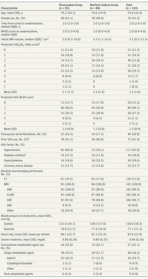
Baseline demographic and clinical characteristics were well balanced except that there were more patients in the rivar-oxaban group than the warfarin group with a history of type 2 diabetes (24 [25.3%] vs 10 [11.4%]), and patients in the rivar-oxaban group had a smaller median initial ischemic lesion vol-ume on DWI than did patients in the warfarin group (2.6 cm3
[interquartile range, 0.3-10.8] vs 5.5 cm3
[interquartile range, 1.1-14.5]) (Table 1). The median interval from stroke onset to ran-domization was 2 days (interquartile range, 2.0-3.0) (eFigure 1 inSupplement 2), the mean (SD) prestroke CHA2DS2-VASc score
was 2.5 (1.6) (Table 1), the mean (SD) prestroke HAS-BLED score was 1.3 (0.9), and the median National Institutes of Health Stroke Scale score at the time of randomization was 2.0 (interquar-tile range, 0.0-4.0). There was a history of VKA use within 30 days before randomization in 75 patients (41.0%), and 57 pa-tients (31.1%) received concomitant antiplatelet therapy dur-ing the trial.
Of the modified ITT population, the proportion of pa-tients taking more than 80% of the study medications was 98.9% in the rivaroxaban group (n = 94) and 97.7% in the war-farin group (n = 86) at a mean (SD) of 5 (2) days and 98.9% in the rivaroxaban group (n = 94) and 98.9% in the warfarin group Figure. Study Flowchart
195 Patients randomized
101 Randomized to receive
rivaroxaban 94 Randomized to receive warfarin sodium
3 Withdrew consent and did not receive treatment
4 Did not receive treatment 3 Withdrew consent 1 Withdrawn by investigators 98 Received treatment and
were included in the safety population
3 Withdrew and did not undergo follow-up MRI 2 Withdrawn by investigators 1 Withdrew consent
95 Underwent follow-up MRI and were included in the modified intent-to-treat population
2 Protocol violations 1 Did not meet the inclusion
criteria
1 Took <80% of assigned treatment
93 Included in the per-protocol population
90 Received treatment and were included in the safety population
2 Withdrew consent and did not undergo follow-up MRI
88 Underwent follow-up MRI and were included in the modified intent-to-treat population
1 Protocol violation (took <80% of assigned treatment)
87 Were included in the per-protocol population
MRI indicates magnetic resonance imaging.
Table 1. Baseline Characteristics of the Patients Included in the Modified Intent-to-Treat Population
Characteristic
Rivaroxaban Group (n = 95)
Warfarin Sodium Group (n = 88)
Total (n = 183)
Age, mean (SD), y 70.2 (10.1) 70.6 (10.9) 70.4 (10.4)
Female sex, No. (%) 40 (42.1) 36 (40.9) 76 (41.5)
Time from onset to randomization, median (IQR), d
2.0 (2.0-3.0) 2.0 (2.0-3.0) 2.0 (2.0-3.0)
NIHSS score at randomization, median (IQR)
2.0 (1.0-4.0) 2.0 (0.0-4.0) 2.0 (0.0-4.0)
Initial DWI volume, median (IQR),acm3 2.6 (0.3-10.8) 5.5 (1.1-14.5) 3.5 (0.5-12.3)
Prestroke CHA2DS2-VASc scoreb
0 11 (11.6) 10 (11.4) 21 (11.5) 1 16 (16.8) 15 (17.0) 31 (16.9) 2 14 (14.7) 26 (29.5) 40 (21.9) 3 20 (21.1) 17 (19.3) 37 (20.2) 4 22 (23.2) 14 (15.9) 36 (19.7) 5 8 (8.4) 6 (6.8) 14 (7.7) 6 3 (3.2) 0 3 (1.6) 7 1 (1.1) 0 1 (0.5) Mean (SD) 2.7 (1.7) 2.3 (1.4) 2.5 (1.6)
Prestroke HAS-BLED scorec
0 13 (13.7) 15 (17.0) 28 (15.3) 1 46 (48.4) 44 (50.0) 90 (49.2) 2 25 (26.3) 25 (28.4) 50 (27.3) 3 9 (9.5) 4 (4.5) 13 (7.1) 4 2 (2.1) 0 2 (1.1) Mean (SD) 1.4 (0.9) 1.2 (0.8) 1.3 (0.9)
Paroxysmal atrial fibrillation, No. (%) 25 (26.3) 24 (27.3) 49 (26.8)
Prior VKA use, No. (%)d 39 (41.1) 36 (40.9) 75 (41.0)
Risk factor, No. (%)
Hypertension 65 (68.4) 52 (59.1) 117 (63.9)
Diabetes mellituse 24 (25.3) 10 (11.4) 34 (18.6)
Hyperlipidemia 16 (16.8) 18 (20.5) 34 (18.6)
Coronary artery disease 13 (13.7) 12 (13.6) 25 (13.7)
Baseline neuroimaging performed, No. (%) CT 67 (70.5) 63 (71.6) 130 (71.0) MRI 95 (100.0) 88 (100.0) 183 (100.0) DWI 95 (100.0) 87 (98.9) 182 (99.5) FLAIR 95 (100.0) 87 (98.9) 182 (99.5) GRE 87 (91.6) 79 (89.8) 166 (90.7) SWI 9 (9.5) 9 (10.2) 18 (9.8) Other 18 (18.9) 20 (22.7) 38 (20.8)
Blood pressure at study entry, mean (SD), mm Hg
Systolic 131.8 (19.1) 128.0 (17.0) 130.0 (18.2)
Diastolic 76.8 (11.7) 77.4 (10.9) 77.1 (11.3)
Heart rate, mean (SD), beats per minute 84.5 (22.7) 83.3 (23.3) 83.9 (22.9)
Serum creatinine, mean (SD), mg/dL 0.89 (0.24) 0.89 (0.25) 0.89 (0.24)
Concomitant antiplatelet agent use, No. (%)
34 (35.8) 23 (26.1) 57 (31.1)
Single antiplatelet agent 28 (29.5) 20 (22.7) 48 (26.2)
Aspirin 25 (26.3) 11 (12.5) 36 (19.7)
Clopidogrel bisulfate 2 (2.1) 7 (8.0) 9 (4.9)
Other 1 (1.1) 2 (2.3) 3 (1.6)
Dual antiplatelet agents 6 (6.3) 3 (3.4) 9 (4.9)
Abbreviations: CHA2DS2-VASc,
congestive heart failure, hypertension, ageⱖ75 years (doubled), diabetes, stroke (doubled), vascular disease, age 65-74 years, sex category (female); CT, computed tomography; DWI, diffusion-weighted imaging; FLAIR, fluid-attenuated inversion recovery; GRE, gradient-recalled echo; HAS-BLED, hypertension, abnormal renal/liver function, stroke, bleeding history or predisposition, labile international normalized ratio, elderly, drugs/alcohol concomitantly; IQR, interquartile range;
MRI, magnetic resonance imaging; NIHSS, National Institutes of Health Stroke Scale; SWI, susceptibility-weighted imaging; VKA, vitamin K antagonist.
SI conversion factor: To convert creatinine to micromoles per liter, multiply by 88.4.
aP = .04. b
The CHA2DS2-VASc scores range
from 0 to 9, with higher scores indicating a greater risk of stroke: congestive heart failure, hypertension, diabetes, 65 to 74 years of age, female sex, and vascular disease are each assigned 1 point, and prior stroke or transient ischemic attack and being 75 years of age or older are assigned 2 points.
c
The HAS-BLED scores range from 0 to 9, with higher scores indicating a greater bleeding risk: hypertension, abnormal renal function, abnormal liver function, prior stroke history, bleeding, labile international normalized ratios, elderly (>65 years of age), prior alcohol or drug use, and medication use predisposing to bleeding are each assigned 1 point.
dUse of VKA within 30 days before
randomization.
e
(n = 87) at 4 weeks. After the end of the trial, 73.7% of pa-tients (n = 70) in the rivaroxaban group continued ban treatment, and 26.3% (n = 25) transitioned from rivaroxa-ban to warfarin. In the warfarin group, the proportion of patients who achieved the target INR of 2.0-3.0 was 40.9% (n = 36) at 5 days, 53.4% (n = 47) at 2 weeks, and 46.6% (n = 41) at 4 weeks. The mean (SD) INR value in the warfarin group was 2.04 (0.62) at 5 days, 2.67 (0.92) at 2 weeks, and 2.39 (0.83) at 4 weeks (eFigure 2 inSupplement 2).
The primary end point occurred in 47 patients (49.5%) in the rivaroxaban group and 48 patients (54.5%) in the warfa-rin group in the modified ITT population (relative risk, 0.91; 95% CI, 0.69-1.20, P = .49) (Table 2). After adjusting for age, sex, initial ischemic lesion volume on DWI, diabetes, prior VKA, concomitant antiplatelet use, and center, the difference was not significant (relative risk, 0.97; 95% CI, 0.79-1.18; P = .73). Sensitivity analyses assuming worst-case and best-case sce-narios for patients who received study drug but did not un-dergo follow-up MRI showed similar findings (eTable 4 in Supplement 2). In the per-protocol population, the 2 groups showed no difference in the primary end point rate (rivaroxa-ban, 46 of 93 [49.5%] vs warfarin, 47 of 87 [54.0%]; relative risk, 0.92; 95% CI, 0.69-1.21; P = .54) (eTable 5 inSupplement 2).
A new ischemic lesion was seen in 28 patients (29.5%) in the rivaroxaban group and 31 of 87 patients (35.6%) in the war-farin group (relative risk, 0.83; 95% CI, 0.54-1.26; P = .38) (Table 2). Each group had 1 clinical ischemic stroke recur-rence, which was presumed to be cardioembolism. The pro-portions of new ischemic lesions greater than 10 mm and mul-tiple lesions did not differ between the 2 groups (eTable 6 in Supplement 2).
New intracranial hemorrhage was seen in 30 patients (31.6%) in the rivaroxaban group and 25 of 87 patients (28.7%) in the warfarin group (relative risk, 1.10; 95% CI, 0.70-1.71;
P = .68) (Table 2). There was no symptomatic intracranial
hem-orrhage in the 2 groups; all intracranial hemhem-orrhages were asymptomatic hemorrhagic transformations within or adja-cent to the qualifying ischemic lesion. Of the hemorrhagic transformations, 49 of 55 (89.1%) were minimal or mild hem-orrhagic transformation without mass effect. Although there was no statistically significant difference, parenchymal hematoma with mass effect was observed more frequently in the warfarin group than in the rivaroxaban group, while type I hemorrhagic infarction was more frequent in the riva-roxaban group than in the warfarin group (eTable 7 in Supplement 2).
Table 2. End Points in the Modified Intent-to-Treat Population
End Point Rivaroxaban Group, No. (%) (n = 95) Warfarin Sodium Group, No. (%) (n = 88) Risk Difference (95% CI) Relative Risk (95% CI) P Value Adjusted Relative Risk (95% CI)a P Value Intracranial hemorrhage or recurrent ischemic lesion on results of 4-wk MRI (primary end point)
47 (49.5) 48 (54.5) –5.07 (–19.52 to 9.49) 0.91 (0.69 to 1.20) .49 0.97 (0.79 to 1.18) .73
Recurrent ischemic lesion
on results of 4-wk MRIb 28 (29.5) 31 (35.6) –6.16(–20.48 to 8.45) 0.83(0.54 to 1.26) .38 0.85(0.56 to 1.30) .45 Intracranial hemorrhage on results of 4-wk MRIb 30 (31.6) 25 (28.7) 2.84 (–11.68 to 17.29) 1.10 (0.70 to 1.71) .68 1.17 (0.74 to 1.85) .50 Clinical recurrent ischemic stroke 1 (1.1) 1 (1.1) –0.08 (–14.54 to 14.42) 0.93 (0.06 to 14.59) >.99 NA NA Symptomatic hemorrhagic conversion or hemorrhagic stroke 0 0 NA NA >.99 NA NA Major bleeding 1 (1.1) 0 1.05 (–13.44 to 15.53) NA >.99 NA NA Systemic embolism 0 0 NA NA >.99 NA NA
Acute coronary syndrome 0 0 NA NA >.99 NA NA
Composite of stroke, MI, or vascular death 1 (1.1) 1 (1.1) –0.08 (–14.54 to 14.42) 0.93 (0.06 to 14.59) >.99 NA NA
Composite of stroke, MI, vascular death, or major bleeding 2 (2.1) 1 (1.1) 0.97 (–13.50 to 15.46) 1.85 (0.17 to 20.08) >.99 NA NA Composite of clinical ischemic events 1 (1.1) 1 (1.1) –0.08 (–14.54 to 14.42) 0.93 (0.06 to 14.59) >.99 NA NA Duration of hospitalization, median (IQR), d 4.0 (2.0-6.0) 6.0 (4.0-8.0) NA NA <.001 NA .002 mRS score 0–1 at 4 wkc 79 (84.0) 64 (74.4) 9.62 (–5.06 to 23.95) 1.13 (0.97 to 1.31) .11 1.04 (0.83 to 1.29) .73 Abbreviations: IQR, interquartile range; MI, myocardial infarction;
MRI, magnetic resonance imaging; mRS, modified Rankin Scale; NA, not applicable.
aAdjusted for age, sex, initial ischemic lesion volume on diffusion-weighted
imaging, diabetes, prior use of vitamin K antagonist, concomitant use of antiplatelet agent, and center.
b
Of the patients in the warfarin group who were included in the intent-to-treat population and were evaluated for the primary end point, recurrent ischemic
lesion on results of 4-week MRI was not evaluated in 1 patient, and new intracranial hemorrhage on 4-week MRI was not evaluated in 1 patient (detailed description in eTable 4 inSupplement 2). Sensitivity analyses assuming worst-case and best-case scenarios for these patients are provided in eTable 4 inSupplement 2.
cOf the intent-to-treat population, the mRS score at 4 weeks was not available
The treatment effects were generally consistent across post hoc subgroups for the primary end point and individual com-ponents. However, there was a significant interaction be-tween prior VKA use and treatment effect for the recurrent is-chemic lesion end point, suggesting that rivaroxaban was associated with fewer recurrent ischemic lesions in patients with no prior VKA use than was warfarin (eTables 8-10 in Supplement 2).
One patient in the rivaroxaban group developed intraocu-lar bleeding leading to transient visual disturbance, which was categorized as major bleeding. However, it did not prolong hos-pitalization, and the patient recovered without sequelae. There was no acute coronary syndrome during the trial. The propor-tions of modified Rankin Scale scores of 0 to 1 at 4 weeks did not differ between the rivaroxaban and warfarin groups (79 of 94 [84.0%] vs 64 of 86 [74.4%]; relative risk, 1.13; 95% CI, 0.97-1.31; P = .11) (Table 2). However, median hospitalization length was significantly shorter in the rivaroxaban group than in the warfarin group (4.0 days [interquartile range, 2.0-6.0 days] vs 6.0 days [interquartile range, 4.0-8.0 days]; P < .001) (Table 2). In the per-protocol population analysis, the secondary end point results were similar to those of the modified ITT popu-lation analysis (eTable 5 inSupplement 2).
The rates of adverse events, adverse drug reactions, and serious adverse events were similar between the 2 groups (Table 3). There were no deaths during the trial.
Discussion
In this first trial to compare NOAC and VKA in patients with AF-related acute ischemic stroke, there was no difference be-tween rivaroxaban and warfarin in the combined surrogate end point of new ischemic lesion or new intracranial hemorrhage on results of follow-up MRI at 4 weeks. In addition, there were no differences between the 2 groups in the individual compo-nents of new ischemic lesion and new intracranial hemor-rhage. No patient had symptomatic intracranial hemorrhage, and 1 patient (1.1%) in each group had clinical recurrence of is-chemic stroke. Therefore, either rivaroxaban or warfarin ini-tiated within 5 days of stroke onset was comparably safe and effective for preventing clinical recurrence of ischemic stroke in patients with AF and mild acute ischemic stroke. However, initiation of rivaroxaban instead of warfarin reduced hospi-talization length by almost 2 days.
A recent European practical guide recommends the “1-3-6-12 day rule” for the initiation of NOACs after transient ische-mic attack or acute ischeische-mic stroke.16However, the
recom-mendations are based on expert opinion and are not supported by clinical trial data. An analysis of the Virtual International Stroke Trials Archive (VISTA) database found that the early ini-tiation of anticoagulants (2-3 days after stroke) was associ-ated with substantially fewer recurrent events during the fol-lowing weeks without an increased risk of symptomatic intracerebral hemorrhages.17
However, the finding is subject to substantial confounding by indication. In a prospective ob-servational study that enrolled 1029 consecutive patients with acute ischemic stroke and known or newly diagnosed AF with-out contraindications to anticoagulation, compared with ini-tiation of treatment before 4 days or more than 14 days after stroke onset, initiation of anticoagulants within 4 to 14 days of stroke onset was associated with a significant reduction in the composite of stroke, transient ischemic attack, sympto-matic systemic embolism, symptosympto-matic cerebral bleeding, and major extracranial bleeding within 90 days from acute stroke.18
In a recent small, prospective observational study of 60 pa-tients initiating rivaroxaban at a median time of 3 days after mild to moderate cardioembolic stroke or transient ischemic attack, no patient developed symptomatic hemorrhagic trans-formation, and 8 patients had asymptomatic new hemor-rhagic transformation or worsening of hemorhemor-rhagic transfor-mation on results of follow-up MRI obtained 7 days after initiation of rivaroxaban, suggesting the safety of rivaroxa-ban treatment in the acute stage.19
Our findings support the recommendations and are generally in accordance with the findings of the earlier observational studies.
Given the lower risk of intracranial hemorrhage with NOACs vs warfarin for long-term prevention therapy, pa-tients with AF-related acute ischemic stroke who were treated with rivaroxaban vs those treated with warfarin were ex-pected to have a lower risk of new intracranial hemorrhage, including hemorrhagic transformation. However, the risk was not different between the 2 treatment groups. Although, of in-tracranial hemorrhage types, minimal or mild hemorrhagic transformation was more frequent with rivaroxaban whereas parenchymal hematoma was more frequent with warfarin, our study was not adequately powered to detect the difference in the types of intracranial hemorrhage.
Although there were no statistically significant differ-ences, the rivaroxaban group had fewer recurrent ischemic Table 3. Adverse Events in the Safety Population
Characteristic No. (%) Risk Difference, % (95% CI) P Value Rivaroxaban Group (n = 98)
Warfarin Sodium Group (n = 90)
≥1 Adverse events 46 (46.9) 51 (56.7) −9.73
(−23.84 to 4.66)
.18
≥1 Adverse drug reactions 9 (9.2) 13 (14.4) −5.26
(−19.41 to 9.11)
.26
≥1 Serious adverse events 5 (5.1) 5 (5.6) −0.45
(−14.76 to 13.82) >.99 Withdrawal owing to adverse events 1 (1.0) 0 1.02 (−13.32 to 15.32) >.99 Death 0 0 NA >.99
lesions and more new intracranial hemorrhages compared with the warfarin group. Full anticoagulation would be immedi-ately achieved with rivaroxaban and delayed with warfarin, which might contribute to fewer ischemic lesions and more hemorrhagic lesions with rivaroxaban in the acute stage when the risks of both recurrent ischemia and intracranial hemor-rhage would be greatest. The post hoc analysis finding of a greater difference in the recurrent ischemic lesion end point in patients with no recent VKA use than in those with recent VKA use might support our speculation. However, the results from the post hoc analysis and multiple comparisons would limit the significance of the finding.
Although there were no differences in the imaging and clinical end points between the 2 groups, initiation of oral an-ticoagulation with rivaroxaban vs warfarin significantly re-duced the length of hospitalization for acute stroke. More rapid achievement of full anticoagulation with rivaroxaban likely en-ables patients to be discharged earlier. Previous studies showed that, for long-term therapy, use of NOACs vs warfarin was cost-effective.20-24Our trial showed that rivaroxaban would
reduce the cost of hospitalization for acute stroke in patients who are deemed suitable for early anticoagulation, but the cost-saving effect might not be applicable to different health care settings.
In the current study, we found that about 50% of patients had asymptomatic new ischemic lesions or intracranial hem-orrhage on results of MRI within 4 weeks after stroke, which was greater than expected. Despite the early use of anticoagu-lation, about 30% of the patients had a new ischemic lesion. In a prior study, 40% of patients who had a cardioembolic stroke within 6 hours and were treated with antiplatelet drugs or anticoagulation had a recurrent ischemic lesion on MRI re-sults within the first week.15Our findings, along with those
of the earlier study, indicate that the risk of early recurrent cerebral ischemia is high in patients with AF-related acute ischemic stroke. However, intracranial hemorrhage was also observed in about 30% of patients in our study. All of the intracranial hemorrhages were asymptomatic and most were minor hemorrhagic transformations, which might spontane-ously develop rather than be caused by anticoagulation. Because we had no placebo group, we were unable to assess whether rivaroxaban or warfarin increased the risk of asymp-tomatic hemorrhagic transformation. Although the ischemic and hemorrhagic lesions were clinically silent in most patients, their effect on cognitive function was not explored in this study and would be of interest as part of a future investigation. In addition, the high frequency of both new ischemic and hemorrhagic lesions emphasizes the impor-tance of a proper balance between the efficacy and safety of
an antithrombotic strategy in patients with AF and acute cerebral ischemia.
We selected a low dose of 10 mg of rivaroxaban for the first 5 days because no safety data on rivaroxaban in patients with acute ischemic stroke were available, and all patients re-ceived aspirin before randomization and the residual effect of aspirin would remain for 5 to 7 days after rivaroxaban initia-tion. In a pharmacodynamics study in healthy Chinese indi-viduals, the inhibition of factor Xa activity, as measured by the median percentage change from baseline, was about 38% with a single dose of rivaroxaban, 10 mg, and about 46% with a single dose of rivaroxaban, 20 mg. Therefore, a single dose of rivar-oxaban, 10 mg, might achieve about 83% of the factor Xa ac-tivity inhibition as a single dose of rivaroxaban, 20 mg.25
When NOAC is compared with warfarin, the anticoagula-tion quality of warfarin therapy must be adequately main-tained. Because of the short trial duration, rather than time in therapeutic range, we assessed the mean INR and the propor-tion of INR values ranging from 2.0 to 3.0. Although the mean INR values in the warfarin arm were within 2.0 to 3.0 at each time point, the proportion of INR values from 2.0 to 3.0 ranged between 40.9% (n = 36 of 88) and 53.4% (n = 47 of 88), even though we followed a recommended warfarin dosing pro-gram. In the pivotal NOAC trials, the time in therapeutic range of Korean patients ranged between 48% and 56%.26-29Our
find-ings, along with those of the large trials, indicate that Korean patients have difficulty in achieving good-quality anticoagu-lation with warfarin therapy.
Limitations
This was an open-label study, which was at risk of a reporting bias for clinical events. However, the primary end point was an imaging surrogate marker evaluated in a blinded fashion. The quality of anticoagulation with warfarin in our study was less than optimal. Therefore, our findings might not be appli-cable to populations with high-quality anticoagulation with warfarin therapy. Because this study selected patients with mild stroke severity who were considered at low risk of intra-cranial hemorrhage with early oral anticoagulation, the find-ings would not be applicable to those with moderate to se-vere stroke.
Conclusions
Both rivaroxaban and warfarin initiated within 5 days of stroke onset in patients with mild AF-related acute ischemic stroke were safe and effective for preventing early clinical stroke recurrence.
ARTICLE INFORMATION
Accepted for Publication: June 12, 2017. Published Online: September 11, 2017. doi:10.1001/jamaneurol.2017.2161
Author Affiliations: Department of Neurology, Ilsan Paik Hospital, Inje University, Goyang, South Korea (Hong); Department of Neurology, University of Ulsan College of Medicine, Asan Medical Center,
Seoul, South Korea (Kwon, D.-W. Kang); Department of Neurology, Korea University Ansan Hospital, Korea University College of Medicine, Ansan, South Korea (S. H. Lee); Clinical Research Center, Asan Medical Center, Seoul, South Korea (J. S. Lee); Department of Neurology, Ewha Women’s University School of Medicine, Seoul, South Korea (Y.-J. Kim, Song); Department of Neurology, Yonsei University College of Medicine,
Seoul, South Korea (Y. D. Kim); Department of Neurology, Chonnam National University Medical School, Gwangju, South Korea (Park); Department of Neurology, Busan Paik Hospital, Inje University, Busan, South Korea (E.-G. Kim); Department of Neurology, Dong-A University Hospital, Busan, South Korea (Cha); Department of Neurology, Pusan National University Hospital, Busan, South Korea (Sung); Department of Neurology, Seoul
National University Hospital, Seoul, South Korea (Yoon); Department of Neurology, Samsung Medical Center, Sungkyunkwan University School of Medicine, Seoul, South Korea (Bang, Seo); Department of Neurology and Cerebrovascular Center, Kyungpook National University School of Medicine and Hospital, Daegu, South Korea (Hwang); Department of Neurology, Chosun University School of Medicine, Gwangju, South Korea (Ahn, H. G. Kang); Department of Neurology, Hallym University Sacred Heart Hospital, Anyang, South Korea (Yu).
Author Contributions: Drs Hong and Kwon had full access to all the data in the study and take responsibility for the integrity of the data and the accuracy of the data analysis. Drs Hong and Kwon contributed equally as first authors to this article. Study concept and design: Hong, Kwon. Acquisition, analysis, or interpretation of data: All authors.
Drafting of the manuscript: Hong, Kwon. Critical revision of the manuscript for important intellectual content: All authors.
Statistical analysis: Hong, Kwon, J. S. Lee. Obtained funding: Kwon.
Administrative, technical, or material support: Hong, Kwon, J. S. Lee.
Study supervision: Hong, Kwon.
Conflict of Interest Disclosures: Dr Hong reported receiving grants from Bayer Korea Ltd during the conduct of the study; grants from Boehringer Ingelheim, Bayer, and Ostuka Korea; grants and personal fees from Pfizer Korea, Bayer Korea, and Boehringer Ingelheim Korea; and personal fees from Sanofi Korea, Chong Kun Dang Pharm, Dong Wha Pharm, United Pharm, and Daichi Sankyo Korea outside the submitted work. Dr Kwon reported receiving grants from Bayer Korea Ltd and Korean Health Technology R&D Project (Ministry of Health & Welfare, Republic of Korea [HI14C1061 and HI10C2020]) during the conduct of the study; grants from Pfizer and Boehringer Ingelheim; grants and personal fees from Otsuka Korea; and personal fees from AstraZeneca Korea, Bayer Korea, Novartis Korea, Pfizer Korea, and Daichi Sankyo Korea outside the submitted work. Drs J. S. Lee, Y.-J. Kim, Song, Y. D. Kim, Park, E.-G. Kim, Bang, and Ahn reported receiving grants from Bayer Korea Ltd during the conduct of the study. Dr Cha reported receiving grants from Bayer Korea Ltd during the conduct of the study; grants from Otsuka Korea, Bayer Korea, ESAI-Korea, Daewoong
Pharmaceutical Co Ltd, Daichi Sankyo, Pfizer, Sanofi Korea, and AstraZeneca Korea; and personal fees from Bayer Korea, Boehringer Ingelheim Korea, Pfizer Korea, AstraZeneca Korea, Bayer Korea, Novartis Korea, Otsuka Korea, Pfizer Korea, and Daichi Sankyo Korea outside the submitted work. Dr Sung reported receiving grants from Bayer Korea Ltd during the conduct of the study and personal fees from Daiichi Sankyo Korea and Pfizer Pharmaceuticals Korea Ltd outside the submitted work. Dr Yoon reported receiving grants from Bayer Korea Ltd during the conduct of the study and grants from Bayer HealthCare outside the submitted work. Dr Seo reported receiving grants from Bayer Korea Ltd during the conduct of the study; grants from Korea Otsuka, Daewoong Pharmaceutical Co Ltd, Myung In Pharmaceutical Co Ltd, and Korea United Pharm Inc; grants and personal fees from Bayer Korea, Pfizer Pharmaceuticals, Sanofi Korea, and Boehringer Ingelheim Korea; and personal fees from Daiichi
Sankyo Korea outside the submitted work. Dr Hwang reported receiving grants from Bayer Korea Ltd during the conduct of the study and grants from Kyungpook National University outside the submitted work. Dr D.-W. Kang reported receiving grants from National Research Foundation of Korea funded by the Korea government (NRF-2014R1A2A1A11051280) and Korea Health Technology R&D Project (Ministry for Health & Welfare, Republic of Korea [HI12C1847 and HI14C1983]) outside the submitted work. Dr K.-H. Yu reported receiving grants from Bayer Korea Ltd during the conduct of the study and personal fees from Bristol-Myers Squibb and Bayer outside the submitted work. No other conflicts were reported. Funding/Support: This study was supported by Bayer Korea Ltd and grants HI14C1061 and HI10C2020 from the Korean Health Technology R&D Project, Ministry of Health & Welfare, Republic of Korea.
Role of the Funder/Sponsor: Bayer Korea Ltd suggested rivaroxaban dose selection. The funding sources had no other role in the design and conduct of the study; collection, management, analysis, and interpretation of the data; preparation, review, or approval of the manuscript; and decision to submit the manuscript for publication.
Group Information: Members of the Phase 2 Exploratory Clinical Study to Assess the Effects of Xarelto (Rivaroxaban) Versus Warfarin on Ischemia, Bleeding, and Hospital Stay in Acute Cerebral Infarction Patients With Non-valvular Atrial Fibrillation (Triple AXEL) Study Group include Sun U. Kwon, MD, Dong-Wha Kang, MD, Keun-Sik Hong, MD, Sang Hun Lee, MD, Ji Sung Lee, PhD, Yong-Jae Kim, MD, Tae-Jin Song, MD, Young Dae Kim, MD, Man-Seok Park, MD, Eung-Gyu Kim, MD, Jae-Kwan Cha, MD, Sang Min Sung, MD, Byung-Woo Yoon, MD, Oh Young Bang, MD, Woo-Keun Seo, MD, Yang-Ha Hwang, MD, Seong Hwan Ahn, MD, Hyun Goo Kang, MD, and Kyung-Ho Yu, MD.
REFERENCES
1. Paciaroni M, Agnelli G, Micheli S, Caso V. Efficacy and safety of anticoagulant treatment in acute cardioembolic stroke: a meta-analysis of randomized controlled trials.Stroke. 2007;38(2): 423-430.
2. Sandercock P, Collins R, Counsell C, et al; International Stroke Trial Collaborative Group. The International Stroke Trial (IST): a randomised trial of aspirin, subcutaneous heparin, both, or neither among 19435 patients with acute ischaemic stroke. Lancet. 1997;349(9065):1569-1581.
3. CAST (Chinese Acute Stroke Trial) Collaborative Group. CAST: randomised placebo-controlled trial of early aspirin use in 20,000 patients with acute ischaemic stroke.Lancet. 1997;349(9066):1641-1649. 4. Berge E, Abdelnoor M, Nakstad PH, Sandset PM. Low molecular-weight heparin versus aspirin in patients with acute ischaemic stroke and atrial fibrillation: a double-blind randomised study: HAEST Study Group: Heparin in Acute Embolic Stroke Trial.Lancet. 2000;355(9211):1205-1210. 5. Hart RG, Palacio S, Pearce LA. Atrial fibrillation, stroke, and acute antithrombotic therapy: analysis of randomized clinical trials.Stroke. 2002;33(11): 2722-2727.
6. EAFT (European Atrial Fibrillation Trial) Study Group. Secondary prevention in non-rheumatic
atrial fibrillation after transient ischaemic attack or minor stroke.Lancet. 1993;342(8882):1255-1262. 7. Connolly SJ, Ezekowitz MD, Yusuf S, et al; RE-LY Steering Committee and Investigators. Dabigatran versus warfarin in patients with atrial fibrillation. N Engl J Med. 2009;361(12):1139-1151. 8. Patel MR, Mahaffey KW, Garg J, et al; ROCKET AF Investigators. Rivaroxaban versus warfarin in nonvalvular atrial fibrillation.N Engl J Med. 2011; 365(10):883-891.
9. Granger CB, Alexander JH, McMurray JJ, et al; ARISTOTLE Committees and Investigators. Apixaban versus warfarin in patients with atrial fibrillation.N Engl J Med. 2011;365(11):981-992. 10. Giugliano RP, Ruff CT, Braunwald E, et al; ENGAGE AF-TIMI 48 Investigators. Edoxaban versus warfarin in patients with atrial fibrillation. N Engl J Med. 2013;369(22):2093-2104. 11. Hong KS, Choi YJ, Kwon SU; Triple AXEL Investigators. Rationale and design of Triple AXEL: trial for early anticoagulation in acute ischemic stroke patients with nonvalvular atrial fibrillation. Int J Stroke. 2015;10(1):128-133.
12. World Medical Association. World Medical Association Declaration of Helsinki: ethical principles for medical research involving human subjects.JAMA. 2013;310(20):2191-2194. 13. Bang OY, Hong KS, Heo JH, et al. New oral anticoagulants may be particularly useful for Asian stroke patients.J Stroke. 2014;16(2):73-80. 14. Schulman S, Kearon C; Subcommittee on Control of Anticoagulation of the Scientific and Standardization Committee of the International Society on Thrombosis and Haemostasis. Definition of major bleeding in clinical investigations of antihemostatic medicinal products in non-surgical patients.J Thromb Haemost. 2005;3(4):692-694. 15. Kang DW, Latour LL, Chalela JA, Dambrosia J, Warach S. Early ischemic lesion recurrence within a week after acute ischemic stroke.Ann Neurol. 2003;54(1):66-74.
16. Heidbuchel H, Verhamme P, Alings M, et al. Updated European Heart Rhythm Association practical guide on the use of non–vitamin K antagonist anticoagulants in patients with non-valvular atrial fibrillation.Europace. 2015;17 (10):1467-1507.
17. Abdul-Rahim AH, Fulton RL, Frank B, et al; VISTA Collaborators. Association of improved outcome in acute ischaemic stroke patients with atrial fibrillation who receive early antithrombotic therapy: analysis from VISTA.Eur J Neurol. 2015;22 (7):1048-1055.
18. Paciaroni M, Agnelli G, Falocci N, et al. Early recurrence and cerebral bleeding in patients with acute ischemic stroke and atrial fibrillation: effect of anticoagulation and its timing: the RAF Study.Stroke. 2015;46(8):2175-2182.
19. Gioia LC, Kate M, Sivakumar L, et al. Early rivaroxaban use after cardioembolic stroke may not result in hemorrhagic transformation: a prospective magnetic resonance imaging study.Stroke. 2016; 47(7):1917-1919.
20. Lee S, Anglade MW, Pham D, Pisacane R, Kluger J, Coleman CI. Cost-effectiveness of rivaroxaban compared to warfarin for stroke prevention in atrial fibrillation.Am J Cardiol. 2012; 110(6):845-851.
21. Dorian P, Kongnakorn T, Phatak H, et al. Cost-effectiveness of apixaban vs current standard of care for stroke prevention in patients with atrial fibrillation.Eur Heart J. 2014;35(28):1897-1906. 22. Harrington AR, Armstrong EP, Nolan PE Jr, Malone DC. Cost-effectiveness of apixaban, dabigatran, rivaroxaban, and warfarin for stroke prevention in atrial fibrillation.Stroke. 2013;44(6): 1676-1681.
23. Shah SV, Gage BF. Cost-effectiveness of dabigatran for stroke prophylaxis in atrial fibrillation.Circulation. 2011;123(22):2562-2570. 24. Magnuson EA, Vilain K, Wang K, et al; ENGAGE AF–TIMI 48 Trial Investigators. Cost-effectiveness of edoxaban vs warfarin in patients with atrial fibrillation based on results of the ENGAGE AF-TIMI 48 trial.Am Heart J. 2015;170(6):1140-1150.
25. Zhao X, Sun P, Zhou Y, et al. Safety, pharmacokinetics and pharmacodynamics of single/multiple doses of the oral, direct factor Xa inhibitor rivaroxaban in healthy Chinese subjects.Br J Clin Pharmacol. 2009;68(1):77-88.
26. Wallentin L, Yusuf S, Ezekowitz MD, et al; RE-LY investigators. Efficacy and safety of dabigatran compared with warfarin at different levels of international normalised ratio control for stroke prevention in atrial fibrillation: an analysis of the RE-LY trial.Lancet. 2010;376(9745):975-983. 27. Singer DE, Hellkamp AS, Piccini JP, et al; ROCKET AF Investigators. Impact of global geographic region on time in therapeutic range on warfarin anticoagulant therapy: data from the
ROCKET AF clinical trial.J Am Heart Assoc. 2013;2 (1):e000067.
28. Wallentin L, Lopes RD, Hanna M, et al; Apixaban for Reduction in Stroke and Other Thromboembolic Events in Atrial Fibrillation (ARISTOTLE) Investigators. Efficacy and safety of apixaban compared with warfarin at different levels of predicted international normalized ratio control for stroke prevention in atrial fibrillation.Circulation. 2013;127(22):2166-2176.
29. Shimada YJ, Yamashita T, Koretsune Y, et al. Effects of regional differences in Asia on efficacy and safety of edoxaban compared with warfarin—insights from the ENGAGE AF-TIMI 48 Trial.Circ J. 2015;79(12):2560-2567.