R E S E A R C H
Open Access
Prognostic factors for maxillary sinus
mucosal thickening following Le Fort I
osteotomy: a retrospective analysis
Masashi Iwamoto
1*, Miki Watanabe
2, Masae Yamamoto
1, Masato Narita
2, Takashi Kamio
2, Takashi Takaki
2,
Takahiko Shibahara
2and Akira Katakura
1Abstract
Background: Le Fort I osteotomy is one of the surgical procedures now routinely and safely performed. It is possible to move the maxilla in three dimensions, but it is necessary to separate the bones around the maxillary sinus. Therefore, with surgery, maxillary sinus mucosal thickening occurs. By knowing the changes in the sinus mucosa after surgery and the factors affecting it, it is possible to better predict the outcomes of surgery and contribute to safer surgery. In this study, thickening of maxillary sinus mucosa before and after surgery in Le Fort I osteotomy was evaluated using multidetector-row computed tomography (MDCT) images, and the changes in mucosal thickening and the related factors were examined.
Methods: Using MDCT images, the maxillary sinus mucosa of 125 patients who had undergone Le Fort I osteotomy was retrospectively evaluated before surgery, 1 month after surgery, and 1 year after surgery. On the MDCT images, the maxillary sinus was judged as mucosal thickening and classified into three grades according to the proportion occupying the maxillary sinus. In the evaluation of factors related to mucosal thickening, the following eight factors were examined: sex, age, diagnosis, operating time, amount of postoperative bleeding, with/without bone graft, with/without multisegmental osteotomy, and with/without macrolide therapy after surgery.
Results: The mean age at the time of surgery was 25.6 ± 8 years. Of all 125 patients, 66 had bilateral thickening, 19 had unilateral thickening, and 40 had no thickening. Factors that were significantly related to mucosal thickening were the operative time for the maxilla, bone grafts, and macrolide therapy after surgery.
Conclusions: Operative time for the maxilla, bone grafts, and macrolide therapy after surgery were found to be related to mucosal thickening. In addition, MDCT scanning 1 month after surgery was considered to be appropriate for evaluation of maxillary sinus mucosal thickening.
Keywords: Le Fort I osteotomy, Maxillary sinus mucosal thickening, Orthognathic surgery, Prognostic factors Background
In orthognathic surgery for patients with jaw deformity, Le Fort I osteotomy in combination with a mandibular osteotomy is one of the surgical procedures that is now routinely and safely performed at many facilities. In Le Fort I osteotomy, it is possible to move the maxilla in three dimensions, but it is necessary to separate the bones around the maxillary sinus. Therefore, following Le Fort I
osteotomy, inflammatory changes in the maxillary sinus mucosa, so-called maxillary sinus mucosal thickening, occur. Inflammatory changes in the maxillary sinus mu-cosa can sometimes be a risk factor for infection. How-ever, there has been no study of the sinus mucosa after surgery, and experience suggests that the changes appear to resolve. By knowing the changes in the sinus mucosa after surgery and the factors affecting them, it is possible to better predict the outcomes of surgery and contribute to safer surgery. In this study, the thickening of maxillary sinus mucosa before and after surgery in Le Fort I osteot-omy was evaluated using multidetector row CT (MDCT) * Correspondence:[email protected]
1Department of Oral Pathobiological Science and Surgery, Tokyo Dental
College, 2-9-18 Kandamisaki-cho, Chiyoda-ku, Tokyo 101-0061, Japan Full list of author information is available at the end of the article
© The Author(s). 2019 Open Access This article is distributed under the terms of the Creative Commons Attribution 4.0 International License (http://creativecommons.org/licenses/by/4.0/), which permits unrestricted use, distribution, and reproduction in any medium, provided you give appropriate credit to the original author(s) and the source, provide a link to the Creative Commons license, and indicate if changes were made.
images, and the changes in mucosal thickening and re-lated factors were examined.
Methods Patients
This retrospective study followed the guidelines of the Helsinki Declaration. It involved 125 patients who had undergone Le Fort I osteotomy at Tokyo Dental
College Chiba Hospital (present—Tokyo Dental
Col-lege Chiba Dental Center) in the 4 years from January 2011 to December 2014. Patients who had a jaw de-formity with cleft lip and palate syndrome and who had marked mucosal thickening and maxillary sinus-itis on preoperative imaging examinations were ex-cluded. The details of skeletal diagnosis and surgery
were shown in Table 1.
Grading of maxillary sinus mucosal thickening
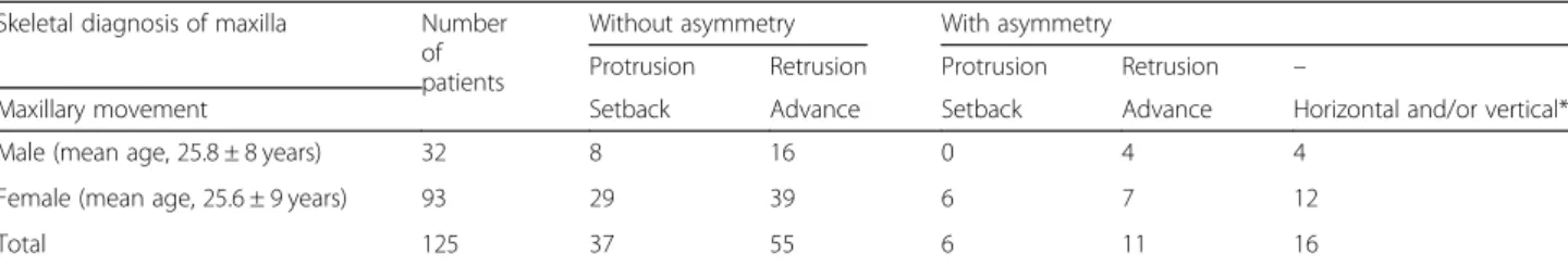
In all cases, imaging examinations of the head and neck (extraoral and intraoral radiography, MDCT examin-ation, MRI examination) were performed before orthog-nathic surgery, and they were diagnosed by oral and maxillofacial radiologists. For all patients, MDCT scan-ning was performed with an MDCT scanner, SOMATOM Definition AS 64 (Siemens, Erlangen, Germany). Using MDCT images, the maxillary sinus mucosa of 125 pa-tients was retrospectively evaluated before surgery, 1 month after surgery, and 1 year after surgery. On the MDCT images, a region showing a CT value (about 80 HU) similar to the soft tissue present in the maxillary sinus was judged as mucosal thickening and classified into three grades according to the proportion occupying the maxillary sinus (Fig.1). Grading of maxillary sinus muco-sal thickening was performed with reference to the evalu-ation method of the maxillary sinus mucosa by Yoshiura et al. [1], Carmeli et al. [2], and Bolger et al. [3].
The classification was as follows: grade 1, thickening of the maxillary sinus occupying the maxillary sinus vol-ume is one third or less; grade 2, thickening occupying one third to two thirds of the maxillary sinus; and grade 3, thickening occupying more than two thirds of the maxillary sinus.
Factors related to postoperative maxillary sinus mucosal thickening
In the evaluation of factors related to mucosal thicken-ing, the following eight factors were examined: sex, age (mean age at the time of 1 month after surgery), diagno-sis (skeletal diagnodiagno-sis of the maxilla), operating time (total operating time and operating time at the end of Le Fort I osteotomy), amount of postoperative bleeding (total bleeding and bleeding at the end of Le Fort I oste-otomy), with/without bone graft in Le Fort I osteotomy, with/without multisegmental osteotomy in Le Fort I osteotomy, and with/without macrolide therapy after surgery. The ultrasonic surgical method (piezoelectric surgery) was mainly used for the separation of the max-illa [4, 5]. In addition, macrolide therapy was imple-mented in accordance with previously reported research and guidelines [6–8].
Ethical considerations
The postoperative MDCT examination including assess-ment of the maxillary sinus mucosa was thoroughly ex-plained before the examination, and written consent was obtained from all patients. This study was approved by the Tokyo Dental College Institutional Review Board (Ethics Review Board Approval Number 803), and all participants provided their written, informed consent.
Statistical analysis
Open source statistical software R version 3.2.3 was used for statistical analysis [9]. The factors were analyzed using pairedt tests and chi-squared tests, as appropriate, comparing patients with and without mucosal thicken-ing.p values < 0.05 were considered significant.
Results
Table 1 shows the mean age and summary of male and
female patients by diagnosis, by movement direction of the maxilla, divided by with/without asymmetry. There
were three times as many females as males. Table 2
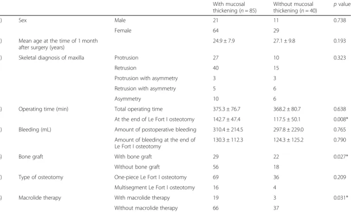
shows the characteristics of the patients in the two treat-ment groups with and without maxillary sinus mucosal thickening. Maxillary sinus mucosal thickening was ob-served in 85 (68%) patients on MDCT images at 1 month postoperatively. In 66 patients who showed
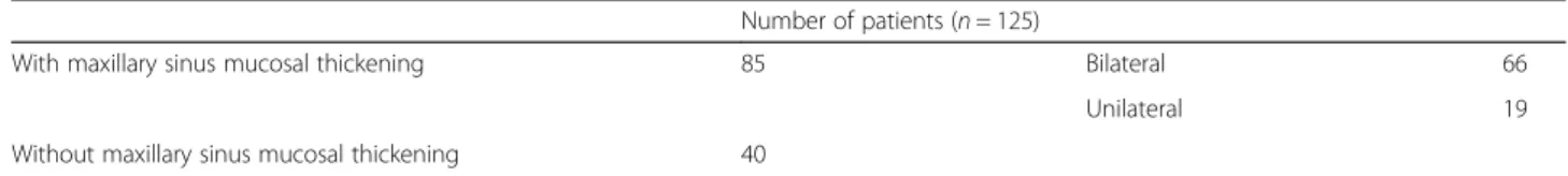
Table 1 Summary of the patients who underwent Le Fort I osteotomy
Skeletal diagnosis of maxilla Number of patients
Without asymmetry With asymmetry
Protrusion Retrusion Protrusion Retrusion –
Maxillary movement Setback Advance Setback Advance Horizontal and/or vertical*
Male (mean age, 25.8 ± 8 years) 32 8 16 0 4 4
Female (mean age, 25.6 ± 9 years) 93 29 39 6 7 12
Total 125 37 55 6 11 16
bilateral mucosal thickening, 92 sinuses were grade 1, 31 were grade 2, and 9 were grade 3. In 19 patients with unilateral mucosal thickening, 14 sinuses were grade 1, 4 were grade 2, and 1 was grade 3 (Table3). Of the 19 pa-tients with unilateral mucosal thickening, 2 were asym-metrical, but no correlations were found between the 2 cases and the movement direction and movement amount of the maxilla. The details of the evaluations of factors related to mucosal thickening and the analysis re-sults are shown in Table 4. There was a significant dif-ference in operating time at the end of Le Fort I osteotomy, with/without bone graft, and with/without macrolide therapy.
Discussion
Since the application of Le Fort I osteotomy to
orthog-nathic surgery in 1927 by Wassmund [10],
improve-ments were made by many surgeons, and it became a surgical operation that was mostly established by Obwe-geser [11]. Today, it is a procedure that is frequently performed due to its diversity of movement directions.
In Le Fort I osteotomy, the bone around the maxillary sinus is separated. Therefore, after surgery, blood accu-mulates in the sinus, and inflammatory changes (sinus mucosal thickening, edematous swelling) occur. It is also thought that the blood is absorbed with the progress of time, and the thickening of the sinus mucosa also disap-pears. It is easy to imagine that the risk of onset of max-illary sinusitis will be high if the blood reservoir or thickening of the mucosa persists for a long time. In the past, maxillary sinusitis has occurred, but fortunately, in
the present evaluation period, there were no cases of maxillary sinusitis. Although mucosal thickening was not observed on the MDCT images of Le Fort I oste-otomy after 1 year, mucosal thickening was observed in 68% in the first month after operation. Due to the characteristics of surgery, maxillary sinusitis may occur. Many reports on events after Le Fort I osteot-omy are mostly related to abnormal fractures and
bleeding [12–15]. However, to the best of our
know-ledge, there are very few reports on the incidence of maxillary sinusitis after Le Fort I osteotomy. Although
Panula et al. [16] reported it in 6 of 655 patients,
Kramer et al. [17] reported it in 11 of 1000 patients
and Chow et al. [18] reported it in 3 of 125 patients; thus, the incidence and factors related to postopera-tive maxillary sinusitis have not yet been clarified.
In the present results, correlations with mucosal thick-ening were suggested, and factors that showed a signifi-cant difference were the operative time for the maxilla, bone grafts, and macrolide therapy after surgery. In 40 patients, no mucosal thickening was shown. One of the reasons for this may be that there was less blood reten-tion in the maxillary sinus after surgery.
Due to the significant difference in operating time dur-ing Le Fort I osteotomy, it was suggested that a safer and faster procedure leads to the prevention of mucosal thickening. We believe that using the ultrasonic surgical method (piezoelectric surgery) and preoperative simula-tion by patient-specific 3D models made with a 3D printer contribute to safer and faster surgery [19]. De-pending on the amount of movement of the maxilla,
a
b
c
Fig. 1 Grading of maxillary sinus mucosal thickening on MDCT images. a Grade 1, thickening of the maxillary sinus occupying the maxillary sinus volume is one third or less. b Grade 2, thickening occupying one third to two thirds of the maxillary sinus. c Grade 3, thickening occupying more than two thirds of the maxillary sinus
Table 2 With/without maxillary sinus mucosal thickening on MDCT images 1 month after surgery
Number of patients (n = 125)
With maxillary sinus mucosal thickening 85 Bilateral 66
Unilateral 19
gaps between the bones may increase. Bone grafting pro-motes the formation of surrounding bone, and conse-quently, it was thought to contribute to the reduction of mucosal thickening. Furthermore, the results of this study support the role of postoperative macrolide ther-apy in reducing mucosal thickening. Opinions are di-vided on the timing of postoperative MDCT scanning to evaluate the maxillary sinus mucosa. Frequent MDCT scanning for the purpose of observation should not be done, since X-ray exposure should be avoided. There-fore, it is necessary to establish the validity of perform-ing MDCT imagperform-ing. Currently, MDCT scannperform-ing is performed 1 month after surgery in our practice. Al-though it aims mainly to evaluate the condition of the bone, it also evaluates inflammatory changes of the max-illary sinus mucosa at the same time. If maxmax-illary sinus mucosal thickening has been prolonged at that time, it is
thought that the risk of infection remains high, and macrolide therapy is continued. Three-dimensional evaluation of the maxillary sinus by MDCT scanning is not performed to track the inflammatory changes of the maxillary sinus mucosa after scanning at 1 month
postoperatively. If CT is performed, it is a
two-dimensional evaluation such as Waters’ view. Therefore, it is difficult to demonstrate the dynamics over time. Therefore, while the period of continuing macrolide therapy is empirical, it is about 1 to 2 months. It is natural to observe the patient’s status carefully after surgery. In addition, it was demonstrated that MDCT 1 month after surgery looking for maxillary sinusitis can be helpful for deciding whether to con-tinue postoperative macrolide therapy.
Conclusions
In observation of maxillary sinus mucosal thickening using preoperative and postoperative MDCT images, shortening of the operative time, bone grafting, and macrolide therapy contributed to the prevention and re-duction of mucosal hypertrophy following Le Fort I oste-otomy. In addition, the usefulness of MDCT 1 month after surgery for determining whether to continue macrolide therapy was shown.
Table 3 Breakdown of maxillary sinus mucosal thickening in 125 patients 1 month after surgery
Number of maxillary sinuses (n = 151)
Grade 1 Grade 2 Grade 3
Bilateral 92 31 9
Unilateral 14 4 1
Table 4 Details of the evaluation of factors affecting mucosal thickening and the analysis results at 1 month after surgery
With mucosal thickening (n = 85) Without mucosal thickening (n = 40) p value 1) Sex Male 21 11 0.738 Female 64 29
2) Mean age at the time of 1 month after surgery (years)
24.9 ± 7.9 27.1 ± 9.8 0.193
3) Skeletal diagnosis of maxilla Protrusion 27 10 0.323
Retrusion 40 15
Protrusion with asymmetry 3 3
Retrusion with asymmetry 5 6
Asymmetry 10 6
4) Operating time (min) Total operating time 375.3 ± 76.7 368.2 ± 80.7 0.638
At the end of Le Fort I osteotomy 142.7 ± 47.4 117.5 ± 50.1 0.008*
5) Bleeding (mL) Amount of postoperative bleeding 310.4 ± 214.5 297.8 ± 229.0 0.765
Amount of bleeding at the end of Le Fort I osteotomy
130.3 ± 112.3 124.3 ± 125.2 0.790
6) Bone graft With bone graft 29 22 0.027*
Without bone graft 56 18
7) Type of osteotomy One-piece Le Fort I osteotomy 69 36 0.209
Multisegment Le Fort I osteotomy 16 4
8) Macrolide therapy With macrolide therapy 19 3 0.031*
Without macrolide therapy 66 37
*
Abbreviations
HU:Hounsfield units; MDCT: Multidetector-row computed tomography; MRI: Magnetic resonance imaging
Acknowledgements Not applicable. Funding
No specific funding sources to declare for this research. Availability of data and materials
Readers interested in the data should contact the authors. Authors’ contributions
MI conceived the study and drafted the study outline. TT and TK collected the requisite data. MI, MY, YK, TT, TS, MN, and AT carried out the analyses. MI and TK interpreted the data and drafted the manuscript. All authors have read and have given final approval of the final version to be published. Ethics approval and consent to participate
The postoperative MDCT examination including assessment of maxillary sinus mucosa was thoroughly explained before the examination, and written consent was obtained from all patients. This study was approved by the Tokyo Dental College Institutional Review Board (Ethics Review Board Approval Number 803), and all participants provided their written, informed consent.
Consent for publication
For all patients, we have obtained written agreement on the use of CT images. Also, we are careful not to identify individuals from images and data. Competing interests
The authors declare that they have no competing interests.
Publisher’s Note
Springer Nature remains neutral with regard to jurisdictional claims in published maps and institutional affiliations.
Author details
1
Department of Oral Pathobiological Science and Surgery, Tokyo Dental College, 2-9-18 Kandamisaki-cho, Chiyoda-ku, Tokyo 101-0061, Japan.
2Department of Oral and Maxillofacial Surgery, Tokyo Dental College, 2-9-18
Kandamisaki-cho, Chiyoda-ku, Tokyo 101-0061, Japan. Received: 25 January 2019 Accepted: 19 February 2019
References
1. Bolger WE, Woodruff WW Jr, Morehead J, Parsons DS (1990) Maxillary sinus hypoplasia: classification and description of associated uncinate process hypoplasia. Otolaryngol Head Neck Surg 103:759–765
2. Yoshiura K, Ban S, Hijiya T, Yuasa K, Miwa K, Ariji E et al (1993) Analysis of maxillary sinusitis using computed tomography. Dentomaxillofac Radiol 22: 86–92
3. Carmeli G, Artzi Z, Kozlovsky A, Segev Y, Landsberg R (2011) Antral computerized tomography pre-operative evaluation: relationship between mucosal thickening and maxillary sinus function. Clin Oral Impl Res 22:78–82 4. Takaki T, Ootake Y, Kogou T, Hirota M, Yamamoto M et al (2014) Use of
ultrasonic new shape blade in orthognathic surgery: review of 138 patients. J Oral Maxillofac Surg 72:e134
5. Matsuda H, Furuya Y, Sasaki H, Takanashi T, Morioka T et al (2015) Comparison of surface morphology and healing in rat calvaria bone defects between ultrasonic surgical method and rotary cutting method. J Hard Tissue Biol 24:267–276
6. Sasaki J, Kaneko A, Karakida K, Shiiki K, Sakamoto H et al (1995) Comparative clinical study of azithromycin with tosufloxacin tosilate in the treatment of acute odontogenic infection. Jpn J Antibiot 48:1093–1118
7. Shinkai M, Henke MO, Rubin BK (2008) Macrolide antibiotics as
immunomodulatory medications: proposed mechanisms of action. Pharmac Therapeutics 117:393–405
8. Mandal R, Patel N, Ferguson BJ (2012) Role of antibiotics in sinusitis. Curr Opin Infect Dis 25:183–192
9. R Core Team (2018) R: a language and environment for statistical computing. R Foundation for Statistical Computing, Vienna https://www.R-project.org/. Accessed 1 Dec 2018.
10. Drommer RB (1986) The history of the“Le Fort I osteotomy”. J Oral Maxillofac Surg 14:119–122
11. Obwegeser H (1969) Surgical correction of small or retrodisplaced maxillae. Plast Recon Surg 43:351–365
12. Lanigan DT, West RA (1984) Management of postoperative hemorrhage following the Le Fort I maxillary osteotomy. J Oral Maxillofac Surg 42:367–375 13. Kim SG, Park SS (2007) Incidence of complications and problems related to
orthognathic surgery. J Oral Maxillofac Surg 65:2438–2444 14. O’Regan B, Bharadwaj G (2007) Prospective study of the incidence of
serious posterior maxillary haemorrhage during a tuberosity osteotomy in low level Le Fort I operations. Br J Oral Maxillofac Surg 45:538–542 15. Bhaskaran AA, Courtney DJ, Anand P, Harding SA (2010) A complication of
Le Fort I osteotomy. Int J Oral Maxillofac Surg 39:292–307 16. Panula K, Finne K, Oikarinen K (2001) Incidence of complications and
problems related to orthognathic surgery: a review of 655 patients. J Oral Maxillofac Surg 59:1128–1136
17. Kramer FJ, Baethge C, Swennen G, Teltzrow T, Schulze A et al (2004) Intra-and perioperative complications of the Le Fort I osteotomy: a prospective evaluation of 1000 patients. J Craniofac Surg 15:971–977
18. Chow LK, Singh B, Chiu WK, Samman N (2007) Prevalence of postoperative complications after orthognathic surgery: a 15-year review. J Oral Maxillofac Surg 65:984–992
19. Kamio T, Hayashi K, Onda T, Takaki T, Shibahara T et al (2018) Utilizing a low-cost desktop 3D printer to develop a“one-stop 3D printing lab” for oral and maxillofacial surgery and dentistry fields. 3D Print Med 4.1:6