The Cellular Localization of GnRH and LHR in Aged
Female Mice
Young-Jong Kim, Byung-Joon Park, Won-Jae Lee and Seung-Joon Kim College of Veterinary Medicine, Kyungpook National University, Daegu 41566, Korea
Abstract
Gonadotropin releasing hormone (GnRH) centrally plays a role in control of the hypothalamic- pituitary-gonadal axis-related hormone secretions in the reproductive neuroendocrine system. In addition, hormone receptors like luteinizing hormone receptor (LHR) are important element for hormones to take effect in target organ. However, ageing-dependent changes in terms of the distribution of GnRH neurons in the brain and LHR expression in the acyclic ovary have not been fully understood yet. Therefore, we comparatively investigated those ageing-dependent changes using young (1-5 months), middle (11-14 months) and old (21-27 months) aged female mice. Whereas a number of GnRH positive fibers and neurons with monopolar or bipolar morphology were abundantly observed in the brain of the young and middle aged mice, a few GnRH positive neurons with multiple dendrites were observed in the old aged mice. In addition, acyclic ovary without repeated development and degeneration of the follicles was shown in the old aged mice than others. LHR expression was localized in theca cells, granulosa cell, corpora lutea and atretic follicle in the ovaries from young and middle aged mice, in contrast, old aged mice had few positive LHR expression on the follicles due to acyclic ovary. However, the whole protein level of LHR was higher in the ovary of old aged mice than others. These results are expected to be used as an important basis on the relationship between GnRH and LHR in old aged animals as well as in further research for reproduction failure.
Received 13 December 2018 Revised 18 December 2018 Accepted 19 December 2018
Key Words : Gonadotropin releasing hormone, Luteinizing hormone receptor, Acyclic ovary, Ageing
INTRODUCTION
The reproductive neuroendocrine system is a complicated mechanism that consists of the brain (hypothalamus), pituitary, target glands (thyroid, adrenals and gonads) and other hormone- producing tissues [Meites et al., 1987]. Especially, the gonadotropin releasing hormone (GnRH) is secreted from the neurons in the hypothalamus, and it centrally controls the hypothalamic- pituitary-gonadal axis (HPG axis). These GnRH neurons and fibers are mainly located in the anteroventral third ventricle region in the preoptic area (PA) and the organum vasculosum of the lamina terminalis in the brain; the PA is divided into the medial preoptic area (MEPA) and lateral preoptic area (LPA), and the MEPA is generally formed by small- and medium-sized neurons whose function is associated with production of GnRH [Castañeyra-Ruiz et al., 2013; Hasegawa et al., 2005; Koutcherov et al., 2013]. Once secreted via the pituitary portal vessels, GnRH is responsible for the synthesis and secretion of follicle- stimulating hormone (FSH) and luteinizing hormone (LH) in the anterior pituitary; thereafter, both hormones are mainly involved in follicular growth and oocyte maturation [Jin and Yang, 2014].
In addition, several hormone receptors such as GnRH receptor (GnRHR), FSH receptor (FSHR) and LH receptor (LHR) are important element for the aforementioned hormones to take effect in target organ [Jin and Yang, 2014]. Among them, LH binds to its receptor (LHR) placing on the surface of theca cells and granulosa cells (GCs), resulting in hormone production (progesterone, androstenedione and testosterone), ovulation, luteinization and corpus luteum formation [Dufau, 1998; Zhang et al., 2001]. Therefore, the regulation of LHR expression is highly important for cycling of the ovary.
Even though several studies have shown ageing-dependent alterations in distribution of GnRH neurons and ovarian follicle development, the change of LHR expression in the reproduction system during aged-physiologic condition (especially acyclic ovary) has not been clearly understood yet. Of particular, the reproductive system is one of the first biological systems to explain age-related decline, and dysfunction of some of these system may disturb normal regulation of hormone cycle [Darrell, 2005; Meites et al., 1987]. Therefore, the aim of the current work was to uncover the age-related changes of GnRH expression in the brain and folliculogenesis with LHR expression in the ovary in the aged female mice.
MATERIAL AND METHOD
Animals, ethics and chemicalsC57BL/6J female mice (age: 1-27 months) were purchased (Daehan Biolink, Cheongju, Korea) and housed in a room maintained for temperature at 23±2°C, humidity at 50-80% and approximately 12h light/dark cycle. The standard feed (Jeil Feed Co., Ltd., Daejeon, Korea) and municipal water irradiated by ultraviolet light were provided to the animals. This experiment were performed in accordance with Kyungpook National University Guide for the Care and Use of Laboratory Animals. For comparing with GnRH and LHR expression by aging, the brain and ovary tissues were collected from different age groups: young (1-5 months), middle (11-14 months) and old aged mice (21-27 months). All chemicals were purchased from Sigma-Aldrich Chemical Company (St. Louis, MO, USA), unless otherwise specified.
Tissue processing
The female mice were anesthetized by ether inhalation. The whole blood was collected and centrifugated with 12,000 rpm for 20 min. The serum was isolated and kept at -20°C. The euthanized mice were subjected to cardiac perfusion fixation with flushing by a 4% paraformaldehyde (PFA). The brain and ovary tissues were collected, dissected, stored in 10% pH 7.4 formalin for 1 week and embedded in paraffin. Tissues were cut into 5 µm sections using a microtome (Leica Microsystems, Germany) and stained with hematoxylin and eosin (H&E) or used for immunohistochemical (IHC) analysis.
Classification of Ovarian follicles
The H&E-stained ovarian follicles were classified in accordance with the previous article [Myers et al., 2004]; in brief, primary follicles or secondary follicles or early antral follicles or preovulatory follicles or atretic follicles were identified by a single layer of cuboidal granulosa cells or surrounding of more than one layer of cuboidal granulose cells without visible antrum or 1-2 small areas of antrum or the largest follicle with cumulus granulosa cell layer or follicles with some pyknotic cells showing oocyte contraction and frequent breakdown of germinal vesicle, respectively.
Immunohistochemistry
The 5µm sections of paraffin-embedded tissues of brain and ovary were deparaffinized, washed in distilled water (DW) twice, heated in citrate buffer (0.01 M, pH 6.0) in a microwave for 5 min, cooled in room temperate (RT) for 20 min and treated with 0.3% hydrogen peroxide to block endogenous peroxidase activity for 20 min. Thereafter, the slides were cleaned with DW, washed with TBS-T (Tris-buffered saline, pH 7.4; 10 mM Tris-HCl, 150 mM NaCl, 0.1% Triton X-100), washed with TBS (pH 7.4; 10 mM Tris-HCl, 150 mM NaCl), incubated with 10% normal goat serum (Histostain plus kit, Invitrogen, USA) for 1 hour, incubated with primary antibody of rabbit polyclonal anti-LHR (ABIN904836, Antikoerper, Germany) to ovary specimen or anti-GnRH antibody (AB5617, Abcam, UK) to brain specimen at 4°C for overnight; as a negative control, primary antibody was replaced with DW. Thereafter, the incubated slides were washed with TBS, incubated with secondary antibody (Histostain plus kit, Invitrogen, USA) for 45 min, washed with TBS and incubated with avidin biotin peroxidase complex (Histostain plus kit, Invitrogen, USA) for 45 min. The slides were reacted with 3,3'-diaminobenzidine (DAB) kit (Vector Laboratories, USA).
Western blotting
The ovary tissues from middle (14 months) and old aged mice (21 and 27 months) were homogenized, lysed with a buffer including 40 mM Tris-HCl (pH 7.4) supplemented with protease inhibitors (leupeptin, 10 µg/ml), aprotinin (10 µg/ml) and phenylmethanesulphonylfluoride (1 mM) and centrifuged at 14,000 rpm at 4°C for 20 min. Equal amounts of protein were separated by electrophoresis on 10% sodium dodecyl sulfate- polyacrylamide (SDS-PAGE) gel. Gels were transferred to nitrocellulose membranes (Whatman GmbH, Germany), thereafter, the membranes were blocked by incubation with 5% non-fat milk in TBS for 2 hours. Subsequently, the membranes were incubated for overnight with rabbit polyclonal anti-LHR antibody or anti-beta-actin (ACTB) antibody (Sigma Aldrich), washed with TBS containing 0.1% Tween-20 and incubated with horseradish peroxidaseconjugated anti-rabbit IgG (Vector Laboratories) for 1 hour. Immunoreactive bands were detected using enhanced chemiluminescence (ECL) kit (Amersham, USA). The optical density of each band was measured by laser scanner. To analyze relative expression of LHR, Image J (NIH, USA) was used to
Statistical analysis
One-way ANOVA was conducted using PASW 18 (SPSS, USA) with Duncan’s post-hoc test to analyze significant differences. Data were presented as the mean ± standard deviation (SD). A p value of <0.05 was considered to be statistically significant.
RESULT
Distribution of GnRH positive expression in the brain The brain tissues of young, middle and old aged mice were processed for Immunohistochemical analysis of GnRH expression (Figure 1). A number of GnRH positive fibers and neurons were abundantly observed in anteroventral periventricular nucleus (AVPV) and medial preoptic area (MEPA) region with showing monopolar or bipolar morphology and uncomplicated dendrites in the young aged mice (Fig. 1A and 1D); these positive distributions of GnRH were maintained until middle age (Fig. 1B and 1E). The scoring for GnRH positive neurons also indicated the strong expression of GnRH from young age to middle age (Table 1). However, a few GnRH positive neurons were distributed in the AVPV and MEPA region in the old aged mice, in addition, multiple dendrites of GnRH neurons were densely exhibited (Figure 1C and 1F). The negative or weak expression of GnRH in the brain of the old aged mice was quantitatively assessed by scoring (Table 1).
Table 1. Localization and intensity of GnRH expression in brain
Mice Region GnRH neuron
Young aged AVPV +++
MEPA +++
Middle aged AVPV ++ or +++
MEPA ++ or +++
Old aged AVPV +
MEPA + or
--, negative; +, weak; ++, moderate; +++, intense Ovarian follicular morphology in old mice
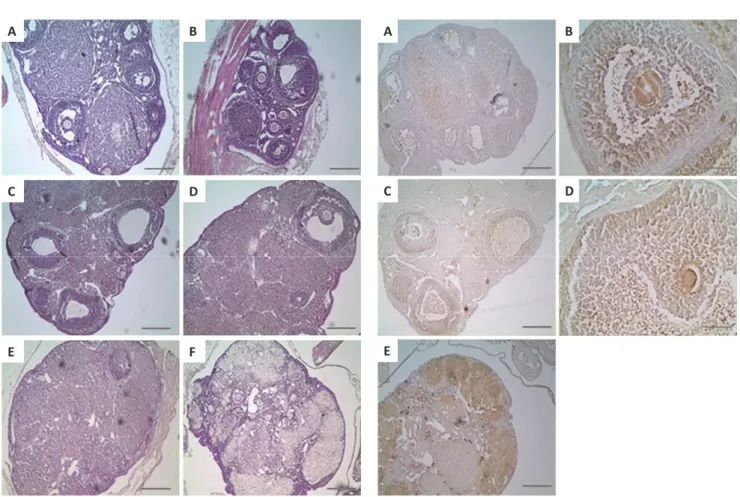
In normal estrous cycle of young and middle aged mice, various type of follicles were shown in the ovary, such as repeated development and degeneration of the follicles (Figure 2A-2D); the
observed in both groups. However, ageing might impact on the ovarian follicular development, shown in old aged mice (Figure 2E and 2F); in comparison with young and middle aged mice, the number of primary, secondary, antral and pre-ovulatory follicles was decreased and luteinized cells were observed in old aged mice, which indicated acyclic ovary.
Immunohistochemical analysis of LHR positive expression in the ovary
In the ovaries from young and middle aged mice, LHR expression was localized in theca cells, granulosa cell, corpora lutea and atretic follicle (Figure 3A-3D); the expression of LHR was strongly found in adjacent granulose cell surrounding the oocyte of antral follicles of early antral phase (Figure 3B). In case of the ovaries from old aged mice, LHR expression under folliculogenesis during the primary, secondary, antral and preovulatory follicle was drastically reduced due to the acyclic ovary, but high LHR expression was identified across the ovary tissue (Figure 3E); because the ovaries in the old aged mice were acyclic, more magnified images for follicles were not further investigated.
Western blot analysis of LHR expression in acyclic old mice. Because there were no considerable differences (folliculogenesis and LHR expression) in the ovaries between young and middle
aged mice (Figure 2 and 3), LHR expression was compared with both ovaries from middle aged (14 months) and old aged (21 and 27 months) mice by western blotting (Figure 4A). When the expression level of LHR was normalized against ACTB, the results demonstrated that LHR expression was significantly (p<0.05) upregulated in acyclic ovary of old aged mice (Figure 4B).
DISCUSSION
Since ageing-related decline in the body is highly related with decrease of the reproductive potential, several studies have tried to much better understand their relationship. In laboratory rats and mice, their life span is expected as 2-3 years, onset of estrous cycles (puberty) usually begins at 35-40 days, irregular reproductive cycles increase from middle age (8-12 months), and estrus cycle ceases (acyclic ovary) at old age (17-21 months) [Meites et al., 1987; Wise, 1993]. In this study, we similarly demonstrated ageing-dependent changes with respect to reduction of GnRH positive cells in the brain, acyclic ovary with decrease of folliculogenesis and luteinized cells, and elevation of LHR expression in the ovary in the old (21-27 months) aged mice, in comparison with the young (1-5 months) and middle (11-14 months) aged mice.
It is well addressed that GnRH is secreted from the hypothalamus Figure 1. The positive immunoreactions of GnRH antibody in the anteroventral periventricular nucleus (AVPV; A, B and C) and
medial preoptic area (MEPA; D, E and F) depending on ageing. While the GnRH immunoreaction was high positive in AVPV (A) and MEPA (D) of young aged mice, the intensity of GnRH immunoreaction was weak in AVPV (B) and MEPA (E) of middle aged mice and in AVPV (C) and MEPA (F) of old aged mice. In addition, the shape of GnRH neuron was shorter and distributed sporadically depending on ageing in AVPV and MEPA (arrows in A-F). Scale bars= 50 µm.
and plays a pivotal role in regulation of the reproductive axis driving puberty and ovulation [Darrell, 2005]. The function of GnRH neurons is associated with anatomy; although their cell bodies are scattered throughout the basal forebrain, most GnRH neurons project axons to the external zone of the median eminence to secret GnRH into the portal vasculature to control release of FSH and LH [Herbison, 2005; Prevot and Hanchate, 2010; Silverman and Livne, 1994]. In aging rodents, the declines in the reproductive system was related with decreased ability of GnRH release from the hypothalamus, obviously due to reduced activity of the hypothalamic catecholamines (CAs) [Wise, 1983].
factor for reduction of GnRH secretion [Darrell, 2005; Miller et al., 1990]. In agreement with the previous report, GnRH neurons and fibers in the young and middle aged mice in the present study were densely located in the AVPV (Figure 1A and 1B) and MEPA (Figure 1D and 1E) in the anteroventral third ventricle region, and formed monopolar or bipolar morphology and uncomplicated dendrites. However, the population of GnRH neurons and fibers have decreased at the same brain region as well as formed multiple dendrites (Figure C and F, and Table 1). Therefore, the present result indicated that the secretion of GnRH reduced in the old aged mice than others. At the pituitary, GnRH stimulates Figure 2. The morphology on the follicular in young (A and
B), middle (C and D) and old (E and F) aged mice. In the ovary of young and middle mice, primary-/ secondary-/early antral-/preovulatory follicles as well as atretic follicle with corpus luteum were well presented (A-D). However, old aged mice had few normal ovarian follicles and a few atretic antral follicles (E and F). Scale bars=200 µm.
Figure 3. The distribution of LHR positive cells on the ovarian follicles in young (A and B), middle (C and D) and old (E and F) aged mice. The expression of LHR was localized in the theca-/ granulosa cell in the preovulatory follicle (B) and atretic follicle (D) on ovaries of young and middle aged mice (A-D). Moreover, stronger expression of LHR was observed in old aged mice than those of young and middle aged mice (E). Scale bars=200 µm (A, C and E) or 50 µm (B and D).
pulses; while low-frequency GnRH pulse leads to FSH release, high-frequency GnRH pulses stimulate LH release [Jayes, 1997; Rubin, 2000; Yin and Gore, 2006]. In addition, aged hypothalamus in the rat also attenuated LH surge due to alteration of GnRH secretion [Darrell, 2005]. These findings explained the degradation of GnRH system took place to delay and attenuation of the LH secretion [Yin and Gore, 2006]. Therefore, decreased population of GnRH neurons in the old aged mice in the present study might result in decrease of LH secretion at the pituitary. Furthermore, reduction of GnRH affected on release of FSH and LH at the pituitary, which related to develop ovarian follicles through neuroendocrine system. In our present study, we identified that few ovarian follicles (acyclic ovary) were observed in old aged mice, with showing few number of primary, secondary, antral and pre-ovulatory follicles (Figure 2E and 2F), possibly due to attenuated release of FSH and LH.
In ovarian follicle development, LHR on granulosa cells, theca cells and corpora lutea of the dominant follicle plays a pivotal role in the physiological LH-mediated effects in terms of the final stages of follicular growth, final maturation of the oocyte, ovulation and luteinization of the follicular wall [Nogueira et al., 2010]. In rodents, pre-antral follicles in regards with the primordial, primary and secondary follicles were found to lack of LHR mRNA expression [Bukovský et al., 1993]. Thereafter, LHR was expressed mainly in the theca cells during the early antral phase [Bukovský et al., 1993; Zhang et al., 2001]. Finally, LHR mRNA expression appears in granulosa cells is in the late
antral follicles stage, and it reaches the highest levels at the pre-ovulatory stage [Yung et al., 2014]. These ovarian follicle development with LHR expression from the appearance of the follicular antrum to the complete acquisition of ovulatory capacity is essentially modulated by GnRH, gonadotrophin (FSH and LH) and ovarian steroid hormones (estradiol and progesterone) [Mihm and Bleach, 2004]. In addition, granulosa cells from antral follicles turned to be more responsive to LH by increase of the total number of LHR via stimulation of FSH and estradiol during follicular maturation [Castañeyra-Ruiz et al., 2013; Prevot and Hanchate, 2010; Rubin, 2000]. Likewise, in the present study, LHR expression was localized in theca cells, granulosa cell, corpora lutea and atretic follicle in the ovaries from young and middle aged mice (Figure 3A-3D). However, old aged mice had few positive LHR expression on granulosa cell and theca cells because of undeveloped follicles in response to alteration of GnRH and gonadotrophin (FSH and LH) in comparison with the young and middle aged mice; however, overall LHR expression across whole ovary was stronger in the old aged mice than the middle aged mice (Figure 3 and 4). Similarly, old aged infertile human patients also demonstrated upregulation of LHR in comparison with young oocyte donors and middle aged infertile human patients [Wu et al., 2015].
Viability and numbers of follicles, ovulation and release of ovarian steroid hormones at the level of the ovary are changed with aging. Therefore, a single level of HPG axis or a single change of a hormone level cannot fully explain the entire Figure 4. The LHR expression in the ovary tissue of middle and old aged mice by western blot analysis. Representative images of
the western blot analysis for LHR and ACTB were shown (A). The relative ratio of LHR against ACTB presented gradual elevation of LHR expression following ageing (B). The graph was presented as mean±SD. 14 Mo or 21 Mo or 27 Mo indicated 14 month (middle aged) or 21 month (old aged) or 27 month (old aged) aged mice, respectively. *P<0.05 VS 14 month aged mice.
process of ageing-dependent alterations in the reproductive system because the reproductive neuroendocrine system is a complicated mechanism that consists of the brain, pituitary, target glands and other hormone-producing tissues. In the present study, we demonstrated the GnRH neurons in the brain and folliculogenesis in the ovary were reduced depending on aging, but LHR expression were increased in acyclic ovary. These results are expected to be used as an important basis on the relationship between GnRH and LHR in old aged animals as well as in further research for reproduction failure.
REFERENCE
Bukovský A, Chen TT, Wimalasena J and Caudle MR. 1993. Cellular localization of luteinizing hormone receptor immunoreactivity in the ovaries of immature, gonadotropin-primed and normal cycling rats. Biol. Reprod. 48:1367-1382.
Castañeyra-Ruiz L, González-Marrero I, Castañeyra-Ruiz A, González-Toledo JM, Castañeyra-Ruiz M, de Paz-Carmona H, Castañeyra-Perdomo A and Carmona-Calero EM. 2013. Luteinizing hormone-releasing hormone distribution in the anterior hypothalamus of the female rats. ISRN Anat. 2013:870721.
Darrell W. 2005. The aging reproductive neuroendocrine axis. Steroids. 70:273 283.
Dufau ML. 1998. The luteinizing hormone receptor. Annu. Rev. Physiol. 60: 461-496.
Hasegawa H, Ishiwata T, Saito T, Yazawa T, Aihara Y and Meeusen R. 2005. Inhibition of the preoptic area and anterior hypothalamus by tetrodotoxin alters thermoregulatory functions in exercising rats. J. Appl. Physiol. 98:1458-1462.
Herbison AE. 2005. Physiology of the GnRH neuronal network. 3rd ed, Physiology of reproduction, pp. 1415 1482.
Jayes FC. 1997. Role of gonadotropin-releasing hormone pulse frequency in differential regulation of gonadotropins in the gilt. Biol. Reprod. 56:1012-1019.
Jin JM and Yang WX. 2014. Molecular regulation of hypothalamus- pituitary gonads axis in males. Gene. 551:15-25.
Koutcherov Y, Mai JK and Paxinos G. 2013. Hypothalamus of the human fetus. J. Chem. Neuroanat. 26:253-270.
Meites J, Goya R and Takahashi S. 1987. Why the neuroendocrine system is important in aging processes. Exp. Gerontol. 22:1-15.
Mihm M and Bleach EC. 2003. Endocrine regulation of ovarian antral follicle development in cattle. Anim. Reprod. Sci. 78:217-237.
Miller MM, Joshi D, Billiar RB and Nelson JF. 1990. Loss of LH-RHs Neurons in the Rostral Forebrain of Old Female C57BL/6J Mice. Neurobiol. Aging. 11:217-221.
Myers M, Britt KL, Wreford NG, Ebling FJ and Kerr JB. 2004. Methods for quantifying follicular numbers within the mouse ovary. Reproduction. 127:569-80.
Nogueira MFG, Fernandes P, Ereno RL, Simões RAL, Buratini Junior J and Barros CM. 2010. Luteinizing Hormone Receptor (LHR): basic concepts in cattle and other mammals. Anim. Reprod. 7:51-64.
Prevot V and Hanchate NK. 2010. Function-related structural plasticity of the GnRH system: a role for neuronal-glial-endothelial interactions. Front. Neuroendocrinol. 31:241 258.
Rubin BS. 2000. Hypothalamic alterations and reproductive aging in female rats: evidence of altered luteinizing hormone- releasing hormone neuronal function. Biol. Reprod. 63:968 76.
Silverman A and Livne I. 1994. The gonadotrophin-releasing hormone (GnRH), neuronal systems: immunocytochemistry and in situ hybridization. 2nd ed, The physiology of reproduction, pp.1683 1706.
Wise PM. 1983. Aging of the female reproductive system. Review of Biological Research in Aging, pp.195-222.
Wise PM. 1993. Neuroendocrine aging: its impact on the reproductive system of the female rat. J. Reprod. Fertil. Suppl. 46:35 46.
Wu YG, Barad DH, Kushnir VA, Lazzaroni E, Wang Q, Albertini DF, Gleicher N. 2015. Aging-related premature luteinization of granulosa cells is avoided by early oocyte retrieval. J. Endocrinol. 226:167-180.
Yin W and Gore AC. 2006. Neuroendocrine control of reproductive aging: roles of GnRH neurons. Reproduction. 131:403 414.
Yung Y, Aviel-Ronen S, Maman E, Rubinstein N, Avivi C, Orvieto R and Hourvitz A. Localization of luteinizing hormone receptor protein in the human ovary. 2014. Mol. Hum. Reprod. 20:844-849.
Zhang M, Shi H, Segaloff DL and Van Voorhis BJ. 2001. Expression and localization of luteinizing hormone receptor in the female mouse reproductive tract. Biol. Reprod. 64:179-187.