Expression of Th17 and related cytokines
according to clinical
activity of Behçet’s disease
by
Park Mi-jin
Major in Molecular Medicine
Department of Biomedical Sciences
The Graduate School, Ajou University
Expression of Th17 and related cytokines
according to clinical
activity of Behçet’s disease
by
Park Mi-jin
A Dissertation Submitted to The Graduate School of
Ajou University in Partial Fulfillment of the Requirements
for the Degree of
Master of Biomedical Sciences
Supervised by
Eun-So Lee,
M.D., Ph.D
.
Major in Molecular Medicine
Department of Biomedical Sciences
The Graduate School, Ajou University
This certifies that the dissertation
of Mi-Jin Park is approved.
SUPERVISORY COMMITTEE
Eun-So Lee
Sun Park
Seong Hyang Sohn
The Graduate School, Ajou University
December, 23rd, 2010
감사의 글
본 논문이 완성 될 때까지 많은 조언과 도움을 주신 지도 교수님이신 이은소 교수님께 진심으로 감사 드립니다. 또한 연구를 하는 동안 많은 가르침과 도움을 주신 박선 교수님과 손성향 교수님께도 진심으로 감사 드립니다. 그리고 연구에 많은 도움을 주신 나도영 선생님, 이미진 선생님, 윤수진 선생님, 피부과학교실의 모든 선생님들께 감사 드립니다. 항상 아낌없는 사랑으로 지원을 해 주신 부모님과 뒤에서 많은 격려를 해 주는 가족들에게도 감사의 마음을 전합니다. 2011 년 1 월 저자씀- ABSTRACT –
Expression of Th17 and related cytokines according to
clinical activity of Behçet’s disease
Background: Behçet’s disease (BD) is a multisystem inflammatory disorder characterized
by recurrent oro-genital ulcers, and cutaneous, ocular, arthritics, gastrointestinal, vascular, and neurovascular involvement. The etiology and pathogenesis of BD have remained unclear, however it has been assumed that genetic factors, infectious, and immune mechanisms are involved in the onset of this disease. In human autoimmunity, the T helper(Th) 1 immune responses have been traditionally associated with the induction and progression of disease. Recently, a number of other T cell populations were discovered, and IL(Interleukin)-17 producing Th17 cells were recognized as novel group of T cells which play a major role in autoimmunity. A recent study has shown elevated production of IL-17, IL-23, and IFN (Interferon)-γ by peripheral blood mononuclear cells (PBMCs) besides increased frequencies of IL-17 and IFN-γ producing T cells in BD patients with active uveitis. It was also reported that increased expression of IL-23 p19 mRNA in erythema nodosum-like lesions in patients with active BD.
Purpose: In this study, we aimed at investigating the expression Th17 and related cytokines
active and inactive BD were enrolled in this study. The disease control and healthy control (HC) group consisted of age- and sex-matched 10 recurrent aphthous ulcer (RAU) patients without any other evident disease and 10 healthy volunteers, respectively. PBMCs of subjects were cultured and stained with the appropriate fluorescent antibody for analysis by flow cytometry. To measure the IL-17, IL-23 and IFN-γ concentrations in serum and in culture supernatants, ELISA was performed. To investigate the mRNA expression level of IL-12p35, IL-12/23p40 and IL-23p19 in PBMCs, real time polymerase chain reaction (RT-PCR) was performed.
Results: In the active BD group, CD4+ and CD8+ T cells from patients expressed
significantly higher level of IL-17 and IFN-γ, compared with the cells from the control group. High serum and supernatant concentration of IL-17 and IFN-γ was in active BD group compared with HC group and RAU group. In addition, mRNA expression of 23p19, IL-12p35 and IL-12/23p40 in PBMCs was up-regulated in total BD patient groups. Expression of IL-23p19 mRNA was significantly higher in the active BD group compared with HC groups.
Conclusion: Increased expression of the IL-17 and IFN-γ was observed in this study and
these results suggest that Th17 cells has a important implication in immune modulation of BD. Future studies clarifying the further immunopathologic role of Th17 pathway in BD are needed.
TABLE OF CONTENTS
ABSTRACT --- i
TABLE OF CONTENTS --- iii
LIST OF FIGURES --- iv
LIST OF TABLES --- v
I. INTRODUCTION --- 1
II. MATERIALS AND METHODS --- 3
A. Subjects --- 3
B. Methods --- 4
1. Cell preparation --- 4
2. Cell culture and stimulation of PBMCs --- 4
3. Flow cytometry --- 5
4. ELISA --- 5
5. RNA preparation and Real time PCR --- 5
6. Statistical analysis --- 6
III. RESULTS --- 7
IV. DISCUSSION --- 25
V. CONCLUSION --- 29
LIST OF FIGURES
Fig. 1. Expression of IL-17 in PBMCs. --- 10
Fig. 2. Intracellular expression of IL-17 in CD4+ and CD8+ T cells. --- 12
Fig. 3. Intracellular expression of IFN-γ in CD4+ and CD8+ T cells. --- 14
Fig. 4. Intracellular expression of IL-17 in CD4+ and CD8+CD45RO+ T cells. --- 17
Fig. 5. Level of IL-17 in sera (A) and culture supernatants at 2 and 5 days (B). --- 20
Fig. 6. Level of IFN-γ in culture supernatants at 2 and 5 days. --- 21
Fig. 7. Level of IL-23 in sera. --- 21
Fig. 8. Relative mRNA expression of IL-23p19 in the PBMCs. --- 23
Fig. 9. Relative mRNA expression of IL-12p35 in the PBMCs. --- 23
LIST OF TABLES
Table 1. Baseline demographics and characteristics of BD group, RAU group and HC
group. --- 7
I. INTRODUCTION
Behçet’s disease (BD) is a multisystemic inflammatory disorder characterized by recurrent oro-genital ulcers, and cutaneous, ocular, arthritic, gastrointestinal, vascular, and neurovascular involvement (Sakane et al., 1999). BD is particularly common in the Far East and the Mediterranean basin, and is frequency noted between the 30th and 45th degree latitudes in Asian and European populations, corresponding to the Old Silk Road, an ancient trading route stretching between the Mediterranean, the Middle East and the Far East. The etiology and pathogenesis of BD have remained unclear; however, it has been assumed thatgenetic factors, as well as infectious, and immune mechanisms are involved in the onset of this disease (Yurdakul and Yazici, 2008; Mendoza-Pinto et al., 2010).
CD4+ T helper (Th) cells are important mediators in immune responses, acting to coordinate the other cellular components of the immune system (Dong, 2008). Several reports have documented that peripheral blood mononuclear cells (PBMCs) from BD patients predominantly produce Th1 type cytokines, mainly interferon (IFN)-γ and tumor necrosis factor (TNF)-α, and this response is more significant in the active clinical stage, suggesting that BD can be categorized as a Th1-mediated disorder (Raziuddin et al., 1998). There are many studies that present Th1 dominant immune response in BD (Imamura et al., 2005; Nagafuchi et al., 2005; Direskeneli, 2006; Chi et al., 2008).
Recently, a number of other T cell populations were discovered, and IL-17 producing Th17 cells were recognized as a novel group of T cells which play a major role in autoimmunity (Brand, 2009). Th17 cells are characterized by their production of a large amount of IL-17, IL-6, and TNF-α, but a low level of IFN-γ. The development and
maintenance of Th17 cells have been linked to IL-23 which is a key initiating cytokine in the development of autoimmunity (Chi et al., 2008). In previous study, the levels of IL-23, IL-17, and IFN-γ are elevated in BD patients with active uveitis, and they suggest that the IL-23/IL-17 pathway together with IFN-γ is associated with the active intraocular inflammation in BD patients (Chi et al., 2008). Another study showed increased IL-23 p19 mRNA levels were observed in erythema nodosum (EN)-like lesions of patients with BD and suggested that IL-23 may be associated with the development of the EN-like lesions in patients with active BD (Lew et al., 2008). However, it is not yet clear whether the IL-23/IL-17 pathway is involved in the development of BD. With regard to the IL-23/Th17 axis, targeting the common subunit, p40, of IL-12 and IL-23 has shown clinical benefits in autoimmune-type diseases, such as Crohn’s disease and psoriasis (Di Cesare et al., 2009). Treatment with a monoclonal antibody against p40, which is common to IL-12 and IL-23, has shown efficacy in psoriasis.
To evaluate the further immunopathological implications of Th17 cells and related cytokines in BD according to clinical activity, I investigated intracellular expression of IL-17 and IFN-γ on PBMCs and the concentration of IL-17, IL-23 and IFN-γ from serum and culture supernatants. In addition, to clarify the mRNA expression level of 23p19, IL-12p35 and IL-12/23p40, real-time polymerase chain reaction (PCR) was performed.
II. MATERIALS AND METHODS
A. SUBJECTS
The patient population consisted of 22 patients with BD (9 women and 13 men; mean ± S.D. age, 43.0 ± 6.1 years), who presented for the first time or were monitored at the department of dermatology, Ajou university hospital from april 2010 to september 2010. Diagnosis was made based on the criteria established by the international study group or the Japanese group. (International Study Group for Behçet's Disease, 1990; Kurokawa et al., 2004). BD Patients were divided into two groups. Patients in the active group had at least one of the BD symptoms despite treatment and those in the inactive group were in well-controlled state by taking anti-inflammatory medication at the time of visit. The disease control and healthy control (HC) group consisted of age- and sex-matched 10 recurrent aphthous ulcer (RAU) patients without any other evident disease and 10 healthy volunteers, respectively. Informed consent was obtained from patients prior to enrolling them into the study. This study was approved by the Institutional Review Board (IRB number: AJIRB-GN3-07-098).
B. METHODS
1. Cell preparation
Peripheral blood mononuclear cells (PBMCs) were prepared from heparinized blood samples by Ficoll Hypaque density gradients centrifugation (Ficoll paqueTM plus; StemCell Technologies, Vancouver, BC, Cananda). Cells were washed with phosphate buffered saline (PBS; Sigma, St. Louis, MO, U.S.A.) with 2% heat-inactivated fetal bovine serum (FBS; Gibco-BRL, Grand Island, NY, U.S.A.)
2. Cell culture and stimulation of PBMCs
I evaluated the change of expression of surface molecules and intracellular cytokine in PBMC. However, IL-17 and IFN-γ expression in the PBMCs were undetectable. So, I was performed cell stimulation. PBMCs were cultured in medium (RPMI 1640 medium supplemented with 2mM L-glutamine, 100 U/ml penicillin and 100 μg/ml streptomycin) (RPMI 1640; Gibco-BRL, Grand Island, NY, U.S.A.) with 10% FBS at 37℃ and 5% CO2.
IFN-γ was detected in the cells stimulated with CD3 (5 μg/ml, clone OKT3) and anti-CD28 antibodies (1 μg/ml, clone anti-CD28.6) (eBiosciences, San Diego, CA, U.S.A.) for 48 hours. 17 was detected in the cells stimulation with 1β (10 ng/ml), 6 (20 ng/ml), IL-23 (100 ng/ml), TNF-α (10 ng/ml) and TGF-β (1 ng/ml) (R&D systems Inc., Minneapolis, MN, U.S.A.) in addition to anti-CD3 and anti-CD28 antibodies for 5 days. During the final 4 hours, 10 μg/ml of Brefeldin A (eBiosciences, San Diego, CA, U.S.A.) was added to the cultured PBMCs.
3. Flow cytometry
For examining cell expression of the indicated related Th17 cytokine, cells were labeled with the following fluorescence conjugated antibodies, APC anti-CD4 (#555349), PE-Cy7 anti-CD8 (#557746), PE anti-CD45RO (#555493) (BD Biosciences Pharmingen, San Diego, CA, U.S.A), PerCP-Cy5.5 anti-IL-17 (#45-7179), FITC anti-IFN-γ (#11-7319) antibodies (eBiosciences, San Diego, CA, U.S.A.). Staining was carried out with an intracellular staining kit (eBioscience, San Diego, CA, U.S.A.) according to the manufacturer’s instructions. Cell surface and intracellularexpression of each molecule was then analyzed by multi-color flow cytometry (BD FACS ARIA III, San Jose, CA, U.S.A.)
4. ELISA
To measure the IL-17, IL-23 and IFN-γ concentrations in serum and in culture supernatants (48 hours, 5 days), ELISA was performed. ELISA kits (R&D systems Inc., Minneapolis, MN, U.S.A.) were used for measuring IL-17 (#DY317), IL-23 (#D2300B) and IFN-γ (#DY285) according to the manufacturer’s protocol and all samples were processed in duplicates.
5. RNA preparation and real-time polymerase chain reaction (PCR)
Total RNA was isolated from fresh PBMCs and stimulated PBMCs with trizol (Invitrogen, Carlsbad, CA, U.S.A.), according to the manufacturer’s instructions. Reverse transcription of RNA was performed using dNTP and oligo(dT) primer (Invitrogen, Carlsbad, CA, U.S.A.). The samples were incubated for 5 min at 65°C. The cDNA was then amplified in a 20μl final volume using SuperscriptTM
III (Invitrogen, Carlsbad, CA, U.S.A.) following the recommendations of the manufacturer. SuperscriptTM III contains 10X RT Buffer, 25mM
MgCl2, 0.1M DTT, RNaseOUT TM
Recombinant RNase inhibitor, and SuperscriptTM III RT (200 unit/μl). The reaction was incubated at 50°C for 50 min, and terminated by 85°C for 5 min and 37℃ 20 min and stored at -20°C. Obtained cDNA was analyzed by real-time PCR with the ABI Prism 7000 Sequence Detection System (Applied Biosystems, Foster, CA, U.S.A.) according to the protocol provided by the manufacturer and the 2–△△Ct method. I normalized each set of samples using the difference in threshold cycles (△Ct) between the sample gene and endogeneous control gene, glyceraldehyde-3-phosphate dehydrogenase (GAPDH): △Ct = (△Ctsample –△CtGAPDH). The calibrator sample (△Ct calibration) was
assigned from the HC group. Relative mRNA levels were calculated by the expression 2–△△Ct, where △△Ct = (△Ctsample –△Ctcalibration). Primers and internal probes for IL-12p35
(Hs01073447_m1), IL-12/23p40 (Hs01011518_m1), IL-23p19 (Hs00372324_m1), and GAPDH (Hs99999905_m1) were purchased as assays on demand primer-probe sets.
6. Statistical analysis
Statistical analysis was performed using SPSS 12.0 software (SPSS Inc., Chicago, IL, U.S.A.). Differences in the expression of cytokines in PBMCs, serum and culture supernatants between the groups were analyzed by the Mann-Whitney U test and one-way ANOVA. A p-value of <0.05 was considered as statistically significant.
III. RESULTS
A. Participant characteristics
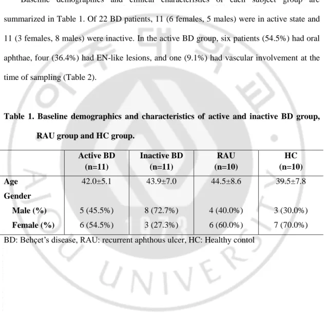
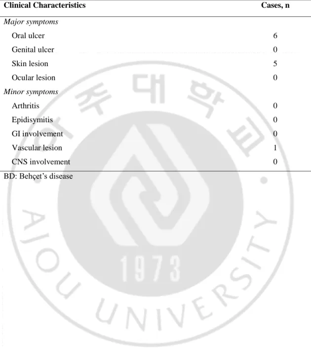
Baseline demographics and clinical characteristics of each subject group are summarized in Table 1. Of 22 BD patients, 11 (6 females, 5 males) were in active state and 11 (3 females, 8 males) were inactive. In the active BD group, six patients (54.5%) had oral aphthae, four (36.4%) had EN-like lesions, and one (9.1%) had vascular involvement at the time of sampling (Table 2).
Table 1. Baseline demographics and characteristics of active and inactive BD group, RAU group and HC group.
Active BD (n=11) Inactive BD (n=11) RAU (n=10) HC (n=10) Age 42.0±5.1 43.9±7.0 44.5±8.6 39.5±7.8 Gender Male (%) 5 (45.5%) 8 (72.7%) 4 (40.0%) 3 (30.0%) Female (%) 6 (54.5%) 3 (27.3%) 6 (60.0%) 7 (70.0%) BD: Behçet’s disease, RAU: recurrent aphthous ulcer, HC: Healthy contol
Table 2. Clinical characteristics of active BD group (n=11) at the time of sampling. Clinical Characteristics Cases, n
Major symptoms Oral ulcer 6 Genital ulcer 0 Skin lesion 5 Ocular lesion 0 Minor symptoms Arthritis 0 Epidisymitis 0 GI involvement 0 Vascular lesion 1 CNS involvement 0 BD: Behçet’s disease
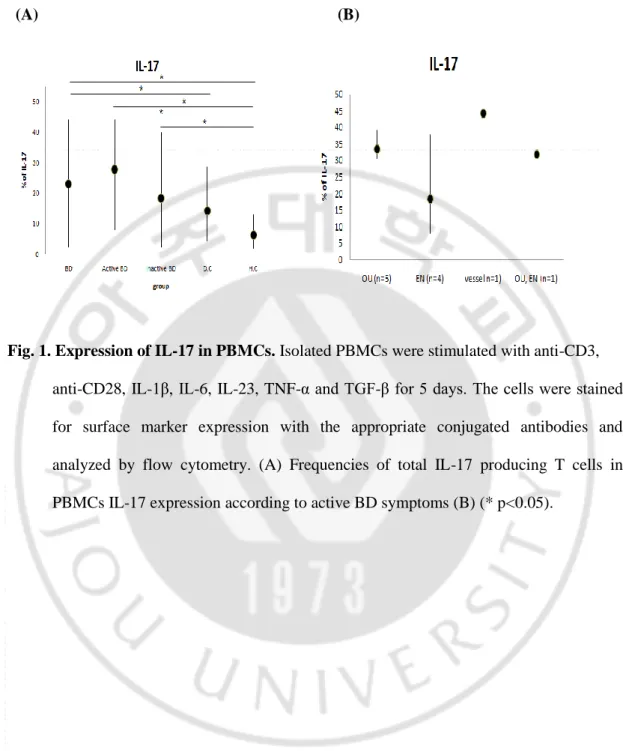
B. Total IL-17 expression in PBMCs
I investigated total IL-17 expression on PBMCs of BD patients using flow cytometry. The percentage of IL-17 producing T cells was based from its proportion in total cells. In the unstimulated state, there was no significant difference in the intracellular expression of IL-17 producing T cells among the study groups (data not shown). PBMCs were stimulated with with anti-CD3, anti-CD28, IL-1β, IL-6, IL-23, TNF-α and TGF-β for 5 days. After stimulation, increased frequency of IL-17 producing T cells in PBMCs was observed in all study groups. The frequencies of IL-17 producing T cells were significantly higher in BD group compared with HC and RAU groups (Fig. 1A). Active BD and inactive BD groups, there was no significant difference. IL-17 producing T cells were significantly increased in the active BD group compared with HC and RAU groups though these significant differences were found in the inactive BD group compared with HC group only (Fig. 1A). The frequencies of IL-17 producing T cells according to clinical symptoms at the time of visit did not how any difference between the symptoms.
(A) (B)
Fig. 1. Expression of IL-17 in PBMCs. Isolated PBMCs were stimulated with anti-CD3,
anti-CD28, IL-1β, IL-6, IL-23, TNF-α and TGF-β for 5 days. The cells were stained for surface marker expression with the appropriate conjugated antibodies and analyzed by flow cytometry. (A) Frequencies of total IL-17 producing T cells in PBMCs IL-17 expression according to active BD symptoms (B) (* p<0.05).
C. IL-17 and IFN-γ in CD4+ and CD8+ T cells subpopulations
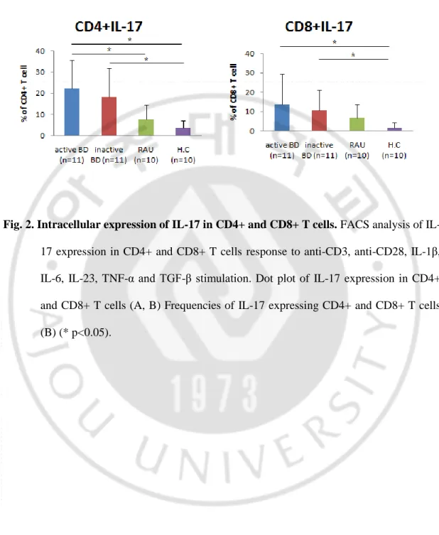
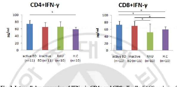
In the unstimulated state, there were no significant difference in the intracellular expressionof IL-17 and IFN-γ in CD4+ and CD8+T cells among the study groups (data not shown). At 5 days after stimulation, impaired up-regulation of intracellular IL-17 in CD4+ and CD8+ T cells in response to anti-CD3, anti-CD28, IL-1β, IL-6, IL-23, TNF-α and TGF-β stimulation was observed in all study groups. The frequencies of IL-17 producing CD4+ T cells were significantly increased in active BD group compared to with HC and RAU groups. The frequencies of IL-17 producing CD4+ T cells were significantly increased in inactive BD group compared to with HC group. In addition, the frequencies of IL-17 producing CD8+ T cells were significantly increased in active BD and inactive BD groups compared to with HC group (Fig. 2B, C). At 2 days after stimulation, impaired up-regulation of intracellular IFN-γ on CD4+ and CD8+ T cells in response to anti-CD3 and anti-CD28 stimulation was observed in all study groups. The frequencies of IFN-γ producing CD4+ T cells were significantly increased in active BD group compared with HC group. The frequencies of IFN-γ producing CD8+ T cells were significantly increased in active BD and inactive BD groups compared with HC and RAU groups (Fig. 3B, C).
(A)
(C)
Fig. 2. Intracellular expression of IL-17 in CD4+ and CD8+ T cells. FACS analysis of IL-
17 expression in CD4+ and CD8+ T cells response to anti-CD3, anti-CD28, IL-1β, IL-6, IL-23, TNF-α and TGF-β stimulation. Dot plot of IL-17 expression in CD4+ and CD8+ T cells (A, B) Frequencies of IL-17 expressing CD4+ and CD8+ T cells (B) (* p<0.05).
(A)
(C)
Fig. 3. Intracellular expression of IFN-γ in CD4+ and CD8+ T cells. FACS analysis of
IFN-γ expression in CD4+ and CD8+ T cells response to anti-CD3 and anti-CD28 stimulation. Dot plot of IFN-γ expression in CD4+ and CD8+ T cells (A, B) Frequencies of IFN-γ expressing CD4+ and CD8+ T cells (C) (* p<0.05).
D. IL-17 in CD4+CD45RO+ and CD8+CD45RO+ T cells subpopulations.
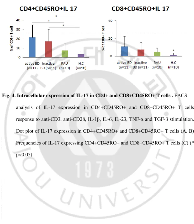
IL-17 is principally produced by CD45RO+ memory T cells. To evaluate the IL-17 producing CD45RO+ memory T cells in BD, PBMCs were stained for surface marker expression with the appropriate conjugated antibodies and analyzed by multi-color flow cytometry. The frequencies of IL-17 producing CD4+CD45RO+ T cells were significantly increased in active BD group compared with HC and RAU groups. The frequencies of IL-17 producing CD4+CD45RO+ T cells were significantly increased in inactive BD group compared only with HC group (Fig. 4B, C). On the other hand, the frequencies of IL-17 producing CD8+CD45RO+ T cells were no significant difference BD and RAU groups. Active BD and inactive BD groups were significantly higher than HC group (Fig. 4B, C).
(A)
(C)
Fig. 4. Intracellular expression of IL-17 in CD4+ and CD8+CD45RO+ T cells . FACS
analysis of IL-17 expression in CD4+CD45RO+ and CD8+CD45RO+ T cells response to anti-CD3, anti-CD28, IL-1β, IL-6, IL-23, TNF-α and TGF-β stimulation. Dot plot of IL-17 expression in CD4+CD45RO+ and CD8+CD45RO+ T cells (A, B) Frequencies of IL-17 expressing CD4+CD45RO+ and CD8+CD45RO+ T cells (C) (* p<0.05).
E. Levels of IL-17, IFN-γ and IL-23
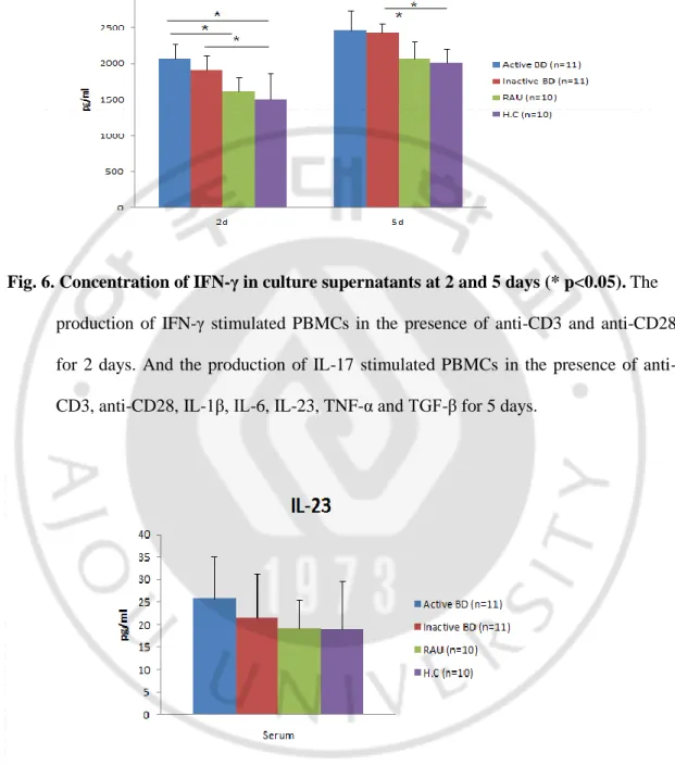
IL-17 levels in the serum were significantly up-regulated in active BD group compared to with HC and RAU groups (Fig. 5A). The expression of IL-17 in the supernatants of cultured PBMCs in time of 2 days after stimulation was significantly higher in the active BD group compared with the HC group. The expression of IL-17 in the supernatants of cultured PBMCs in time of 5 days after stimulation was significantly higher in the active BD group compared with the HC and RAU groups through the expression of IL-17 in the supernatants of cultured PBMCs in time of 5 days after stimulation was significantly higher in the inactive BD group compared with the HC group (Fig. 5A). IFN-γ level was undetectable in the sera of all patients and control groups (data not shown). The expression of IFN-γ in the supernatants of cultured PBMCs in time of 2 days after stimulation were significantly higher in the active BD group compared with the HC and RAU groups, although the expression of IFN-γ in the supernatants of cultured PBMCs in time of 5 days after stimulation were significantly higher in the inactive BD group compared only with the HC group. The expression of IFN-γ in the supernatants of cultured PBMCs in time of 5 days after stimulation were significantly higher in the active BD and inactive BD groups compared with the HC and RAU groups (Fig. 6). IL-23 levels in the serum showed no significant difference among study groups (Fig. 7). 2 and 5 days in culture supernatant IL-23 levels were not measured.
(A) (B)
Fig. 5. Level of IL-17 in sera (A) and culture supernatants at 2 and 5 days (B) (* p<0.05).
The production of IL-17 stimulated PBMCs in the presence of anti-CD3 and anti-CD28 for 2 days. And the production of IL-17 stimulated PBMCs in the presence of anti-CD3, anti-CD28, IL-1β, IL-6, IL-23, TNF-α and TGF-β for 5 days.
Fig. 6. Concentration of IFN-γ in culture supernatants at 2 and 5 days (* p<0.05). The
production of IFN-γ stimulated PBMCs in the presence of anti-CD3 and anti-CD28 for 2 days. And the production of IL-17 stimulated PBMCs in the presence of anti-CD3, anti-CD28, IL-1β, IL-6, IL-23, TNF-α and TGF-β for 5 days.
Fig. 7. Level of IL-23 in sera. IL-23 levels in the serum showed no significant difference
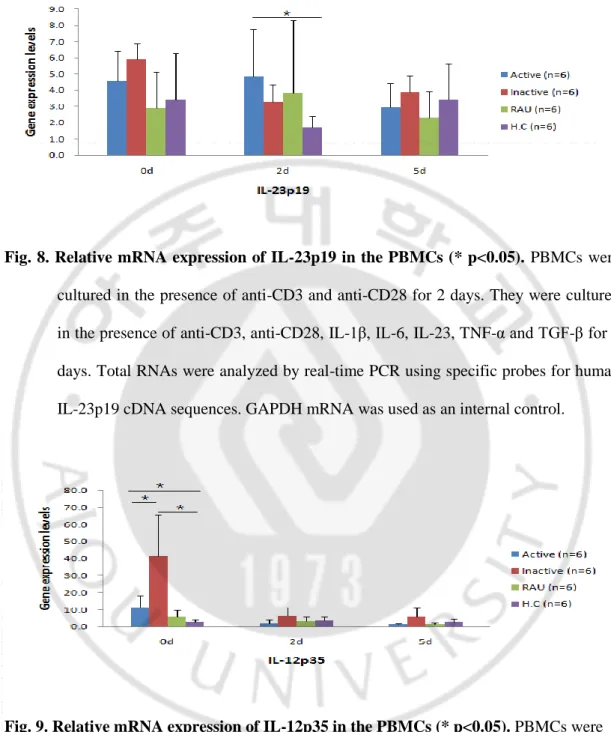
F. mRNA expression levels of IL-23p19, IL-12p35 and IL-12/23p40
IL-23p19 mRNA expression was not different among the study groups in the resting state. Expression of IL-23p19 mRNA was significantly higher in the active BD group compared with HC group at 2 days after stimulation but there were no significant differences of IL-12p35 mRNA levels among the study groups at 5 days after stimulation. By contrast, expression of IL-12p35 mRNA was significantly higher in active BD group compared with inactive HC group. And IL-12p35 mRNA was significantly higher in inactive BD group compared with active BD and HC groups. At 2 and 5 days after stimulation, there were no significant differences of 12p35 mRNA levels among the study groups. Expression of IL-12/23p40 mRNA was significantly higher in the inactive BD group compared with HC group at 5 days after stimulation. The data for IL-23/23p40 after 2 days and without stimulation were corrupt due to prolonged exposure to room temperature. 0 and 2 days IL-12/23p40 the measure, but the results can not be used (Data not shown).
Fig. 8. Relative mRNA expression of IL-23p19 in the PBMCs (* p<0.05). PBMCs were
cultured in the presence of anti-CD3 and anti-CD28 for 2 days. They were cultured in the presence of anti-CD3, anti-CD28, IL-1β, IL-6, IL-23, TNF-α and TGF-β for 5 days. Total RNAs were analyzed by real-time PCR using specific probes for human IL-23p19 cDNA sequences. GAPDH mRNA was used as an internal control.
Fig. 9. Relative mRNA expression of IL-12p35 in the PBMCs (* p<0.05). PBMCs were
cultured in the presence of anti-CD3 and anti-CD28 for 2 days. They were cultured in the presence of anti-CD3, anti-CD28, IL-1β, IL-6, IL-23, TNF-α and TGF-β for 5 days. Total RNAs were analyzed by real-time PCR using specific probes for human IL-12p35 cDNA sequences. GAPDH mRNA was used as an internal control.
Fig. 10. Relative mRNA expression of IL-12/23p40 in the PBMCs (* p<0.05). PBMCs
were cultured in the presence of anti-CD3 and anti-CD28 for 2 days. They were cultured in the presence of anti-CD3, anti-CD28, IL-1β, IL-6, IL-23, TNF-α and TGF-β for 5 days. Total RNAs were analyzed by real-time PCR using specific probes for human IL-12/23p40 cDNA sequences. GAPDH mRNA was used as an internal control.
V. DISCUSSION
Until recently, Th cells were thought to be a binary system, consisting of Th1 and Th2 cells. However, interleukin-17 (IL-17) expressing T cells, which are now widely known as Th17 cells, were proposed to be a third lineage of Th cells (Dong, 2008). Efforts have been made in characterizing human Th17 cells and the factors involved in their differentiation and in understanding the role of these cells in protective immunity and autoimmune diseases (Sallusto and Lanzavecchia, 2009). Th17 cells, are characterized by the master transcription factor orphan hormone receptor gamma t (RORγt), the surface markers IL-23R and chemokine (C-C motif) receptor (CCR) 6, and by production of the proinflammatory cytokines IL-17A, IL-17F, IL-21, IL-22 and IL-26, and the chemokine CCL20 (Brand, 2009). Although human Th17-cell development is less well understood, IL-23 have been shown to mediate the differentiation of human Th17 cells (Dong, 2008). Diseases such as psoriasis, RA, multiple sclerosis (MS), Crohn’s disease have in common the local chronic inflammatory reaction with production of inflammatory cytokines, leading to matrix destruction and defective repair (Firestein, 2003). IL-17 inhibition in the treatment of inflammatory conditions remains to be defined. As of today positive results have been obtained with a monoclonal against IL-17A in patients with psoriasis (Miossec, 2009).
Previous reports showed inconsistent results about Th17 related cytokines in patients with BD. Significantly up-regulated IL-17 and IL-23 producing T cells were found in BD patients with active uveitis (Chi et al., 2008). In contrast, these findings were not appeared in patients with BD who have gastrointestinal involvement (Ferrante et al., 2010).
CD4+CD45RO+ T cells was increased in patients with BD compared with control groups. Consistently, increased serum concentration level of IL-17 and IFN-γ was appeared in the BD group. In addition, mRNA expression of IL-23p19, IL-12p35 and IL-12/23p40 in PBMCs was up-regulated in total BD patient groups. All these findings suggest that IL-23 and IL-17 are associated with active inflammation in BD patients.
IL-17 (also known as IL-17A) produced by Th17 cells has been associated for many years with host defend against infectious agents and with autoimmune diseases. IL-17 is mainly produced by activated CD4+ T cells; predominantly produced by CD4+CD45RO+ (memory) T cells (Colin et al., 2010), but can be induced in CD8+ T cells, natural killer (NK) T cells and possibly other cells (Nistala and Wedderburn, 2009).
IL-17 expression has been associated with many inflammatory diseases, such as rheumatoid arthritis, asthma, systemic lupus erythematosus (SLE) and allograft rejection (Moseley et al., 2003). Similarly, increased expression of IL-17 was observed in BD patients with acute uveitis (Chi et al., 2008). Consistent with previous studies, the expression of IL-17 in the PBMCs was increased in patients with BD compared with control groups in this study.
Moreover, this study revealed that frequencies of IL-17 producing CD4+CD45RO+ T cells were significantly increased in patients with BD compared with control groups. These results are consistent with those reported by chi et al (Chi et al., 2008). Considering the high pathogenicity of IL-17 producing Th cells in the development and maintenance of autoimmune diseases, it is likely that a large amount of IL-17 producing CD4+CD45RO+ memory T cells correlates with the active inflammation in BD patients.
occurred.
I also evaluated the expression of IL-17 in PBMCs according to the symptom of BD patients. However, sample size of each symptom group was small, therefore, it is difficult to evaluate the tendency of IL-17 expression related BD symptom. Future study with large sample size is needed to identify the exact level of IL-17 and the correlation of IL-17 and each symptom of BD patients.
IL-23 has been linked to the pathogenesis of autoimmune inflammation. IL-23 deficiency was shown to protect against EAE and collagen-induced arthritis (Murphy et al., 2003). The expression of IL-23 in clinical samples of Crohn’s disease (CD) rheumatoid arthritis (RA) supports a possible role of IL-23 in common human autoimmune diseases (Di Cesare et al., 2009). Therefore, the role of IL-23 in the pathogenesis of BD was evaluated in this study. Although, there was no difference of IL-23 concentration in the serum among all study groups in resting state, mRNA expression of IL-23 was up-regulated in patients with BD compared with control groups. From these results, I suggest that up-regulated IL-23 is associated with the active inflammation seen in BD. However, IL-23 concentration in the culture supernatants was not detected in this study. Therefore, further study is needed to evaluate the changes of IL-23 level in each study group after stimulation.
IFN-γ was considered an important mediator involved in the development of BD (Hamzaoui et al., 2002; Direskeneli, 2006). Thl cells produce IL-2, IFN-γ, and lymphotoxin and mainly support cell-mediated immune responses. It has been hypothesized that IL-12 differentially regulates the effector response in infectious and autoimmune diseases and that in the latter case it may function independently of IFN-γ (Sugi-Ikai et al., 1998). IL-12 is a potent cofactor stimulating growth, IFN-γ synthesis, and cell adhesion of already differentiated Th1 cells (Adorini, 1999). Previous reports showed inconsistent results about
increased production of Thl cytokines such as IL-21 and IFN- γ has also been observed in patients with BD (Sugi-Ikai et al., 1998). From these results, I suggest that up-regulated IL-12 is associated with the active inflammation seen in BD.
V. CONCLUSION
Increased expression of IL-17 and IFN-γ in the PBMCs and in the CD4+ and CD8+ T cells was increased in patients with BD compared with control groups. And the expression of IL-17 and IFN-γ in the serum and supernatants of cultured PBMCs was significantly higher in the BD group compared with the control groups. These results suggest that Th17 cells has a important implication in immune modulation of BD. Future studies clarifying the further immunopathologic role of Th17 pathway in BD are needed. Novel insights into the immunopathogenesis of BD will lead to development of new effective therapies with highly selective mechanisms of action targeting key immunologic cytokines and receptors.
REFERENCES
1. Criteria for diagnosis of Behçet’s disease International study group for Behçet’s disease
Lancet 335: 1078-1080, 1990
2. Adorini L: Interleukin-12, a key cytokine in Th1-mediated autoimmune diseases. Cell Mol
Life Sci 55: 1610-1625, 1999
3. Brand S: Crohn's disease: Th1, Th17 or both? The change of a paradigm: new
immunological and genetic insights implicate Th17 cells in the pathogenesis of Crohn's disease. Gut 58: 1152-1167, 2009
4. Chi W, Zhu X, Yang P, Liu X, Lin X, Zhou H, Huang X, Kijlstra A: Upregulated IL-23 and IL-17 in Behcet patients with active uveitis. Invest Ophthalmol Vis Sci 49: 3058-3064, 2008
5. Colin EM, Asmawidjaja PS, van Hamburg JP, Mus AM, van Driel M, Hazes JM, van Leeuwen JP, Lubberts E: 1,25-dihydroxyvitamin D3 modulates Th17 polarization and interleukin-22 expression by memory T cells from patients with early rheumatoid arthritis.
Arthritis Rheum 62: 132-142, 2010
7. Direskeneli H: Autoimmunity vs autoinflammation in Behçet’s disease: do we oversimplify a complex disorder? Rheumatology (Oxford) 45: 1461-1465, 2006
8. Dong C: TH17 cells in development: an updated view of their molecular identity and genetic programming. Nat Rev Immunol 8: 337-348, 2008
9. Ferrante A, Ciccia F, Principato A, Giardina AR, Impastato R, Peralta S, Triolo G: A Th1 but not a Th17 response is present in the gastrointestinal involvement of Behçet’s disease.
Clin Exp Rheumatol 28: S27-30, 2010
10. Firestein GS: Evolving concepts of rheumatoid arthritis. Nature 423: 356-361, 2003
11. Hamzaoui K, Hamzaoui A, Guemira F, Bessioud M, Hamza M, Ayed K: Cytokine profile in Behçet’s disease patients. Relationship with disease activity. Scand J Rheumatol 31: 205-210, 2002
12. Imamura Y, Kurokawa MS, Yoshikawa H, Nara K, Takada E, Masuda C, Tsukikawa S, Ozaki S, Matsuda T, Suzuki N: Involvement of Th1 cells and heat shock protein 60 in the pathogenesis of intestinal Behçet’s disease. Clin Exp Immunol 139: 371-378, 2005
13. Kurokawa MS, Yoshikawa H, Suzuki N: Behçet’s disease. Semin Respir Crit Care Med 25: 557-568, 2004
14. Lew W, Chang JY, Jung JY, Bang D: Increased expression of interleukin-23 p19 mRNA in erythema nodosum-like lesions of Behçet’s disease. Br J Dermatol 158: 505-511, 2008
15. Mendoza-Pinto C, Garcia-Carrasco M, Jimenez-Hernandez M, Jimenez Hernandez C, Riebeling-Navarro C, Nava Zavala A, Vera Recabarren M, Espinosa G, Jara Quezada J, Cervera R: Etiopathogenesis of Behçet’s disease. Autoimmun Rev 9: 241-245, 2010
16. Miossec P: IL-17 and Th17 cells in human inflammatory diseases. Microbes Infect 11: 625-630, 2009
17. Moseley TA, Haudenschild DR, Rose L, Reddi AH: Interleukin-17 family and IL-17 receptors. Cytokine Growth Factor Rev 14: 155-174, 2003
18. Murphy CA, Langrish CL, Chen Y, Blumenschein W, McClanahan T, Kastelein RA, Sedgwick JD, Cua DJ: Divergent pro- and antiinflammatory roles for IL-23 and IL-12 in joint autoimmune inflammation. J Exp Med 198: 1951-1957, 2003
19. Nagafuchi H, Takeno M, Yoshikawa H, Kurokawa MS, Nara K, Takada E, Masuda C, Mizoguchi M, Suzuki N: Excessive expression of Txk, a member of the Tec family of tyrosine kinases, contributes to excessive Th1 cytokine production by T lymphocytes in patients with Behçet’s disease. Clin Exp Immunol 139: 363-370, 2005
21. Raziuddin S, al-Dalaan A, Bahabri S, Siraj AK, al-Sedairy S: Divergent cytokine production profile in Behçet’s disease. Altered Th1/Th2 cell cytokine pattern. J
Rheumatol 25: 329-333, 1998
22. Sakane T, Takeno M, Suzuki N, Inaba G: Behçet’s disease. N Engl J Med 341: 1284- 1291, 1999
23. Sallusto F, Lanzavecchia A: Human Th17 cells in infection and autoimmunity. Microbes
Infect 11: 620-624, 2009
24. Sugi-Ikai N, Nakazawa M, Nakamura S, Ohno S, Minami M: Increased frequencies of interleukin-2- and interferon-gamma-producing T cells in patients with active Behçet’s disease. Invest Ophthalmol Vis Sci 39: 996-1004, 1998
25. Yurdakul S, Yazici H: Behçet’s syndrome. Best Pract Res Clin Rheumatol 22: 793-809, 2008
- 국문요약 - 베체트병에서 질병 활성도에 따른 Th17의 발현 분석 아주대학교 대학원 의생명과학과 박 미 진 (지도교수: 이 은 소) 연구 배경: 베체트병은 구강궤양, 외음부궤양, 포도막염 등을 특징으로 하며, 점 막, 피부, 눈, 심장, 혈관, 관절, 신장, 소화기, 신경계까지 침범 될 수 있는 만성, 재발성 전신 염증 질환이다. 현재까지 정확한 원인은 밝혀져 있지 않으나 유전적, 환경적, 감염 및 면역학적 요인들이 복합적으로 관련하여 지속적 염증 상태를 유 발하는 것으로 알려져 있다. 면역학적으로는 T lymphocytes가 체내 외 여러 항원 들에 대한 면역 과반응을 유도하여 베체트병 증상의 활성화에 중요한 역할을 하 고 있다. 환자들의 병변과 혈청 내 Th1 cytokine인 IL-2, IFN-γ, IL-12의 증가가 관 찰되고 특히 IL-12가 질병의 활성도와 비례하는 결과 등으로 볼 때 베체트병은 Th1 패턴이 우세한 질병군으로 생각되고 있다. Th17 세포는 염증의 주요 원인인 염증성 사이토카인 IL-17을 분비하고 대부분 CD4+CD45RO+ T 세포에서 생성된 다. 최근 베체트병 환자의 포도막염 병변에서 IL-17과 IFN-γ의 증가가 확인 되었 고, 결절성 홍반 병변 조직에서 IL-23p19 mRNA의 발현 증가가 확인된 바 있다.
세포와 이와 연관있는 cytokine의 발현과 베체트병의 관련성을 분석하고자 하였다.
연구 방법: 실험 대상은 베체트병 환자 22명, 질병 대조군으로 재발성 아프타성 궤양 환자 10명, 정상 대조군 10명으로 하였다. 모집된 각 그룹으로부터 얻은 혈액에서 분리된 PBMC를 자극 및 배양한 후 flow cytometry를 실시 하였고 혈청 및 세포 배양액의 IL-17, IL-23, IFN-γ의 농도 측정을 위해 ELISA 실시 하였다. 또한, IL-23p19, IL-12p35, IL-12/23p40의 mRNA 발현을 측정하기 위해 real-time PCR을 시행하였다. 연구 결과: PBMC의 CD4+ T 세포에서 자극 5일 후 IL-17의 발현과 자극 2일 후 IFN-γ의 발현은 정상대조군 및 재발성 아프타성 구강 궤양 환자군과 비교하여 활동성, 비활동성 베체트병 환자군에서 유의하게 높은 결과를 보였다. CD8+ T 세포에서 IL-17과 IFN-γ의 발현도 활동성, 비활동성 베체트병 환자군 모두에서 정상대조군과 비교하여 높은 결과를 보였다. 혈청과 PBMC를 자극 후 배양한 배양액에서의 IL-17과 IFN-γ 농도 역시 정상대조군 및 환자대조군과 비교하여 활동성, 비활동성 베체트병 환자군에서 유의하게 높은 결과를 보였다. 하지만 자극을 주지 않은 23은 유의한 차이를 나타내지 않았다. mRNA 발현에서 23p19는 2일째에서는 활동성 베체트병 환자군이 정상대조군보다 높았다. IL-12p35는 자극을 주지 않은 0시간에서 활동성 베체트군은 정상대조군보다, 비활동성 베체트군은 활동성 베체트병군과 정상대조군에 비해서 높게 나타났다. IL-12/23p40은 5일째에서 비활동성 베체트군이 정상대조군보다 높았다.
증면하였고 이를 통해 IL-17의 과발현이 베체트병에 관련이 있음을 확인하였다. 하지만 IL-17, IFN-γ와 관련있는 IL-12, IL-23은 전체 베체트병 환자에서는 비교적 높게 나타났지만 증상 유무와는 크게 상관이 없을 것으로 생각되며 향후 이에 따른 추가적인 실험이 필요할 것으로 보인다.