Research
C The Korean Society for Biomaterials
Preparation of PLGA Microparticles by a double Emulsion Solvent
Technique for Sustained Release of (-)-epigallocatechin Gallate (EGCG)
and Their Growth Inhibitory Effect on Rat Aortic Smooth Muscle Cells
Mi Hee Lee1,2, Hye Ryeon Lim1, Tek Hyung Lee1, and Jong-Chul Park1,2*
1Department of Medical Engineering, 2Brain Korea 21 Project for Medical Science, Yonsei University College of Medi-cine, 134 Shinchon-dong, Seodaemun-gu, Seoul 120-752, Korea
(Received December 20, 2008/Acccepted February 3, 2009)
In this study, we fabricated porous PLGA microparticles loaded with EGCG by a modified water-in-oil-in-water (W/ O/W) double emulsion solvent evaporation method. The physicochemical properties of the EGCG-loaded PLGA microparticles were studied using scanning electron microscopy (SEM), Fourier transformed-infrared (FT-IR) spectros-copy and in vitro release measurements. A remarkable burst effect was observed on first stage however the released amount was reduced largely with time. This result showed that the release of EGCG increased by its loading amount. Also, EGCG - loaded PLGA particles inhibited the growth of rat aortic smooth muslce cells (RASMC). This study demonstrates that PLGA microparticle system is potential as an efficient EGCG carrier in medical therapy. Key words: (-)-epigallocatechin gallate, PLGA microparticles, sustained release, rat aortic smooth muscle cell, water-in-oil-in-water (W/O/W) double emulsion method
Introduction
everal epidemiological studies have suggested that green tea has pharmacological effects associated with a reduced risk of degenerative disease such as cancer and cardiovascular disease. Among the green tea polyphenols, epigallocatechin-3-gallate (EGCG) is the major component of green tea that mediates cardiovascular-related effects such as anti-prolifera-tive,1,2) anti-atherogenic3) and anti-thrombotic.4) Furthermore, recent studies have demonstrated that EGCG has ability of preventing angioplasty-induced restenosis, suppressing vascular smooth muscle cells (SMCs) proliferation and migration4,5) and inhibiting the activities of some matrix metalloproteinases (MMPs).6) On these bases, it can be assumed that EGCG has a protective effect on the vascular dysfunction and intimal hyperplasia after vascular damage. Local delivery of water-sol-uble EGCG may be needed for a successful vascularization from injured vessels.
For the purpose of a sustained drug release, a drug delivery system using biodegradable polymer microparticles has been focused with its potentials.7,8) PLGA has been often used for drug delivery systems because of its proven safety and efficacy in sustained release. Drugs may be incorporated into PLGA
microparticles by a variety of techniques including encapsula-tion method. Among the encapsulaencapsula-tion techniques, the o/o solvent evaporation9) and the w/o/w solvent extraction techniques10-12) are two of the most convenient ways for the encapsulation of drug within PLGA microparticles. According to the used encapsulation methods, PLGA molecular weight, and stabilizers the drug encapsulation efficiency, diffusion of drug, morphological features of the PLGA microparticles and burst release can be determined.
In this study, the EGCG-loaded microparticles were pre-pared by a double emulsion solvent technique using a biode-gradable copolymer of poly (lactic-co-glycolic acid) (PLGA) and in vitro EGCG release study from PLGA microparticles was performed.
Materials and Methods
Materials
PLGA (50:50) polymer (Mw 72.3 kDa) was purchased from Lakeshore Biomaterials, Inc (AL, USA). EGCG (MW 458.4) was supplied by TeavigoTM (DSM Nutritional Products Ltd., Basel, Switzerland) and its purity exceeded 93%. Dichlo-romethane (DCM), polyvinyl alcohol (PVA), DMSO and iso-propyl alcohol were purchased from Sigma-Aldrich Co., (St. Louis., USA).
S
extraction technique. Briefly, 600 mg PLGA was dissolved in 5 mL of DCM and 0, 15, 30, 60 mg of EGCG were dissolved into 1 mL of distilled water, respectively. After mixing these two solutions vigorously for 10min by a vortex mixer, emulsi-fied solution was re-emulsiemulsi-fied in 50 mL of PVA (0.2%, w/v)., Solution was poured in 50 mL of aqueous isopropyl alcohol (2%, w/v) and stirred with a magnetic stirrer for overnight at room temperature to harden fabricated microparticles. Also, EGCG-free microparticles were prepared accordingly without EGCG. Fabricated PLGA microparticles were rinsed three times with pH 7.4 phosphate buffered saline (PBS) to remove the un-captured EGCG and lyophilized in a freeze-dryer. Dried microparticles were stored in -20oC.
Characterization of PLGA microparticles
Determination of EGCG loading amount and efficiency To determine EGCG amount loaded in the PLGA micropar-ticles, 10 mg of microparticles were dissolved in 5 mL DMSO and analyzed at 312 nm by UV-Vis spectrophotometer. The concentration of EGCG released from particle was calculated from the standard calibration curve of EGCG solution. The relationship between the input amount of EGCG and the EGCG loading content of particle was investigated:
The yield of the produced microparticles and EGCG loading efficiency were calculated as followings;
Yield = Weight of EGCG-loaded PLGA in production/ Total weights of input EGCG and input PLGA× 100
(1) EGCG loading efficiency = Actual loading amount of EGCG/
Input amount of EGCG× 100 (2) PLGA microparticles morphology
Prepared PLGA microparticles were coated by gold and their surfaces and sizes were observed by a scanning elec-tronic microscope (Hitachi S-800, Tokyo, Japan). The micro-particles were mounted and sputter-coated with gold/platinum using an ion coater (E1010, Hitachi) and then observed at an accelerating voltage of 20 kV.
Fourier transformed-infrared (FT-IR) spectroscopy
Fourier transform infrared (FT-IR) spectra of EGCG, PLGA and EGCG- PLGA particle were obtained between 4000 and 650 cm-1 using an FT-IR spectrophotometer (NicoletTM 380, Thermo Fisher Scientific, Inc., Waltham, MA). Samples were scanned 128 times at 8 cm-1 resolution.
In vitro release of EGCG from particle
The 50 mg of EGCG-loaded PLGA microparticles were
sus-ume of buffer at predetermined time intervals. The suspended solution was centrifuged at 25oC and 1600 rpm for 10 min. The concentrations of EGCG from released buffer were deter-mined at 280 nm by UV-Vis spectrophotometer from the stan-dard calibration curve of EGCG solution. The accumulated release percentage of EGCG at each time point was detected from three replicate samples for 23 days.
Cell culture and growth inhibition assay of EGCG-PLGA particle
RASMCs (Rat Aortic smooth muscle cells) were purchased from BioBud (Seoul, Korea) and used between passages 7 and 15. The cells were routinely maintained in Dulbecco’s modi-fied Eagle’s medium (Wellgene, Seoul, Korea) supplemented with 10% FBS (HyClone Laboratories Inc, UT, USA) and a 1% antibiotic antimycotic solution (including 10,000 U penicillin, 10 mg streptomycin, and 25µg amphotericin B per ml, Wellgene) at 37oC in a humidified atmosphere of 5% CO2 in air. In order to examine the growth inhibitory effects of EGCG-loaded PLGA microparticles on RASMCs, the cells were seeded into 48well plates and incubated for 4hr at 37oC in a humidified atmosphere of 5% CO2 in air. After the cells were attached, they were treated with EGCG-loaded PLGA particles (20 mg/ml) and incubated for 1d, 3d, and 5d, respectively. EGCG-loaded PLGA particles were sterilized by low tempera-ture EO gas (3M™ Steri-Vac™ 5XL Gas Sterilizer/Aerators, 3M, St. Paul, MN) before the treatment to RASMC. Cell viability was determined by MTT assay [reduction of 3-(4,5-dimethylth-iazol-2-yl)-2,5-diphenyltetrazolium bromide to a purple forma-zan product].
Results and discussions
Preparation of biodegradable microparticles containing water-soluble bioactive agents has received much attention in recent years, due to their numerous advantages compared to conventional dosage forms, which include improved efficacy, reduced toxicity, and improved patient compliance and con-venience. Various techniques are available to entrap water sol-uble bioactive agents into biodegradable microparticles. Among various methods, the W/O/W double emulsion tech-nique is the most popular method.13)
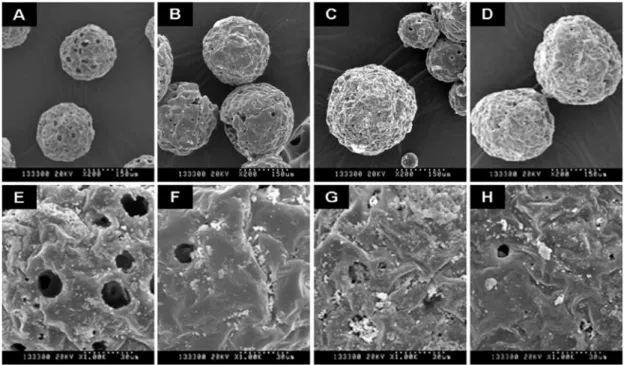
EGCG-loaded PLGA microparticles were prepared by the W1/O/W2 emulsification-solvent evaporation method. The morphology of PLGA microparticles was observed by SEM (Figure 1). Although the size of microparticles was diverse, the average size was from 100µm to 300 µm. Moreover, the PLGA microparticle were spherical with several pores distrib-uted on their surface, irrespective of their dimension (Figure 1
A, B, C, D). However, the pore number decreased with increasing EGCG concentration (Figure 1 E, F, G, H). Porosity of microparticles is known to vary with numerous factors such as polymer molecular mass, co-solvent concentration, dis-persed phase to continuous phase ratio, drug concentration, rate and method of solvent removal.15) Pores are formed because of the inner aqueous phase during microparticle for-mation in the water-in-oil-in-water (W/O/W) method, in com-parsion with the oil-in-water method. In the organic phase, the polymer concentration continuously increases during organic solvent extraction/evaporation. The PLGA begins to precipitate and encapsulate the drug as well as inner water droplets. By elimination of the water during drying, empty holes remain and are uniformly distributed throughout the microparticles, irrespective of their size.14)
However, once the microparticles are dried, in EGCG-loaded particles the empty holes are filled by EGCG that was included in water. We observed that the inside of pore is filled with loaded-EGCG compared with non-treated microparticles (Figure 1 E, F, G, H). Moreover, it has been observed that the
size of pores decreased with the increasing EGCG concentra-tion in EGCG-loaded microparticles.
The actual load of EGCG in PLGA microparticles was calcu-lated by spectrophotometric analysis. The actual loading amount and efficiency of EGCG are presented Table 1. The production yield of PLGA microparticles gradually was decreased by addition of EGCG. The actual EGCG loading was almost identical in all cases. The EGCG loading efficien-cies were found to be 8~13 % with no significant difference with increasing EGCG concentration. Although the loading efficiency has been reported to be related to surfactant con-centration and viscosity in oil phase, stirring time period and stirring rate in the preparation procedure of primary emulsion, stirring rate in secondary emulsion preparation, polymer con-centration, and osmotic pressure, drug loading efficiency and particle size of microparticle can not be completely con-trolled.15,16)
The FT-IR spectra of the representative formulations con-firmed that the EGCG was dissolved in the microparticle (Figure 2). FT-IR spectra of EGCG-loaded particles were dominated by Figure 1. Scanning electron micrographs images (SEM) of PLGA microparticles. (A, E) 0 mg ; (B, F) 15 mg ; (C, G) 30 mg ; (D, H) 60 mg of EGCG was loaded microparticles. Upper panel: 200 magnification, down panel: 1.0 K magnification.
Table 1. Encapsulation efficiency and burst effect of EGCG-loaded PLGA microparticles.
EGCG(mg)/PLGA(mg) Yield Theoretical loaded EGCG (mg) Actual loaded-EGCG (mg) EGCG loading efficiency (%)
0/600 94.58±1.58 - -
-15/600 90.09±0.98 15 1.59±0.39 10.61±2.58
30/600 84.21±0.34 30 3.11±0.53 10.36±1.78
a broad notable band at phenyl-OH group (3600–3150 cm-1), which is the characteristic peak of EGCG. Therefore, the char-acteristic phenyl-OH peak was stretched with increasing EGCG concentration. The change in this peak was similar with EGCG releasing PLCL film prepared by solvent casting.17) It seems that EGCG was encapsulated in the PLGA microparticle, probably by dispersion of EGCG in polymer.
The release experiments in vitro were carried out by spec-trophotometrical method for 23 days. The EGCG release pro-files from the PLGA microparticles in PBS are illustrated in Figure 3 for different concentration of EGCG. The EGCG release profiles were biphasic, remarkable burst effect was observed on the first and second day. However, the released concentration of EGCG was largely reduced from the third day and kept constant for 3 weeks. The highest burst effect and release rate was observed in microparticles with the high-est concentration of EGCG. Burst release from PLGA
micro-particles has been attributed to either the surface or uniform distribution of the associated drug. Therefore, to reach the sustained release the drug should be distributed exclusively inside the microparticle. Porosity increases the surface area of the microparticle, which itself can contribute to the burst.18) A successful microparticle should yield high encapsulation effi-ciency, and desired particle size.
Other studies have already shown that EGCG has pharma-cological activites, including antioxidant, proliferative, anti-thrombotic and anti-migrative effect on smooth muscle cells.1,4,19) In this study, we examined the growth inhibitory effect on RASMC by EGCG-loaded PLGA microparticles (Fig-ure 4). When the cells were treated with EGCG-loaded micro-particles, cell proliferation significantly decreased with increas-ing EGCG concentration dose dependently in comparison with the unloaded PLGA microparticles. Our previous results had shown that proliferation and migration in serum-stimu-lated RASMC was inhibited in more than 200µM EGCG.5) By calculation, PLGA microparticles manufactured by different concentration of EGCG (15:600, 30:600 and 60:600) loaded 1.5 mg, 30 mg and 60 mg, respectively. Our results showed that RASMC proliferation was inhibited by EGCG released from PLGA microparticles for the first 5 days.
Microparticles should provide effective and sustainable release to control the drug release kinetics over periods of days to months. Thus, PLGA microparticles for local delivery of EGCG can be very helpful to optimize the therapeutic effi-ciency and to reduce possible side effects in medical applica-tion of EGCG.
Acknowledgement
This study was supported by a faculty research grant of Yon-sei University College of Medicine for 2006.
Figure 2. FT-IR spectrums of EGCG-loaded PLGA microparti-cles.
Figure 3. In vitro release behavior of EGCG from PLGA microparticles. Released EGCG was observed at 280 nm with control for 23 days (PBS buffer, pH 7.4, 37oC).
Figure 4. Effect of EGCG-loaded PLGA microparticles on RASMC proliferation. The results are reported as means±SD (n = 4). The data are analyzed by a t-test. The values marked with asterisks are significantly different from the unloaded PLGA microparticle (0:600) (*p < 0.05).
Reference
1. L. H. Lu, S. S. Lee and H. C. Huang. “Epigallocatechin suppres-sion of proliferation of vascular smooth muscle cells: correlation with c-jun and JNK.” Br. J. Pharmacol., 124, 1227-1237 (1998). 2. K. C. Hwang, K. H. Lee, Y. Jang, Y. P. Yun and K. H. Chung.
“Epigallocatechin-3-gallate inhibits basic fibroblast growth factor-induced intracellular signaling transduction pathway in rat aortic smooth muscle cells.” J. Cardiovasc. Pharmacol., 39, 271-277 (2002).
3. V. Ivanov, M. W. Roomi, T. Kalinovsky, A. Niedzwiecki and M. Rath. “Anti-atherogenic effects of a mixture of ascorbic acid, lysine, proline, arginine, cysteine, and green tea phenolics in human aortic smooth muscle cells.” J. Cardiovasc. Pharmacol., 49, 140-145 (2007).
4. K. Maeda, M. Kuzuya, X. W. Cheng, T. Asai, S. Kanda, N. Tamaya-Mori, T. Sasaki, T. Shibata and A. Iguchi. “Green tea cat-echins inhibit the cultured smooth muscle cell invasion through the basement barrier.” Atherosclerosis, 166, 23-30 (2003). 5. D. W. Han, H. R. Lim, H. S. Baek, M. H. Lee, S. J. Lee, S. H.
Hyon and J. C. Park. “Inhibitory effects of epigallocatechin-3-O-gallate on serum-stimulated rat aortic smooth muscle cells via nuclear factor-kappaB down-modulation.” Biochem. Biophys. Res. Commun., 345, 148-155 (2006).
6. X. W. Cheng, M. Kuzuya, T. Sasaki, S. Kanda, N. Tamaya-Mori, T. Koike, K. Maeda, E. Nishitani and A. Iguchi. “Green tea cat-echins inhibit neointimal hyperplasia in a rat carotid arterial injury model by TIMP-2 overexpression.” Cardiovasc. Res., 62, 594-602 (2004).
7. M. Dutt and G. K. Khuller. “Liposomes and PLG microparticles as sustained release antitubercular drug carriers--an in vitro-in vivo study.” Int. J. Antimicrob. Agents., 18, 245-252 (2001). 8. C. Raman, C. Berkland, K. Kim and D. W. Pack. “Modeling
small-molecule release from PLG microspheres: effects of poly-mer degradation and nonuniform drug distribution.” J. Control. Release, 103, 149-158 (2005).
9. R. Wada, S. H. Hyon and Y. Ikada. “Lactic acid oligomer micro-spheres containing hydrophilic drugs.” J. Pharm. Sci., 79, 919-924 (1990).
10. M. J. Alonso, R. K. Gupta, C. Min, G. R. Siber and R. Langer “Biodegradable microspheres as controlled-release tetanus tox-oid delivery systems.” Vaccine, 12, 299-306 (1994).
11. S. Cohen, T. Yoshioka, M. Lucarelli, L. H. Hwang and R. Langer “Controlled Delivery Systems for proteins based on poly(Lactic/ Glycolic Acid) Microspheres.” Pharm. Res, 8, 713-720 (1991). 12. C. Yan, J. H. Resau, J. Hewetson, M. West, W. L. Rill and M.
Kende. “Characterization and morphological analysis of protein-loaded poly (lactide-co-glycolide) microparticles prepared by water-in-oil-in-water emulsion technique.” J. Control. Release, 32, 231-241 (1994).
13. F. T. Meng, G. H. Ma, W. Qiu and Z. G. Su. “W/O/W double emulsion technique using ethyl acetate as organic solvent: effects of its diffusion rate on the characteristics of microparticles.” J. Control. Release, 91 407-416 (2003).
14. D. Klose, F. Siepmann, K. Elkharraz, S. Krenzlin and J. Siep-mann. “How porosity and size affect the drug release mecha-nisms from PLGA-based microparticles.” Int. J. Pharmaceutics, 314, 198-206 (2006).
15. F. Ito, H. Fujimori and K. Makino. “Incorporation of water-solu-ble drugs in PLGA microspheres.” Colloids and Surfaces B: Bio-interfaces, 54, 173-178 (2007).
16. F. Ito, H. Fujimori and K. Makino. “Factors affecting the loading efficiency of water-soluble drugs in PLGA microspheres.” Col-loids Surf B Biointerfaces, 61, 25-29 (2008).
17. H. H. Cho, D. W. Han, K. Matsumura, S. Tsutsumi and S. H. Hyon. “The behavior of vascular smooth muscle cells and plate-lets onto epigallocatechin gallate-releasing poly(l-lactide-co-epsi-lon-caprolactone) as stent-coating materials.” Biomaterials, 29, 884-893 (2008).
18. S. D. Allison. “Effect of structural relaxation on the preparation and drug release behavior of poly(lactic-co-glycolic)acid micro-particle drug delivery systems.” J. Pharm. Sci., 97, 2022-2035 (2007).
19. C. S. Hofmann and G. E. Sonenshein. “Green tea polyphenol epigallocatechin-3 gallate induces apoptosis of proliferating vas-cular smooth muscle cells via activation of p53.” FASEB J., 17, 702-704 (2003).