Factors Predictive of High-Risk Adenomas at the Third
Colonoscopy after Initial Adenoma Removal
Evaluating predictive factors for high-risk adenomas at the third colonoscopy based on two prior colonoscopies may help evaluate high-risk adenoma at the third colonoscopy. We analyzed clinical data of 131 patients at Severance Hospital from January 1997 to January 2011. All of them underwent two subsequent colonoscopies after removal of adenomas during an initial colonoscopy. Among 20 patients with high-risk adenoma at the first and second colonoscopies, 10 (50%) patients had high-risk adenoma at the third colonoscopy. Among the 67 patients who had high-risk adenoma only once at the first or second colonoscopy, 15 (22.4%) patients had high-risk adenoma at the third colonoscopy but among the 44 patients without high-risk adenoma at the first and second colonoscopies, only 1 (2.3%) patient had high-risk adenoma at the third colonoscopy (P < 0.001). A multivariate time dependent covariate Cox regression analysis confirmed that high-risk adenoma at the first and/or second colonoscopy (HR, 9.56; 95% CI, 2.37-38.54; P = 0.002) was independent predictor of high-risk adenoma at the third colonoscopy. Given these findings, data from two prior colonoscopies, not one prior examination, may help identify high-risk populations at the third colonoscopy who require careful
colonoscopic surveillance.
Key Words: High-Risk Adenoma; Predictor; Third Colonoscopy; Multiple Surveillance Colonoscopies
Sook Hee Chung,1,2 Soo Jung Park,1 Jae Hee Cheon,1 Mi Sung Park,1 Sung Pil Hong,1 Tae Il Kim,1
and Won Ho Kim1
1Department of Internal Medicine and Institute of
Gastroenterology, Yonsei University College of Medicine, Seoul; 2Department of Internal Medicine
and Institute of Gastroenterology, Ajou University College of Medicine, Suwon, Korea
Received: 8 April 2013 Accepted: 18 July 2013 Address for Correspondence: Soo Jung Park, MD
Department of Internal Medicine and Institute of Gastroenterology, Yonsei University College of Medicine, 50 Yonseiro, Seodaemun-gu, Seoul 120-752, Korea Tel: +82.2-2228-1963, Fax: +82.2-393-6884 E-mail: sjpark@yuhs.ac
http://dx.doi.org/10.3346/jkms.2013.28.9.1345 • J Korean Med Sci 2013; 28: 1345-1350 Gastroenterology & Hepatology
INTRODUCTION
Surveillance colonoscopy plays an important role in the early detection of colon polyps and performing subsequent polypec-tomy, which has been shown to reduce the rate of colorectal cancer development (1). Despite a complete initial polypecto-my, 37%-60% of the patients are found to have recurrent polyps during subsequent examinations (2, 3). According to the cur-rent practice guidelines for determining the optimal follow-up intervals in patients with adenoma (4-6), a subsequent surveil-lance colonoscopy is recommended at ten years for patients without adenoma; between five to ten years for those with low-risk adenomas, defined as one or two small (< 10 mm) adeno-mas; and at three years for those with high-risk adenomas de-fined as advanced adenomas (tubular adenoma ≥ 1 cm, ade-noma with a villous or tubulovillous component, a lesion with high-grade dysplasia or adenocarcinoma) or ≥ three synchro-nous adenomas (6). However, the optimal recommended sur-veillance interval for the third colonoscopy after initial adeno-ma removal has not been established. The findings of the third colonoscopy could be affected by the results of both the first and second colonoscopies. Here, we evaluated risk factors for the presence of high-risk adenomas at the time of a third colo-noscopy based on the findings from two prior colonoscopic
ex-aminations, which will help determine optimal colonoscopic intervals by identifying high-risk patients that require careful colonoscopic surveillance using a time dependent covariate Cox regression analysis.
MATERIALS AND METHODS Study subjects
Between January 1997 and February 2011, our retrospective co-hort included a total of 212 patients who underwent two con-secutive surveillance colonoscopies after an initial polyp remo-val (size ≥ 5 mm and number ≥ 1) at the first colonoscopy at a single tertiary academic medical center. Of these, 23 patients with hyperplastic polyps or benign mucosal lesions, 30 patients with colon cancer, 11 patients with inflammatory bowel disease and 17 patients whose surveillance colonoscopy interval was ≤ 6 months were excluded from this study. We therefore ana-lyzed the clinical and colonoscopic data of the remaining 131 patients. Colonoscopic data were collected at the first, second, and third colonoscopy. Clinical data of each patient including age, sex, body mass index (BMI), and serum levels of albumin, cholesterol, glucose, and bilirubin were collected at the first colonoscopy.
Colonoscopic details
All the examinees followed standard methods of bowel prepa-ration. For the colonoscopy arranged at the morning, examin-ees should drink the 4 L of colyte at the night of one day before colonoscopy. For the colonoscopy arranged at the afternoon, examinees should drink the 2 L of colyte at the night of one day before colonoscopy and remaining 2 L of colyte at the morning of examination day. Experienced endoscopists, all of whom had performed over 1,000 colonoscopies, conducted all exami-nations using a standard colonoscope (CF Q240L, CF Q240I, CF H260AI, or CF Q260AI; Olympus Optical Co., Ltd., Tokyo, Ja-pan), and the number, size, and location of all polyps identified were recorded during the procedure at the first, second, and third colonoscopy. Moreover, all polyps were categorized histo-logically according to the World Health Organization (WHO) classification system (7). The right colon was defined as the ce-cum, ascending colon, hepatic flexure, and the transverse co-lon, whereas the left colon included the splenic flexure, descen-ding colon, sigmoid colon, and the rectum. All polyps less than 5 mm were removed with cold biopsy forceps, while polyps lar-ger than 5 mm were removed by either endoscopic mucosal re-section or snare polypectomy. Large sessile or flat adenomas greater than 20 mm in size were resected by piecemeal endo-scopic mucosal resection or endoendo-scopic submucosal dissection (8, 9).
Statistical analysis
Continuous variables were compared via ANOVA, while cate-gorical data were analyzed using the chi-square test. All statisti-cal analyses were performed using SPSS Statistics (version 18.0.0,
IBM Corp., Armonk, NY, USA). Time dependent covariate Cox regression analysis (SAS version 9.2 Institute Inc., Cary, NC, USA) using colonoscopic data collected at the first and second colo-noscopy was employed to identify time dependent covariates predictive of high-risk adenoma at the third colonoscopy. Cox regression analysis was used to identify clinical covariates pre-dictive of high-risk adenoma at the third colonoscopy. In all cas-es, P values < 0.05 were considered statistically significant. Ethics statement
Institutional review board of Severance Hospital at the Yonsei University College of Medicine approved the study protocol (Ap-proval number: 4-2011-0808). Informed consent was waived by the board.
RESULTS
Baseline characteristics of the study population
Two subsequent surveillance colonoscopies were performed in 131 patients after adenoma (size ≥ 5 mm and number ≥ 1) re-moval at the first colonoscopy. The baseline characteristics of the study population were summarized in Table 1. Their mean age was 65.5 ± 8.5 yr. The mean body mass index and serum levels of albumin, bilirubin, and cholesterol were 24.1 ± 1.8 kg/ m2, 4.4 ± 0.4 mg/dL, 0.8 ± 0.4 mg/dL, and 177.9 ± 38.4 mg/dL,
respectively. The median interval (min-max) between the first and second colonoscopy was 17 (6-101) months, while the me-dian interval (min-max) between the second and third colo-noscopy was 24 (6-90) months.
Endoscopic characteristics at the time of the first, second, and third colonoscopy
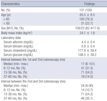
In Fig. 1, there were 131 (100%), 81 (61.8%), and 76 (58.1%) pa-Table 1. Baseline characteristics of the study population and endoscopy
Characteristics Findings No. (%) 131 (100) Age (yr) ≥ 60 < 60 65.5 ± 8.5 100 (76.3) 31 (23.7) Sex (M:F), No. (%) 108/23 (82.4/17.6) Body mass index (kg/m2) 24.1 ± 1.8
Laboratory data Serum albumin (mg/dL) Serum bilirubin (mg/dL) Serum cholesterol (mg/dL) Serum glucose (mg/dL) 4.4 ± 0.4 0.8 ± 0.4 177.9 ± 38.4 106.5 ± 39.2 Interval between the 1st and 2nd colonoscopy (mo)
Median (min.-max.) 6-12 mo, No. (%) 13-36 mo, No. (%) 37-60 mo, No. (%) 17 (6-101) 41 (31.3) 71 (54.2) 19 (14.5) Interval between the 2nd and 3rd colonoscopy (mo)
Median (min.-max.) 6-12 mo, No. (%) 13-36 mo, No. (%) 37-60 mo, No. (%) 24 (6-90) 14 (10.7) 71 (54.2) 46 (35.1) All continuous variables except for median are presented as means ± standard de-viations.
Fig. 1. The numbers of patients with any adenoma and high-risk adenoma at the first, second, and third colonoscopies. *P values < 0.05 compared with the numbers of patients with any adenoma at the first colonoscopy; †P values < 0.05 compared with
the numbers of patients with high-risk adenoma at the first colonoscopy.
Numbers of pa
tients
1st colonoscopy 2nd colonoscopy 3rd colonoscopy 140 120 100 80 60 40 20 0 131 77 81 * 30 * 76 † 26 †
Patients with any adenoma Patients with high-risk adenoma
tients in whom adenomas were detected at the first, second, and third colonoscopies, respectively (P < 0.001). The numbers of patients with high-risk adenomas were 77 (58.8%), 30 (22. 9%), and 26 patients (19.8%) at the first, second, and third colo-noscopies, respectively (P < 0.001) (Fig. 1). The endoscopic out-comes at the first, second, and third colonoscopies are summa-rized in Table 2. The mean numbers of advanced adenomas at the time of the first, second, and third colonoscopies were 1.5 ± 0.9, 1.1 ± 0.3, and 1.0 ± 0.0, respectively (P = 0.016). The num-bers of adenomas that were either high-grade dysplasia or ade-nocarcinoma at the first, second, and third colonoscopies were 10 (12.3%), 0 (0%), and 2 (12.5%), respectively (P < 0.001). There were 17 (21.0%), 5 (22.7%), and 0 (0%) villous or tubulovillous adenomas at the first, second, and third colonoscopies, respec-tively (P < 0.001). The numbers of tubular adenomas ≥ 10 mm at the first, second, and third colonoscopies were 54 (66.7%), 17 (77.3%), and 14 (87.5%), respectively (P < 0.001). There was no significant difference in Ottawa scores, withdrawal times, and cecal intubation rates between the first, second, and third colo-noscopies.
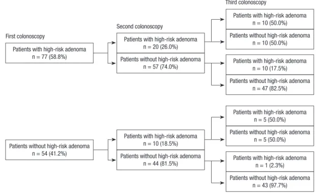
Endoscopic outcomes according to high-risk adenoma In Fig. 2, among the 20 patients with high-risk adenoma at the
first and second colonoscopy, 10 (50%) patients had high-risk adenoma at the third colonoscopy. Among the 67 patients who had high-risk adenoma only once at the first or second colo-noscopy, 15 (22.4%) patients had high-risk adenoma at the third colonoscopy. However, among the 44 patients without any high-risk adenoma at the first and second colonoscopy, only 1 (2.3%) patient had high-risk adenoma at the third colonoscopy (P < 0.001).
Among the 30 patients with high-risk adenoma at the second colonoscopy, 10 out of 20 patients (50%) with high-risk adeno-ma at the first colonoscopy had high-risk adenoadeno-ma at the third colonoscopy, and 5 out of 10 patients (50%) without high-risk adenoma at the first colonoscopy had high-risk adenoma at the third colonoscopy (P > 0.999).
In the absence of high-risk adenoma at the second colonos-copy, there was a higher incidence of high-risk adenoma at the third colonoscopy in presence of high-risk adenoma at the first colonoscopy than absence of high-risk adenoma at the first col-onoscopy (17.5% (10/57) vs 2.3% (1/44), P = 0.021).
Factors predictive of high-risk adenoma at the time of a third colonoscopy
According to the univariate and multivariate time dependent Table 2. Endoscopic outcomes at the time of the first, second, and third colonoscopies
Characteristics First colonoscopy Second colonoscopy Third colonoscopy P value
No. of TAs Total
Mean value per patient*
268 2.1 ± 1.7 151 1.9 ± 1.2 147 1.9 ± 1.7 0.379
Largest diameter of TA* (mm) 9.5 ± 5.6 8.0 ± 3.3 8.9 ± 5.1 < 0.001
No. of patients with TA 68 59 61
Location of TA, No. (%) of patients Right colon Left colon Both 26 (38.2) 26 (38.2) 16 (23.6) 28 (47.5) 19 (32.2) 12 (20.3) 29 (47.5) 19 (31.1) 13 (21.4) 0.411 0.438 0.307 No. of AAs Total
Mean value per patient*
81 1.5 ± 0.9 22 1.1 ± 0.3 16 1.0 ± 0.0 0.016 Largest diameter of AA (mm)* 14.3 ± 6.9 12.3 ± 4.7 14.2 ± 7.7 0.469 Types of AA High grade dysplasia Villous or tubulovillous TA ≥ 10 mm 10 (12.3) 17 (21.0) 54 (66.7) 0 (0) 5 (22.7) 17 (77.3) 2 (12.5) 0 (0) 14 (87.5) < 0.001 < 0.001 < 0.001
No. of patients with AA 63 22 15
Location of AAs, No. (%) of patients Right colon Left colon Both 17 (27.0) 37 (58.7) 9 (14.3) 14 (63.3) 8 (36.4) 0 (0) 11 (73.3) 4 (26.7) 0 (0) 0.508 < 0.001 < 0.001
No. of patients with TA ≥ 3 32 15 15
No. of patients with HRA 77 30 26
Ottawa score Mean value* 0-5, No. (%) 6-10, No. (%) 11-14, No. (%) 7.8 ± 2.4 13 (9.9) 79 (60.3) 39 (29.8) 7.7 ± 2.3 10 (7.6) 85 (64.9) 36 (27.5) 7.3 ± 2.1 13 (9.9) 96 (73.3) 22 (16.8) 0.144
Withdrawal time (min)* 17.5 ± 10.5 17.6 ± 9.3 17.2 ± 10.2 0.950
Cecal intubation rate (%) 100 100 100
covariate Cox regression analysis, a high-risk adenoma at the first or second colonoscopy was only independent predictor of high-risk adenoma at the third colonoscopy (hazard ratio [HR], 9.56; 95% confidential interval [CI], 2.37-38.54; P = 0.002) (Table 3). Upon Cox regression analysis, age, gender, BMI, serum lev-els of albumin, cholesterol, glucose, bilirubin, Ottawa score, and withdrawal time (min) were not significantly related to recur-rence of high-risk adenoma at the third colonoscopy.
DISCUSSION
The findings of the third colonoscopy could be affected by the results of both the first and second colonoscopies. Evaluating the risk factors based on the first and second colonoscopies for high-risk adenoma recurrence at the third colonoscopy would
give better information than if based on the only one prior colo-noscopy. Here we evaluated the factors predictive of high-risk adenomas at the third colonoscopy based on the findings from two prior colonoscopic examination by considering the con-cept of time. In our study the patients with high-risk adenoma at the first and/or second colonoscopy had increased risk of re-curred high-risk adenoma at the third colonoscopy than the patients without high-risk adenoma at the first and second colo-noscopy (HR, 9.56; 95% CI, 2.37-38.54; P = 0.002).
Our results also showed that among the patients who had high-risk adenoma at the second colonoscopy, the presence of high-risk adenoma at the first colonoscopy did not have a sig-nificant effect on the recurrence of high-risk adenoma at the third colonoscopy. However, even if the patients did not have high-risk adenoma at the second colonoscopy, the presence of high-risk adenoma at the first colonoscopy had a significant ef-fect on the recurrence of high-risk adenoma at the third colo-noscopy. Therefore, the patients without high-risk adenoma at the second colonoscopy should be divided according to the finding of first colonoscopy for evaluating recurrence risk of high-risk adenoma at the third colonoscopy. Then patients with high-risk adenoma at the first colonoscopy may need more careful surveillance with shorter intervals than the patients without high-risk adenoma at the first colonoscopy. Likewise, if the patients underwent multiple colonoscopies, all data from previous colonoscopic findings would be checked for planning the surveillance schedule. Even if the results of the most recent colonoscopy were unremarkable, the earlier results of colonos-copy should be considered to distinguish the high-risk patients. Fig. 2. The outcomes of the first, second, and third colonoscopies of patients with and without high- risk adenoma.
Patients with high-risk adenoma n = 77 (58.8%) First colonoscopy
Second colonoscopy
Third colonoscopy
Patients with high-risk adenoma n = 10 (50.0%) Patients without high-risk adenoma
n = 10 (50.0%) Patients with high-risk adenoma
n = 10 (17.5%) Patients without high-risk adenoma
n = 47 (82.5%)
Patients with high-risk adenoma n = 5 (50.0%) Patients without high-risk adenoma
n = 5 (50.0%) Patients with high-risk adenoma
n = 1 (2.3%) Patients without high-risk adenoma
n = 43 (97.7%) Patients with high-risk adenoma
n = 20 (26.0%) Patients without high-risk adenoma
n = 57 (74.0%)
Patients with high-risk adenoma n = 10 (18.5%) Patients without high-risk adenoma
n = 44 (81.5%) Patients without high-risk adenoma
n = 54 (41.2%)
Table 3. Univariate and multivariate time-dependent covariant Cox regression of pre-dictors for the development of high-risk adenomas at the time of the third colonoscopy
Characteristics Univariate Multivariate
HR 95% CI P HR 95% CI P
High-risk adenoma 5.14 2.29-11.55 < 0.001 9.56 2.37-38.54 0.002 Age 1.02 0.97-1.07 0.473 1.02 0.93-1.12 0.703
Sex 0.46 0.11-1.93 0.285 0.00 - 0.996
Body mass index 0.75 0.58-0.98 0.032 0.71 0.50-1.01 0.054 Serum albumin 1.37 0.44-4.25 0.288 Serum cholesterol 0.99 0.98-1.01 0.360 Serum glucose 0.99 0.78-1.01 0.475 Serum bilirubin 0.50 0.12-2.04 0.334 Ottawa score 1.18 0.42-3.31 0.751 Withdrawal time 0.99 0.92-1.08 0.879 HR, hazard ratio; CI, confidential interval.
According to a previous prospective cohort study of patients undergoing multiple surveillance colonoscopies, the result of the first colonoscopy had no significant effect on the recurrence of risk adenoma at the third colonoscopy if there was high-risk adenoma at the second colonoscopy (18.2% of patients with high-risk adenoma at the first colonoscopy vs 20% of patients with low-risk adenoma found at the first colonoscopy (P = 0.780) which was similar to our findings (10). In another study, 15% of patients with normal findings at second colonoscopy and 40% of patients with a neoplasia at the second colonoscopy had a neoplasia at a subsequent examination (11). In our results, 10.9% (11/101) of patients without high-risk adenoma at the second colonoscopy and 50% (15/30) of patients with high-risk adeno-ma at second colonoscopy had high-risk adenoadeno-ma at the third colonoscopy.
To date, various risk factors have been identified in prior stu-dies as predictors for recurrent adenomas or advanced adeno-mas, including size (12), number (13), histology (12), and ad-vanced adenomas (13-19). Nonetheless, other studies suggest-ed that the number of adenomas (14, 15, 20), polyp size (20-22), and histology (14, 20) were not related to advanced adenomas at follow-up colonoscopy. As yet the data regarding the individ-ual predictive factors of high-risk adenomas are inconsistent. Such results imply that no conclusive single characteristics pre-dict the recurrence of high-risk adenoma, especially in the set-ting of multiple surveillance colonoscopies.
There were important strengths of our study. First, in this study we evaluated the presence of high-risk adenoma from two prior colonoscopic examinations as a risk factor of recur-rence of high-risk adenoma at the third colonoscopy. The mean-ing of high-risk adenoma included size, number, and histology of polyps. We evaluated the high-risk adenoma as risk factor to overcome these individual inconclusive characteristics predic-tive of high-risk adenoma recurrence in previous studies (12-14, 20-22). The second strength of our study lies with the multivari-ate time dependent covarimultivari-ate Cox regression analysis of data culled from two prior examinations. The time dependent co-variate Cox regression model is used as a method for analyzing time-to-event data, as it accounts for multiple covariates with values that change according to time and treats time as a func-tioning factor. Our study was unique in using time dependent covariate Cox regression model to analyze risk factor of recur-rence of high-risk adenoma in multiple colonoscopies. Third, despite the nature of this retrospective study, we sought to iden-tify specific predictive factors for high-risk adenomas at subse-quent surveillance colonoscopies by analyzing well-organized electronic medical database from two prior colonoscopies. How-ever, there were limiting points of interpretation for our study. First, the number of enrolled examiners (n = 131) was relatively small, and selection bias was inevitably existed because the pa-tients were enrolled retrospectively from a tertiary medical
cen-ter. Second, the median interval between sequential colonos-copies was shorter than recommended intervals of practice guidelines. The reason why the patients in this study underwent repeat colonoscopies at these shorter intervals may be explain-ed by poor bowel preparation. Mean values of Ottawa score at the first, second and third colonoscopy were 7.8 ± 2.4, 7.7 ± 2.3, and 7.3 ± 2.1. The polyps found at follow up colonoscopy could be a missed lesion or synchronous lesions at previous colonos-copy. Third, even though the short median interval between se-quential colonoscopies, the recurrent rates of adenoma and high risk adenoma were high in this study. This might be caused by previous not enough clean bowel preparation for detection of adenomas.
In conclusion, our results indicated that the patients with high-risk adenoma at the first and/or second colonoscopy had increased risks of high-risk adenoma at the third colonoscopy compared to the patients without high-risk adenoma at first and second colonoscopies. Given these findings, data from two prior colonoscopies, not one prior examination, may help iden-tify high-risk populations at the third colonoscopy who require careful colonoscopic surveillance.
DISCLOSURE
The authors have no conflicts of interest to disclose.
REFERENCES
1. Fukutomi Y, Moriwaki H, Nagase S, Tajika M, Naito T, Miwa Y, Yamada Y, Araki H, Okuno M, Nagura K, et al. Metachronous colon tumors: risk factors and rationale for the surveillance colonoscopy after initial polyp-ectomy. J Cancer Res Clin Oncol 2002; 128: 569-74.
2. Yood MU, Oliveria S, Boyer JG, Wells K, Stang P, Johnson CC. Colon pol-yp recurrence in a managed care population. Arch Intern Med 2003; 163: 422-6.
3. Neugut AI, Jacobson JS, Ahsan H, Santos J, Garbowski GC, Forde KA, Treat MR, Waye J. Incidence and recurrence rates of colorectal adeno-mas: a prospective study. Gastroenterology 1995; 108: 402-8.
4. Atkin WS, Saunders BP; British Society for Gastroenterology; Associa-tion of Coloproctology for Great Britain and Ireland. Surveillance guide-lines after removal of colorectal adenomatous polyps. Gut 2002; 51: V6-9. 5. Davila RE, Rajan E, Baron TH, Adler DG, Egan JV, Faigel DO, Gan SI,
Hirota WK, Leighton JA, Lichtenstein D, et al. ASGE guideline: colorec-tal cancer screening and surveillance. Gastrointest Endosc 2006; 63: 546-57.
6. Winawer SJ, Zauber AG, Fletcher RH, Stillman JS, O’brien MJ, Levin B, Smith RA, Lieberman DA, Burt RW, Levin TR, et al. Guidelines for colo-noscopy surveillance after polypectomy: a consensus update by the US Multi-Society Task Force on Colorectal Cancer and the American Cancer Society. CA Cancer J Clin 2006; 56: 143-59.
7. Hamilton SR, Aaltonen LA. World Health Organization classification of tumours: pathology and genetics of tumours of the digestive system. Lyon:
IARC Press, 2000.
8. Saito Y, Uraoka T, Yamaguchi Y, Hotta K, Sakamoto N, Ikematsu H, Fu-kuzawa M, Kobayashi N, Nasu J, Michida T, et al. A prospective, multi-center study of 1111 colorectal endoscopic submucosal dissections (with video). Gastrointest Endosc 2010; 72: 1217-25.
9. Tanaka S, Oka S, Chayama K. Colorectal endoscopic submucosal dissec-tion: present status and future perspective, including its differentiation from endoscopic mucosal resection. J Gastroenterol 2008; 43: 641-51. 10. Robertson DJ, Burke CA, Welch HG, Haile RW, Sandler RS, Greenberg
ER, Ahnen DJ, Bresalier RS, Rothstein RI, Cole B, et al. Using the results of a baseline and a surveillance colonoscopy to predict recurrent adeno-mas with high-risk characteristics. Ann Intern Med 2009; 151: 103-9. 11. Blumberg D, Opelka FG, Hicks TC, Timmcke AE, Beck DE. Significance
of a normal surveillance colonoscopy in patients with a history of ade-nomatous polyps. Dis Colon Rectum 2000; 43: 1084-91.
12. Bertario L, Russo A, Sala P, Pizzetti P, Ballardini G, Andreola S, Spinelli P. Predictors of metachronous colorectal neoplasms in sporadic adenoma patients. Int J Cancer 2003; 105: 82-7.
13. Noshirwani KC, van Stolk RU, Rybicki LA, Beck GJ. Adenoma size and number are predictive of adenoma recurrence: implications for surveil-lance colonoscopy. Gastrointest Endosc 2000; 51: 433-7.
14. Martínez ME, Sampliner R, Marshall JR, Bhattacharyya AK, Reid ME, Alberts DS. Adenoma characteristics as risk factors for recurrence of ad-vanced adenomas. Gastroenterology 2001; 120: 1077-83.
15. Lorenzo-Zúñiga V, Moreno de Vega V, Domènech E, Mañosa M, Cabré E, Planas R, Boix J. High-definition colonoscopy and risk factors for re-currence of advanced adenomas in patients with a personal history of polyps. Eur J Gastroenterol Hepatol 2011; 23: 425-30.
16. Lieberman DA, Weiss DG, Harford WV, Ahnen DJ, Provenzale D, Son-tag SJ, Schnell TG, Chejfec G, Campbell DR, Kidao J, et al. Five-year co-lon surveillance after screening coco-lonoscopy. Gastroenterology 2007; 133: 1077-85.
17. Nusko G, Hahn EG, Mansmann U. Characteristics of metachronous col-orectal adenomas found during long-term follow-up: analysis of four subsequent generations of adenoma recurrence. Scand J Gastroenterol 2009; 44: 736-44.
18. Jørgensen OD, Kronborg O, Fenger C, Rasmussen M. Influence of long-term colonoscopic surveillance on incidence of colorectal cancer and death from the disease in patients with precursors (adenomas). Acta Oncol 2007; 46: 355-60.
19. Moon CM, Cheon JH, Choi EH, Kim ES, Park JJ, Han SY, Kim DH, Kim TI, Kim WH. Advanced synchronous adenoma but not simple adenoma predicts the future development of metachronous neoplasia in patients with resected colorectal cancer. J Clin Gastroenterol 2010; 44: 495-501. 20. Külling D, Christ AD, Karaaslan N, Fried M, Bauerfeind P. The presence
of more than two index adenomas is the strongest predictor of metachro-nous colon adenomas. Swiss Med Wkly 2002; 132: 139-42.
21. Winawer SJ, Zauber AG, O’Brien MJ, Ho MN, Gottlieb L, Sternberg SS, Waye JD, Bond J, Schapiro M, Stewart ET, et al. Randomized compari-son of surveillance intervals after colonoscopic removal of newly diag-nosed adenomatous polyps: the National Polyp Study Workgroup. N Engl J Med 1993; 328: 901-6.
22. Triantafyllou K, Papatheodoridis GV, Paspatis GA, Vasilakaki TH, Ele-menoglou I, Karamanolis DG. Predictors of the early development of advanced metachronous colon adenomas. Hepatogastroenterology 1997; 44: 533-8.