year) ISR.8,9 Differences in tissue morphology and timing of ISR may be associated with different mechanisms of ISR after DES implantation. However, there are few data about the associations between clinical outcomes after DCB and tissue characteristics or timing of ISR. The aim of this study was to investigate clinical outcomes according to tissue characteristics and timing of DES ISR after DCB angioplasty.
Methods
Study Design and Patient Selection
This study was approved by the Institutional Review
A
lthough the development of drug-eluting stents(DES) has significantly reduced the incidence of in-stent restenosis (ISR) compared with bare metal stents (BMS),1 DES restenosis still occurs in 3–20% of patients.2 For the treatment of ISR, drug-coated balloon (DCB) angioplasty has been shown to have a lower rate of further adverse clinical events compared with uncoated balloons.3,4
Intravascular optical coherence tomography (OCT) evaluation is a useful tool not only for quantitative mea-surement, but also for qualitative assessment of character-istics.5–7 Recent OCT studies suggest that neointimal tissue characteristics differ between early (<1 year) and late (≥1
Received May 24, 2018; revised manuscript received July 21, 2018; accepted July 31, 2018; released online September 7, 2018 Time for primary review: 32 days
Division of Cardiology, Yeungnam University Medical Center, Yeungnam University College of Medicine, Daegu (J.-H.L.); Department of Cardiology, Daegu Catholic University Medical Center, Daegu (H.W.J.); Severance Cardiovascular Hospital (J.-S.K., S.-J.H., C.-M.A., B.-K.K., Y.-G.K., D.C., M.-K.H., Y.J.), Cardiovascular Institute (J.-S.K., S.-J.H., C.-M.A., B.-K.K., Y.-G.K., D.C., M.-K.H., Y.J.), Severance Biomedical Science Institute (M.-K.H., Y.J.), Yonsei University College of Medicine, Seoul, Korea
The first two authors contributed equally to this work (J.-H.L., H.W.J.).
Mailing address: Jung-Sun Kim, MD, PhD, Division of Cardiology, Severance Cardiovascular Hospital, Yonsei University College of Medicine, 250 Seongsanno, Seodaemun-gu, Seoul 120-752, Republic of Korea. E-mail: [email protected]
ISSN-1346-9843 All rights are reserved to the Japanese Circulation Society. For permissions, please e-mail: [email protected]
Different Neointimal Pattern in Early vs. Late In-Stent
Restenosis and Clinical Outcomes After
Drug-Coated Balloon Angioplasty
― An Optical Coherence Tomography Study ―
Jung-Hee Lee, MD; Hae Won Jung, MD; Jung-Sun Kim, MD; Sung-Jin Hong, MD; Chul-Min Ahn, MD; Byeong-Keuk Kim, MD; Young-Guk Ko, MD;
Donghoon Choi, MD; Myeong-Ki Hong, MD; Yangsoo Jang, MD
Background: There are few data of clinical outcomes after drug-coated balloon (DCB) angioplasty according to neointimal
characteristics. This study investigated long-term clinical outcomes according to timing of in-stent restenosis (ISR) and neointimal characteristics in patients with drug-eluting stent (DES) ISR after DCB angioplasty.
Methods and Results: In all, 122 patients (122 ISR lesions), treated with DCB under optical coherence tomography (OCT)
examination before and after DCB, were categorized as early ISR (<12 months; E-ISR; n=21) and late ISR (≥12 months; L-ISR; n=101). Associations between OCT-based neointima characteristics and period of ISR, as well as clinical outcomes after DCB were evaluated. Major adverse cardiac events (MACE) were a composite of cardiac death, non-fatal myocardial infarction, or target lesion revascularization (TLR). Quantitative parameters of the neointima were similar, but qualitative characteristics showed significant differences between the E-ISR and L-ISR groups. The incidence of MACE (33.3% vs. 20.8%; P=0.069) and TLR (33.3% vs. 18.5%; P=0.040) was higher in the E-ISR group. In addition, the incidence of MACE was significantly higher for heterogeneous than non-heterogeneous neointima (43.7% vs. 19.6%; P=0.018), but was not significantly associated with neoatherosclerosis (33.4% vs. 18.4%; P=0.168).
Conclusions: DCB angioplasty is less effective for heterogeneous neointima in DES ISR. OCT-based neointimal evaluation may
be helpful in guiding treatment of DES ISR.
Key Words: Drug-eluting balloon; In-stent restenosis; Optical coherence tomography
cutting balloon (n=15 patients), additional stent implanta-tion was required (n=30 patients), poor image quality or hard to acquired pre-interventional OCT images due to tight restenotic lesions (n=34 patients), and loss to follow-up (n=3 patients). Thus, 122 patients with 122 ISR lesions treated with DCB angioplasty were included in this study. The study cohort was divided into early ISR (<12 months; E-ISR; n=21; first-generation DES n=1, second-generation DES n=20) and late ISR (≥12 months; L-ISR; n=101; Board of Severance Hospital, the Yonsei University, and
written informed consent was obtained from all patients enrolled in the study. Clinical data were obtained for analysis from the Yonsei OCT Registry database (ClinicalTrials.gov ID: NCT01308281) between July 2010 and December 2013 for 204 patients with 208 stented ISR lesions who had not been treated previously. Eighty-two patients with 86 lesions were excluded from the present study for the following reasons: ISR lesions treated with a
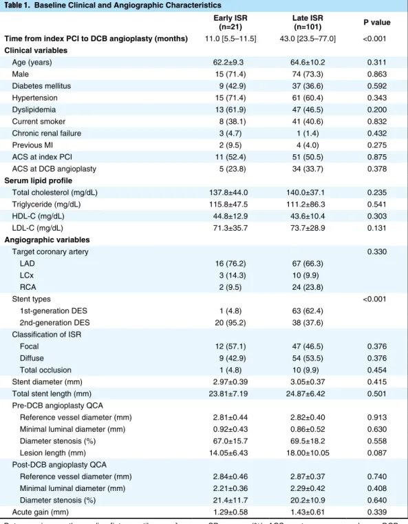
Table 1. Baseline Clinical and Angiographic Characteristics Early ISR
(n=21) Late ISR (n=101) P value Time from index PCI to DCB angioplasty (months) 11.0 [5.5–11.5] 43.0 [23.5–77.0] <0.001
Clinical variables Age (years) 62.2±9.3 64.6±10.2 0.311 Male 15 (71.4) 74 (73.3) 0.863 Diabetes mellitus 9 (42.9) 37 (36.6) 0.592 Hypertension 15 (71.4) 61 (60.4) 0.343 Dyslipidemia 13 (61.9) 47 (46.5) 0.200 Current smoker 8 (38.1) 41 (40.6) 0.832
Chronic renal failure 3 (4.7) 1 (1.4) 0.432
Previous MI 2 (9.5) 4 (4.0) 0.275
ACS at index PCI 11 (52.4) 51 (50.5) 0.875
ACS at DCB angioplasty 5 (23.8) 34 (33.7) 0.378
Serum lipid profile
Total cholesterol (mg/dL) 137.8±44.0 140.0±37.1 0.235
Triglyceride (mg/dL) 115.8±47.5 111.2±86.3 0.541
HDL-C (mg/dL) 44.8±12.9 43.6±10.4 0.303
LDL-C (mg/dL) 71.3±35.7 73.7±28.9 0.131
Angiographic variables
Target coronary artery 0.330
LAD 16 (76.2) 67 (66.3) LCx 3 (14.3) 10 (9.9) RCA 2 (9.5) 24 (23.8) Stent types <0.001 1st-generation DES 1 (4.8) 63 (62.4) 2nd-generation DES 20 (95.2) 38 (37.6) Classification of ISR Focal 12 (57.1) 47 (46.5) 0.376 Diffuse 9 (42.9) 54 (53.5) 0.376 Total occlusion 1 (4.8) 10 (9.9) 0.454 Stent diameter (mm) 2.97±0.39 3.05±0.37 0.415
Total stent length (mm) 23.81±7.19 24.87±6.42 0.501
Pre-DCB angioplasty QCA
Reference vessel diameter (mm) 2.81±0.44 2.82±0.40 0.913
Minimal luminal diameter (mm) 0.92±0.43 0.86±0.52 0.630
Diameter stenosis (%) 67.0±15.7 69.5±18.2 0.558
Lesion length (mm) 14.05±6.43 18.00±10.05 0.087
Post-DCB angioplasty QCA
Reference vessel diameter (mm) 2.84±0.46 2.87±0.37 0.740
Minimal luminal diameter (mm) 2.21±0.36 2.29±0.42 0.408
Diameter stenosis (%) 21.4±11.7 20.2±10.9 0.640
Acute gain (mm) 1.29±0.58 1.43±0.61 0.339
Data are given as the median [interquartile range], mean ± SD, or as n (%). ACS, acute coronary syndrome; DCB, drug-coated balloon; DES, drug-eluting stent; HDL-C, high-density lipoprotein cholesterol; ISR, in-stent restenosis; LAD, left anterior descending artery; LCx, left circumflex artery; LDL-C, low-density lipoprotein cholesterol; MI, myocardial infarction; PCI, percutaneous coronary intervention; QCA, quantitative coronary analysis; RCA, right coronary artery.
Clinical Follow-up
The primary endpoint was major adverse cardiac events (MACE), defined as a composite of cardiovascular death, non-fatal myocardial infarction (MI), and target-lesion revascularization (TLR), based on the Academic Research Consortium.10 Without an explainable non-cardiac cause, all deaths were considered cardiac death. MI was defined based on the third universal definition of MI.11 TLR was defined as any repeat percutaneous intervention of the target lesion or bypass surgery of the target vessel performed for restenosis or other complications of the target lesion. All revascularizations were considered clinically indicated if angiography at follow-up showed a percentage diameter stenosis (%DS) ≥50% as assessed by quantitative coronary angiography (QCA) analysis with either ischemic symptoms or a positive stress test, or a %DS ≥70% as assessed by QCA analysis without either ischemic symptoms or a positive stress test.
Image Analysis of Coronary Angiography and OCT
QCA were performed using an offline computerized QCA system (CAAS System; Pie Medical Imaging, Maastricht, The Netherlands) in an independent core laboratory (Cardiovascular Research Center, Seoul, Korea). All OCT images were analyzed using certified offline software (QIvus; Medis Medical Imaging System, Leiden, The Netherlands) at a core laboratory (Cardiovascular Research Center) by analysts who were blinded to both clinical and angiographic first-generation DES n=63, second-generation DES n=38)
depending on the time of ISR after DES implantation.
Angioplasty Procedure
The decision to perform DCB angioplasty was based on angiographic findings, including a >50% diameter stenosis on a follow-up angiogram with evidence of myocardial ischemia, such as ischemic symptoms or a positive stress test. All patients in the study received at least 100 mg aspirin and a total of 300 mg clopidogrel as a loading dose at least 12 h before DCB angioplasty. After diagnosis of ISR on a conventional angiogram, the OCT procedure was performed using a C7-XR imaging system (LightLab Imaging; St. Jude Medical, St. Paul, MN, USA) before plain balloon angioplasty. The DCB used in the present study was a paclitaxel-coated balloon (Sequent Please; B.Braun, Melsungen, Germany). The DCB angioplasty was performed using current conventional techniques: after predilation with a plain balloon, the DCB was inflated for 60s. An adequate size of the DCB was deter-mined by the physician according to the length of the target ISR lesion and the diameter of the previously implanted stents. After successful intervention, all patients underwent a final OCT procedure. Dual anti-platelet therapy (100 mg aspirin and 75 mg clopidogrel) was prescribed to all patients for at least 1 month after DCB angioplasty.
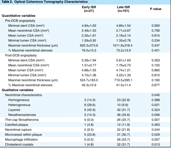
Table 2. Optical Coherence Tomography Characteristics Early ISR
(n=21) Late ISR (n=101) P value Quantitative variables
Pre-DCB angioplasty
Minimal stent CSA (mm2) 4.84±1.63 4.89±1.54 0.900
Mean neointimal CSA (mm2) 2.49±1.55 2.71±3.67 0.790
Mean lumen CSA (mm2) 2.35±1.61 2.18±2.14 0.815
Minimal lumen CSA (mm2) 1.59±0.92 1.33±0.76 0.234
Maximal neointimal thickness (μm) 920.3±373.6 1011.8±318.4 0.247
% Maximal neointimal stenosis 76.0±13.3 73.2±13.9 0.401
Post-DCB angioplasty
Minimal stent CSA (mm2) 5.39±1.94 5.81±1.63 0.303
Mean neointimal CSA (mm2) 1.51±0.77 1.76±0.73 0.155
Mean lumen CSA (mm2) 4.68±1.05 4.74±1.21 0.865
Minimal lumen CSA (mm2) 4.15±1.36 4.22±1.33 0.812
Maximal neointimal thickness (μm) 524.7±163.0 710.5±595.1 0.160
% Maximal neointimal stenosis 40.3±12.9 41.5±11.4 0.677
Qualitative variables Neointimal characteristics 0.040 Homogeneous 3 (14.3) 23 (22.8) 0.388 Heterogeneous 6 (28.6) 10 (9.9) 0.021 Layered 9 (42.9) 32 (31.7) 0.324 Neoatherosclerosis 3 (14.3) 36 (35.6) 0.056 Thin-cap fibroatheroma 0 (0.0) 26 (25.7) 0.007 Calcified plaque 1 (4.8) 12 (11.9) 0.463 Neointimal rupture 2 (9.5) 22 (21.8) 0.244
Microvessel within plaque 5 (23.8) 31 (30.7) 0.529
Macrophage infiltration 0 (0.0) 26 (25.7) 0.007
Cholesterol crystals 1 (4.8) 32 (31.7) 0.013
Statistical Analysis
Data are expressed as number (%), mean ± SD, or median (interquartile range [IQR]). Continuous variables were compared using Student’s t-test, and comparisons of categorical data were performed using Chi-squared statistics or Fisher’s exact test. Event-free survival was analyzed using Kaplan-Meier survival curves, and differences between event-free survival curves were compared with the log-rank test. Univariate analyses were performed using Cox pro-portional hazards regression for traditional cardiac risk factors and the qualitative OCT characteristics. Variables achieving P<0.10 in the univariate analysis were entered into the multivariate analysis model to determine the independent predictors for MACE. P<0.05 was considered statistically significant. Statistical analyses were performed using SPSS version 20.0.0 (IBM, Armonk, NY, USA).
Results
Baseline Characteristics
Among the total study population, 21 patients (17.2%) were categorized as E-ISR and 101 patients (82.8%) were categorized as L-ISR. There were significant differences in the median time interval from index percutaneous coro-nary intervention to DCB angioplasty between the E-ISR and L-ISR groups (11.0 vs. 43.0 months, respectively; P<0.001). The patients’ baseline clinical and angiographic information. Cross-sectional OCT images before
angio-plasty were measured at 1-mm intervals for quantitative measurements. Stent and luminal cross-sectional area (CSA) was analyzed, and neointimal CSA was calculated as the stent CSA minus luminal CSA. The segment with minimal lumen area and greatest neointimal proliferation could be the representative site of lesions for future clinical follow-up.12 Therefore, the stented segments at the minimal lumen CSA and greatest neointimal CSA were assessed qualitatively to characterize the neointimal tissue as either homogeneous (a uniform signal-rich band without focal variation or attenuation), heterogeneous (focally changing optical properties and various backscattering patterns), layered neointima (layers with different optical properties, namely an adluminal high-scattering layer and an abluminal low-scattering layer),6,8,12 and neoatherosclerosis (lipid-laden plaque, thin-cap fibroatheroma [defined as a plaque with a lipid content in ≥2 quadrants and the thinnest part of a fibrous cap ≤65 μm], neointimal rupture, and/or calcification within the neointima).13 Neointimal morphologic charac-teristics were evaluated qualitatively by 2 observers who were blinded to the patient’s data and angiographic results. Inter- and intraobserver agreements for the assessment of neointimal tissue characteristics in our core laboratory have been reported previously.14
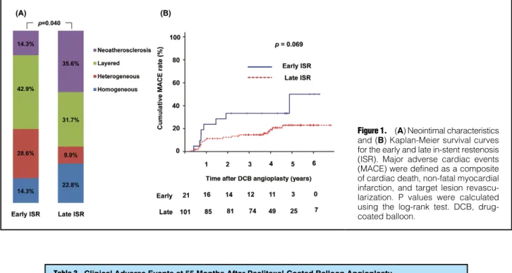
Figure 1. (A) Neointimal characteristics and (B) Kaplan-Meier survival curves for the early and late in-stent restenosis (ISR). Major adverse cardiac events (MACE) were defined as a composite of cardiac death, non-fatal myocardial infarction, and target lesion revascu-larization. P values were calculated using the log-rank test. DCB, drug-coated balloon.
Table 3. Clinical Adverse Events at 55 Months After Paclitaxel-Coated Balloon Angioplasty Early ISR
(n=21) Late ISR (n=101) P value
MACE 7 (33.3) 18 (20.8) 0.069
Cardiac death 0 (0.0) 0 (0.0) 1.0
Non-fatal MI 0 (0.0) 4 (4.6) 0.786
TLR 7 (33.3) 16 (18.5) 0.040
All-cause mortality 0 (0.0) 1 (1.1) 0.474 Data are given as n (%). ISR, in-stent restenosis; MACE, major adverse cardiac events, defined as a composite of cardiac death, non-fatal myocardial infarction (MI), or target-lesion revascularization (TLR).
between the 2 groups. However, lesion length tended to be shorter in the E-ISR than L-ISR group (14.05±6.43 vs. 18.00±10.05 mm; P=0.087). Post-DCB angioplasty QCA and acute gain after procedure did not differ significantly between the 2 groups.
characteristics are summarized in Table 1. There were no differences in comorbidities or medications between the 2 groups. Second-generation DESs were more frequently used in E-ISR than L-ISR group (95.2% vs. 37.6%; P<0.001). There were no significant differences in QCA parameters
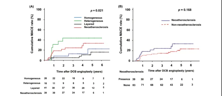
Figure 2. Kaplan-Meier survival curves according to (A) neointimal characteristics and (B) neoatherosclerosis. Major adverse cardiac events (MACE) were defined as a composite of cardiac death, non-fatal myocardial infarction, and target lesion revascularization. P values were calculated using the log-rank test. DCB, drug-coated balloon.
Figure 3. Representative (A,C) optical coherence tomography (OCT) and (B,D) angiography images for homogeneous (A,B) and heterogeneous (C,D) patterns of in-stent restenosis (ISR) before and after drug-coated balloon (DCB) angiography. (B) Images obtained at follow-up of the homogeneous pattern of ISR show a patent stent (arrows). (D) However, in the case of a heterogeneous pattern of ISR, follow-up images show a critical narrowing at the DCB-treated site (arrows).
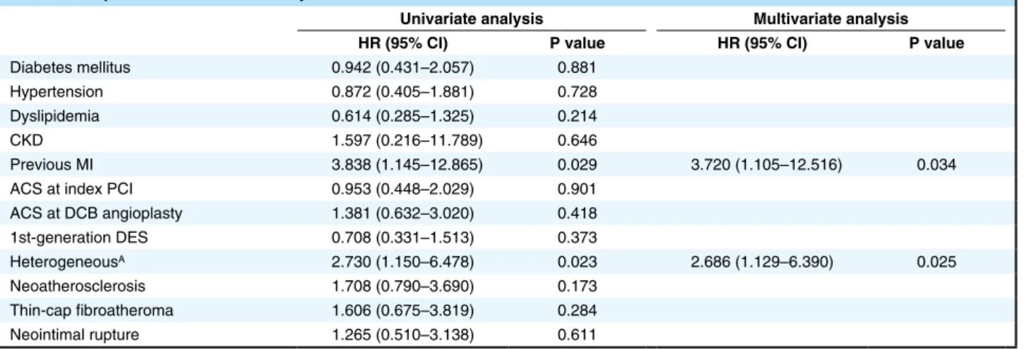
regression analysis revealed that the following baseline and qualitative OCT variables were associated with an increased risk of MACE: heterogeneous pattern (hazard ratio [HR] 2.686; 95% confidence interval [CI] 1.129–6.390; P=0.025) and previous MI (HR 3.720; 95% CI 1.105– 12.516; P=0.034; Table 4).
Discussion
The major findings of this study were as follows: (1) neointimal characteristics, as assessed by OCT, differed between the E-ISR and L-ISR groups, with patients in the E-ISR showing a high rate of long-term adverse clinical outcomes after DCB angioplasty; (2) neointimal charac-teristics were associated with clinical outcomes after DCB treatment in DES ISR, especially repeated revasculariza-tion; and (3) the presence of a heterogeneous neointima was an independent predictor of MACE.
Mechanism of ISR
One of the most important predictors for ISR is stent underexpansion. A recent study by Song et al9 showed that early ISR within 1 year was related to minimum stent CSA <4.0 mm2. However, another study by Kang et al15 sug-gested that intimal hyperplasia could be a major cause of ISR for stents ≤28 mm long. Although stent underexpansion within a longer stented segment could be associated with future adverse clinical outcomes, this is a preventable mechanism underlying ISR development with optimal stent implantation. In the present study, quantitative OCT data showed that minimal stent CSA was similar between the E-ISR and L-ISR group (4.84±1.63 vs. 4.89±1.54 mm2,
respectively; P=0.900). These results suggest that stent underexpansion was not main mechanism for the develop-ment of ISR in our study population. Another important mechanism of stent failure after DES implantation is neointimal growth. In-stent neoatherosclerosis, defined as an accumulation of lipid-laden foamy macrophages with or without necrotic core formation and/or calcification within the neointima, is also well known as an important mechanism underlying late DES failure.16 A retrospective OCT analysis demonstrated stent age ≥48 months was the most powerful predictor for neoatherosclerosis.17 Two
OCT Findings
OCT findings of the restenotic stented segments are summarized in Table 2. The quantitative variables before and after DCB did not differ significantly between the 2 groups. However, in terms of qualitative characteristics, the site of the minimum lumen area exhibited significant differences. A heterogeneous neointima was more frequently observed in the E-ISR than L-ISR group (28.6% vs. 9.9%; P=0.021), whereas neoatherosclerosis tended to be more frequent in the L-ISR than E-ISR group (35.6% vs. 14.3%; P=0.056). Thin-cap fibroatheroma (0.0% vs. 25.7%; P=0.007), macrophage infiltration (0.0% vs. 25.7%; P=0.007), and cholesterol crystals (4.8% vs. 31.7%; P=0.013) were also more frequent in the L-ISR than E-ISR. Figure 1A shows that the incidence of heterogeneous neointima decreased, but the incidence of neoatherosclerosis was higher in L-ISR groups compared to E-ISR group.
Clinical Outcomes
Over the median follow-up duration of 55.3 months (IQR 43.1–66.0 months), 25 MACE occurred. The long-term clinical outcomes of the E-ISR vs. L-ISR groups after DCB angioplasty are summarized in Table 3. MACE tended to be more frequent in the E-ISR than L-ISR group (33.3% vs. 20.8%; P=0.069; Figure 1B). The difference in MACE between the 2 groups was driven by the rate of TLR (33.3% vs. 18.5% in the E-ISR and L-ISR groups, respectively; P=0.040). Clinical adverse events according to neointimal characteristics of the minimum lumen area are summarized in Table S1. There were also significant differences in the rate of MACE according to neointima characteristics (Figure 2A). The heterogeneous neointima group had a significantly higher rate of MACE than the non-heterogeneous group (43.7% vs. 19.6%; P=0.018), whereas the incidence of MACE was not significantly associated with the presence of neoatherosclerosis (33.4% vs. 18.4% for patients with and without neoatherosclerosis, respectively; P=0.168; Figure 2B). Figure 3 shows represen-tative OCT and angiographic images after DCB angioplasty in groups with different clinical outcomes. However, the rate of MACE was not associated with the generation of the DES used or the type of first-generation DES (Figure S1). Multivariate Cox proportional hazards
Table 4. Independent Predictors for Major Adverse Cardiac Events
Univariate analysis Multivariate analysis HR (95% CI) P value HR (95% CI) P value
Diabetes mellitus 0.942 (0.431–2.057) 0.881
Hypertension 0.872 (0.405–1.881) 0.728
Dyslipidemia 0.614 (0.285–1.325) 0.214
CKD 1.597 (0.216–11.789) 0.646
Previous MI 3.838 (1.145–12.865) 0.029 3.720 (1.105–12.516) 0.034
ACS at index PCI 0.953 (0.448–2.029) 0.901
ACS at DCB angioplasty 1.381 (0.632–3.020) 0.418 1st-generation DES 0.708 (0.331–1.513) 0.373 HeterogeneousA 2.730 (1.150–6.478) 0.023 2.686 (1.129–6.390) 0.025 Neoatherosclerosis 1.708 (0.790–3.690) 0.173 Thin-cap fibroatheroma 1.606 (0.675–3.819) 0.284 Neointimal rupture 1.265 (0.510–3.138) 0.611
ACompared with non-heterogeneous. ACS, acute coronary syndrome; CI, confidence interval; CKD, chronic kidney disease; DCB, drug-coated
associated with the formation of an immature neointima due to an abnormal inflammatory reaction with the stent strut or underlying unstable clinical characteristics. Further-more, a heterogeneous neointima is one of the independent predictors for MACE (HR 2.686; 95% CI 1.129–6.390; P=0.025).
A previous OCT study of clinical outcomes for ISR reported similar results to those seen in the present study, namely that when comparing DCB with conventional balloon angioplasty and DES, the incidence of ISR was numerically higher for DCB (38.5% vs. 20.0% and 18.8%, respectively), as was the incidence of TLR (34.6% vs. 20.0% and 18.8%, respectively).18 It is possible that a heterogeneous neointima is one of the indicators of further adverse events after DCB for the treatment of DES ISR due to its intrinsic instability. Because we did not conduct follow-up OCT examinations, further studies are required to evaluate this hypothesis.
Several studies have reported adverse local effects at the vessel wall after DCB for patients presenting with ISR.19,20 Furthermore, the long-term effective treatment modality for DES ISR has not been fully investigated. A previous OCT study about qualitative assessment of ISR reported that vascular response was poor in patients with a homo-geneous neointima compared with those with a layered or heterogeneous pattern (29.5±11.9% vs. 42.2±15.7% and 46.3±12.9%, respectively; P<0.01).21 Another OCT study of neointimal patterns with serial follow-up OCT suggested intensive reduction in LDL-C can prevent neointimal pattern changes from a homogeneous to non-homogeneous pattern.22 These studies suggest that OCT-based neointimal evaluation could be important in making treatment deci-sions for patients with ISR. A prospective randomized trial demonstrated that for the treatment of patients presenting with DES ISR, everolimus-eluting stents (EES) provided superior angiographic results as well as superior clinical outcome (composite of cardiac death, MI, TLR) (10% vs. 18%; P=0.04) compared with DCB.23 To our knowledge, the present study is the first to compare long-term clinical outcomes of DCB according to the neointima pattern and duration from DES implantation to ISR. Based on the findings of this study, we suggest that OCT-based neointimal evaluation and different treatment strategies, such as intensive medical treatment or implantation of another DES, should be used for the treatment of ISR after DES implantation.
Study Limitations
The present study has several limitations. First, this study was based on single-center OCT registry data with a relatively small population, and the study has intrinsic limitations related to its retrospective design. The imbalance of stent types between groups, driven by temporal differ-ences in stent usage, is a limitation related to the retrospec-tive study design. However, the first-generation DES was not associated with a higher incidence of MACE after DCB angioplasty compared with the second-generation DES (22.5% vs. 24.0%; P=0.400). Furthermore, Cox proportional hazards regression analysis showed that first-generation DES was not a significant predictor for future MACE (HR 0.708; 95% CI 0.331–1.513; P=0.373). Second, we excluded some patients who had poor image quality or hard to acquire pre-interventional OCT images due to tight restenotic lesions, so this could have led to selection bias. However, these cases accounted for a recent OCT studies of neointimal characteristics after
second-generation DES implantation also showed that there were significant differences in neointimal morphologic characteristics between early and late ISR, and that neoatherosclerosis was commonly associated with late ISR.8,9 In the present study, there was a tendency for more frequent observation of neoatherosclerosis in the L-ISR than E-ISR group (35.6% vs. 14.3%, respectively; P=0.056), whereas a heterogeneous neointima was more frequently observed in the E-ISR than L-ISR group (28.6% vs. 9.9%, respectively; P=0.021). Our findings are consistent with those of previous studies regarding the possibility of different mechanisms in the development of early vs. late ISR.8,9 Although the serum lipid profile may affect neointimal plaque characteristics and stent age, cholesterol levels did not different significantly in the present study. Our data showed that total cholesterol (TC) and low-density lipo-protein cholesterol (LDL-C) concentrations tended to be higher in the neoatherosclerosis group than in the hetero-geneous group (TC 149.0±41.0 vs. 128.1±35.8 mg/dL, respectively [P=0.067]; LDL-C 76.9±32.2 vs. 64.1±27.1 mg/dL, respectively [P=0.181]). However, the relationship between lipid profile and neointimal characteristics could not be fully defined in this study.
Clinical Outcomes After DCB
In the present study, the incidence of TLR was significantly higher in the E-ISR than L-ISR group over the long-term follow-up (33.3% vs. 18.5%; P=0.040). Although the inci-dence of non-fatal MI was numerically lower in the E-ISR than L-ISR group (0.0% vs. 4.6%; P=0.786), half of these cases were non-target vessel MI. This finding may be attributed to the native lesion progression, as well as the development of neoatherosclerosis in the late period after stent implantation.
DCB have been reported to be highly effective for the treatment ISR, superior to conventional balloon angio-plasty.3,4 In the present study, quantitative OCT data also showed sufficient mechanical effect comparing pre- and post-DCB angioplasty. However, long-term effects after DCB differed between E-ISR and L-ISR.
Although baseline characteristics of the study population were similar except for stent type, there were significant difference in the neointimal characteristics between the 2 groups. Interestingly, a heterogeneous neointima was more frequently observed in the E-ISR than L-ISR group (28.6% vs. 9.9%; P=0.021), which is a similar finding to that reported previously in a study evaluating ISR neointimal characteristics of early vs. late ISR.9 Furthermore, in the E-ISR group, those with a heterogeneous neointima (n=6) showed a higher prevalence of TLR after DCB angioplasty (83.3%).
Evaluating clinical adverse events according to neointimal characteristics also revealed that a heterogeneous neointima was significantly associated with MACE compared with a non-heterogeneous neointima (43.7% vs. 19.6%; P=0.018). A previous pathologic study revealed that neointimal abnormalities, such as fibrin deposits or peristrut inflam-mation, were frequently observed in heterogeneous neointima, as assessed by OCT.7 A clinical study about the relationship between neointimal characteristics assessed by OCT and clinical outcomes suggested that a heterogeneous neointima was significantly associated with both old age and initial acute coronary syndrome presentation.12 These findings suggest that a heterogeneous neointima could be
standardized definitions. Circulation 2007; 115: 2344 – 2351. 11. Thygesen K, Alpert JS, Jaffe AS, Simoons ML, Chaitman BR,
White HD. Third universal definition of myocardial infarction. J Am Coll Cardiol 2012; 60: 1581 – 1598.
12. Kim JS, Lee JH, Shin DH, Kim BK, Ko YG, Choi D, et al. Long-term outcomes of neointimal hyperplasia without neo-atherosclerosis after drug-eluting stent implantation. JACC Cardiovasc Imaging 2014; 7: 788 – 795.
13. Kang SJ, Mintz GS, Akasaka T, Park DW, Lee JY, Kim WJ, et al. Optical coherence tomographic analysis of in-stent neoathero-sclerosis after drug-eluting stent implantation. Circulation 2011; 123: 2954 – 2963.
14. Lee SJ, Kim BK, Kim JS, Ko YG, Choi D, Jang Y, et al. Evaluation of neointimal morphology of lesions with or without in-stent restenosis: An optical coherence tomography study. Clin Cardiol 2011; 34: 633 – 639.
15. Kang SJ, Mintz GS, Park DW, Lee SW, Kim YH, Lee CW, et al. Mechanisms of in-stent restenosis after drug-eluting stent implantation: Intravascular ultrasound analysis. Circ Cardiovasc Interv 2011; 4: 9 – 14.
16. Nakazawa G, Otsuka F, Nakano M, Vorpahl M, Yazdani SK, Ladich E, et al. The pathology of neoatherosclerosis in human coronary implants bare-metal and drug-eluting stents. J Am Coll Cardiol 2011; 57: 1314 – 1322.
17. Yonetsu T, Kato K, Kim SJ, Xing L, Jia H, McNulty I, et al. Predictors for neoatherosclerosis: A retrospective observational study from the optical coherence tomography registry. Circ Cardiovasc Imaging 2012; 5: 660 – 666.
18. Tada T, Kadota K, Hosogi S, Miyake K, Ohya M, Amano H, et al. Association between tissue characteristics assessed with optical coherence tomography and mid-term results after percutaneous coronary intervention for in-stent restenosis lesions: A comparison between balloon angioplasty, paclitaxel-coated balloon dilatation, and drug-eluting stent implantation. Eur Heart J Cardiovasc Imaging 2015; 16: 1101 – 1111.
19. Kovarnik T, Mintz GS, Sonka M. The late stent malapposition develops also after paclitaxel balloon predilatation before bare-metal stent implantation: Case description. Eur Heart J 2011; 32: 1432.
20. Linares Vicente JA, Lukic A, Gonzalo Lopez N, Ruiz Arroyo JR. Atypical “black hole” phenomenon after treatment of sirolimus stent restenosis with a paclitaxel-coated balloon. EuroIntervention 2012; 7: 1479.
21. Nagoshi R, Shinke T, Otake H, Shite J, Matsumoto D, Kawamori H, et al. Qualitative and quantitative assessment of stent restenosis by optical coherence tomography: Comparison between drug-eluting and bare-metal stents. Circ J 2013; 77: 652 – 660.
22. Jang JY, Kim JS, Shin DH, Shin DH, Kim BK, Ko YG, et al. Favorable effect of optimal lipid-lowering therapy on neointimal tissue characteristics after drug-eluting stent implantation: Qualitative optical coherence tomographic analysis. Atherosclerosis 2015; 242: 553 – 559.
23. Alfonso F, Perez-Vizcayno MJ, Cardenas A, García del Blanco B, García-Touchard A, López-Minguéz JR, et al. A prospective randomized trial of drug-eluting balloons versus everolimus-eluting stents in patients with in-stent restenosis of drug-everolimus-eluting stents: The RIBS IV randomized clinical trial. J Am Coll Cardiol 2015; 66: 23 – 33.
24. Lutter C, Mori H, Yahagi K, Ladich E, Joner M, Kutys R, et al. Histopathological differential diagnosis of optical coherence tomographic image interpretation after stenting. JACC Cardiovasc Interv 2016; 9: 2511 – 2523.
Supplementary Files Supplementary File 1
Figure S1. Kaplan-Meier survival curves according to the genera-tion of eluting stents and the type of first generagenera-tion drug-eluting stent Kaplan-Meier survival curves accoridng to the generation fo drug-eluting stents (A) and the type of drug-eluting stents (B).
Table S1. Clinical adverse events at 55 months after drug-coated balloon angioplasty
Please find supplementary file(s); http://dx.doi.org/10.1253/circj.CJ-18-0619
relatively small proportion of cases, and we included lesions that had an adequate amount of neointimal tissue for accurate OCT analysis. Third, the neointimal tissue characteristics need to be validated with histology, and the current intravascular OCT system may be limited in its ability to properly evaluate the qualitative characteristics of the neointima. A recent histopathological OCT study showed that neointimal histologic characteristics exhibited considerable variability in tissue components, which were not consistent with characteristic OCT features, except in the case of restenotic tissue.24 So, careful attention must be paid when interpreting OCT images for evaluation of the neointima.
Conclusions
The incidence of heterogeneous neointima was higher in the E-ISR than L-ISR group after DES implantation. DCB angioplasty is less effective for the heterogeneous neointima pattern in DES ISR. The data strongly suggest the need for OCT-based neointimal evaluation and different treatment strategies for the management of patients with ISR.
Acknowledgments
This study was supported by grants from the Korea Healthcare Technology Research & Development Project, Ministry for Health & Welfare, Republic of Korea (A085136 and HI15C1277), the National Research Foundation of Korea (NRF), funded by the Korean Government (MSIT; No.2017R1A2B2003191), and the Cardiovascular Research Center, Seoul, Korea.
References
1. Stone GW, Ellis SG, Cannon L, Mann JT, Greenberg JD, Spriggs D, et al. Comparison of a polymer-based paclitaxel-eluting stent with a bare metal stent in patients with complex coronary artery disease: A randomized controlled trial. JAMA 2005; 294: 1215 – 1223.
2. Dangas GD, Claessen BE, Caixeta A, Sanidas EA, Mintz GS, Mehran R. In-stent restenosis in the drug-eluting stent era. J Am Coll Cardiol 2010; 56: 1897 – 1907.
3. Scheller B, Hehrlein C, Bocksch W, Rutsch W, Haghi D, Dietz U, et al. Treatment of coronary in-stent restenosis with a paclitaxel-coated balloon catheter. N Engl J Med 2006; 355: 2113 – 2124. 4. Rittger H, Brachmann J, Sinha AM, Waliszewski M, Ohlow M,
Brugger A, et al. A randomized, multicenter, single-blinded trial comparing paclitaxel-coated balloon angioplasty with plain balloon angioplasty in drug-eluting stent restenosis: The PEPCAD-DES study. J Am Coll Cardiol 2012; 59: 1377 – 1382. 5. Kim JS, Hong MK, Shin DH, Kim BK, Ko YG, Choi D, et al.
Quantitative and qualitative changes in DES-related neointimal tissue based on serial OCT. JACC Cardiovasc Imaging 2012; 5: 1147 – 1155.
6. Gonzalo N, Serruys PW, Okamura T, van Beusekom HM, Garcia-Garcia HM, van Soest G, et al. Optical coherence tomography patterns of stent restenosis. Am Heart J 2009; 158: 284 – 293.
7. Lee SY, Hong MK, Jang Y. Formation and transformation of neointima after drug-eluting stent implantation: Insight from optical coherence tomogrphic studies. Korenan Circ J 2017; 47: 823 – 832.
8. Jinnouchi H, Kuramitsu S, Shinozaki T, Tomoi Y, Hiromasa T, Kobayashi Y, et al. Difference of tissue characteristics between early and late restenosis after second-generation drug-eluting stents implantation: An optical coherence tomography study. Circ J 2017; 81: 450 – 457.
9. Song L, Mintz GS, Yin D, Yamamoto MH, Chin CY, Matsumura M, et al. Characteristics of early versus late in-stent restenosis in second-generation drug-eluting stents: An optical coherence tomography study. EuroIntervention 2017; 13: 294 – 302. 10. Cutlip DE, Windecker S, Mehran R, Boam A, Cohen DJ, van Es