저작자표시-비영리-변경금지 2.0 대한민국 이용자는 아래의 조건을 따르는 경우에 한하여 자유롭게 l 이 저작물을 복제, 배포, 전송, 전시, 공연 및 방송할 수 있습니다. 다음과 같은 조건을 따라야 합니다: l 귀하는, 이 저작물의 재이용이나 배포의 경우, 이 저작물에 적용된 이용허락조건 을 명확하게 나타내어야 합니다. l 저작권자로부터 별도의 허가를 받으면 이러한 조건들은 적용되지 않습니다. 저작권법에 따른 이용자의 권리는 위의 내용에 의하여 영향을 받지 않습니다. 이것은 이용허락규약(Legal Code)을 이해하기 쉽게 요약한 것입니다. Disclaimer 저작자표시. 귀하는 원저작자를 표시하여야 합니다. 비영리. 귀하는 이 저작물을 영리 목적으로 이용할 수 없습니다. 변경금지. 귀하는 이 저작물을 개작, 변형 또는 가공할 수 없습니다.
A Thesis for the Degree of Master of Engineering
HBNP Incorporated, Aligned Nanofiber
Scaffolds for Osteogenesis
골재생을 위한 말뼈 분말이 포함된 방향성 나노섬유
스캐폴드
LIM, JAE WOON
임 재 운
February 2018
Department of Biosystems & Biomaterials Science and Engineering Major of Biosystems Engineering
The Graduate School Seoul National University
Seoul national university
HBNP Incorporated, Aligned Nanofiber Scaffolds for
Osteogenesis
골재생을 위한 말뼈 분말이 포함된 방향성 나노섬유 스캐폴드 지도교수 정 종 훈 이 논문을 공학석사학위논문으로 제출함 2018년 2월 서울대학교 대학원 바이오시스템∙
소재학부 바이오시스템공학 전공 임 재 운 임재운의 석사학위논문을 인준함 2018년 1월 위 원 장 김 기 석 (인) 부 위 원 장 정 종 훈 (인) 위 원 선 우 훈 (인)iii
HBNP Incorporated, Aligned Nanofiber Scaffolds for Osteogenesis
Lim, Jae Woon
Department of Biosystems & Biomaterials Science and Engineering Major of Biosystems Engineering
The Graduate School Seoul National University
Various scaffolds have been attempted on osteogenesis and osteogenic differentiation. Recently poly (ε-caprolactone) has been employed in various in vivo medical devices. This semi-crystalline polymer have been combined with other polymers or additives to improve various properties. It was also used to fabricate effective nanostructures using various methods such as lithography and electrospinning. Several approaches that add nanosized hydroxyapatite (nHA) into electrospun PCL nanofibers showed great performances. However, the nHA used to the previous studies are synthetic HA, because nanosized xenogenic HA have not been developed. Recently, we developed new horse bone-derived nanopowder (HBNP) from waste horse bone. Because xenogenic HA has better osteogenic ability than synthetic HA, HBNP-incorporated nanofibrous scaffold is anticipated to have better healing efficacy than formerly developed HA-incorporated nanofibrous scaffolds. In this study, we developed electrospun, HBNP-incorporated nanofibrous scaffolds (NSs). Furthermore, aligned NSs as well as random
HBNP-iv
NSs were fabricated using rotating drum. Eight kinds of specimen were prepared by varying HBNP/nHA percentage in PCL solution. Scanning electron microscopy showed that fibers produced using rotating drum are well aligned to certain direction. As high resolution transmission electron microscopy (HFTEM) suggests, HBNP and nHA particles are well incorporated into the fiber. To determine the effect of HBNP and aligned nanofiber structure on bone progenitor cell, dental pulp stem cells (DPSCs) were cultured on the NSs and analyzed by WST assay. From proliferation test, it turned out that aligned, HBNP incorporated scaffold promoted proliferation and viability of DPSCs. In future study, we are looking forward to carry on additional differentiation test and in vivo proliferation and differentiation test in the end. Keyword : Tissue engineering, Osteogenesis, Bioceramic, Horse bone
v
Contents
Abstract --- iii
Contents --- v
List of Figures --- vii
List of Terms and Abbreviations --- ix
1. Introduction --- 1
2. Objectives --- 5
3. Literature Review --- 6
3.1 Characteristics of PCL and making scaffolds --- 6
3.2 Electrospun nanofiber structure --- 7
3.3 Improvement of PCL electrospun nanofiber scaffold --- 8
3.3.1 Aligned structure --- 8
3.3.2 nHA addition, coating and mineralization --- 9
3.3.3 HBNP as nHA substitute --- 10
4. Material and Methods --- 11
4.1 Cell selection and culture --- 11
4.2 Scaffold fabrication --- 11
4.2.1 PCL Solution --- 11
4.2.2 PDMS --- 11
4.2.3. Sample making --- 12
vi
4.2.3.2 Aligned scaffold --- 13
4.2.4 Nanofiber with PDMS platform --- 14
4.3 Characterization of nanofiber scaffolds by FESEM, TEM --- 15
4.4 Determining chemical properties of nanofiber scaffolds by XRD --- 15
4.5 Determining cell viability and proliferation on the scaffold in vitro --- 15
4.5.1. Cell culture on the scaffold --- 15
4.5.1.1 Culture media --- 16
4.5.2 WST assay --- 16
4.6 Determining differentiation on the scaffold in vitro --- 17
4.6.1 Alizarin red staining --- 17
4.6.2 Immunocytochemistry --- 18
4.7 Statics Analysis --- 19
5. Results --- 20
5.1 Characterization of nanofiber scaffolds --- 20
5.1.1 SEM results and fiber orientation analysis --- 20
5.1.2 TEM results --- 23
5.1.3 XRD results --- 23
5.2 In vitro results --- 24
5.2.1 Cell viability tests --- 24
5.2.2 Differentiation tests --- 26
6. Discussion --- 34
7. References --- 36
vii
List of figures
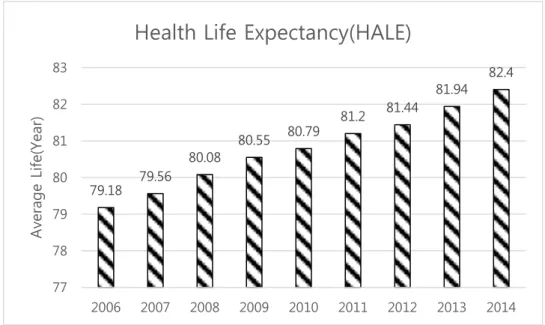
Figure 1. Average life span of Korea, from 2006 to 2014. --- 1
Figure 2. Annual bone fracture diagnosis population transition --- 2
Figure 3. Overall scheme of entire thesis. --- 3
Figure 4. Scheme of electrospun random pattern scaffold production --- 13
Figure 5. Scheme of electrospun aligned pattern scaffold production --- 14
Figure 6. Scheme of nanofiber-PDMS platform production --- 14
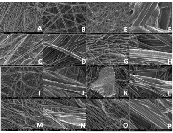
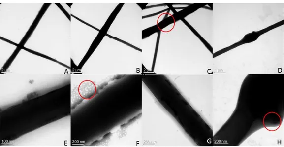
Figure 7. Overall FESEM image of HBNP-NSs. A, B: PCL only random scaffold in 5k, 50k scale, C, D: PCL only aligned scaffold in 5k, 50k, E, F: nHA PCL random scaffold in 5k, 50k, G, H: nHA PCL aligned scaffold in 5k, 50k, I, J: HBNP 10mg PCL random scaffold in 5k, 50k, K, L: HBNP 10mg PCL aligned scaffold in 5k, 50k, M, N: HBNP 100mg PCL random scaffold in 5k, 50k, O, P: HBNP 100mg PCL aligned scaffold in 5k, 50k. --- 20
Figure 8. FESEM Fiber diameter difference between control and experimental groups. B: PCL only, random group, F: nHA 1%, random group, J: HBNP 1%, random group, L: HBNP 10%, random group --- 21
Figure 9. FESEM fiber orientation-degree graph between random and aligned group. --- 22
Figure 10. TEM images of experimental groups. A, B: PCL only group, C, D: HBNP 1% group, E, F: nHA 1% group, G, H: HBNP 10% group. --- 23
Figure 11. XRD result of nanofiber scaffold. --- 24
Figure 12. WST results of cells seed on nanofiber scaffolds by day. (Statically significant difference between groups with non-duplicating alphabets. For example, ab and c have statically significant difference p<0.01, n=5) --- 25 Figure 13. Alizarin Red staining pictures and destaining results of cells seeded on nanofiber
viii
scaffolds by week. A. Pictures of Alizarin Red stained scaffolds after 2 week of differentiation. B. Pictures of Alizarin Red stained scaffolds after 3 week of differentiation. C. Pictures of Alizarin Red stained scaffolds after 4 week of differentiation. D. Calcium deposition of Alizarin Red destained solution, week 2. E. Calcium deposition of Alizarin Red destained solution, week 3. F. Calcium deposition of Alizarin Red destained solution, week 4.
(*: Statically significant difference between groups. p<0.05, n=5) --- 27
Figure 14. Immunocytochemistry (ICC) fluorescent images by the week, scaffold variation and filter. DAPI targeted cell nuclear, FITC targeted osteopontin (OPN), TRITC-conjugated phalloidin targeted cellular skeleton actin. A, B. Pictures of immunostained scaffolds after 3 week of differentiation. C, D. Pictures of immunostained scaffolds after 4 week of differentiation. Abbreviations: DAPI, 4',6-diamidino-2-phenylindole, OPN, Osteopontin, PCL, Poly(ε-carprolactone), nHA, Nano-sized Hydroxyapatite, HBNP, Horse bone nanopowder, 1%, 1% (w/v), 10%, 10%(w/v) --- 31
ix
List of Terms and Abbreviations
BSA: Bovine serum albumin
DAPI: 4',6-diamidino-2-phenylindole
DPBS: Dulbecco’s Phosphate Buffered Saline DPSC: Dental pulp stem cell
FITC: Fluorescein isothiocyanate HALE: Healthy life expectancy HBNP: Horse bone nanopowder
HBNP-NS: HBNP-incorporated nanofiber scaffold ICC: Immunocytochemistry
nHA: Nanosized Hydroxyapatite OPN: Osteopontin
PBS: Phosphate Buffered Saline
PBST: Phosphate Buffered Saline with Tween 20 PDMS: Polydimethylsiloxane
TRITC: Tetramethylrhodamine
1
1. Introduction
By the improvement of medical technology and social welfare, the average life span of Korean citizen has steadily increased. But compared to average life span, quality of life isn’t very promising. This is due to shortage of healthy life expectancy (HALE) to average life span. HALE is calculated by, to know expected life duration of an individual is expected to live healthy. According to 2012 HALE census of major countries, HALE of Korea is 73. This is 8 year less compared to that time’s average life span of 81.44(Figure 1), meaning individuals cannot enjoy satisfying quality of life during those years.
Figure 1. Average HALE of major countries by years, from 2006 to 2014.
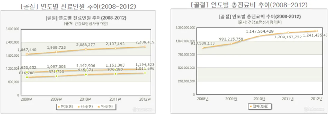
The main cause of HALE shortening is disease, especially osteoporosis and bone fracture as senior population increase by following average life span. By statistics of 2008 to 2012, number of both men and women bone fracture patient are increasing, and total medical
79.18 79.56 80.08 80.55 80.79 81.2 81.44 81.94 82.4 77 78 79 80 81 82 83 2006 2007 2008 2009 2010 2011 2012 2013 2014 A vera g e L ife( Yea r)
2
expenses also steadily increased (Figure 2).
Figure 2. Annual bone fracture diagnosis population transition in Korea
As previously seen, bone fractures tribute to HALE shortening. If a treatment which can heal the wound faster in inexpensive way is developed, it would be beneficial to patients and country itself by improving quality of life and saving budgets on medical issues. Scaffold can be a solution. It is biocompatible structure which can provide space for various cells to grow inside, ultimately targeting to be transplanted in vivo to tissue level. Scaffold can be produced by various methods like salt leaching, fiber bonding, 3D printing, selective laser sintering, and multiple stage jet-solidification etc.
And although not included in previous examples, electrospinning can also be used to produce scaffold. Polymer solution pressured at certain rate is applied with nano dimeter sized fibers are jetted from end of the needle in cone aspect, which is called Taylor cone. Fiber is then laminated to grounded metal collector under the needle, forming non-woven layer. Structures produced by electrospinning is basically have very high surface area, and
3
porous enough to induce beneficial cell to cell interaction. Also by varying conditions, thickness, fiber diameter, composition, porosity and other properties of nanofiber and nanopatch can be controlled. In addition, electrospinning is relatively low priced and less complicated compared to other methods to produce nano level fibrous scaffold which are introduced above. One of the most frequently used polymer in biomedical field is Poly (ε-carprolactone) (PCL). It is semi crystalline, aliphatic compound and low biodegradation rate of 12 month to 24 month. Since FDA approved PCL for its biodegradability and biocompatibility, it is suitable as material for scaffold which have to apply in vivo.
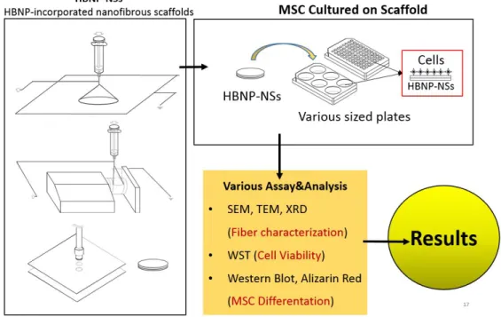
Figure 3. Overall scheme of entire thesis.
4
scaffold, MSC, Mesenchymal stem cell, SEM, Scanning electron microscope, TEM, Transmission electron microscope, XRD, X-ray diffraction, WST, Water Soluble Tetrazolium
However, there are several aspects to overcome for applying electrospun PCL scaffold to bone regeneration. PCL lacks biocompatibility and ability to induce osteogenesis. To improve these characteristics, researchers proposed coating nanosized hydroxyapatite (nHA) on electrospun scaffold, which is main composition of bone. In reality, nHA coated, electrospun scaffold showed increased biocompatibility and osteoinductivity in vitro. Evidently, biocompatibility and osteoinductivity of electrospun PCL scaffold improved by coating nHA. Nevertheless, cost of nHA is considerably high compared to PCL even though it is subside compound. Graft materials can be used like nHA are xenograft, allograft, and autograft. As to the latter, negative effects like immune response, but have difficulty obtaining the material itself. There is already technology from previous research of processing horse bone, which is slaughterhouse byproduct, into nHA-like horse bone nano sized powder (HBNP) by sintering in high temperature. By sintering, many side effects including immune response can be minimized. HBNP have lower cost compared to other commercial nHA products but still can expect beneficial effects of nHA, and raw material supply is sufficient.
HBNP will be incorporated to PCL electrospun scaffold, and varying effects of HBNP on promoting osteogenic effect by concentration and alignment will be examined as seen in overall scheme (Figure 3).
5
2. Objectives
In this study, PCL solution with evenly distributed HBNP is electrospun into aligned structure, fabricating incorporated nanofibrous scaffolds (NSs). HBNP-NSs aim to improve osteoinductivity, and acceleration of bone tissue itself. The objectives of this research are as follows.
(1) Fabricate aligned HBNP-NSs.
(2) Determine whether aligned HBNP-NSs have significantly better cell, bone differentiation inducing ability to traditional PCL electrospun scaffolds with varied condition.
6
3. Literature Review
3.1 Characteristics of PCL and making scaffolds
PCL is one of the oldest polymers synthesized by Carothers group. It was not issue of interest in early days, but when researchers searched for synthesized polymer degradable by bacteria, it became commonly, commercially available. PCL is hydrophobic, semi crystalline polymer and its crystallinity decreases as molecular weight increases. It is very soluble and have low melting point of 59-64°C, and have good characteristics towards additives, showing potential to be used in biomedical field. It can be manufactured into nanosized sphere, nano diametered fiber, fabrication, scaffold and various forms of structures [1, 2, 3].
Bone is main support system of human body, complex structure of combined mineral and tissue having excellent tensile and compressive strength. Mineral inside form hard phase, and tissues and cells form organic phase and influence bone repair, tensile force, and flexibility.
When bone fracture occurs, the main purpose of treatment is to stabilize fractured bone area as it was before the injury. By inflammation, necrotized cells are removed and cartilage tissue which is soft tissue take its place, and hard tissue of bone tissue replaces cartilage. However bone repair can fail when damage is severe or infection and other causes are intervened, bone implant being proposed for solution [4].
7
area and non-reactive to surrounding environment. But by non-specific immune response encapsulation occurred on the surface of material and resulted in aseptic dislocation. Therefore bone implant development afterwards target to material and surrounding bone to meld together. One of these various attempts was scaffold designed to have adequate biocompatibility, porosity, micro and nano scale structure, degradation rate, physical property, growth factor delivery [5]. Especially, as porosity increases, cell seeding and transfer becomes easier, and as pore size decreases, tissues can grow in the scaffold easier. However, increase of porosity accompany decrease of physical properties. Therefore, scaffold should be designed to accomplish both physical properties matching surrounding tissue and beneficial effects due to porosity. Also aligned nanostructure can influence protein motif unit and promote cell proliferation and differentiation, and such element can be considered.
3.2 Electrospun nanofiber structure
Electrospinning uses electric field to electrificate polymer and form jet of solution called Taylor cone. As jet passes through air solvent evaporates and fiber can be electrically collected or pass by to the metal screen, creating nanodiametered fibers [6]. Voltage, position of fiber collecting metal, and time of applying voltage can be varied to control fiber diameter, layer thickness, and alignment and can produce nanofiber in more simple way than traditional methods. Hydrogel based biopolymer scaffold have sufficient moisture, but their in vivo physical properties lacks to be applied in hard tissue like bone or cartilage. And degradation point cannot be controlled evenly. These disadvantages can be solved by using PCL 3D scaffold produced by electrospinning. PCL is already tested in
8
skin, bone, cartilage, liver, protein carriers and many other fields, and PCL included scaffold proved promoting regeneration in cartilage [7], and bone [8] in past researches. In this study electrospinning can be effective strategy since the objective is to develop scaffold for bone regeneration.
3.3 Improvement of PCL electrospun nanofiber scaffold
Electrospun fibers provide nano sized structure with big surface per area, porosity, and accessibility for cells. Additionally, directed property of tissue structure is important in musculoskeletal system. Tendons and ligaments showed 200 to 500 fold higher tensile parameter in horizontal direction than in perpendicular direction. Joint cartilage and meniscus also showed directional properties [9]. Also scaffold can provide cell great surface and porosity to grow in without toxicity, but PCL itself does not affect cell proliferation by chemical interaction with cells.
3.3.1 Aligned structure
When provided with ECM-similar structure and substrates of originated tissue, stem cells tend to proliferate and differentiate into similar cell types. One of the main constituent of ECM is collagen, which is aligned in certain direction and provide mechanical strength to bone structure. Focused on these points, many researches went on imitating ECM properties by using various fabrication method and studied the effect of fiber structure on proliferation and differentiation. For example, aligned electrospun scaffold fabricated by rotating collector showed higher paxillin expression than random fibers, meaning aligned fiber showed higher cell adhesion than control group [10]. Also ALP assay and alizarin red
9
staining results of MC-3T3-E1 cultured on aligned and random fiber showed higher ALP activity and calcium accumulation in aligned structure [11].
From the studies above, aligned structures can hypothesized as enhancing osteoinductivity and osteogenesis in electrospun scaffolds.
3.3.2 nHA addition, coating and mineralization
Hydroxyapatite (HA) is a kind of bioceramic, and was proven suitable for providing adequate physical properties for orthopedic treatment with considerable osteoinductivity [12]. As previously explained, polymer nanofiber is non-toxic, and the network structure can enchance osteoinduction, proliferation, osteogenesis despite osteoinductivity of PCL material itself is poor. There were several attempts to enhance osteoinductivity of scaffolds by adding HA. For example, poly (L-lactic acid) (PLLA) electrospun scaffold added with HA and collagen showed higher cell viability than PLLA-only scaffold [13]. Also, Akkouch et al. fabricated collagen and poly (lactide-co-ε-caprolactone) (PLCL)
into scaffold by porogen leaching [14]. From the MTT assay results, HA added scaffolds showed higher cell growth to control groups without HA. In RT-PCR, Osteocalcin and Alkaline phosphatase (ALP) expression also increased in HA added groups, meaning osteogenesis behavior of cells increased by HA.
Judging from studies above, adding HA or HA similar material to PCL electrospun scaffold can be hypothesized of contributing osteogenesis and can still maintain advantages on osteogenesis from aligned nanostructure.
10
3.3.3 HBNP as nHA substitute
As explained before, bone grafts are used when natural osteogenesis fails. There are three varieties of bone graft materials: Autograft, allograft, xenograft. Autograft is the most effective in enhancing osteogenesis, but physical properties doesn’t match the needs in load bearing sites. Allograft is more osteoinductive than xenograft, but organ donor number is low and risk of infection exists. Xenograft material is easy to achieve since most of xenograft material is byproducts of slaughterhouse, but chance of cattle originated disease infection such as BSE hinders its usage. Nevertheless infection possibility of xenograft materials can be lowered by sterilizing, or by using in form of demineralized bone matter (DBM) which is demineralized putty.
Thus nHA can be used to solve problems of traditional bone graft materials. When nHA is combined with polymer scaffold, viability of osteogenic progenitor cell or mesenchymal stem cell increases, and expression of proteins associated with osteogenesis also increased compared to normal polymer-only scaffold. And it can be resolution to infection and immune responses of standard bone graft materials. However, synthetic nHA is expensive and lack novelty because several researches already used nHA for orthopedic treatment and osteogenesis enhancing.
Horse Bone-derived Natural Calcium Phosphate powders (HBNP) can be substitute of nHA. Manufactured from horse bone pretreated with oxidant to remove organic matter, by sintering in high temperature and grinding the treated material using various milling machine. Compared to HA or commercial bone graft products like Bio-Oss, HBNP shows similar or even better properties on cytotoxicity and cell viability depending on sintering
11
temperature due to mineral composition and inner conformation identical to that of natural bone. Also HBNP show osteoinductivity in addition to osteoconductivity and stimulating osteoblasts, which property other animal originated calcium phosphate powder and similar products didn’t show in past researches [19]. Therefore in means of utilizing knowledge and legacy of previous research and cutting down cost, adapting HBNP in this experiment can be a genuine and better choice.
4. Material and Methods
4.1 Cell selection and culture
For in vitro experiment, passage 5 dental pulp stem cell (DPSC) was used. Dental pulp stem cell is one of the variations of human mesenchymal stem cell (hMSC) and have ability to differentiate into osteocytes and many other variation of cells consisting dental tissue, matching needs with this study to observe proliferation and differentiation of osteocyte.
4.2 Scaffold fabrication
4.2.1 PCL Solution
PCL pellet of molecular weight 80,000(Sigma-Aldrich) 1.00g is put into 20ml glass vial, 10ml solvent made from chloroform to DMF ratio of 3:1 is added to previous vial and stirred in 300RPM for one day. Afterward, 10mg nHA, 10mg, 100mg HBNP is added to each vials to make 1% nHA (w/w to PCL), 1% HBNP, 10% HBNP PCL solution.
12
Liquid PDMS agent A (Sylgard 184A, Sewang Hitech Silicone) 6ml was put on square dish and added with 10% (w/w) of solidifying agent (Sylgard 184B, Sewang Hitech Silicone). Mixture was solidified on 60°C heating plate for a day.
4.2.3. Sample making
For positive control, nHA 1% PCL solution was used. And for negative control PCL only solution was used. For even distribution of additives, each vial was sonicated in sonicating bath before electrospinning. KDScientific 101 syringe pump and commercial 10ml syringe was used in electrospinning process. To prevent additives influencing between each group, nozzle was washed with 70% EtOH every time before and after electrospinning.
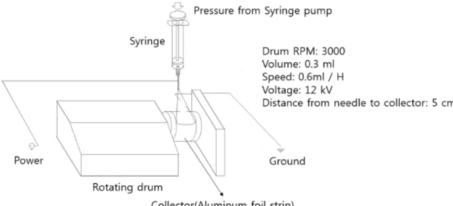
4.2.3.1 Random scaffold
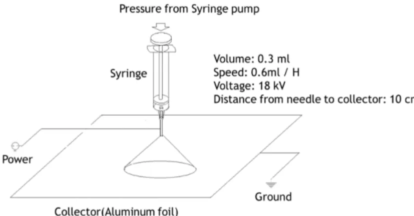
100mmⅹ100mm Size aluminum foil was used to collect electrospun fiber. Fiber was collected for 30 minute in condition of voltage 18kV, solution volume 0.3ml, syringe pump rate at 0.6ml/hr (Figure 4). To remove remaining solvent, HBNP-NSs was put under fume hood of room temperature for at least one day.
13
Figure 4. Scheme of electrospun random pattern scaffold production
4.2.3.2 Aligned scaffold
40mmⅹ150mm Size aluminum foil was used to collect elctrospun fiber. To achieve fiber alignment, attach collector on grounded drum rotating at rate of 3000 RPM. Fiber was collected for 30 minute in condition of voltage 12kV, solution volume 0.3ml, syringe pump rate at 0.6ml/hr (Figure 5). After electrospinning, HBNP-NSs were relived with tension by pulling each end of strip of HBNP-NSs with large sized metal clips. To remove remaining solvent, HBNP-NSs were put under fume hood of room temperature for at least one day.
14
Figure 5. Scheme of electrospun aligned pattern scaffold production
4.2.4 Nanofiber with PDMS platform
Figure 6. Scheme of nanofiber-PDMS platform production
HBNP-NSs were attached to hardened PDMS. Afterwards, biopsy punch was used to make circular shaped scaffold samples size depending on uses (Figure 6). Scaffolds were washed three times with 70% ethanol then three times with DPBS, and exposed under UV light for at least 30 minutes to sterilize before seeding cells.
Biopsy Punch
Nanofiber Layer
15
4.3 Characterization of nanofiber scaffolds by FESEM, TEM
Eight groups of sample were prepared: PCL random/aligned, nHA 1% random/aligned, HBNP 1% PCL random/aligned, HBNP 10% PCL random/aligned. For the FESEM image of each experimental group, sample was put in room temperature for at least one day to remove remaining solvent and moisture. After removing solvent sample was sputter coated or put on grid, then examined by using SUPRA 55VP field emission scanning electron microscope or JEM1010 transmission electron microscope. Fiber orientation was examined by OrientationJ plugin of ImageJ 1.51k. 8 random sites from 5,000 magnified image were selected from picture and processed.
4.4 Determining chemical properties of nanofiber scaffolds by
XRD
Four groups of sample were prepared: PCL, nHA 0.1% (w/v), HBNP 0.1%, HBNP 1%. Samples were examined by XRD instrument in NICEM (D8 Advance, Bruker) to determine chemical properties.
4.5 Determining cell viability and proliferation on the scaffold in
vitro
4.5.1. Cell culture on the scaffold
Human dental pulp cell was used in in vitro experiments including proliferation and differentiation tests. Dental pulp cells are mesenchymal stem cells derived from dental
16
pulp inside tooth and available to differentiate into various types of cells including osteoblast and other bone-like cells, which is this study is targeting to examine. Every cells were passage 5 at the period of seeding to assure osteogenesis ability of stem cell.
4.5.1.1 Culture media
Proliferation media was made by mixing 10% fetal bovine serum (FBS) (Welgene, S 001-01), 2% antibody to minimum essential medium, alpha modification (α-MEM) media (Welgene, LM008-52). Differentiation media was made by adding 0.1µM dexamethasone, 10µM β–glycerophosphate, 100 µM ascorbic acid to proliferation media.
4.5.2 WST assay
Water-soluble tetrazolium salt (WST) was used for agent. Tetrazolium salts show different absorbing wavelength varying on cell’s viability by reacting with byproducts of mitochondria which indicates cell metabolism activity, and can omit solubilizing process which is essential in traditional MTT reagent, thus saving time in process. It also have less toxicity on cells, and provide more effective signals than standard MTT reagent.
1 × 10⁴ Passage 5 dental pulp cells were seeded to 6mm circular HBNP-NSs scaffolds
in 96 well plate. 20µl of cell dispersed proliferation media was pre-seeded to each well and incubated for 30 minute in incubator, then added with 180µl proliferation media. Every well was washed three times with 70% ethanol, then three times with Dulbecco’s phosphate buffered saline (DPBS) and exposed under UV ray lamp of clean bench for 30 minute to remove contaminants.
17
For procedure, WST agent was added to proliferation media assuring concentration to be 10× in room without direct exposure to light. To remove media, each well was aspirated and washed three times with DPBS. 200 µl of WST reagent-media solution was put into each well, then plate was wrapped with aluminum foil to seal from light and put in incubator. After 1 hour 100 µl of each well’s media was transferred to a new 96 well plate and measured in ELISA machine in 450 nm wavelength setting.
4.6 Determining differentiation on the scaffold in vitro
HBNP-NSs made with 6mm diameter circular biopsy punch was used for differentiation assays. As explained in WST procedures, each well of plate was washed three times first with 70% alcohol then three times with DPBS to remove contaminants.
4.6.1 Alizarin red staining
Alizarin red is used to identify calcium in tissue sections and cultured cells in vitro since it forms Alizarin Red S-calcium complex in a chelation process and end product is a bright red stain. The reaction is not strictly specific for calcium, but these elements usually do not occur in sufficient concentration to interfere with the staining, thus suitable for identifying calcium deposition by differentiation [18].
3 × 10³ Passage 5 dental pulp cells were seeded to 6mm nanofiber-PDMS scaffolds in
96 well plate, 20µl of cell dispersed proliferation media was pre-seeded to each well and incubated for 30 minute in incubator, then added with 180µl proliferation media. Every well was washed three times with 70% ethanol, then three times with Dulbecco’s phosphate buffered saline (DPBS) and exposed under UV ray lamp of clean bench for 30
18
minute to remove contaminants.
For procedure, media was aspirated from the plate and washed each well for three times with DPBS. Then 100µl 4°C cold paraformaldehyde solution was added to each well for 1 hour, then aspirated and washed with deionized water for three times. After fixation, 200µl Alizarin red S solution was added in each well and incubated in room temperature for 30 minute, then washed with distilled water three times and added with 200µl destaining solution in each well. 100µl of destaining solution was transferred to each well and measure in 570 nm condition after incubating in room temperature for 15 minute.
4.6.2 Immunocytochemistry
Although osteogenesis can be determined by Alizarin Red staining, but there can be some uncertainty in results since basic principal of the assay is chelation process between staining agent and metal ions. Therefore immunocytochemistry staining, which can directly target marker proteins produced by osteogenic genes, can be used as more precise method to determine cell’s osteogenic behavior on metal incorporated scaffold.
3 × 10⁴ Passage 5 dental pulp cells were seeded to 24mm diameter nanofiber-PDMS
scaffolds in 24 well plate. Before seeding, every well was washed three times with 70% ethanol, then three times with Dulbecco’s phosphate buffered saline (DPBS) and exposed under UV ray in clean bench to remove undesired microbes and possible contaminants before seeding. 200µl of cell dispersed proliferation media was pre-seeded to scaffolds each well and incubated for 30 minute in incubator, then added with 1ml proliferation media. After two days when cells are confluent and ready for differentiation, osteo-differentiation media was provided to cells. Media was changed in every three days.
19
For procedure, media was aspirated from the plate and washed each well for three times with DPBS. Then 300µl 4°C cold paraformaldehyde solution was added to each well for 1 hour, then aspirated and washed with deionized water for three times. After fixation and washing two times with PBST, 1% triton X solution was added to each well for 15 minute to permeabilize. After washing thrice with PBST, 1% bovine serum albumin (BSA) solution was added in each well and incubated in room temperature for 1 hour to block. Appropriate concentration of primary antibody (Polyclonal anti-human osteopontin goat IgG, R&D Systems AF1433), secondary antibody (Anti-goat IgG fluorescein isothiocyanate (FITC) conjugated, tetramethylrhodamine (TRITC)-conjugated phalloidin), 4',6-diamidino-2-phenylindole (DAPI) solution was treated for 1 hours(5 minutes for DAPI) continuously with suitable concentration, washed thrice with PBST between and after treatment. All scaffolds were mounted on cover glass after staining was done, and imaged with fluorescent microscope.
4.7 Statistical analysis
Statistical significance of quantitative data such as WST and Alizarin red destaining was analyzed by performing Duncan’s multiple range test between control (PCL random group) and other experimental groups. At p < 0.05, differences between groups were considered to be statistically significant.
20
5. Results
5.1 Characterization of nanofiber scaffolds
5.1.1 FESEM results and fiber orientation analysis
Figure 7. Overall FESEM image of HBNP-NSs. A, B: PCL only random scaffold in 5k, 50k scale, C, D: PCL only aligned scaffold in 5k, 50k, E, F: nHA PCL random scaffold in 5k, 50k, G, H: nHA PCL aligned scaffold in 5k, 50k, I, J: HBNP 10mg PCL random scaffold in 5k, 50k, K, L: HBNP 10mg PCL aligned scaffold in 5k, 50k, M, N: HBNP 100mg PCL random scaffold in 5k, 50k, O, P: HBNP 100mg PCL aligned scaffold in 5k, 50k.
21
nanopowder, R, Random, A, Aligned, 1%, 1% (w/v), 10%, 10% (w/v)
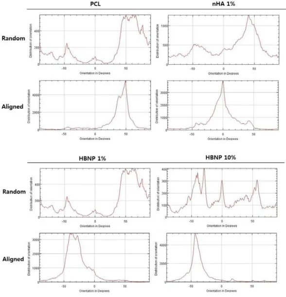
As shown in Figure 7 and 8, SEM images of nanofibers showed thickening diameter size as bioceramic amounts increased, but remained micro-sized in any experimental group. In fiber orientation analysis by OrientationJ (Figure 9), aligned scaffolds showed distribution of orientations concentrated over 1000 in certain orientation in degrees, while random patterned scaffolds showed distribution of orientation under 1000 with varying orientation in degrees. These results suggest HBNP-NSs made in this study obtained alignment while incorporating bioceramic in fiber structure.
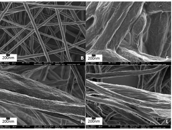
Figure 8. FESEM Fiber diameter difference between control and experimental groups. B: PCL only, random group, F: nHA 1%, random group, J: HBNP 1%, random group, L: HBNP 10%, random
22
group Abbreviations: PCL, Poly (ε-carprolactone), nHA, Nano-sized Hydroxyapatite, HBNP, Horse bone nanopowder, R, Random, A, Aligned, 1%, 1% (w/v), 10%, 10% (w/v)
Figure 9. FESEM fiber orientation-degree graph between random and aligned group.
Abbreviations: PCL, Poly(ε-carprolactone), nHA, Nano-sized Hydroxyapatite, HBNP, Horse bone nanopowder, R, Random, A, Aligned, 10, 1% (w/v), 100, 10%(w/v)
23
5.1.2 TEM results
In Figure 10, Images of scaffolds with bioceramics showed particles in and around fibers compared to control groups, showing that bioceramics successfully included in nanofibers, supporting hypothesis that bioceramics are well incorporated in fibers obtaining nano scale size of fibers.
Figure 10. TEM images of experimental groups. A, B: PCL only group, C, D: HBNP 1% group, E, F: nHA 1% group, G, H: HBNP 10% group.
24
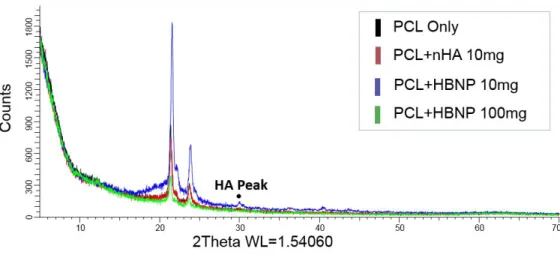
Figure 11. XRD result of nanofiber scaffold. Every groups with bioceramic showed HA-specific peak.
Abbreviations: PCL, Poly(ε-carprolactone), nHA, Nano-sized Hydroxyapatite, HBNP, Horse bone nanopowder, 10, 1% (w/v), 100, 10%(w/v)
From Figure 11, every scaffold group showed HA peak although showing some difference, except for PCL only group. This results suggest that bioceramics maintained chemical properties even after being electrospun with PCL.
5.2 In vitro results
25 A.
26
Figure 12. WST results of cells seed on nanofiber scaffolds by day. A. WST result after day 1. B. WST result after day 3. C. WST result after day 7. (*: Statically significant difference between groups. p<0.05, n=5)
Abbreviations: TCPS, Tissue Culture grade Polystyrene, PCL, Poly(ε-carprolactone), nHA, Nano-sized Hydroxyapatite, HBNP, Horse bone nanopowder, R, Random, A, Aligned, 10, 1% (w/v), 100, 10%(w/v)
In figure 12, WST results suggested groups with random structure and without bioceramics showed significantly larger cell metabolic activity in vitro while PCL only, random group had larger O.D values than other groups in day 7 as in Figure 12, C.
5.2.2 Differentiation tests
27 A.
28 C.
29 E.
30
Figure 13. Alizarin Red staining pictures and destaining results of cells seeded on nanofiber scaffolds by week. A. Pictures of Alizarin Red stained scaffolds after 2 week of differentiation. B. Pictures of Alizarin Red stained scaffolds after 3 week of differentiation. C. Pictures of Alizarin Red stained scaffolds after 4 week of differentiation. D. Calcium deposition of Alizarin Red destained solution, week 2. E. Calcium deposition of Alizarin Red destained solution, week 3. F. Calcium deposition of Alizarin Red destained solution, week 4.
(*: Statically significant difference between groups. p<0.05, n=5)
Abbreviations: PCL, Poly(ε-carprolactone), nHA, Nano-sized Hydroxyapatite, HBNP, Horse bone nanopowder, R, Random, A, Aligned, 10, 1% (w/v), 100, 10%(w/v)
In Alizarin Red staining assay, Cells differentiated on scaffolds became with red color as week passed and calcium deposition by differentiation increased, shown in Figure 13 A to C. Groups with aligned fiber structure and bioceramics showed stronger figment color compared to controls. Destaining results of HBNP-NSs showed statically significant difference in 10% HBNP group, compared with control. Although there was no significantly difference between control and other groups in week 2, HBNP 10% groups, especially 10% HBNP A group showed statically bigger results in week 3 and week 4 (Figure 13 D, E). Other groups didn’t show significant difference to control, but showed increasing tendency of osteogenesis by alignment and bioceramic in week 4 (Figure 13 F).
32
Figure 14. Immunocytochemistry (ICC) fluorescent images by the week, scaffold variation and filter. DAPI targeted cell nuclear, FITC targeted osteopontin (OPN), TRITC-conjugated phalloidin
33
targeted cellular skeleton actin. A, B. Pictures of immunostained scaffolds after 3 week of differentiation. C, D. Pictures of immunostained scaffolds after 4 week of differentiation. Abbreviations: DAPI, 4',6-diamidino-2-phenylindole, OPN, Osteopontin, PCL, Poly(ε-carprolactone), nHA, Nano-sized Hydroxyapatite, HBNP, Horse bone nanopowder, 1%, 1% (w/v), 10%, 10%(w/v)
Figure 14 showed TRITC-conjugated phalloidin (Actin) images and DAPI images show cells are present in overall image, suggesting that FITC images by OPN is tracking protein itself, not scaffold fibers. While random scaffolds showed nodule-like or non-patterned OPN images in ICC, aligned scaffolds showed aligned patterns and following actin and fiber alignment if possible. In most of groups OPN showed more vivid color in aligned groups than that of random groups, even after being merged. FITC stain in merged images suggest that OPN expression increased as structure became aligned and amount of bioceramics increased, proving not only calcium deposition but also osteogenic marker protein expression increased in time lapse.
34
6. Discussion
Characteristics of HBNP-NSs by FESEM, TEM, and XRD data showed that HBNP-NSs maintained chemical properties of bioceramic while obtaining aligned structure compared to past studies.
Cell viability assay on HBNP-NSs suggest that groups with bioceramic promote cell growth in early stage, but all other groups with modified structure and composition turned out to be proliferate less than control (PCL R). Bioceramic, including HBNP was effective at least in early proliferation. But aligned structure didn’t have any influence on promoting cell growth.
Assays on differentiation showed some significant results to support the hypothesis. Pictures presented osteogenesis progressed in most of groups, especially other groups than control, PCL only random HBNP-NSs had more deposited and vivid figment. Destaining data showed these tendency by sorted, quantitative way. In week 3, 10% random and aligned HBNP-NSs showed significantly larger O.D values from control while 10% aligned group alone showed significantly larger calcium deposition in week 4. Judging from week 3 and 4 data, 10% HBNP-NSs groups promoted osteogenesis better than control and other groups supporting the hypothesis. Alizarin red destaining results showed aligned structure and bioceramic, especially HBNP showed synergetic effect in 10% w/w concentration to PCL promoted osteogenesis than other groups, presenting possibilities of synergetic effect between alignment and bioceramic addition.
35
osteogenesis by staining calcium ion, uncertainties like absorption into fiber or staining bioceramics included in scaffold fabrication make it hard to be used as evidences of osteogenesis alone. ICC can target osteogenic marker protein directly by using antibodies and more precise in determining osteogenesis by scaffolds. While cellular nuclear and cytoskeleton actin was present in every image growing in certain direction, osteogenic marker protein OPN was distributed in overall images. And in merged images, OPN expression was stronger in aligned, bioceramic included groups.
Although this study did not have quantitative assay directly on marker protein or DNA, quantitative assays like Western Blotting are on plans of future study. Quantitative differentiation assay in this study targeted on calcium deposition but Western blot can target osteogenic marker proteins like osteocalcin and osteopontin, being more direct than Alizarin red staining and destaining. Also, further researches like differentiation of other dental tissues like nerves, ligaments on this scaffold and application of HBNP-NSs on bioreactors are considered as well.
36
7. References
1. Akkouch, A., et al. (2011). "A novel collagen/hydroxyapatite/poly(lactide-co-epsilon-caprolactone) biodegradable and bioactive 3D porous scaffold for bone regeneration." J Biomed Mater Res A 96(4): 693-704.
2. Chen, X., et al. (2013). "Regulation of the osteogenesis of pre-osteoblasts by spatial arrangement of electrospun nanofibers in two- and three-dimensional environments." Nanomedicine 9(8): 1283-1292.
3. Chen, Y., et al. (2006). "PLLA scaffolds with biomimetic apatite coating and biomimetic apatite/collagen composite coating to enhance osteoblast-like cells attachment and activity." Surface and Coatings Technology 201(3-4): 575-580.
4. Clemens van Blitterswijk, P. T., Anders Lindahl, Jeffrey Hubbell, David F. Williams, Ranieri Cancedda, Joost D. de Bruijn and Jérôme Sohier (2014). "Tissue Engineering."
5. Doshi, J. a. R., Darrell (1995). "Electrospinning Process and Applications of Electrospun Fibers." Journal of Electrostatics 35: 151-160.
6. Du, C. (1998). "Tissue response to nanohydroxyapatite/collagen compositeimplants in marrow cavity." Journal of Biomedical Materials Research 42(4): 540-548.
7. FC, D. (1998). "Osteotransductive bone cements." Proceedings of the Institution of Mechanical Engineers, Part H- Journal of Engineering in Medicine 212: 428-435.
8. Gao, X., et al. (2015). "Osteoinductive peptide-functionalized nanofibers with highly ordered structure as biomimetic scaffolds for bone tissue engineering." Int J Nanomedicine 10: 7109-7128.
9. Jang, K.-J., et al. (2014). "Development and Characterization of Horse Bone-derived Natural Calcium Phosphate Powders." Journal of Biosystems Engineering 39(2): 122-133.
10. Kim, G. H. (2008). "Electrospun PCL nanofibers with anisotropic mechanical properties as a biomedical scaffold." Biomed Mater 3(2): 025010.
11. Li, W.-J. (2003). "Biological response of chondrocytes cultured in threedimensional nanofibrous poly(-caprolactone) scaffolds." Journal of Biomedical Materials 67A(4): 1105-1114.
12. Luckachan, G. E. and C. K. S. Pillai (2011). "Biodegradable Polymers- A Review on Recent Trends and Emerging Perspectives." Journal of Polymers and the Environment 19(3): 637-676. 13. Nair, L. S. and C. T. Laurencin (2007). "Biodegradable polymers as biomaterials." Progress in
Polymer Science 32(8-9): 762-798.
14. Okada, M. (2002). "Chemical syntheses of biodegradable polymers." Progress in Polymer Science 27: 87-133.
15. Polo-Corrales, L. (2014). "Scaffold Design for Bone Regeneration." J Nanosci Nanotechnol 14(1): 15-56.
37
AND ALIZARIN RED STAINS FOR CALCIUM." The Journal of Histochemistry and Cytochemistry 17: 15.
17. Wang, M. (2006). "Composite Scaffolds for Bone Tissue Engineering." American Journal of Biochemistry and Biotechnology 2(2): 80-84.
18. Puchtler H, M. S., Terry M (1968). "ON THE HISTORY AND MECHANISM OF ALIZARIN AND ALIZARIN RED STAINS FOR CALCIUM." The Journal of Histochemistry and Cytochemistry 17: 15.
19. Guizzardi S, Montanari C, Migliaccio S, Strocchi R, Solmi R, Martini D, Ruggeri A. "Qualitative Assessment of Natural Apatite In Vitro and In Vivo." J Biomed Mater Res. 53(3) 227-34
38
말뼈 분말이 포함된 방향성 나노섬유
스캐폴드
서울대학교 대학원
바이오시스템
∙소재학부 바이오시스템공학전공
임 재 운
초록
대한민국이 고령화 사회로 진입함에 따라 골절 환자의 수 역시 증가했고, 이 로 인한 가계 부담과 국가적 차원의 비용 역시 증가하였다. 그 중에서도 자가 치료가 어려운 중증 골절은 골이식재를 포함한 다양한 방법을 활용해서 치료 를 하게 되는데, 골이식재는 다양한 종류가 있으며 그 중에서 이종골 이식재39
가 비교적 비용이 저렴하고 재료를 취득하기 쉬우나 실제로 개발하기까지에 는 많은 단점이 있었다. 당 연구실에서는 점차 증가하는 말 도축두수로 인해 발생하는 부산물을 농가에 이익이 되는 방향으로 활용하기 위해 말뼈를 사용 해 이종골 이식재를 개발하는 연구를 한 적이 있었으며, 말뼈를 활용한 골이 식재(Horse Bone Nanopowder, HBNP)의 개발로 이어졌다. HBNP는 다른 골이식재에 비해서 in vitro 세포 활성 측면에서 비슷하거나 더 나은 효과가 있었으며 화학적 성질에서도 기존 골이식재와 크게 다르지 않은 조성이라는 것이 증명되었다. 하지만 HBNP 역시 골이식재라면 근본적으로 가지고 있는 물성 문제에서 벗어날 수 없었다. 기본적으로 골이식재의 경우 즉시 이식되었 을 때 분말 내지는 페이스트 형태이기 때문에 외부에서 충격을 줬을 경우 파 손되기가 쉽기 때문이다. 이러한 물성 문제는 전기방사법을 통해 해결할 수 있다. 나노섬유가 축적된 부직포 형태의 스캐폴드를 제작할 수 있으며, 제작 과정에서 발생하는 나노섬 유 집적체의 다공성, 세포가 분화하는 환경을 유사하게 제공해주는 등 구조적 특징을 이용해 내부에서 자랄 세포에게 이로운 영향을 미치는 것과 동시에 내부에 골이식재를 포함시켜서 그 화학적 성질도 동시에 유지할 수 있다. 한 편 전기방사법으로 나노섬유를 형성하는 고분자 중에서는 PCL이 목적에 가 장 적합하다. FDA로부터 인체 사용 승인을 받은 물질이며, 따라서 분해 산물 이나 물질 자체가 인체에 유독하지 않다. 또한 인장성이 크기 때문에 어느 정 도의 강도를 요구하는 연구 목적에도 부합한다.
40
이러한 배경을 통해 도출해낸 연구의 목적은 중증 골절을 치료하기 위해 소 재로 PCL에 HBNP를 첨가한 방향성 전기방사 스캐폴드(Electrospun, HBNP-incorporated nanofibrous scaffolds, HBNP-NSs)를 생산하는 것이 다. 만들어진 스캐폴드 내부에 HBNP가 포함되었는지, 방향성이 부여되어 있 는지 확인하기 위해 FESEM과 TEM으로 섬유의 형태를 확인하고 XRD를 통 해 수산화인회석(HA) 특이적인 피크 여부를 확인했다. WST assay와 Alizarin Red staining을 통해 제작한 스캐폴드에 중간엽 줄기세포인 인간치 수세포(Human Dental Pulp Stem Cell, hDPSC)를 배양하여 시간 경과에 따 른 세포활성과 골분화 정도를 측정했다. 섬유의 특성을 확인한 결과 바이오세라믹은 섬유 내부에 포함되어 직경을 증 가시켰으나 여전히 나노 단위의 직경을 유지하였으며, 전기방사를 통해 획득 한 방향성을 유지하면서 고유의 화학적 성질 역시 유지하였다. 한편, HBNP-NSs 위에 배양된 hDPSC는 대조군에 비해 세포 활성에서 큰 차이를 보이지 않아 세포가 자라는 데 지장을 주지 않는다는 것을 증명했으며 세포 분화 정 도를 확인했을 때는 방향성이 있는 그룹이 그렇지 않은 그룹보다, 바이오세라 믹이 포함된 그룹이 그렇지 않은 그룹보다 더 높은 골분화 수준을 보였다. 비 록 가설에서 제시한대로 방향성과 바이오세라믹이 시너지 효과를 보인다는 것을 입증하지는 못했으나 골분화를 촉진한 것은 확인할 수 있었으며, 차후 연구에서는 골분화 정도를 더 정확하게 확인할 수 있는 실험법의 도입과 동 물 실험을 통해 스캐폴드의 효과를 입증하는 것을 목표로 하고 있다.
41