A visible phagemid system for the estimation of
Cre-mediated recombination efficiency
by
Chie-Hyung Lee
A Dissertation Submitted to The Graduate School of Ajou University in Partial Fullfilment of the Requarements for the Degree of
DOCTOR OF PHILOSOPHY
Supervised by Ho-Joon Shin, Ph.D.
Department of Medical Sciences The Graduate School, Ajou University
- ABSTRACT -
A visible phagemid system for the estimation of Cre-mediated
recombination efficiency
Purposes:The Cre- lox system is generally employed to increase the size of phage antibody libraries. However, estimation of library sizes after Cre- mediated recombination is difficult since time-consuming nucleotide sequence analyses are required. We aimed to develop a visible phagemid vector system that facilitates the estimation of recombination efficiency between VH and VL genes.
Materials & Methods : Two phagemids (pRGA and pRGB) were constructed. To
induce recombination BS1365 bacterial cells expressing Cre recombinase were co-infected using the two phagemids. Cre- mediated recombination between two phagemids was verified by PCR, analysis by restriction enzymes, and DNA sequencing. To confirm the function of scFv proteins expressed from phagemids, total bacterial cell fractions were used for Western blotting and ELISA. Recombination efficiency was calculated simply by counting the number of blue colonies on X-gal-containing medium.
Results: It was found that intermolecular recombination between VH and VL genes
and acquirement of β-galactosidase (β-gal) activity occurred simultaneously in BS1365 bacterial cells. Molecular analyses of plasmids isolated from blue colonies verified a novel VH/VL combination. Recombination efficiency was calculated from
the frequency of a novel recombinant phagemid, and expressed as a percentage difference between the number of blue and total colonies. The calculated
from phagemids were detected as a band of 97 kDa and showed specific binding activity to corresponding antigens.
Conclusions: We developed a visible phagemid system to measure the
recombination efficiency of VH/VL genes without sequence analysis. Our results
suggest that this newly developed visible phagemid system may be reliably used for the measurement of recombination efficiency, which would enable precise evaluation of the diversity of phage antibody libraries.
Keywords: Antibody library size, Visible phagemid system, Cre- mediated
TABLE OF CONTENTS
TITLE PAGE --- 1 ABSTRACT--- 2 TABLE OF CONTENTS--- 4 LIST OF FIGURES--- 6 LIST OF TABLES--- 7 I. INTRODUCTION--- 8II. MATERIALS & METHODS--- 11
A. Construction of phagemid vectors --- 11
B. Cre- mediated recombination--- 15
C. Transformation and X-gal staining--- 15
D. PCR analysis --- 16
E. Isolation of recombinants--- 17
F. Expression and functional analysis of scFv proteins--- 17
III. RESULTS--- 19
A. Construction of phagemid vectors --- 19
B. Recombination in Cre-expressing bacteria --- 19
C. Analysis of plasmids following recombination--- 22
D. Recombination efficiency- --- 24
IV. DISCUSSION--- 30
V. CONCLUSIONS--- 33
BIBLIOGRAPHY--- 34
LIST OF FIGURES
Fig. 1. Phagemid structure and Cre-mediated recombination between phagemids
--- 13
Fig. 2. Analysis of recombination in Cre-expressing bacterial cells --- 21
Fig. 3. Analysis of plasmids prepared from blue colonies --- 23
LIST OF TABLES
I. INTRODUCTION
Phage display is a technique used to establish antibody libraries in vitro without immunization and select antigen-specific antibodies, usually single chain Fv or Fab.1, 2 It is difficult to obtain an antibody library size over 1x10¹⁰ using traditional phage display whereby antibody diversity is created by random combinatorial linkage of VH and VL genes and cloning, due to the limited efficiency of bacterial
transformation (109 cfu/ml). Evidence showing that a larger initial phage antibody library results in significantly higher possibility of selection for high-affinity antigen-specific antibodies has encouraged further attempts to increase initial antibody library sizes.8, 17, 21
Site-specific recombination is an alternative to generating antibody libraries, circumventing the limitations of transformation in traditional phage display. To date, a single lambda recombinase-based technique4 and several Cre recombinase-based strategies have been employed to generate combinatorial phage antibody libraries.3, 5,
20, 22, 23
Cre recombinase derived from bacteriophage P1 catalyzes recombination between two 34 bp loxP recognition sites. The loxP site consists of two 13 bp inverted repeats separated by an 8 bp spacer region involved in cleavage and re-ligation of each strand of DNA.6, 7 Depending on the orientation and location of the
loxP sites, Cre-mediated recombination leads to different results. Intramolecular
DNA sequence flanked by the sites, and that between two loxP sites of inverted orientation leads to inversion. Intermolecular recombination entails the reciprocal exchange of DNA sequences flanked by mutant lox sites (Sauer, 1998).
In approaches using the Cre/lox system as a tool to generate diverse phage antibody libraries, the assembly of each VH and VL library created by molecular
cloning has been exploited in two different ways, specifically, two-vector and one-vector systems.16 In the two- vector system, VH and VL genes were separately cloned
into two vectors, followed by induction of combinatorial assembly of both genes by Cre. However, this system was limited by remaining non-functional byproducts that did not participate in recombination, and irreversibility of the procedure.5 In the more recent one- vector system, both VH and VL genes were cloned in a single phagemid,
resulting in a library of primary functional scFv. This was followed by Cre/lox recombination in Cre-expressing bacteria, and intermolecular shuffling of VL or VH
genes between phagemids to obtain a novel VL-VH combination. The one-vector
system has advantages over the two-vector technique, due to the absence of non-functional byproducts and reversibility of the recombination procedure.15 Despite the advantages of the one-vector system, it is difficult to estimate the final library sizes following recombination using this technique. Library size may be roughly estimated only by calculating the rate of appearance of novel recombinant phagemids through intensive sequence analyses of several hundred regions of the V gene.15 Sequence analysis of a large number of DNA samples is both time- and labor- consuming.
one-vector system to measure the recombination efficiency of VH/VL genes without
sequence analysis. Our vector system was designed so that intermolecular recombination between VH and VL genes and acquirement of β-galactosidase (β-gal)
activity occurred simultaneously. We detected recombinants by X-gal staining, and measured recombination efficiency in Cre-expressing bacteria. Recombination efficiency was calculated by simply counting the number of colored bacterial colonies. By using this method to measure recombination efficiency, increase in final antibody library size after Cre- mediated recombination would be readily evaluated.
II. MATERIALS AND METHODS
A. Construction of phagemid vectors
Phagemids, pRGA and pRGB (Fig.1), are constructed by modifying pCANTAB-5E (Amersham Biosciences, Piscataway, NJ). In both phagemids, the linker fragment (Gly4Ser1)3 within pCANTAB-5E was replaced with the loxP511
peptide-(Gly4Ser1)1 sequence. Following PCR using overlapping and complementary
oligonucleotides (5’ primer, 5’- ATA ACT TCG TAT AAT GTA TAC TAT ACG AAG TTA TCT GGA GGT GGC GGT TCT AGA GAC CTT GTG ATG TCA CAG TCT CC -3’; 3’ primer, 5’- ACC TCC AGA TAA CTT CGT ATA GTA TAC ATT ATA CGA AGT TAT GCT AGC AGA GGA GAC GGT GAC TGA GGT TCC- 3’) a scFv containing the loxP511 peptide-(Gly4Ser1)1 sequence was cloned into the pIg20
vector as a XmaI/NcoI fragment. The scFv was subcloned into pCANTAB-5E using
SfiI/NotI restriction sites. Next, two synthetic sequences containing (His)6
tag-thrombin cleavage site and E tag are added to the carboxy-terminus of the scFv and g3p genes respectively, resulting in pRG. In pRG, contiguous BglII/EcoRI and
SpeI/MluI sites are cloned as a NdeI/DraIII fragment downstream of the E-tag by
overlapping PCR, leading to the generation of pRG1 and pRG2, respectively. To construct pRGA from pRG1, an EcoRI/DraIII overlap PCR fragment containing the loxPwt sequence was cloned into pRG1 digested with the corresponding restriction
enzymes. Next, the structural β-gal gene lacking N-terminal 5 amino acids (derived from pGEM-3Zf (+) (Promega Co, Madison, WI) was introduced by cloning between
BglII and EcoRI sites. To construct pRGB, an NdeI/SpeI overlap PCR fragment
containing the loxPwt sequence was clo ned into the corresponding restriction sites of pRG2, and a tac promoter region plus 15 bases encoding the N-terminal 5 amino acids of β-gal was inserted between SpeI and MluI sites of pRG2. Finally, the 3D8 (D) VH - S2C11 (S) VL gene was subcloned into scFv to generate pRGA with D-S
scFv, and the S2C11 VH - 3D8 VL gene was inserted to obtain pRGB with S-D scFv.
Variable region genes originated from mouse monoclonal 3D8 antibody-specific DNA and S2C11 antibody specific for the surface antigen of hepatitis B virus (HBs).
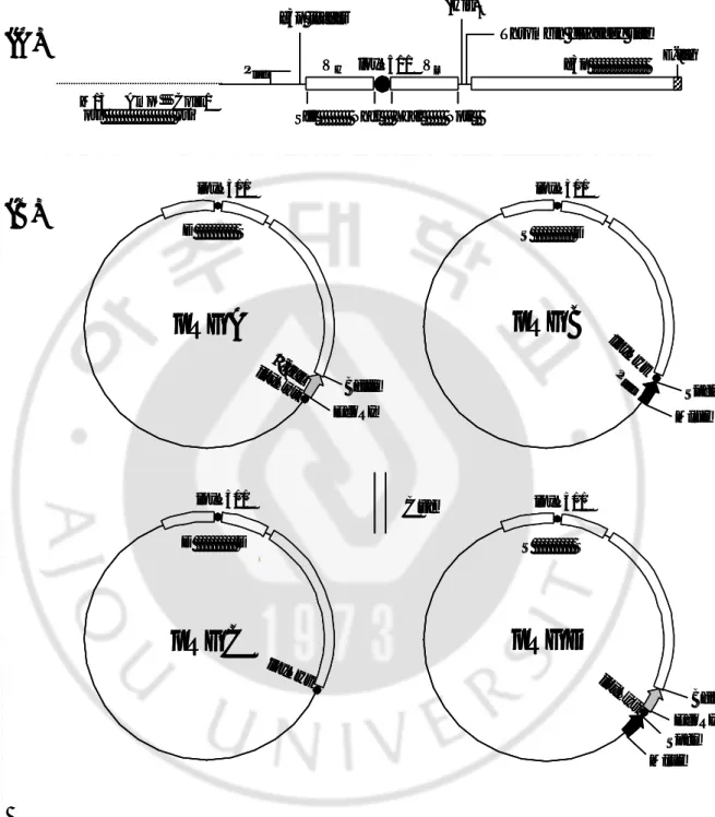
Fig. 1. Phagemid structure and Cre-mediated recombination between
phagemids. (A) Shared structure with phagemids. (B) Scheme for reciprocal
exchange of the VL- g3p region by Cre- mediated recombination. Recombination of (His)6
Thrombin cleavage site
M13 AmpR ColE1
ori ori SfiI NheI XbaI NotI
E-tag VH loxP511 VL g3p Plac g3p leader
(A)
pRGC
loxP511 D DpRGA
loxP511 loxPwt BglII EcoRI D S -galpRGB
S D SpeI MluI loxP511 loxPwt P tac(B)
pRGD
EcoRI BglII MluI loxP511 loxPwt S S Cre SpeI loxPwt (His)6Thrombin cleavage site
M13 AmpR ColE1
ori ori SfiI NheI XbaI NotI
E-tag VH loxP511 VL g3p Plac g3p leader
(A)
(His)6Thrombin cleavage site
M13 AmpR ColE1
ori ori SfiI NheI XbaI NotI
E-tag VH loxP511 VL g 3 p Pl a c g 3 p l e a d e r
(A)
pRGC
l o x P 5 1 1 D DpRGA
l o x P 5 1 1 loxPwt B g l I I E c o R I D S -g a lpRGB
S D S p e I M l u I l o x P 5 1 1 loxPwt P t ac(B)
pRGD
E c o R I B g l I I M l u I l o x P 5 1 1 loxPwt S S C r e S p e I loxPwtpRGC
l o x P 5 1 1 D DpRGA
l o x P 5 1 1 loxPwt B g l I I E c o R I D S -g a lpRGB
S D S p e I M l u I l o x P 5 1 1 loxPwt P t ac(B)
pRGD
E c o R I B g l I I M l u I l o x P 5 1 1 loxPwt S S C r e S p e I loxPwtparental pRGA and pRGB via compatible loxP sites leads to the generation of pRGC and pRGD. pRGD permits lacZ’ fragment translation from the opposite-oriented tac promoter. D and S refer to 3D8 and S2C11, respectively.
B. Cre-mediated recombination
TG1 cells harboring pRGA or pRGB are separately superinfected by M13KO7 helper phage (Amersham Biosciences, Piscataway, NJ) to package phagemid DNA. Titers of the rescued phage containing pRGA or pRGB are measured as AmpR colony forming units (CFU). To induce in vivo recombination, E.
coli BS1365 cells (a gift from Dr. Brian Sauer) constitutively expressing Cre
recombinase are used. BS1365 cells are grown at 370C in 10 ml LB containing 100
µg/ml kanamycin and 1% glucose to an OD600 of 0.5. Phages containing pRGA and
pRGB are added to midlog BS1365 cells at a multiplicity of infection (MOI) of 100. Cells are left for 1 h at 37oC without shaking for infection, and ampicillin was added (100 µg/ml). Recombination was allowed for 2 h at 37 oC with shaking. Next, plasmid DNA was isolated from BS1365 using spin columns (Qiagen Inc, Stanford Valencia, CA), and used to transform E. coli JM109 cells.
C. Transformation and X-gal staining
JM109 bacterial cells which utilize α-complementation of lacZ’ are transformed with DNA isolated from BS1365. Transformed bacterial cells are spread onto nitrocellulose membranes that are placed on the surface of SOB agar plates and incubated at 370C overnight. Glucose (2%) was added to SOB agar to suppress
cytotoxic g3p expression during bacterial growth. Colonies grown on the membrane are transferred onto an LB agar plate containing X-gal (20 µg/ml), IPTG (1 mM) and ampicillin (100 µg/ml), and left for several hours at room temperature to allow the blue color to develop. Blue and white colonies are separately harvested and grown in LB broth. Plasmid DNA was isolated using a Qiagen kit and analyzed for recombination.
D. PCR analysis
Cre-mediated recombination between two phagemids was verified by PCR using 4 primers. Backward DVH (5’- CTT ACA ATG ATG GTA CTA AG -3’) and
forward DVL (5’- ACT GTT GAA CAG ACT CTG -3’) are designed to hybridize to
CDR of 3D8VH and 3D8VL respectively. Backward SVH (5’- CTT CAT TTA TTA
CTC CGA TG -3’) and forward SVL (5’- CGT GAA TGG GTT ACT ACT CC -3’)
are employed for hybridization to CDR of S2C11 VH and S2C11 VL. Four primer
combinations (DVH-SVL, SVH-DVL, DVH-DVL and SVH-SVL) are used for
amplification of the scFv gene. Recombinant plasmid DNA isolated from BS1365 and blue and white colonies (JM109) are used as templates for PCR (30 cycles of 1 min at 940C, 1 min at 550C, and 1 min at 720C).
E. Isolation of recombinants
pRGC and pRGD plasmids resulting from recombination between pRGA and pRGB are isolated for confirmation of the correct sequence. To isolate pRGC, recombinant DNA extracted from BS1365 was digested with three restriction enzymes (BglII, MluI and SpeI) which linearize plasmids except pRGC. Among these, BglII linearizes pRGA and pRGD, MluI linearizes pRGB and pRGD, and SpeI linearizes pRGD as Ill as a small plasmid generated by intramolecular recombination between incompletely non-compatible loxP sites (loxP and loxP511). After bacterial transformation with a plasmid mixture treated with the three enzymes, several colonies are cultured in broth, and aliquots (1 µl) of cell suspension are employed for PCR analysis, using the DVH - DVL primer set. Plasmid DNA was prepared from
cultures containing bands of expected size s. Recombinant pRGD was isolated by culturing blue colonies after X-gal staining. The integrity of pRGC and pRGD was confirmed by sequenc ing, using a cycle sequencing ready reaction kit (Applied Biosystems, Foster, CA).
F. Expression and functional analysis of scFv proteins
Expression of scFv proteins generated from pRGA, pRGB, pRGC and pRGD was induced in JM109 with 1 mM IPTG at 250C overnight. To prepare total
cell protein, cells are lysed in resuspension buffer (20 mM Tris, pH 8.0, 50 mM NaCl, 2 mM EDTA) by sonication. Proteins are subjected to SDS-PAGE on a 10% gel, transferred to a nitrocellulose membrane and probed with rabbit anti-E tag antibody and secondary antibody goat anti-rabbit alkaline conjugate. Proteins are visualized with BCIP/NBT (Sigma-Aldrich Co, Milwaukee, MI). To confirm the function of scFv proteins, total bacterial cell fractions are used for ELISA, performed according to standard protocols. Briefly, Ills of an ELISA plate (Costar, High Wycombe, UK) are coated with 100 µl ssDNA (2 µg/ml) or HBs antigen (5 µg/ml). After blocking Ills with 3% BSA and incubation with the diluted total cell fraction, bound scFv are detected using primary rabbit anti- g3p (1:2,000) (MoBiTec, Göttingen, Germany) and secondary anti-rabbit antibody conjugated to alkaline phosphatase (1: 10,000) (Pierce, Rockford, IL). Reaction revealed with p-NPP substrate (Sigma-Aldrich Co, Milwaukee, MI) was read 495 nm.
III. RESULTS
A. Construction of phagemid vectors
To establish a novel system for the proficient estimation of recombination efficiency, two phagemid vectors are constructed. In both the vectors, a polypeptide of loxP511 (ITSYNVYYTKL)-(Gly4Ser1)1 was used as a linker between VH and VL.
Restriction enzyme sites flanking the loxP511 peptide-(Gly4Ser1)1 are added to
facilitate further VH or VL cloning. The first phagemid (pRGA) contains a β-gal gene
lacking N-terminal MTMIT amino acids upstream of the loxPwt site. The second phagemid (pRGB) contains a tac promoter and a DNA fragment encoding the N-terminal MTMIT amino acids of β-gal downstream of the loxPwt site. In E. coli, β -gal expression is 5- fold more active under control of the tac promoter, compared to the lac promoter.9 Different scFv genes are cloned into each vector to verify recombination between VH/VL genes.
B. Recombination in Cre-expressing bacteria
The 3D8VH - S2CllVL (D-S) and S2C11VH - 3D8VL (S-D) genes are
introduced into pRGA and pRGB, respectively. BS1365 cells expressing Cre recombinase are co- infected using the two phagemids containing scFv genes to
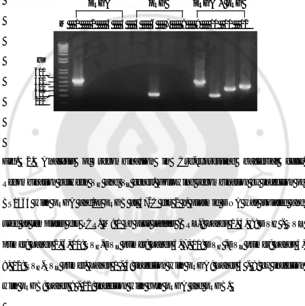
induce recombination. Recombination was analyzed by PCR, using plasmids isolated from BS1365 as templates. PCR analyses to identify scFv genes created by shuffling of the VL genes revealed amplified bands of 551 bp (DVH - SVL primer set), 226 bp
(SVH - DVL), 359 bp (DVH - DVL), and 423 bp (SVH - SVL), corresponding to the
scFv formats within pRGA, pRGB, pRGC, and pRGD, respectively. The data indicate that four different scFv gene formats (D-S, S-D, D-D, and S-S) coexist in the BS1365 bacterial pool, as expected from the consideration that reversible recombination event by Cre recombinase results in coexistence of parent and recombined DNA at equilibrium state (Fig. 2A: lanes 9 - 12).
Fig. 2. Analysis of recombination in Cre-expressing bacterial cells.
Recombination between VH and VL genes. Following recombination by infection of
BS1365 with pRGA and/or pRGB at 370C for 2 h, plasmid DNA was isolated and used as templates for PCR. M: 1 kb plus ladder (BRL); Lane s 1, 5, 9: DVH - SVL primer; Lane s 2, 6, 10: SVH-DVL primer; Lane s 3, 7, 11: DVH -DVL primer; Lanes 4, 8, 12: SVH-SVL primer. Lanes 1 - 4: infection with pRGA; Lanes 5 - 8: by infection
with pRGB; Lanes 9 - 12: infection with both pRGA and pRGB.
M 1 2 3 4 5 6 7 8 9 10 11 12 600 500 400 300 200 pRGA pRGB pRGA + pRGB bp M 1 2 3 4 5 6 7 8 9 10 11 12 600 500 400 300 200 pRGA pRGB pRGA + pRGB bp
C. Analysis of pl asmids following recombination
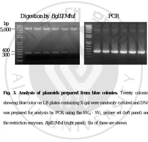
After transformation of JM109 cells with DNA purified from BS1365, blue colonies are observed on X- gal-containing agar plates. Digestion of twenty plasmids purified randomly from these cells by BglII and MluI revealed a 350 bp insert band corresponding to the restored β-gal gene in pRGD. Using the above plasmids as templates and the SVH - SVL primer set, PCR products of 423 bp are observed. An
analysis of six representatives of these twenty plasmids is depicted in Fig. 2B. All plasmids isolated from the blue colonies indeed contained recombinant pRGD comprising S2C11VH - S2C11VL and an active β-gal gene (Fig. 2B). Correct
recombination was confirmed by sequencing of the VH-VL and β-gal genes.
Furthermore, when plasmid DNA isolated from a pool of cells collected by scraping several hundreds of white colonies on a pla te was used for PCR with the SVH - SVL
primer set, no DNA band was observed. Ho wever, DNA bands of expected sizes are amplified using the three primer sets, DVH - SVL, SVH - DVL andDVH - DVL. This
indicates that pRGA, pRGB, and pRGC that are unable to develop blue color did not contain the S2C11VH - S2C11VL gene. Therefore, the frequency of blue colonies
darectly represents the presence of pRGD. Analysis of individual white colonies disclosed that 25% contained an unknown 3.2 kb band, distinguishable from four unimpaared plasmids of around 5.3 kb (data not shown). Deletion of the 1.9 kb fragment corresponding to the VL - g3p region by recombination between loxPwt and
loxP511 sites on a single phagemid would result in the small 3.4 kb band.
Fig. 3. Analysis of plasmids prepared from blue colonies. Twenty colonies
showing blue color on LB plates containing X-gal were randomly cultured and DNA was prepared for analysis by PCR, using the SVH - SVL primer set (left panel) and
the restriction enzymes, BglII/MluI (right panel). Six of these are shown.
Digestion by BglII/MluI
PCR
5,000
300
bp
400
Digestion by BglII/MluI
PCR
5,000
300
bp
400
D. Recombination efficiency
In the presence of Cre, recombination between VH/VL of two phagemids
simultaneously resulted in the generation of a phagemid capable of expressing functional β-gal. In other words, expression of β-gal from a recombinant phagemid was indicative of VH/VL recombina tion. Recombination efficiency was calculated
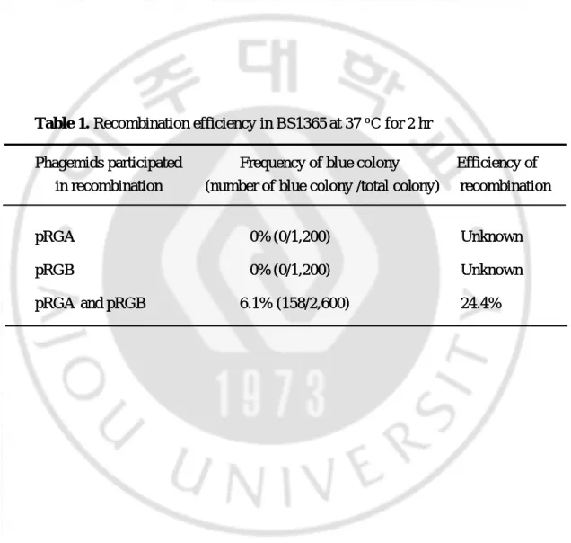
from the frequency of a novel recombinant phagemid, and expressed as a percentage difference between the number of blue and total colonies. If the recombination reaction was fully successful and reached equilibrium (denoted as 100% efficiency), pRGD (equivalent to blue colonies) comprised approximately 25%. Based on this assumption, if the frequency of blue colonies was 10%, this would mean that the recombination efficiency was 40% (10% multiplied by 4). In the case of recombination followed by co- infection of BS1365 with pRGA and pRGB, the percentage of blue colonies was 6.1%, compared to total colonies. Here, recombination efficiency was calculated as approximately 24.4%. When BS1365 cells are infected with either pRGA or pRGB alone, efficiency of recombination was not evaluated (Table 1).
Table 1. Recombination efficiency in BS1365 at 37 oC for 2 hr
Phagemids participated Frequency of blue colony Efficiency of
in recombination (number of blue colony /total colony) recombination
pRGA 0% (0/1,200) Unknown
pRGB 0% (0/1,200) Unknown
pRGA and pRGB 6.1% (158/2,600) 24.4%
Table 1. Recombination efficiency in BS1365 at 37 oC for 2 hr
Phagemids participated Frequency of blue colony Efficiency of
in recombination (number of blue colony /total colony) recombination
pRGA 0% (0/1,200) Unknown
pRGB 0% (0/1,200) Unknown
E. Expression and functional analysis of recombinant scFv proteins
Expression of scFv- g3p fusion protein was induced from the two parent al plasmids (pRGA and pRGB) and two novel recombinant plasmids (pRGC and pRGD). After induction with IPTG, scFv-g3p proteins in total cell extracts are detected by Western blotting using anti- E tag antibody (Fig. 3A). A band with a molecular mass slightly below 97 kDa was detected. This was significantly larger than the size calculated based on the amino acid sequence (64 kDa). A protein size of 64 kDa may be subdivided into 32 kDa for scFv plus additiona l sequences, and 42 kDa for the g3p sequence. The discrepancy between the calculated molecular weight of scFv-g3p and the Western blot is possibly due to the unusual three-dimensional structure of g3p. The g3p protein (calculated as approximately 42 kDa) was observed in a Western blot at 60 – 65 kDa, consistent with earlier reports.19
To ensure that scFv with a loxP511 peptide-(Gly4Ser1)1 linker is functional,
JM109 cells harboring each vector are grown and IPTG-induced cells are lysed. Total cell extracts are used for ELISA (Fig. 3B). Both 3D8 scFv (expressed from pRGC) and S2C11 scFv protein (expressed from pRGD) shoId specific binding to corresponding antigens; the former bound ssDNA while the latter bound HBs antigen. Furthermore, 3D8 scFv bound to ssDNA with a signal comparable to the standard (Gly4Ser1)3 linker (data not shown). The linker used in this study served as a flexible
is due to 3D8 VH, which was shown to bind ssDNA without associating with VL in a
(A)
175 kDa
83 kDa
M 1 2
3 4
-(A)
175 kDa
83 kDa
M 1 2
3 4
-175 kDa
83 kDa
M 1 2
3 4
-2D Graph 15 1 20.0
0.5
1.0
1.5
2.0
A
405
Antigen:
ssDNA HBs
-
1 2 3 4 -
1 2 3 4
(B)
2D Graph 15 1 20.0
0.5
1.0
1.5
2.0
A
405
Antigen:
ssDNA HBs
-
1 2 3 4 -
1 2 3 4
2D Graph 15 1 20.0
0.5
1.0
1.5
2.0
A
405
Antigen:
ssDNA HBs
-
1 2 3 4 -
1 2 3 4
(B)
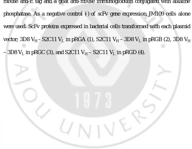
Fig. 4. Expression and functional analysis of scFv proteins. (A) Western blot of
expressed scFv proteins. (B) ELISA for analysis of binding of scFv proteins. Polystyrene microtiter wells were coated with ssDNA and HBs antigen, followed by blocking of wells with 3% BSA. After IPTG induction, total cell extracts prepared from JM109 bacterial cells harboring pRGA, pRGB, pRGC and pRGD were added to each well. ScFv proteins binding to ssDNA or HBs antigen were detected using a mouse anti-E tag and a goat anti- mouse immunoglobulin conjugated with alkaline phosphatase. As a negative control (-) of scFv gene expression, JM109 cells alone were used. ScFv proteins expressed in bacterial cells transformed with each plasmid vector; 3D8 VH - S2C11 VL in pRGA (1), S2C11 VH – 3D8 VL in pRGB (2), 3D8 VH
IV. DISCUSSION
I have developed a visible phagemid system that permits the measurement of recombination efficiency between VH/VL genes by simple count ing of the number of
blue colonies. The ultimate purpose of this new system is to estimate the size of antibody libraries produced using Cre-lox recombination under the one-vector system.15, 16 Recombination efficiency by Cre recombinase is readily measured using our visible phagemid system.
A key factor of our system was the graft of β-gal expression into the Cre-lox system. In the presence of Cre recombinase, assembly of β-gal fragments into the functional gene was accompanied by intermo lecular recombination between VH and
VL genes derived from two separate phagemid vectors. In constructing two novel
vectors, I targeted the loxPwt site to a specific region within the β-gal gene, since the N-terminal region of the alpha peptide of lacZ (lacZ’) is amenable to disruption by foreign DNA fragments, such as multiple cloning sites up to 11-18 amino acids long.13 In fact, replacement of multiple cloning sites of the β-gal gene with the
loxPwt sequence did not interfere with β-gal function. In the established visible phagemid system, only bacterial cells harboring recombinant pRGD are stained with X-gal, signifying undetectable levels of background for false staining. Therefore, it is possible to determine precise recombination efficiency using this system.
frequency < 7%) through recombination in BS1365 (370C, 2 h). This was much lower, compared to other studies utilizing BS1365 as an in vivo recombination reactor. This may be attributed to the fact that experimental conditions are not identical. The first possible explanation is low intermolecular recombination frequency between the two vectors via loxPwt and loxP511 sites. The Cre- mediated reaction includes recognition of the loxP sites by Cre, synapsis to bring the recombining strands into proximity, and formation of a covalent protein-DNA intermediate (tetramer composed of Cre2-Lox dimers).12 Thus, the appropriate physical positioning of lox sites located on different DNA molecules may be unfavorable, compared to cis- linked lox sites. This possibility is supported by a recent report in which inter-chromosome recombination is shown to occur at a lower frequency than intra-chromosome recombination.14
The second possibility for reduced recombination efficiency is high excision rate due to intramolecular recombination between loxPwt/loxP511 in a single vector. In BS1365, intramolecular recomb ination may occur within all four vectors (pRGA, pRGB, pRGC and pRGD), which would reduce the yield of pRGD (an indicator for recombination) despite high occurrence of the recombination reaction. In particular, early intramolecular recombination in either pRGA or pRGB may prevent intermolecular recombination between the two phagemids. In this study, 25% plasmids isolated from white colonies are less than 5.3 kb, probably due to excision of a 1.9 kb DNA fragment. When recombination in BS1365 was performed at 300C overnight, a lower frequency (1.2%) of blue colonies was observed with a three- fold
higher excision rate (data not shown) than that at 370Cfor 2 h (6.1%). This finding supports the second theory for low recombination efficiency.
Several studies on recombination between loxPwt and loxP511 have reported different degrees of recombination (1~10%).7, 11, 18 Though the differences are reasonable in view of the slightly different experimental conditions, evident compatibility exists between the loxPwt and loxP511 sites to some extent. For practical use of this visible phagemid system in the generation of antibody libraries, two mutant lox sites more incompatible than loxP511 are required to prevent excision by intramolecular recombination in a single phagemid vector, which would lead to loss of functional scFv genes. Systematic screening to identify lox mutants incompatible with loxPwt has been reported in both in vitro and in vivo, including Cre-expressing bacterial and mammalian cells.10, 18 A number of lox mutants, such as FAS and 2272, that display a frequency of less than 1% with loxPwt in Cre-expressing bacterial recombination, may be used in lieu of loxP511.
This visible phagemid system may be rationally employed to generate antibody libraries. A critical advantage of this system is that the recombination efficiency value obtained by simple counting of blue colonies is applicable for accurate estimation of the size increase of secondary scFv libraries by Cre-lox recombination without laborious analysis of the scFv sequences. In addition, our system may potentially be employed in future studies that require the determination of efficiency of intermolecular recombination in the Cre-lox system.
V. CONCLUSIONS
I have developed a visible phagemid system that permits the measurement of recombination efficiency between VH/VL genes by simple counting of the number of
blue colonies. The ultimate purpose of this new system is to estimate the size of antibody libraries produced using Cre-lox recombination under the one-vector system. Recombination efficiency by Cre recombinase is readily measured using our visible phagemid system.
BIBLIOGRAPHY
1. Barbas CF, 3rd, Kang AS, Lerner RA, and Benkovic SJ: Assembly of combinatorial antibody libraries on phage surfaces: the gene III site. Proc Natl Acad Sci U S A 88: 7978-82, 1991.
2. Chang CN, Landolfi NF, and Queen C: Expression of antibody Fab domains on bacteriophage surfaces. Potential use for antibody selection. J Immunol 147: 3610-4, 1991.
3. Davies J and Riechmann L: An antibody VH domain with a lox-Cre site integrated into its coding region: bacterial recombination within a single polypeptide chain. FEBS Lett 377: 92-6, 1995.
4. Geoffroy F, Sodoyer R, and Aujame L: A new phage display system to construct multicombinatorial libraries of very large antibody repertoires. Gene 151: 109-13, 1994.
5. Griffiths AD, Williams, S.C., Hartley, O., Tomlinsom, I.M., Waterhouse, P., Crosby, W.L., Kontermann, R.E., Jones, P.T., Low, N.M., Allison, T.J., Prospero, T.D., Hoogenboom, H.R., Nissim, A., Cox, J.P.L., Harrison, L.,
Zaccolo, M., Gherardi, E. and Winter, G.: Isolation of high affinity human antibodies directly from large synthetic repertoires. EMBO J 13: 3245, 1994.
6. Hoess RH and Abremski K: Mechanism of strand cleavage and exchange in the Cre- lox site-specific recombination system. J Mol Biol 181: 351-62, 1985.
7. Hoess RH, Wierzbicki A, and Abremski K: The role of the loxP spacer region in P1 site-specific recombination. Nucleic Acids Res 14: 2287-300, 1986.
8. Knappik A, Ge L, Honegger A, Pack P, Fischer M, Wellnhofer G, Hoess A, Wolle J, Pluckthun A, and Virnekas B: Fully synthetic human combinatorial antibody libraries (HuCAL) based on modular consensus frameworks and CDRs randomized with trinucleotides. J Mol Biol 296: 57-86, 2000.
9. Labes M, Puhler A, and Simon R: A new family of RSF1010-derived expression and lac- fusion broad-host-range vectors for gram- negative bacteria. Gene 89: 37-46, 1990.
10. Langer SJ, Ghafoori AP, Byrd M, and Leinwand L: A genetic screen identifies novel non-compatible loxP sites. Nucleic Acids Res 30: 3067-77, 2002.
11. Lee G and Saito I: Role of nucleotide sequences of loxP spacer region in Cre-mediated recombination. Gene 216: 55-65, 1998.
12. Martin SS, Pulido E, Chu VC, Lechner TS, and Baldwin EP: The order of strand exchanges in Cre-LoxP recombination and its basis suggested by the crystal structure of a Cre-LoxP Holliday junction complex. J Mol Biol 319: 107-27, 2002.
13. Old RWaP, S.B: Principles of gene manipulation: An introduction to genetic engineering. Cambridge, MA, Blackwell Science, 1994,
14. Sauer B: Inducible gene targeting in mice using the Cre/lox system. Methods 14: 381-92, 1998.
15. Sblattero D and Bradbury A: Exploiting recombination in single bacteria to make large phage antibody libraries. Nat Biotechnol 18: 75-80, 2000.
16. Sblattero D, Lou J, Marzari R, and Bradbury A: In vivo recombination as a tool to generate molecular diversity in phage antibody libraries. J Biotechnol 74: 303-15, 2001.
Hemingsen G, Wong C, Gerhart JC, Marks JD, and Lindqvist E: Efficient construction of a large nonimmune phage antibody library: the production of high-affinity human single-chain antibodies to protein antigens. Proc Natl Acad Sci U S A 95: 6157-62, 1998.
18. Siegel RW, Jain R, and Bradbury A: Using an in vivo phagemid system to identify non-compatible loxP sequences. FEBS Lett 505: 467-73, 2001.
19. Tesar M, Beckmann C, Rottgen P, Haase B, Faude U, and Timmis KN: Monoclonal antibody against pIII of filamentous phage: an immunological tool to study pIII fusion protein expression in phage display systems. Immunotechnology 1: 53-64, 1995.
20. Tsurushita N, Fu H, and Warren C: Phage display vectors for in vivo recombination of immunoglobulin heavy and light chain genes to make large combinatorial libraries. Gene 172: 59-63, 1996.
21. Vaughan TJ, Williams AJ, Pritchard K, Osbourn JK, Pope AR, Earnshaw JC, McCafferty J, Hodits RA, Wilton J, and Johnson KS: Human antibodies with sub- nanomolar affinities isolated from a large non- immunized phage display library. Nat Biotechnol 14: 309-14, 1996.
22. Waterhouse P, Griffiths AD, Johnson KS, and Winter G: Combinatorial infection and in vivo recombination: a strategy for making large phage antibody repertoires. Nucleic Acids Res 21: 2265-6, 1993.
23. Zahra DG, Vancov T, Dunn JM, Hawkins NJ, and Ward RL: Selectable in-vivo recombination to increase antibody library size-an improved phage display vector system. Gene 227: 49-54, 1999.
- 국문요약 -
Cre 유도 재조합 효율 향상을 위한 육안 판정
phagemid 방법의 확립
아주대학교 대학원 이 치 형 (지도교수: 신 호 준) 목적: Cre-lox 시스템은 일반적으로 phage 항체 라이브러리의 크기를 증가시키는 데 이용되어 왔다. 그러나 Cre에 의해 유도된 재조합 후 라이브러리의 크기를 판정하는 것은 뉴클레오타이드 염기서열 분석에 의존하기 때문에 어렵다. 이 연구의 목적은 VH와 VL간의 재조합을 육안으로 판정할 수 있는 phagemid 벡터를 제작하는 것이다. 재료 및 방법: 두개의 phagemid(pRGA와 pRGB)를 제작하였다. 두개의 phagemid들을 함께 Cre 재조합 효소를 발현하는 BS1365 대장균내에 감염시켜 재조합을 유도하였다. 벡터간의 재조합 유발 여부는 PCR, 제한효소 처리, DNA 염기서열 분석으로 확인하였다. phagemid에서 발현된 scFv의 기능을 확인하기 위하여, 대장균 세포 전체 단백질을 Western blot과 ELISA에 사용하였다. 재조합 효율은 X-gal을 포함한 배지에서 청색 집락을 세어 계산하었다.Results : BS1365 대장균에서 분자간 VH와 VL간의 재조합과 â-galactosidase
(â-gal) 활성 획득이 함께 이루어졌다. 청색 집락에서 얻은 플라스미드의
phagemid의 빈도로 계산되었고, 전체 집락과 청색 집락간의 백분율로 표시되었다. 계산된 재조합 효율은 24.2%였다. Phagemid에서 발현된 ScFv-g3p 단백질은 97 kDa 크기로 확인되었고, 특정 항원에 대한 결합 능력을 보였다. 결론: 본 연구에서는 염기서열 분석 없이 VH/VL 유전자간의 재조합 효율을 측정할 수 있는 육안 판정 phagemid 시스템을 개발하였다. 본 연구의 결과는 새로 개발된 이 phagemid 시스템이 재조합 효율을 측정하는데 이용될 수 있고, 이것은 phage 항체 라이브러리에 대한 정확한 평가를 가능하게 할 것이다. 핵심되는 말: 항체 라이브러리 크기, 육안 phagemid 시스템, Cre 유도 재조합, 재조합 효율, X-gal 염색, â-galactosidase, scFv