Review Article
Prognostic Role of the MicroRNA-200 Family in Various
Carcinomas: A Systematic Review and Meta-Analysis
Jung Soo Lee,
1Young-Ho Ahn,
2Hye Sung Won,
3Der Sheng Sun,
3Yeo Hyung Kim,
1and Yoon Ho Ko
3,41Department of Rehabilitation Medicine, College of Medicine, The Catholic University of Korea, Seoul, Republic of Korea 2Department of Molecular Medicine and Tissue Injury Defense Research Center, Ewha Womans University School of Medicine,
Seoul, Republic of Korea
3Division of Oncology, Department of Internal Medicine, College of Medicine, The Catholic University of Korea, Seoul, Republic of Korea
4Cancer Research Institute, College of Medicine, The Catholic University of Korea, Seoul, Republic of Korea Correspondence should be addressed to Yoon Ho Ko; [email protected]
Received 5 September 2016; Accepted 26 December 2016; Published 22 February 2017 Academic Editor: Lei Yao
Copyright © 2017 Jung Soo Lee et al. This is an open access article distributed under the Creative Commons Attribution License, which permits unrestricted use, distribution, and reproduction in any medium, provided the original work is properly cited. Background/Aims. The miRNA-200 (miR-200) family may act as key inhibitors of epithelial-to-mesenchymal transition. However, the potential prognostic value of miR-200s in various human malignancies remains controversial. This meta-analysis analyzed the associations between miR-200 levels and survival outcomes in a variety of tumors. Methods. Eligible published studies were identified by searching the Embase, PubMed, CINAHL, and Google scholar databases. Patient clinical data were pooled, and pooled hazard ratios (HRs) with 95% confidence intervals (95% CI) were used to calculate the strength of this association. Results. The pooled HRs suggested that high tissue expression of miR-200 family members was associated with better survival (overall survival [OS]: HR = 0.70, 95% CI 0.54–0.91; progression-free survival [PFS]: HR = 0.63, 95% CI 0.52–0.76) in thirty-four eligible articles. In contrast, higher expression of circulating miR-200 members was significantly associated with poor clinical outcome (OS, HR = 1.68, 95% CI 1.15–2.46; PFS, HR = 2.62, 95% CI 1.68–4.07). Conclusion. The results from this meta-analysis suggest that miR-200 family members are potential prognostic biomarkers in patients with various carcinomas. To apply these findings in the clinic, large prospective studies are needed to validate the prognostic values of miR-200s in individual cancer types.
1. Introduction
MicroRNAs (miRNAs) are a class of small (19–22 nucleotides), endogenous, noncoding, highly conserved, and single-stranded RNAs. miRNAs negatively regulate numerous genes by forming base-pairs with target mRNAs, thereby facili-tating translational silencing or mRNA degradation of tar-geted genes [1]. The miRNA binding sites, complementary
sequences within the 3-untranslated regions of target genes,
are critical for the regulatory effects of miRNAs on gene expression [1]. MiRNAs are implicated in regulating many fundamental and biological processes such as cellular differ-entiation, proliferation, metabolism, cell-cycle control, and apoptosis [2]. MiRNAs frequently reside in fragile sites and
genomic regions involved in various cancers, suggesting that they play a potentially critical and complex role in cancer [3]. Unique miRNA expression profiles have been observed in various cancer types. In addition, miRNAs may act as tumor suppressors or oncogenes in cancer and can influence the response to treatment [4].
The miR-200 family includes five members (miR-200a, miR-200b, miR-200c, miR-141, and miR-429) and can be divided into two clusters based on chromosomal location. The 200b/a/429 cluster is comprised of 200a, miR-200b, and miR-429 and is located on chromosome 1p36. The miR-200c/141 cluster is comprised of miR-200c and miR-141 and is located on chromosome 12p13 [5]. MiR-200b, miR-200c, and miR-429 have the same seed region (nucleotides
Volume 2017, Article ID 1928021, 11 pages https://doi.org/10.1155/2017/1928021
2–7), and miR-200a and miR-141 share a seed region with a difference in only the fourth nucleotide (U to C) among these regions [6]. The miR-200 family was first reported to play a role in olfactory neurogenesis [7]. A number of studies showed that miR-200 family members are aberrantly expressed in multiple human malignancies, suggesting that these miRNAs play a role in tumor pathogenesis during all stages of carcinogenesis. The miR-200 family acts as key inhibitors of epithelial-to-mesenchymal transition (EMT) by directly targeting transcriptional repressors of E-cadherin, ZEB1, and ZEB2 [5]. MiR-200 family members are also likely downregulated during tumor progression. In addition, these miRNAs suppress cell proliferation by inhibiting self-renewal and differentiation of cancer stem cells and modulating cell division and apoptosis. These finding suggest that the miR-200 family members function as tumor suppressor genes. The tumor-suppressive roles of the miR-200 family have also been reported in gastric [8], breast [9], endometrial, [10] pancreatic cancers [11, 12], hepatocellular carcinoma [13], gliomas [14], and lung cancer [15, 16].
EMT, thought to play a fundamental role during tumori-genesis, is associated with poor histological differentiation, local invasiveness, and distant metastasis in various cancers. Thus, expression of miR-200 family members could influence the cancer phenotype and prognosis of cancer patients [5]. However, due to small sample sizes and different detection methods used in previous studies, the prognostic role of miR-200 has not been clearly elucidated. The discovery of molecular prognostic factors could contribute to classifying patients by prognosis and identifying high-risk cases requir-ing aggressive approaches. Meta-analyses offer increasrequir-ing statistical power and resolve any inconsistencies or discrep-ancies among different studies. Therefore, we performed a literature-based meta-analysis of eligible studies to obtain evidence-based results on the prognostic role of miR-200 family members in various types of malignancies.
2. Materials and Methods
2.1. Search Strategy and Selection Criteria. We searched the
CINAHL, Embase, and Google scholar using the defined key-words and PubMed using medical subject headings (MeSH) vocabulary to identify relevant articles up to December 2015. The articles were searched using the following keywords and
MeSH vocabulary (Supplementary Table 1 in
Supplemen-tary Material available online at https://doi.org/10.1155/2017/ 1928021): miR-141, miR-200, or miR-429 combined with prognostic, prognosis, survival, tumor, cancer, neoplasm, or carcinoma. We also conducted a manual search. Articles meeting the following criteria were included: (1) human patient versus animal study on any type of malignant cancer or neoplasm and (2) assessment data on patient survival (overall survival [OS] and progression-free survival [PFS]) and the miR-200 family with multivariate hazard ratios (HRs) included. Exclusion was based on the following criteria: (1) review articles, letters, or abstracts, (2) no appropriate data, and (3) non-English or unpublished articles. The statistical data were reviewed before inclusion in the final selection,
and the study data were extracted based on a predefined standardized form.
2.2. Data Extraction, Quality Assessment, and Statistical Methods. For the meta-analysis, the effect size was evaluated
using multivariate HRs with 95% confidence intervals (95% CIs) for OS or PFS according to high miRNA expression. OS was measured from the time at which the baseline blood or tissue sample was obtained to the date of death from any cause or the date of last follow-up. PFS was measured as the time between the baseline blood and tissue sampling for miRNA analysis and documentation of the first tumor progression, based on clinical and radiological findings or death (events). Two reviewers systematically evaluated the assessment of all selected studies according to the Newcastle-Ottowa Scale for the quality assessment of articles [42]. The study information was collected using a predefined form. The meta-analysis statistics were obtained using Revman (version 5.3.5). Heterogeneity of the combined HRs was assessed using
Cochran’s𝑄 test and Higgins 𝐼-squared statistic. A 𝑃 value
less than 0.1 was considered statistically significant. A random effect model (DerSimonian and Laird method) was applied if heterogeneity was observed among studies (𝑃 < 0.1), while the fixed-effects model was used if no heterogeneity was observed (𝑃 > 0.1). Publication bias was evaluated using the funnel plot with Egger’s bias indicator test [43].
3. Results
3.1. Literature Selection. After removal of duplicates, 895
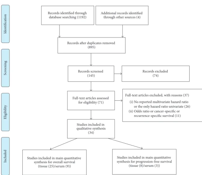
studies were identified from the searches in the PubMed, CINAHL, Embase, and Google scholar databases. 750 studies were excluded using these criteria; unpublished, non-English, letters or abstracts, withdrawn articles, review articles, non-human studies, or irrelevant to the current analysis. Of the remaining 145 studies, 74 were excluded because they did not have the survival data associated with miR-200 family. Of the remaining 71 studies, 26 did not include the data of hazard ratio associated with OS or PFS data, and 11 included odds ratio or univariate Cox regression HRs for survival data. Finally, 34 eligible studies were selected for the final analysis. A flow chart depicting the article selection process is shown in Figure 1.
3.2. Literature Characteristics. The main features of the 34
enrolled studies are systematically summarized in Tables 1 and 2. Briefly, these studies were published between 2011 and 2015, and the study sample sizes ranged from 30 to 373 (median 105.5) patients. A total of 4497 patient samples were included. Patient OS data were reported in 33 studies, PFS data in 11, and both OS and PFS data in 10. All studies were nonrandomized and retrospective except for one prospective study. The malignant neoplasms assessed in these studies included brain, breast, colorectal, endometrial, esophageal, gastric, hepatocellular, non-small-cell lung, ovarian, pancre-atic, and prostate cancers. Nineteen cohorts staged with I– IV cancers were included. Quantitative real-time PCR was performed in 22 studies, in situ hybridization in 2 studies, and two separate techniques in 2 studies to assess miR-200
T a ble 1: C ha rac ter ist ics o f th e eligib le st udies eval u at in g hig h miRN A exp ressio n le ve ls in ti ssue sa m p les an d p at ien t sur vi val da ta . Stu d y (ye ar ) (re f) Co u n tr y C ancer St ag e T est C u t-o ff va lue miRN A Sa m p le size M FD N ew cast le-O tt awa Q u al it y A ss essmen t Sca le Se le ct io n C om p at ibi li ty O u tc om e F en g et al .( 20 15) [13] Chin a H C C N A q R T -PCR/IS H R O C 20 0a 115 120 m ‰‰ ‰ ‰ ‰ Li et al .( 20 15 ) [1 7] Chin a H C C I–IV q R T -PCR/IS H N A 4 29 16 1 9 6 m ‰‰‰‰ ‰ ‰‰‰ L u et al .( 20 15 ) [8] Chin a GC I–IV qR T -PCR M edia n 141 14 1 6 0 m ‰‰‰ ‰ ‰ ‰ W an g et al .( 20 15 ) [1 4 ] Chin a Glio ma I–IV qR T -PCR N A 20 0 b 12 3 5 y ‰‰‰ ‰‰ ‰‰ Y ao et al .(2015) [9] Chin a B C I–III qR T -PCR C o m pa ri so n w /n o rmal gr o u p 20 0b 27 8 10 y ‰‰‰‰ ‰ ‰ ‰ Zh ao et al .( 20 15 ) [1 8] † Chin a NSCL C IIB -III B qR T -PCR M edia n 20 0 c 78 4 0 m ‰‰‰ ‰ ‰ ‰ Ca o et al .( 20 14 ) [1 9] Chin a O C I–IV qR T -PCR M edia n 20 0 a/b/c 10 0 56 m ‰‰‰‰ ‰ ‰‰‰ Diaz et al .( 20 14 ) [20 ] Sp ai n CR C II q R T -PCR M axst at R pac k ag e 4 29 12 7 120 m ‰‰‰ ‰‰ ‰‰ Ki m et al. (2 01 4 ) [2 1] Ko re a NSCL C I–IV q R T -PCR M edia n 20 0c 72 12 5 m ‰‰‰ ‰‰ ‰‰‰ Li et al .( 20 14 ) [22] † Chin a NSCL C IIIB-IV q R T -PCR M inim u m P val u e 20 0 c 15 0 18.5 [9.6 ]m ‰‰‰ ‰‰ ‰‰‰ Li u et al .( 20 14 ) [23 ] Chin a HC C I– IV ISH M ed ian 14 1 21 2 10 0 m ‰‰‰ ‰ ‰ ‰ So n g et al .( 20 14 ) [2 4 ] ∗ Chin a GC I–IV qR T -PCR M edia n/L o w est q u in tile val u es 20 0a/b/c 37 3 112 m ‰‰‰ ‰ ‰ ‰ T eje ro et al .( 20 14 ) [2 5] Sp ai n NSCL C I–III qR T -PCR M axsta t R p ac kag e 14 1/20 0c 15 5 16 0 m ‰‰‰ ‰‰ ‰‰‰ Zha n g et al .(201 4) [26] ∗ Chin a AS T III-IV qR T -PCR M edia n 20 0 b 122 120 m ‰‰‰‰ ‰‰ ‰‰‰ Zh u et al .(201 4) [11] Chin a PC I–IV qR T -PCR M ea n 141 94 20 0 m ‰‰‰‰ ‰ ‰ ‰ Zh u et al .(201 4) [15] Chin a NSCL C I–IV q R T -PCR R O C 4 29 70 30 m ‰‰ ‰ ‰ ‰ Be rg h m an s et al .( 20 13 ) [1 6 ] Eu ro p e NSCL C IV (79 % ) q R T -PCR p redic te d sco re 20 0c 38 6 0 m ‰‰‰ ‰‰ ‰‰ Hu an g et al .( 20 13 ) [2 7] Chin a H C C I-II q R T -PCR M ea n 4 29 13 8 14 0 m ‰‰‰ ‰ ‰‰‰ Li et al .( 20 13 ) [28 ] Chin a CR C I–III qR T -PCR M edia n 4 29 10 7 82 m ‰‰‰‰ ‰ ‰‰‰ T an g et al .( 20 13 ) [29 ] Chin a GC I–IV IS H M edia n 20 0 b/c 126 N A ‰‰‰ ‰ ‰ ‰ T o rr es et al .( 20 13) [10] † Eu ro p e EEC I–IV qR T -PCR M edia n/R O C 20 0c/4 29 [429 ]3 0 15 0 m ‰‰‰‰ ‰ ‰ ‰ X iao et al .( 20 13 ) [3 0] Chin a H C C I–III qR T -PCR M ea n 20 0 a 120 6 0 m ‰‰‰ ‰ ‰‰‰ Zh ao at al .( 20 13 ) [12 ] Chin a PC I–IV qR T -PCR M edia n 141 4 0 50 m ‰‰ ‰ ‰ ‰ Le sk el ¨a et al .(2 011) [3 1] ∗ Sp ai n O C I–IV qR T -PCR M edia n 4 29 [14 1, 20 0a/b/c ,429 ]7 2 12 8 m ‰‰ ‰ ‰ ‰ M ar chini et al .( 20 11) [3 2] ∗ It al y O C I q R T -PCR C o n ta l& O ’Q uig le y met h o d 200b/c (A ) 89 24 0 m ‰‰ ‰ ‰ ‰ 200c (B ) 55 Y u et al .( 20 10 ) [3 3] Ja p an PC I–IV qR T -PCR M edia n 20 0 c 9 9 101 m ‰‰‰ ‰ ‰‰‰ [Va lu e] indica tes th e m icr o RN A typ e o r m axim um fo llo w -u p d ura tio n fo r p rogr essio n-f ree sur vi va l. M F D : m ax im al fo ll o w -up du ra ti on , A ST : as tro cy to m a, B C : bre as t ca n ce r, C RC : co lore ct al can ce r, E E C : en d om et ri oi d en d om et ri al ca rc in om a, E O C : ep it hel ia l o var ia n canc er , E SC : es oph age al sq u amou s ca nc er , GC: gastr ic ca ncer ,H C C :h epa to cell u la r ca rcino ma, O C: o va ria n ca n cer ,PC :p an cr ea tic ca n cer ,P rC: castra tio n -r es is ta n t p ro sta te ca n ce r, N SC L C :no n-sma ll-cell lu n g ca ncer ,R O C ,r ecei ve r o p era tin g cha rac te ri stic an al ysis, N A: no t ava ila b le ,m o: mo n th s, w k: w eeks, an d y: ye ar s. ∗St ud y rep o rtin g b o th o vera ll sur vi va la nd p rogr essio n-f ree sur vi va lda ta . †Stu d y re p or ti ng on ly pro gre ss io n -f re e su rv iv al d at a.
T a ble 2: C ha rac ter ist ics o f th e eligib le st udies eval u at in g hig h miRN A exp ressio n le ve ls in se ru m sa m p les an d p at ien t sur vi val da ta . St ud y (y ea r) C o u n tr y C ancer St ag e T est C u t-o ff va lue miRN A Sa m p le size M FD N ew cast le-O tt awa Q u al it y A ss essmen t Sca le Se lec tio n C o m pa tib ili ty O u tc o m e An tol ´ın et al .( 20 15) [3 4 ] ∗ Sp ai n B C I–IV q R T -PCR R O C 20 0c/1 4 1 57 26 5 [23 5]w ‰‰‰ ‰‰ ‰‰‰ Lin et al .(201 4) [3 5] A u stralia P rC IV qR T -PCR M edia n 20 0 b 97 62 m ‰‰‰ ‰ ‰‰‰ Li u et al .(201 4) [3 6] China H C C I–IV qR T -PCR M edia n 20 0 a 13 6 50 m ‰‰ ‰ ‰ ‰ Zh u et al .(201 4) [15] China N SCL C I–IV qR T -PCR R O C 4 29 70 30 m ‰‰ ‰ ‰ ‰ Y u et al .( 2013) [3 7] C hina ESC III-IV qR T -PCR M edia n 20 0 c 157 50 m ‰‰ ‰‰ ‰‰ T an ak a et al .( 20 13 ) [3 8] ∗ Ja pa n E SC I–IV qR T -PCR C o m pa ri so n w /n o rmal gr o u p 20 0c 6 4 2 y ‰‰‰ ‰ ‰ ‰ T o iy am a et al .(201 4) [3 9] Ja pa n C R C I–IV qR T -PCR R O C 20 0c 32 1 6 0 m ‰‰‰ ‰ ‰ ‰ V allada res-A ye rb es et al .( 20 12) [4 0] ∗ Sp ai n G C I– IV q R T -P C R M ean C o m p ar is on w /nor m al gr oup 20 0 c 52 6 0 m ‰‰‰ ‰‰ ‰‰‰ Chen g et al .(2 011) [41] Chin a CR C I–IV q R T -PCR R O C 14 1 (Ti an ji n ) 15 6/10 2 50/10 0 m ‰‰‰ ‰ ‰‰‰ USA 141 (T exG en) [Va lu e] indica tes micr oRN A typ e o r maxim u m fo llo w-u p d u ra tio n fo r p ro gr essio n -f re e sur vi va l. M F D : m ax im al fo ll o w -up du ra ti on , A ST : as tro cy to m a, B C : bre as t ca n ce r, C RC : co lore ct al can ce r, E E C : en d om et ri oi d en d om et ri al ca rc in om a, E O C : ep it hel ia l o var ia n canc er , E SC : es oph age al sq u amou s ca nc er , GC: gastr ic ca ncer ,H C C :h epa to cell u la r ca rcino ma, O C: o va ria n ca n cer ,PC :p an cr ea tic ca n cer ,P rC: castra tio n -r es is ta n t p ro sta te ca n ce r, N SC L C :no n-sma ll-cell lu n g ca ncer ,R O C ,r ecei ve r o p era tin g cha rac te ri stic an al ysis, N A: no t ava ila b le ,m o: mo n th s, w k: w eeks, an d y: ye ar s. ∗St ud y rep o rtin g b o th o vera ll sur vi va la nd p rogr essio n-f ree sur vi va lda ta .
Studies included in main quantitative synthesis for overall survival
(tissue (25)/serum (9)) In cl uded S cr eenin g E ligib ili ty Iden tifica tio
n Records identified through
database searching (1192)
Records screened (145)
Records excluded (74)
Full-text articles assessed for eligibility (71)
Full-text articles excluded, with reasons (37) (i) No reported multivariate hazard ratio or the only hazard ratio univariate (26) (ii) Odds ratio or cancer-specific or
recurrence-specific survival (11) Studies included in
qualitative synthesis (34)
PRISMA 2009 Flow Diagram
Studies included in main quantitative synthesis for progression-free survival
(tissue (8)/serum (3)) Additional records identified
through other sources (4)
Records after duplicates removed (895)
Figure 1: Flow chart of the selection process of the eligible articles.
family expression. Tissue (in 26 studies), serum (in 9 studies), and both tissue and serum samples (in 1 study) were used to determine miR-200 expression.
3.3. Quality Assessment and Meta-Analysis. We
systemat-ically assessed the quality of all non-randomized studies included in the meta-analysis based on the Newcastle-Ottawa Scale criteria. The following aspects of each study were evaluated based on the (1) selection of the study groups, (2) comparability of the groups, and (3) ascertainment of either the exposure or outcome of interest. These criteria were assessed on a star scoring system, with higher scores given to higher-quality studies. The quality assessment is summarized in Tables 1 and 2.
3.4. Overall Effects of miR-200 Expression in Cancer Tissues on OS and PFS. Because a growing body of evidence suggests
that miRNA function differs between cancer tissue and blood [44, 45], the prognostic role of miR-200 family members
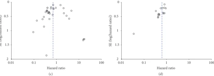
in both tumor tissue and serum was evaluated. Twenty-five studies on miR-200 expression in tissue samples were evaluated for OS analysis (Figure 2(a)) using a
random-effects model due to high heterogeneity (OS,𝑃 < 0.00001,
𝐼2 = 85%). Pooled HRs and 95% CIs were calculated. The
pooled results showed that high miR-200 expression was a favorable prognostic factor in patients with various types of cancer (pooled HR = 0.70, 95% CI 0.54–0.91). In addition, the PFS analysis of seven studies revealed a protective role for increased miR-200 tissue expression (pooled HR = 0.63, 95% CI 0.52–0.76), as determined using a random-effects model
(𝑃 = 0.03, 𝐼2= 44%; Figure 2(b)).
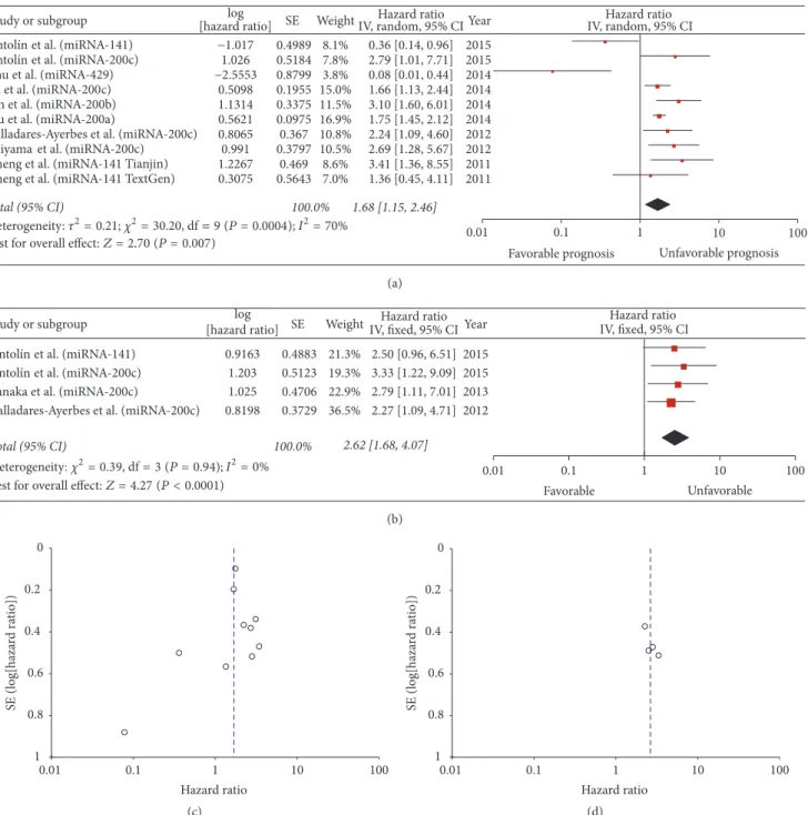
3.5. Overall Effects of Circulating miR-200 Expression on OS and PFS. The prognostic role of circulating miR-200
family members on OS was evaluated in eight studies, and heterogeneity was apparent among studies (𝑃 = 0.0004,
𝐼2 = 70%). We found that higher expression of circulating
Hazard ratio
Study or subgroup [hazard ratio] SE Weight IV, random, 95% CIlog IV, random, 95% CIHazard ratio
2.9% 0.4436 3.3% 0.3509 3.4% 0.3463 2.6% 0.5211 0.2106 3.9% 3.9% 0.2242 3.1% 0.4051 1.5% 0.8584 0.4804 2.8% 3.9% 0.2176 1.2964 0.8% 17.26 [1.36, 219.05] 2014 1.2996 0.8% 16.22 [1.27, 207.13] 2014 0.5% 1.8684 3.2% 0.3819 1.3331 0.8% 0.69 [0.29, 1.64] 2015 0.33 [0.17, 0.66] 2015 0.46 [0.23, 0.91] 2015 0.28 [0.10, 0.77] 2015 2.07 [1.37, 3.13] 2015 0.57 [0.37, 0.88] 2014 0.51 [0.23, 1.12] 2014 0.28 [0.05, 1.52] 2014 2.79 [1.09, 7.15] 2014 0.72 [0.47, 1.10] 2014 15.41 [1.13, 210.15] 2014 0.36 [0.01, 14.16] 2014 0.42 [0.20, 0.89] 2014 4.0% 0.1987 0.5833 2.4% 3.8% 0.2421 4.1% 0.177 3.5% 0.3131 2.1% 0.6506 0.3034 3.6% 3.5% 0.3144 0.3865 3.2% 3.6% 0.2916 4.3% 0.0997 4.0% 0.2005 4.0% 0.2005 0.1028 4.3% 1.2% 1.0502 2.3% 0.5941 3.3% 0.3585 2.3% 0.5951 3.2% −0.3754 −1.0989 −0.7715 −1.2814 0.7275 −0.5684 −0.6831 −1.2641 1.025 −0.3285 2.8484 2.7862 −1.0114 −0.8629 2.735 −0.0726 1.3002 −0.2784 −0.5447 −0.9361 −1.9951 1.5347 −1.0017 0.7724 −0.9636 −0.3271 −0.9163 −0.9163 0.4101 −2.3645 −0.7195 −0.7324 −1.4106 −0.7885 0.3763 0.70 [0.54, 0.91] Wang et al. (miRNA-200b)
Feng et al. (miRNA-200a) Yao et al. (miRNA-200b) Lu et al. (miRNA-141) Li et al. (miRNA-429) Li et al. (miRNA-200c) Diaz et al. (miRNA-429) Tejero et al. (miRNA-141 & 200c) Song et al. (miRNA-200a) Cao et al. (miRNA-200a) Cao et al. (miRNA-200c) Zhu et al. (miRNA-429) Cao et al. (miRNA-200b) Song et al. (miRNA-200b) Kim et al. (miRNA-200c) Song et al. (miRNA-200c) Liu et al. (miRNA-141) Zhu et al. (miRNA-141) Zhao et al. (miRNA-141) Huang et al. (miRNA-429) Torres et al. (miRNA-200c) Xiao et al. (miRNA-200a) Torres et al. (miRNA-429) Tang et al. (miRNA-200c) Tang et al. (miRNA-200b) Berghmans et al. (miRNA-200c) Marchini et al. (miRNA-200 Marchini et al. (miRNA-200b) Marchini et al. (miRNA-200 Yu et al. (miRNA-200c) 0.93 [0.63, 1.37] 3.67 [1.17, 11.51] 2014 0.76 [0.47, 1.22] 2014 0.58 [0.41, 0.82] 2014 0.39 [0.21, 0.72] 2014 0.14 [0.04, 0.49] 2013 4.64 [2.56, 8.41] 2013 0.37 [0.20, 0.68] 2013 2.16 [1.01, 4.62] 2013 0.38 [0.22, 0.68] 2013 0.72 [0.59, 0.88] 2013 0.40 [0.27, 0.59] 2013 0.40 [0.27, 0.59] 2013 1.51 [1.23, 1.84] 2013 0.09 [0.01, 0.74] 2011 0.49 [0.15, 1.56] 2011 0.48 [0.24, 0.97] 2011 0.24 [0.08, 0.78] 2011 0.45 [0.22, 0.95] 2010 Total (95% CI) 100.0% Year 100 10 Unfavorable prognosis 1 0.1 0.01 Favorable prognosis 2014
Test for overall effect:Z = 2.68 (P = 0.007)
Heterogeneity:2= 0.40; 2= 216.44, df== 33 (P < 0.00001);I2= 85%
Li et al. (miRNA-429)
Leskel̈a et al. (miRNA-429)
Zhang et al. (miRNA-200b )∗
Zhang et al. (miRNA-200b)†
§ c) ‡ c) (a) −0.7372 0.444 3.8% −0.3439 0.3732 4.9% −0.1985 0.1946 10.6% −1.0757 0.3066 6.5% −0.0584 0.21 9.9% −0.5906 0.2129 9.8% −0.4005 0.2031 10.2% −0.1985 0.0785 16.4% −0.8699 0.5379 2.8% −0.848 0.5117 3.0% −0.7419 0.4209 4.1% −3.3524 1.1067 0.7% −0.8065 0.4128 4.2% −0.8544 0.4421 3.8% −0.3001 0.3955 4.5% −0.1989 0.3822 4.8%
Zhang et al. (miRNA-200 Zhang et al. (miRNA-200 Song et al. (miRNA-200b) Zhao et al. (miRNA-200c) Song et al. (miRNA-200c) Li et al. (miRNA-200c) Song et al. (miRNA-200a) Torres et al. (miRNA-429) Marchini et al. (miRNA-200 Marchini et al. (miRNA-200b) Marchini et al. (miRNA-200
Total (95% CI) 100.0% 0.48 [0.20, 1.14] 2015 0.71 [0.34, 1.47] 2015 0.82 [0.56, 1.20] 2014 0.34 [0.19, 0.62] 2014 0.94 [0.63, 1.42] 2014 0.55 [0.36, 0.84] 2014 0.67 [0.45, 1.00] 2014 0.82 [0.70, 0.96] 2013 0.42 [0.15, 1.20] 2011 0.43 [0.16, 1.17] 2011 0.48 [0.21, 1.09] 2011 0.04 [0.00, 0.31] 2011 0.45 [0.20, 1.00] 2011 0.43 [0.18, 1.01] 2011 0.74 [0.34, 1.61] 2011 0.82 [0.39, 1.73] 2011 0.63 [0.52, 0.76]
Study or subgroup [hazard ratio]log SE Weight IV, random, 95% CIHazard ratio IV, random, 95% CIHazard ratio
0.1 0.01 Favorable 1 10 100 Unfavorable Year
Leskel ̈aet al. (miRNA-200a)
Leskel ̈aet al. (miRNA-200b)
Leskel ̈a
Leskel ̈a
Leskel ̈aet al. (miRNA-429)
et al. (miRNA-200c) et al. (miRNA-141)
Test for overall effect:Z = 4.82 (P < 0.00001)
Heterogeneity:2= 0.05; 2= 26.62, df == 15 (P = 0.03); I2= 44% b)∗ b)† § c) ‡ c) (b) Figure 2: Continued.
0.01 1000.1 1 10 Hazard ratio 2 1.5 1 0.5 0 SE (log[haza rd ra tio]) (c) 0.01 1000.1 1 10 Hazard ratio 2 1.5 1 0.5 0 SE (log[haza rd ra tio]) (d)
Figure 2: Forest plot of hazard ratios for the prediction of overall (a) and progression-free survival (b) by high-expressing miR-200 family members in tissue samples. Funnel plot showing publication bias of the overall (c) and progression-free survival (d) prediction by high-expressing miR-200 family members in tissue samples.∗Sample from grade IV astrocytoma.†Sample from grade III astrocytoma.‡,§Samples from different tissue collection.
95% CI 1.15–2.46; Figure 3(a)). PFS analysis of three studies (Figure 3(b)) demonstrated a significant association between circulating miR-200 levels and PFS (pooled HR = 2.62, 95% CI 1.68–4.07).
3.6. Subgroup Analyses of OS and PFS. To evaluate intrastudy
inconsistencies and heterogeneity, the studies were stratified by the variables shown in Table 1. The heterogeneity decreased in meta-analyses of OS and PFS when the studies were stratified by the primary tumor site and individual miRNA. Pooled analyses of the brain tumor and pancreatic cancer subgroups indicated that tissue miR-200 family expression was positively correlated with OS (pooled HR = 0.51, 95% CI 0.32–0.82 in brain tumor subgroup; pooled HR = 0.35, 95% CI 0.21–0.60 in pancreatic cancer subgroup), with low
heterogeneity among the studies analyzed (𝑃 = 0.71, 𝐼2= 0%
in brain tumor subgroup;𝑃 = 0.26, 𝐼2 = 26% in pancreatic
cancer subgroup; Supplementary Figure 1A). In the stratified analyses of PFS, increased tissue miR-200 expression was sig-nificantly associated with increased PFS in the ovarian cancer subgroup (pooled HR = 0.50, 95% CI 0.35–0.72) with low
het-erogeneity (𝑃 = 0.26, 𝐼2 = 21%; Supplementary Figure 1B).
In contrast, a pooled analysis of the colorectal cancer sub-group showed that serum miR-200 expression was negatively correlated with OS (pooled HR = 2.50, 95% CI 1.50–4.18)
with low heterogeneity (𝑃 = 0.44, 𝐼2 = 0%; Supplementary
Figure 2A). In the breast cancer subgroup, circulating miR-200 expression showed a significantly negative correlation with PFS (pooled HR = 2.87, 95% CI 1.43–5.73) with low
heterogeneity (𝑃 = 0.69, 𝐼2= 0%, Supplementary Figure 2B).
Among the subgroup analyses stratified by individual miRNAs, a pooled analysis of the miR-141 subgroup indicated that increased tissue expression was significantly correlated with enhanced OS (pooled HR = 0.38, 95% CI 0.23–0.64), which was determined using a random-effects model given the moderate heterogeneity among the studies (𝑃 = 0.09,
𝐼2 = 53%; Supplementary Figure 3A). In addition, the
high miR-200b subgroup showed a longer PFS than that of the low miR-200b subgroup (pooled HR = 0.71, 95% CI 0.54–0.94), which was determined using a fixed-effects model given the low heterogeneity among the studies (𝑃 =
0.68, 𝐼2 = 0%; Supplementary Figure 3B). In contrast, the
analysis stratified by circulating miRNA levels showed that circulating miR-200c expression was negatively correlated with OS (pooled HR = 1.97, 95% CI 1.47–2.65; Supplementary Figure 4A) and PFS (pooled HR = 2.65, 95% CI 1.61–4.35) which was determined using a fixed-effects model given the
low heterogeneity among the studies (𝑃 = 0.83, 𝐼2 = 0%;
Supplementary Figure 4B).
4. Discussion
MiRNAs have numerous advantages over mRNAs for predi-cating clinical outcomes in cancer patients, because miRNAs are posttranscriptional regulators of multiple target genes and are involved in various cellular pathways [1]. Thus, miR-NAs potentially regulate complex biological processes and biomarkers involved in cancer prognosis [4]. Although the miR-200 family is a determinant of epithelial cell phenotypes, its prognostic role has not yet been elucidated. In addition, increasing evidence suggests that miRNAs have different roles in tumor tissues and blood [44, 45], and thus the prognostic roles of miR-200 family members in both tumor and serum samples were analyzed in this study. This systemic review and meta-analysis showed that elevated cancer tissue expression of miR-200 was associated with longer survival in patients with multiple carcinoma types. In contrast, high lev-els of miR-200 in serum were associated with poor prognosis. Recently, two meta-analyses on the prognostic value of miR-200 were published. Shi and Zhang [46] evaluated seven ovarian cancer studies and showed that high expression of miR-200c may predict improved survival (OS: HR = 0.34, 95% CI 0.20–0.58; PFS: HR = 0.64, 95% CI 0.50–0.82). However, this study focused on ovarian cancer and cannot be
8.1% −1.017 0.4989 1.026 0.5184 7.8% 3.8% −2.5553 0.8799 0.5098 0.1955 15.0% 1.1314 0.3375 11.5% 0.5621 0.0975 16.9% 0.8065 0.367 10.8% 0.991 0.3797 10.5% 1.2267 0.469 8.6% 0.3075 0.5643 7.0%
Antol et al. (miRNA-141) Zhu et al. (miRNA-429) Lin et al. (miRNA-200b) Liu et al. (miRNA-200a) Toiyama et al. (miRNA-200c) Cheng et al. (miRNA-141 Tianjin) Cheng et al. (miRNA-141 TextGen)
Total (95% CI) 100.0% 0.36 [0.14, 0.96] 2015 2.79 [1.01, 7.71] 2015 0.08 [0.01, 0.44] 2014 1.66 [1.13, 2.44] 2014 3.10 [1.60, 6.01] 2014 1.75 [1.45, 2.12] 2014 2.24 [1.09, 4.60] 2012 2.69 [1.28, 5.67] 2012 3.41 [1.36, 8.55] 2011 1.36 [0.45, 4.11] 2011 1.68 [1.15, 2.46]
Study or subgroup [hazard ratio]log SE WeightIV, random, 95% CIHazard ratio Year IV, random, 95% CIHazard ratio
Test for overall effect:Z = 2.70 (P = 0.007) 10 100
Unfavorable prognosis 1 0.1 0.01 Favorable prognosis Heterogeneity:2= 0.21; 2= 30.20, df == 9 (P = 0.0004); I2= 70% Yu et al. (miRNA-200c) ´ın
Antol et al. (miRNA-200c)´ın
Valladares-Ayerbes et al. (miRNA-200c)
(a)
0.9163 0.4883
1.203 0.5123
1.025 0.4706
0.8198 0.3729
Tanaka et al. (miRNA-200c)
21.3% 19.3% 22.9% 36.5% 100.0% 2.62 [1.68, 4.07] 2015 2015 2013 2012 Total (95% CI) 2.50 [0.96, 6.51] 3.33 [1.22, 9.09] 2.79 [1.11, 7.01] 2.27 [1.09, 4.71]
Test for overall effect:Z = 4.27 (P < 0.0001)
Study or subgroup [hazard ratio] SE Weightlog IV, fixed, 95% CIHazard ratio Year IV, fixed, 95% CIHazard ratio
0.1 0.01 Favorable 10 100 1 Unfavorable Heterogeneity:2= 0.39, df = 3 (P = 0.94); I2= 0%
Antol et al. (miRNA-141)´ın
Antol et al. (miRNA-200c)´ın
Valladares-Ayerbes et al. (miRNA-200c)
(b) 0.01 1000.1 1 10 Hazard ratio 1 0.8 0.6 0.4 0.2 0 SE (log[haza rd ra tio]) (c) 0.01 1000.1 1 10 Hazard ratio 1 0.8 0.6 0.4 0.2 0 SE (log[haza rd ra tio]) (d)
Figure 3: Forest plot of hazard ratios for the prediction of overall (a) and progression-free survival (b) by high-expressing miR-200 family members in serum samples. Funnel plot showing publication bias of the overall (c) and progression-free survival (d) prediction by high-expressing miR-200 family members in serum samples.
applied to other cancer types due to population heterogeneity and a small sample size. Wu et al. [47] found that miR-200c was not significantly correlated with either OS (HR = 1.41,
95% CI 0.95–2.10;𝑃 = 0.09) or PFS (HR = 1.12, 95% CI
0.68–1.84;𝑃 = 0.67) in various types of cancer. However,
considering that some miRNAs have similar functions as their target genes, evaluating a set of miRNAs is preferable compared with a single miRNA to increase the prediction power. For example, Song et al. identified a signature of 17
miRNAs, which included the miR-200 family, in patients with gastric cancer [24]. This miRNA risk signature remained a strong predictor of survival (𝑃 = 0.015 and 𝑃 = 0.006 for OS and PFS, resp.) in a multivariate analysis, compared with analysis of an individual miR-200 family member. This suggests that a panel of miRNAs is a better predictor of survival than is an individual miRNA. Therefore, we evaluated all five miR-200 family members instead of a single miRNA in this meta-analysis.
The results of this meta-analysis showed a pooled HR of 0.70 (95% CI 0.54–0.91), demonstrating that increased miR-200 family expression in cancer tissues is associated with a favorable outcome (𝑃 = 0.007). Furthermore, in a subgroup analysis based on tumor type, a statistically significant differ-ence in OS was observed between brain and pancreatic cancer subgroups, with pooled HRs of 0.51 and 0.35, respectively. Subgroup analyses also showed that miR-141 and miR-200b were associated with favorable OS, with pooled HRs of 0.40 and 0.58, respectively. The miR-200 family has regulatory functions in diverse biological processes. Zhu et al. described miR-141 as a significant tumor suppressor in pancreatic can-cer, as it interferes with the proliferative pathway mediated by Yes-associated protein-1 [11]. In addition, the miR-200 family inhibits EMT by regulating a number of target genes such as ZEB1 and ZEB2 [5]. MiR-200c strongly suppressed mammary duct formation from normal mammary stem cells and tumor formation from breast cancer stem cells in vivo by targeting B lymphoma Mo-MLV insertion region 1 homolog, a regulator of stem cell self-renewal [48]. In addition, downregulation of miR-200 family members has been associated with resistance to cytotoxic chemotherapeutic agents and EGFR inhibitors [16, 22, 31, 49, 50]. In addition, this may be mediated by two antiapoptotic factors, B-cell lymphoma 2 and X-linked inhibitor of apoptosis protein [51]. Taken together, the miR-200 family can affect cancer progression by regulating various cell signaling and genetic pathways.
Interestingly, the miR-200 levels in plasma and tumor tissues had opposing associations with survival in this study. The pooled outcome from the OS and PFS analyses revealed HRs of 1.68 (𝑃 = 0.007) and 2.62 (𝑃 < 0.001), respectively, showing that increased circulating miR-200 family expres-sion is associated with unfavorable survival. Similarly, Wu et al.’s meta-analysis indicated that higher blood levels of miR-200c were significantly associated with poor OS (HR = 2.10,
95% CI 1.52–2.90,𝑃 < 0.00001), but there was no significant
association in tumor tissue (HR = 1.41, 95% CI 0.95–2.10; 𝑃 = 0.09) [47]. MiR-200 family members are increased in the blood of patients with breast [34], prostate [35], esophageal [37], gastric [40], ovarian [52], and metastatic colorectal can-cers [41]. MiR-200 expression is correlated with metastasis and relapse in breast cancer [34]. Moreover, expression of miRNA, including miR-200, may be an early predictor of chemotherapy outcomes in prostate and esophageal cancers [35, 37]. In 258 cases of colorectal cancer [41], high levels of plasma miR-141 were associated with unfavorable OS (HR = 2.40, 95% CI 1.182–4.86). The reason for the discrepancies between cancer tissue and circulating levels is likely explained by the different functions of miRNAs in extracellular vesicles compared with tissue miRNAs. Le et al. reported that miR-200 family members are secreted by highly metastatic epithe-lial breast cancer cells and that the secretion of these miRNAs results in increased metastatic potential in xenograft models [45]. The authors proposed that the miR-200 family is po-tentially involved in promoting the last step of the metastatic cascade in the development of macroscopic metastatic masses at distant sites.
It is unknown whether miRNA expression in the systemic circulation reflects their expression in cancer tissues. Some
studies have shown no correlation between miR-200 levels in serum and tumor tissues [53]. However, Tsujiura et al. found that the levels of plasma oncomiRNAs, including miR-21 and miR-106b, may reflect tumor miRNA levels [54]. Fur-thermore, a previous meta-analysis of miR-21 demonstrated that high miR-21 expression in both tissues and the circu-lation predicted poor outcomes [55]. Clinically, circulating biomarkers have numerous advantages, including easy access for monitoring, and their evaluation is therefore preferred for predicting early diagnosis, prognosis, and individualized treatments. However, there are still many barriers to over-come before utilizing circulating miRNAs as diagnostic or prognostic biomarkers in the clinic. These barriers include clarifying miRNA correlations between tumor tissues and cir-culation, normalizing data from different studies using refer-ence genes [56] or internal controls [57], and developing sen-sitive, specific, reliable, reproducible, and inexpensive detec-tion methods. In addidetec-tion, circulating miRNA expression can be significantly altered by physiological or pathological con-ditions, such as pregnancy, heart failure, or sepsis [57]. There-fore, further clarification on the clinical roles of circulating miR-200 family members in well-designed prospective stud-ies is needed.
Our meta-analysis has several limitations. Marked het-erogeneity among the subjects was present in the OS and PFS groups. The heterogeneity of the population was likely due to differences in sample size, baseline patient characteristics (e.g., age, cancer type, tumor stage, and treatment type), follow-up duration, detection methods, and cut-off values. Thus, we only selected high-quality studies using a quality assessment based on the Newcastle-Ottawa Scale. When the studies were stratified by tumor type, heterogeneity was no longer detected in the brain tumor and pancreatic cancer subgroups (𝑃 = 0.71 and 𝑃 = 0.26, resp.).
In conclusion, our meta-analysis suggests that the miR-200 family members are potential biomarkers and accurate prognostic predictors in patients with various carcinomas. The decreased tumor expression of the miR-200 family was significantly associated with poor survival in patients with brain, pancreas, and ovarian cancers. In contrast, low circulating miR-200 levels were associated with a positive prognosis in patients with colon and breast cancers. For future clinical application, large prospective studies are needed to validate the prognostic values of circulating miR-200 in individual cancer types.
Competing Interests
The authors declare that they have no competing interests.
Authors’ Contributions
Jung Soo Lee and Yoon Ho Ko performed acquisition, analy-sis, and interpretation of data and drafted the article; Young-Ho Ahn, Der Sheng Sun, Yeo Hyung Kim, and Hye Sung Won revised the article for important intellectual content; all the authors performed final approval of the version to be published.
Acknowledgments
This study was supported by Basic Science Research Program through the National Research Foundation of Korea (NRF) funded by the Ministry of Science, ICT & Future Planning (NRF-2015R1C1A1A01054591) (Yoon Ho Ko)
References
[1] D. P. Bartel, “MicroRNAs: genomics, biogenesis, mechanism, and function,” Cell, vol. 116, no. 2, pp. 281–297, 2004.
[2] H.-W. Hwang and J. T. Mendell, “MicroRNAs in cell prolifera-tion, cell death, and tumorigenesis,” British Journal of Cancer, vol. 94, no. 6, pp. 776–780, 2006.
[3] G. A. Calin, C. Sevignani, C. D. Dumitru et al., “Human microRNA genes are frequently located at fragile sites and genomic regions involved in cancers,” Proceedings of the National Academy of Sciences of the United States of America, vol. 101, no. 9, pp. 2999–3004, 2004.
[4] Y. W. Kong, D. Ferland-McCollough, T. J. Jackson, and M. Bushell, “MicroRNAs in cancer management,” The Lancet Oncology, vol. 13, no. 6, pp. e249–e258, 2012.
[5] M. Korpal and Y. Kang, “The emerging role of miR-200 family of microRNAs in epithelial-mesenchymal transition and cancer metastasis,” RNA Biology, vol. 5, no. 3, pp. 115–119, 2008. [6] B. P. Lewis, C. B. Burge, and D. P. Bartel, “Conserved seed
pairing, often flanked by adenosines, indicates that thousands of human genes are microRNA targets,” Cell, vol. 120, no. 1, pp. 15–20, 2005.
[7] P. S. Choi, L. Zakhary, W.-Y. Choi et al., “Members of the miRNA-200 family regulate olfactory neurogenesis,” Neuron, vol. 57, no. 1, pp. 41–55, 2008.
[8] Y. B. Lu, J. J. Hu, W. J. Sun, X. H. Duan, and X. Chen, “Prognostic value of miR-141 downregulation in gastric cancer,” Genetics and Molecular Research, vol. 14, no. 4, pp. 17305–17311, 2015. [9] Y. Yao, J. Hu, Z. Shen et al., “MiR-200b expression in breast
cancer: a prognostic marker and act on cell proliferation and apoptosis by targeting Sp1,” Journal of Cellular and Molecular Medicine, vol. 19, no. 4, pp. 760–769, 2015.
[10] A. Torres, K. Torres, A. Pesci et al., “Diagnostic and prognostic significance of miRNA signatures in tissues and plasma of endometrioid endometrial carcinoma patients,” International Journal of Cancer, vol. 132, no. 7, pp. 1633–1645, 2013.
[11] Z.-M. Zhu, Y.-F. Xu, Q.-J. Su et al., “Prognostic significance of microRNA-141 expression and its tumor suppressor function in human pancreatic ductal adenocarcinoma,” Molecular and Cellular Biochemistry, vol. 388, no. 1-2, pp. 39–49, 2014. [12] G. Zhao, B. Wang, Y. Liu et al., “MiRNA-141, downregulated
in pancreatic cancer, inhibits cell proliferation and invasion by directly targeting MAP4K4,” Molecular Cancer Therapeutics, vol. 12, no. 11, pp. 2569–2580, 2013.
[13] J. Feng, J. Wang, M. Chen et al., “MiR-200a suppresses cell growth and migration by targeting MACC1 and predicts prog-nosis in hepatocellular carcinoma,” Oncology Reports, vol. 33, no. 2, pp. 713–720, 2015.
[14] B. Wang, M. Li, Z. Wu et al., “Associations between SOX2 and miR-200b expression with the clinicopathological characteris-tics and prognosis of patients with glioma,” Experimental and Therapeutic Medicine, vol. 10, no. 1, pp. 88–96, 2015.
[15] W. Zhu, J. He, D. Chen et al., “Expression of 29c, miR-93, and miR-429 as potential biomarkers for detection of early
stage non-small lung cancer,” PLOS ONE, vol. 9, no. 2, Article ID e87780, 2014.
[16] T. Berghmans, L. Ameye, L. Willems et al., “Identification of microRNA-based signatures for response and survival for non-small cell lung cancer treated with cisplatin-vinorelbine A ELCWP prospective study,” Lung Cancer, vol. 82, no. 2, pp. 340– 345, 2013.
[17] L. Li, J. Tang, B. Zhang et al., “Epigenetic modification of MiR-429 promotes liver tumour-initiating cell properties by targeting Rb binding protein 4,” Gut, vol. 64, no. 1, pp. 156–167, 2015.
[18] J. Zhao, Y. Zhao, Z. Wang, Y. Xuan, Y. Luo, and W. Jiao, “Loss expression of micro ribonucleic acid (miRNA)-200c induces adverse post-surgical prognosis of advanced stage non-small cell lung carcinoma and its potential relationship with ET𝐴R messenger RNA,” Thoracic Cancer, vol. 6, no. 4, pp. 421–426, 2015.
[19] Q. Cao, K. Lu, S. Dai, Y. Hu, and W. Fan, “Clinicopathological and prognostic implications of the miR-200 family in patients with epithelial ovarian cancer,” International Journal of Clinical and Experimental Pathology, vol. 7, no. 5, pp. 2392–2401, 2014. [20] T. Diaz, R. Tejero, I. Moreno et al., “Role of miR-200 family
members in survival of colorectal cancer patients treated with fluoropyrimidines,” Journal of Surgical Oncology, vol. 109, no. 7, pp. 676–683, 2014.
[21] M. K. Kim, S. B. Jung, J.-S. Kim et al., “Expression of microRNA miR-126 and miR-200c is associated with prognosis in patients with non-small cell lung cancer,” Virchows Archiv, vol. 465, no. 4, pp. 463–471, 2014.
[22] J. Li, X. Li, S. Ren et al., “miR-200c overexpression is associated with better efficacy of EGFR-TKIs in non-small cell lung cancer patients with EGFR wild-type,” Oncotarget, vol. 5, no. 17, pp. 7902–7916, 2014.
[23] Y. Liu, Y. Ding, J. Huang et al., “MiR-141 suppresses the migration and invasion of HCC cells by targeting Tiam1,” PLoS ONE, vol. 9, no. 2, Article ID e88393, 2014.
[24] F. Song, D. Yang, B. Liu et al., “Integrated microRNA network analyses identify a poor-prognosis subtype of gastric cancer characterized by the miR-200 family,” Clinical Cancer Research, vol. 20, no. 4, pp. 878–889, 2014.
[25] R. Tejero, A. Navarro, M. Campayo et al., “MiR-141 and miR-200c as markers of overall survival in early stage non-small cell lung cancer adenocarcinoma,” PLoS ONE, vol. 9, no. 7, Article ID e101899, 2014.
[26] J.-Q. Zhang, Q.-H. Yao, Y.-Q. Kuang et al., “Prognostic value of coexistence of abnormal expression of micro-RNA-200b and cyclic adenosine monophosphate-responsive element-binding protein 1 in human astrocytoma,” Human Pathology, vol. 45, no. 10, pp. 2154–2161, 2014.
[27] X.-Y. Huang, J.-G. Yao, H.-D. Huang et al., “MicroRNA-429 modulates hepatocellular carcinoma prognosis and tumorige-nesis,” Gastroenterology Research and Practice, vol. 2013, Article ID 804128, 10 pages, 2013.
[28] J. Li, L. Du, Y. Yang et al., “MiR-429 is an independent prog-nostic factor in colorectal cancer and exerts its anti-apoptotic function by targeting SOX2,” Cancer Letters, vol. 329, no. 1, pp. 84–90, 2013.
[29] H. Tang, M. Deng, Y. Tang et al., “miR-200b and miR-200c as prognostic factors and mediators of gastric cancer cell progression,” Clinical Cancer Research, vol. 19, no. 20, pp. 5602– 5612, 2013.
[30] F. Xiao, W. Zhang, L. Zhou et al., “MicroRNA-200a is an independent prognostic factor of hepatocellular carcinoma and induces cell cycle arrest by targeting CDK6,” Oncology Reports, vol. 30, no. 5, pp. 2203–2210, 2013.
[31] S. Leskel¨a, L. J. Leandro-Garc´ıa, M. Mendiola et al., “The miR-200 family controls𝛽-tubulin III expression and is associated with paclitaxel-based treatment response and progression-free survival in ovarian cancer patients,” Endocrine-Related Cancer, vol. 18, no. 1, pp. 85–95, 2011.
[32] S. Marchini, D. Cavalieri, R. Fruscio et al., “Association between miR-200c and the survival of patients with stage I epithelial ovarian cancer: a retrospective study of two independent tumour tissue collections,” The Lancet Oncology, vol. 12, no. 3, pp. 273–285, 2011.
[33] J. Yu, K. Ohuchida, K. Mizumoto et al., “MicroRNA, hsa-miR-200c, is an independent prognostic factor in pancreatic cancer and its upregulation inhibits pancreatic cancer invasion but increases cell proliferation,” Molecular Cancer, vol. 9, article 169, 2010.
[34] S. Antol´ın, L. Calvo, M. Blanco-Calvo et al., “Circulating miR-200c and miR-141 and outcomes in patients with breast cancer,” BMC Cancer, vol. 15, no. 1, article 297, 2015.
[35] H.-M. Lin, L. Castillo, K. L. Mahon et al., “Circulating microR-NAs are associated with docetaxel chemotherapy outcome in castration-resistant prostate cancer,” British Journal of Cancer, vol. 110, no. 10, pp. 2462–2471, 2014.
[36] M. Liu, J. Liu, L. Wang et al., “Association of serum microRNA expression in hepatocellular carcinomas treated with transarte-rial chemoembolization and patient survival,” PLoS ONE, vol. 9, no. 10, Article ID e109347, 2014.
[37] H. Yu, B. Duan, L. Jiang et al., “Serum miR-200c and clinical outcome of patients with advanced esophageal squamous can-cer receiving platinum-based chemotherapy,” American Journal of Translational Research, vol. 6, no. 1, pp. 71–77, 2013.
[38] K. Tanaka, H. Miyata, M. Yamasaki et al., “Circulating miR-200c levels significantly predict response to chemotherapy and prognosis of patients undergoing neoadjuvant chemotherapy for esophageal cancer,” Annals of Surgical Oncology, vol. 20, supplement 3, pp. S607–S615, 2013.
[39] Y. Toiyama, K. Hur, K. Tanaka et al., “Serum miR-200c is a novel prognostic and metastasis-predictive biomarker in patients with colorectal cancer,” Annals of Surgery, vol. 259, no. 4, pp. 735–743, 2014.
[40] M. Valladares-Ayerbes, M. Reboredo, V. Medina-Villaamil et al., “Circulating miR-200c as a diagnostic and prognostic biomarker for gastric cancer,” Journal of Translational Medicine, vol. 10, article 186, 2012.
[41] H. Cheng, L. Zhang, D. E. Cogdell et al., “Circulating plasma MiR-141 is a novel biomarker for metastatic colon cancer and predicts poor prognosis,” PLoS ONE, vol. 6, no. 3, Article ID e17745, 2011.
[42] A. Stang, “Critical evaluation of the Newcastle-Ottawa scale for the assessment of the quality of nonrandomized studies in meta-analyses,” European Journal of Epidemiology, vol. 25, no. 9, pp. 603–605, 2010.
[43] M. Egger, G. D. Smith, M. Schneider, and C. Minder, “Bias in meta-analysis detected by a simple, graphical test,” British Medical Journal, vol. 315, no. 7109, pp. 629–634, 1997.
[44] J. Zhu, Z. Zheng, J. Wang et al., “Different miRNA expression profiles between human breast cancer tumors and serum,” Frontiers in Genetics, vol. 5, article 149, 2014.
[45] M. T. N. Le, P. Hamar, C. Guo et al., “MiR-200-containing extracellular vesicles promote breast cancer cell metastasis,” Journal of Clinical Investigation, vol. 124, no. 12, pp. 5109–5128, 2014.
[46] C. Shi and Z. Zhang, “The prognostic value of the miR-200 family in ovarian cancer: a meta-analysis,” Acta Obstetricia et Gynecologica Scandinavica, vol. 95, no. 5, pp. 505–512, 2016. [47] J. Wu, Z. Fang, J. Xu, W. Zhu, Y. Li, and Y. Yu, “Prognostic value
and clinicopathology significance of microrna-200c expression in cancer: a meta-analysis,” PLOS ONE, vol. 10, no. 6, Article ID e0128642, 2015.
[48] Y. Shimono, M. Zabala, R. W. Cho et al., “Downregulation of miRNA-200c links breast cancer stem cells with normal stem cells,” Cell, vol. 138, no. 3, pp. 592–603, 2009.
[49] R. Hamano, H. Miyata, M. Yamasaki et al., “Overexpression of miR-200c induces chemoresistance in esophageal cancers mediated through activation of the Akt signaling pathway,” Clinical Cancer Research, vol. 17, no. 9, pp. 3029–3038, 2011. [50] J. L. Bryant, J. Britson, J. M. Balko et al., “A microRNA gene
expression signature predicts response to erlotinib in epithelial cancer cell lines and targets EMT,” British Journal of Cancer, vol. 106, no. 1, pp. 148–156, 2012.
[51] W. Zhu, H. Xu, D. Zhu et al., “miR-200bc/429 cluster modulates multidrug resistance of human cancer cell lines by targeting BCL2 and XIAP,” Cancer Chemotherapy and Pharmacology, vol. 69, no. 3, pp. 723–731, 2012.
[52] C. W. S. Kan, M. A. Hahn, G. B. Gard et al., “Elevated levels of circulating microRNA-200 family members correlate with serous epithelial ovarian cancer,” BMC Cancer, vol. 12, article 627, 2012.
[53] X.-G. Liu, W.-Y. Zhu, Y.-Y. Huang et al., “High expression of serum miR-21 and tumor miR-200c associated with poor prognosis in patients with lung cancer,” Medical Oncology, vol. 29, no. 2, pp. 618–626, 2012.
[54] M. Tsujiura, D. Ichikawa, S. Komatsu et al., “Circulating microRNAs in plasma of patients with gastric cancers,” British Journal of Cancer, vol. 102, no. 7, pp. 1174–1179, 2010.
[55] X. Zhou, X. Wang, Z. Huang et al., “Prognostic value of miR-21 in various cancers: an updating meta-analysis,” PLoS ONE, vol. 9, no. 7, Article ID e102413, 2014.
[56] G.-H. Liu, Z.-G. Zhou, R. Chen et al., “Serum 21 and miR-92a as biomarkers in the diagnosis and prognosis of colorectal cancer,” Tumor Biology, vol. 34, no. 4, pp. 2175–2181, 2013. [57] F. Wang, G. Long, C. Zhao et al., “Atherosclerosis-related
circulating miRNAs as novel and sensitive predictors for acute myocardial infarction,” PLOS ONE, vol. 9, no. 9, Article ID e105734, 2014.
Submit your manuscripts at
https://www.hindawi.com
Stem Cells
International
Hindawi Publishing Corporation
http://www.hindawi.com Volume 2014
Hindawi Publishing Corporation
http://www.hindawi.com Volume 2014
INFLAMMATION
Hindawi Publishing Corporation
http://www.hindawi.com Volume 2014
Behavioural
Neurology
Endocrinology
International Journal ofHindawi Publishing Corporation
http://www.hindawi.com Volume 2014
Hindawi Publishing Corporation
http://www.hindawi.com Volume 2014
Disease Markers
Hindawi Publishing Corporation
http://www.hindawi.com Volume 2014
BioMed
Research International
Oncology
Journal ofHindawi Publishing Corporation
http://www.hindawi.com Volume 2014
Hindawi Publishing Corporation
http://www.hindawi.com Volume 2014 Oxidative Medicine and Cellular Longevity Hindawi Publishing Corporation
http://www.hindawi.com Volume 2014
PPAR Research
The Scientific
World Journal
Hindawi Publishing Corporationhttp://www.hindawi.com Volume 2014
Immunology Research
Hindawi Publishing Corporation
http://www.hindawi.com Volume 2014
Journal of
Obesity
Journal ofHindawi Publishing Corporation
http://www.hindawi.com Volume 2014
Hindawi Publishing Corporation
http://www.hindawi.com Volume 2014 Computational and Mathematical Methods in Medicine
Ophthalmology
Journal ofHindawi Publishing Corporation
http://www.hindawi.com Volume 2014
Diabetes Research
Journal ofHindawi Publishing Corporation
http://www.hindawi.com Volume 2014
Hindawi Publishing Corporation
http://www.hindawi.com Volume 2014
Research and Treatment
AIDS
Hindawi Publishing Corporation
http://www.hindawi.com Volume 2014
Gastroenterology Research and Practice
Hindawi Publishing Corporation
http://www.hindawi.com Volume 2014