OPEN
Cellular localization of NRF2 determines the
self-renewal and osteogenic differentiation potential
of human MSCs via the P53
–SIRT1 axis
DS Yoon
1, Y Choi
1,2and JW Lee*
,1,2NRF2 (nuclear factor erythroid-derived 2-like 2) plays an important role in defense against oxidative stress at the cellular level.
Recently, the roles of NRF2 in embryonic and adult stem cells have been reported, but its role in maintaining self-renewal and
differentiation potential remains unknown. We studied the mechanisms of NRF2 action in mesenchymal stem cells (MSCs) derived
from human bone marrow. We found that the cellular localization of NRF2 changed during prolonged cell passage and osteogenic
differentiation. Blocking the nuclear import of NRF2 using ochratoxin A (OTA) induced the loss of the self-renewal and osteogenic
potential of early-passage (EP) MSCs. Conversely, reinforcing the nuclear import of NRF2 using tert-butylhydroquinone (t-BHQ)
improved the self-renewal capacity and maintained the differentiation potential in the osteogenic lineage of EP MSCs. Real-time
quantitative PCR and western blot analysis showed that NRF2 positively regulates sirtuin 1 (SIRT1) at the mRNA and protein levels
via the negative regulation of p53. The self-renewal and osteogenic potential suppressed in OTA-treated or NRF2-targeting small
hairpin RNA (shRNA)-infected EP MSCs were rescued by introducing small interfering RNA (siRNA) targeting p53. t-BHQ treatment
in late-passage (LP) MSCs, which lost their self-renewal and osteogenic potential, reversed these effects. In LP MSCs treated with
t-BHQ for
∼ 7 days, the phosphorylation and nuclear localization of NRF2 improved and SIRT1 protein level increased, whereas p53
protein levels decreased. Therefore, our results suggest that NRF2 plays an important role in regulating p53 and SIRT1 to maintain
MSC stemness. This study is the first to establish a functional link between NRF2 and SIRT1 expression in the maintenance of MSC
self-renewal and differentiation potential.
Cell Death and Disease (2016) 7, e2093; doi:10.1038/cddis.2016.3; published online 11 February 2016
Mesenchymal stem cells (MSCs) grown in ex vivo conditions
become senescent during prolonged cell passage.
Conse-quently, long-term cultured stem cells are not used for tissue
regeneration because they do not function as stem cells after
reaching therapeutically effective amounts during in vitro
cultivation. Appropriate culture methods are very important to
maintain MSC stem cell potential because their self-renewal
and multipotency decrease during ex vivo cultivation.
1–5The
decrease in the self-renewal and differentiation potentials
during ex vivo conditions may be related to exposure to
exogenous oxidative stress or to reactive oxygen species
(ROS) that are endogenously generated in in vitro cultured
cells.
6,7Hypoxic conditions have been extensively examined
with respect to the maintenance of the self-renewal and
multipotency of human MSCs during long-term cultivation.
8–11Indeed, in vivo MSC niches where the cells reside exhibit
low-oxygen tension (2–8%).
12A recent study has also
reinforced the importance of hypoxic conditions for the
maintenance of MSC stemness.
13Therefore, hypoxic
condi-tions may be essential for the maintenance of the early
characteristics of ex vivo cultured MSCs. In hypoxic
environments, the osteogenic and adipogenic potentials of
human bone marrow-derived MSCs (BM-MSCs) decrease.
13However, their self-renewal capacity is maintained.
14,15These
results indicate that hypoxic environments in MSC cultivation
maintain the undifferentiated state of MSCs without affecting
their differentiation potential, promoting stemness over
differ-entiation. However, no studies have examined the master
regulators related to the maintenance of stemness in MSC
cultivation in hypoxic environments in vitro.
Nuclear factor erythroid-derived 2-like 2 (NRF2) plays an
important role in defense against oxidative stresses.
16,17It
induces the expression of antioxidant genes that protect many
cell types from oxidative stress caused by tissue damage,
such as inflammation and injury.
18Under normal condition,
NRF2 remains in the cytoplasm, and is degraded via
ubiquitination triggered by binding with kelch
like-ECH-associated protein 1 (KEAP1).
19Under oxidative stress,
NRF2 is phosphorylated and moves from the cytoplasm to
the nucleus by disrupting the complex with KEAP1.
20,21In this
regard, the regulation of NRF2 cellular localization may be
important to overcome MSC aging during prolonged cell
1
Department of Orthopaedic Surgery, Yonsei University College of Medicine, Seoul, South Korea and2Brain Korea 21 PLUS Project for Medical Science, Yonsei University, Seoul, South Korea
*Corresponding author: JW Lee, Department of Orthopedic Surgery-29, Yonsei University College of Medicine, 250, Seongsanno, Seodaemun-gu, Seoul 120-752, South Korea. Tel: +82 2 2228 2190; Fax: +82 2 363 1139; E-mail: [email protected]
Received 23.9.15; revised 18.11.15; accepted 23.12.15; Edited by Y Shi
Abbreviations: BM-MSC, bone marrow-mesenchymal stem cell; ROS, reactive oxygen species; NRF2, nuclear factor erythroid-derived 2-like 2; CFU-F, colony-forming unit fibroblast; DAPI, 4,6-diamidino-2-phenyindole; FITC, fluorescein isothiocyanate; t-BHQ, tert-butylhydroquinone; HO-1, heme oxygenase-1; NQO-1, NAD(P)H dehydrogenase (quinone 1); OTA, ochratoxin A; RUNX2, Runt-related transcription factor 2; SIRT1, sirtuin 1; siRNA, small interfering RNA; HIC1, hypermethylated in cancer 1
Citation: Cell Death and Disease (2016) 7, e2093; doi:10.1038/cddis.2016.3
&
2016 Macmillan Publishers Limited All rights reserved 2041-4889/16 www.nature.com/cddispassages in vitro because oxidative stress induces premature
senescence of MSCs.
22NRF2 overexpression protects MSCs
against cell death and apoptosis caused by oxidative stress
and retains multi-differentiation potential.
23A recent study has
shown that NRF2 controls the self-renewal and pluripotency
of human embryonic stem cells.
24These results imply that
NRF2 may regulate MSC stemness. However, no studies to
date have clarified the relationship between NRF2 and MSC
stemness or interactions between NRF2 and stemness
genes. Enhancing NRF2 activity may mimic the effects of
hypoxic environments, similar to an in vivo MSC niche in
ex vivo cultured MSCs.
Here, we identify NRF2 as a regulator of sirtuin 1 (SIRT1).
NRF2 phosphorylation was decreased and NRF2 was exported
from the nucleus in late-passage (LP) and differentiated MSCs.
The inhibition of NRF2 activity in early-passage (EP) MSCs
suppressed the self-renewal and differentiation capacities.
NRF2 activation in EP or LP MSCs enhanced the
self-renewal capacity, but suppressed the differentiation potential.
NRF2 increased SIRT1 protein expression by decreasing
p53 protein levels. RNA interference targeting p53 in
NRF2-knockdown MSCs rescued SIRT1 protein levels as well
as the self-renewal and differentiation potentials. Our data
indicate that NRF2 plays an important role in the maintenance
of MSC stemness via p53–SIRT1 regulation.
Results
NRF2 phosphorylation and activity decreases during
long-term
cultivation
and
differentiation
in
human
BM-MSCs. We hypothesized that NRF2 expression or activity
decrease during the long-term cultivation or osteogenic
differentiation of MSCs. Accordingly, we observed the mRNA
and protein levels of NRF2 in EP or LP MSCs as well as in
those that were undifferentiated or differentiated to the
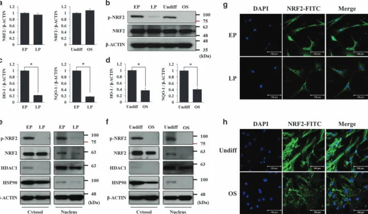
osteogenic lineage. As shown in Figures 1a and b, the mRNA
and protein levels of NRF2 did not change during prolonged
cell passage or osteogenic differentiation. The nuclear import
of NRF2 is blocked by its dephosphorylation, resulting in a
decrease in NRF2 activity.
25Consistent with this, we found that
the levels of NRF2 phosphorylation decreased during these
processes (Figure 1b, upper panel). We also confirmed that
the mRNA levels of HO-1 (heme oxygenase-1) and NQO-1
(NAD(P)H dehydrogenase (quinone 1)), which are
down-stream target genes of NRF2, decreased during these
processes (Figures 1c and d). Next, the cellular localization
of NRF2 and its phosphorylated form were analyzed by
cytosolic and nuclear fractionated western blots and
immuno-cytochemistry. The protein levels of nuclear and
phosphory-lated NRF2 were nearly detected in LP or differentiated MSCs
compared with EP or undifferentiated MSCs (Figures 1e and f).
Immunocytochemistry verified the absence of nuclear NRF2 in
the LP and differentiated MSCs (Figures 1g and h). These
results indicate that NRF2 phosphorylation and activity may be
important factors in the maintenance of the early
character-istics of MSCs, whereas the mRNA and protein levels did not
change during prolonged cell passages and the osteogenic
differentiation process. It is also thought that a decrease in
NRF2 activity may be related to loss of EP MSC stemness
during these processes. LP MSCs lose their antioxidant ability,
which is accompanied by NRF2 dephosphorylation, resulting
in higher susceptibility to oxidative stresses than that of
EP MSCs.
Regulation of NRF2 activity affects the self-renewal and
osteogenic differentiation potentials of EP MSCs. To
confirm whether NRF2 activity affects the self-renewal and
differentiation potentials of MSCs, ochratoxin A (OTA), which
is a NRF2 inhibitor,
26was used to suppress NRF2 activity in
EP MSCs. Treatment with 10
μM OTA successfully
sup-pressed the nuclear import of NRF2 and its phosphorylated
form (Figure 2a). Immunofluorescence also showed the
suppression of nuclear import (Figure 2b), and the mRNA
levels of HO-1 and NQO-1 decreased in response to OTA
treatment (Figure 2c). To assess the effects of OTA on the
self-renewal capacity of EP MSCs, proliferation and
colony-forming unit fibroblast (CFU-F) assays were performed.
There was no difference in the proliferation rates between
control EP MSCs and OTA-treated EP MSCs at day 3, but the
gap between the two groups gradually widened up to day 7
(Figure 2d). OTA significantly reduced the colony-forming
ability of EP MSCs cultured for 12 days (Figure 2e) and blocked
the ability to differentiate into the osteogenic lineage (Figure 2f).
To exclude the nonspecific effects of the compounds, small
interfering RNA (siRNA) knockdown of NRF2 and its effects on
self-renewal and differentiation of EP MSCs were evaluated,
thus resulting in the same effects as OTA treatment
(Supple-mentary Figure S1). These results showed that the
self-renewal and differentiation ability of EP MSCs are inhibited by
blocking the nuclear import and phosphorylation of NRF2.
tert-Butylhydroquinone (t-BHQ) is an activator of NRF2 and
regulates NRF2 stabilization by preventing binding with
KEAP1.
27We treated EP MSCs with 10
μM t-BHQ in order
to evaluate the self-renewal and osteogenic differentiation
potentials. We observed an increase in NRF2 phosphorylation
in the nucleus when EP MSCs were exposed to t-BHQ at a
dose of 10
μM for 12 h (Figure 3a). It was difficult to observe
the difference in the nucleic NRF2 protein levels between
control EP MSCs and t-BHQ-treated EP MSCs using
immunofluorescence because nucleic NRF2 was generally
abundant (Figure 3b). Nevertheless, the mRNA levels of HO-1
and NQO-1 were significantly higher in t-BHQ-treated EP
MSCs than in control cells (Figure 3c). t-BHQ enhanced the
proliferation rate and increased the number of colony-forming
cells in t-BHQ-treated EP-MSCs compared with control
EP-MSCs, as expected (Figures 3d and e). Unexpectedly,
the osteogenic differentiation of EP MSCs was blocked by
10
μM t-BHQ (Figure 3f). In general, pluripotent or stemness
genes, such as SOX2, NANOG, and OCT3/4, enable the
maintenance of self-renewal and undifferentiated states in
stem cells.
28Therefore, we inferred that the inhibition of
osteogenic differentiation caused by NRF2 activation in EP
MSCs was due to NRF2-mediated stemness gene regulation.
Enhanced NRF2 activity maintains the undifferentiated
state of EP MSCs by increasing SIRT1 expression at the
mRNA and protein levels. Three groups were examined.
The first group (GROUP I) was EP-MSCs grown for 10 days
in osteogenic induction medium with each vehicle for OTA or
NRF2 regulates SIRT1 to maintain MSC stemness DS Yoonet al
t-BHQ. The second group (GROUP II) was EP-MSCs grown
for 10 days in osteogenic induction medium with OTA or
t-BHQ at a dose of 10
μM. The third group (GROUP III) was
EP-MSCs grown for 7 days in an osteogenic induction
medium with OTA or t-BHQ at a dose of 10
μM. The cells
were grown for an additional 3 days in osteogenic induction
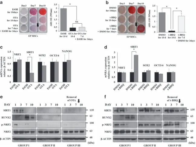
medium without OTA or t-BHQ. As shown in Figure 4a,
OTA-treated EP MSCs did not differentiate to the osteogenic
lineage. The removal of OTA on day 7 of differentiation did not
rescue the osteogenic potential of EP MSCs. t-BHQ-treated
EP MSCs also did not differentiate to the osteogenic lineage.
However, the removal of t-BHQ on day 7 significantly rescued
the osteogenic potential (Figure 4b). Next, we performed
quantitative real-time PCR (qRT-PCR) to confirm the
regula-tion of stemness genes by NRF2. As menregula-tioned previously,
SOX2, NANOG, and OCT3/4 are very important stemness
genes for the maintenance of MSC self-renewal and
multipotency,
4,29–31and SIRT1 is also thought to regulate
MSC stemness.
32The regulation of NRF2 activity by OTA or
t-BHQ treatment in EP MSCs did not affect the mRNA
expression of SOX2, OCT4, or NANOG. Among the
stem-ness genes, only SIRT1 expression was significantly affected
by OTA or t-BHQ treatment (Figures 4c and d). We previously
reported that among the general stemness genes, SOX2 may
be a key factor for maintaining self-renewal and multipotency.
Moreover, SOX2 is regulated via post-translational
modifica-tion such as acetylamodifica-tion in MSCs, and SIRT1, a lysine
deacetylase, directly modulates SOX2 through the inhibition
of SOX2 degradation via ubiquitination, although the mRNA
level of SOX2 is not changed.
32Thus, we selected SIRT1 as
a candidate gene that may be regulated by NRF2 activity. We
examined whether the t-BHQ-mediated interference of
osteogenic differentiation was due to the maintenance of
the undifferentiated state of EP MSCs by SIRT1. The p-NRF2
and SIRT1 protein levels decreased, whereas the protein
level of Runt-related transcription factor 2 (RUNX2), a master
transcription factor involved in osteogenic differentiation,
increased during the osteogenic differentiation process
(GROUP I) (Figure 4e, left). For the sustained OTA treatment
during the osteogenic differentiation of EP MSCs (GROUP II),
p-NRF2, SIRT1, and RUNX2 were not observed during the
differentiation processes (Figure 4e, middle). After the
removal of OTA on day 7 of the differentiation process,
p-NRF2, SIRT1, and RUNX2 expression levels were not
rescued (Figure 4e, right). Sustained t-BHQ treatment
maintained the protein levels of p-NRF2 and SIRT1, whereas
Figure 1 The phosphorylated level and nuclear localization of NRF2 decrease during prolonged cell passages and osteogenic differentiation. (a) EP, LP, undifferentiated, or differentiated MSCs to the osteogenic lineage were harvested at each stage to prepare cell lysates. The mRNA expression of NRF2 was analyzed by qRT-PCR, and (b) the protein levels of NRF2 and phosphorylated NRF2 were analyzed by western blot analysis. The expression level ofβ-CATENIN was used as a loading control. For the same cDNA, the mRNA expression levels of HO-1 (c) and NQO-1 (d), which are downstream targets of NRF2, were also analyzed by qRT-PCR. *Po0.05 compared with control EP or undifferentiated MSCs. (e) In the same conditions, the cell lysates were prepared and fractionated into nuclear and cytosolic extracts according to the manufacturer’s instructions. The protein levels of NRF2 and phosphorylated NRF2 were analyzed by western blot analysis in the EP and LP MSCs (e) or undifferentiated or osteogenic (OS)-differentiated MSCs (f). The protein level of HSP90 was used as a loading control for cytosolic extracts, and the protein level of HDAC1 was used as a loading control for nuclear extracts. Immunofluorescence was performed to observe the nuclear and cytosolic localization of NRF2 in the EP and LP MSCs (g) or undifferentiated or OS-differentiated MSCs (h). The nucleus was stained with DAPI, and NRF2 was stained with FITC-conjugated secondary antibody. The images were obtained using confocal microscopy. Scale bar= 100 μm. EP, early-passage (passages 1–3) MSCs; LP, late-passage (passages 7–10) MSCs; OS, MSCs differentiated to the osteogenic lineage for 10 days; Undiff, undifferentiated MSCs
NRF2 regulates SIRT1 to maintain MSC stemness DS Yoonet al
the p-NRF2 and SIRT1 protein levels decreased and the
RUNX2 protein level increased after the removal of t-BHQ on
day 7 of the differentiation process (Figure 4f). These results
suggest that NRF2 localization or activity is an important
regulator in the SIRT1-mediated maintenance of MSC
stemness. In this experiment, the inhibition of NRF2 activity
by OTA induced the loss of the self-renewal and osteogenic
differentiation potential, whereas NRF2 activation by t-BHQ
enhanced the self-renewal and osteogenic differentiation
potentials of EP MSCs. Therefore, the cellular localization of
NRF2 in MSCs may be a crucial factor that determines stem
cell characteristics.
p53 is a negative mediator of NRF2-induced SIRT1
stabilization. SIRT1 transcription is negatively regulated by
p53 and hypermethylated cancer 1 (HIC1).
33HIC1 directly
represses SIRT1 transcription via binding the SIRT1
promoter.
34The SIRT1 promoter also contains two p53
binding sites, and p53 functions as a transcriptional repressor
of SIRT1.
35Based on these results, we hypothesized that the
decrease in the SIRT1 protein levels by the inhibition of NRF2
activity is due to an increase in HIC1 or p53. NRF2 induces
the degradation of tumor-suppressor genes, such as p53 or
HIC1, through direct interactions between tumor-suppressor
genes and NRF2 downstream target genes.
36,37We
confirmed that SIRT1 was transcriptionally regulated by the
inhibition of activity or the knockdown of NRF2. As shown in
Figure 5a, OTA treatment at doses of 1 or 10
μM reduced the
mRNA levels of HO-1, NQO-1, and SIRT1 dose dependently
in EP MSCs. The western blot results also showed that OTA
treatment significantly decreased the protein levels of
p-NRF2 and SIRT1. In contrast, the p53 protein level, but
not HIC1, was highly increased by OTA treatment in EP
MSCs (Figure 5b). Using small hairpin RNA (shRNA)
Figure 2 OTA induces nuclear export of NRF2 and decreases the self-renewal capacity and osteogenic differentiation in EP MSCs. (a) EP-MSCs were incubated in basal growth medium (DMEM-LG containing 10% FBS) in the presence of EtOH or OTA (10μM) for 16 h. The cell lysates were prepared for EtOH-treated or OTA-treated EP MSCs, and then fractionated into nuclear and cytosolic extracts. The protein levels of NRF2 and phosphorylated NRF2 were analyzed by western blot analysis for EtOH-treated or OTA-treated EP MSCs. The protein level of LDH was used as a loading control for cytosolic extracts, and the protein level of LAMIN-B was used as a loading control for nuclear extracts. The expression level ofβ-CATENIN was also used as a loading control for both cytosolic and nuclear extracts. EtOH was used as a vehicle for OTA. (b) Immunofluorescence was performed to observe the nuclear and cytosolic localization of NRF2 in the EtOH-treated or OTA (10μM)-treated EP MSCs. The nuclei were stained with DAPI, and NRF2 was stained with Alexa Fluor 568 (Yellow)-conjugated secondary antibody. The images were obtained using confocal microscopy. Scale bar= 100 μm. (c) The mRNA expression levels of HO-1 and NQO-1 were also analyzed by qRT-PCR. *Po0.05 compared with control EtOH-treated MSCs. (d) A cell proliferation assay was performed to determine the proliferative capacities of EtOH- or OTA (10μM)-treated EP MSCs using an EZ-Cytox Kit. Each experiment was performed in triplicate (n = 3). (e) EP-MSCs (1 × 10³cells per well in 100-mm dishes) treated with EtOH or OTA (10μM) were incubated in basal growth medium for 12 days. The colony-forming abilities were compared for EP MSCs treated with EtOH or OTA using crystal violet (CV) staining, and the numbers of colony-forming cells were counted in triplicate by three observers (n= 3). *Po0.05 compared with EtOH-treated EP MSCs. (f) EP MSCs (8 × 10⁴cells per well in 12-well plates) treated with EtOH or OTA (10
μM) were incubated in osteogenic medium for 10 days. Alizarin red S staining was performed to detect mineral deposition at day 10. For quantitative analysis, absorbance was measured at 595 nm following destaining with 10% cetylpyridinium for 30 min. *Po0.05 compared with EtOH-treated EP MSCs
NRF2 regulates SIRT1 to maintain MSC stemness DS Yoonet al
targeting NRF2, the mRNA expression levels of HO-1,
NQO-1, and SIRT1 decreased significantly in
shNRF2-infected EP MSCs (Figure 5c). NRF2 knockdown in EP
MSCs induced a decrease in the SIRT1 protein level and an
increase in the p53 protein level, but did not affect HIC1
(Figure 5d). These results imply that p53 induced by NRF2
inhibition may suppress the level of SIRT1 expression in EP
MSCs. To verify this hypothesis, we purchased siRNA
targeting the human p53 or HIC1 genes. In the western blot
analysis, OTA decreased the protein levels of p-NRF2 and
SIRT1, and increased the p53 protein level. In the same
conditions, p53 knockdown using siRNA rescued the SIRT1
protein level, even though the p-NRF2 protein level was still
decreased in these OTA-treated EP MSCs (Figure 5e).
Knockdown of HIC1 did not affect the SIRT1 protein level in
the same conditions (Figure 5f). Knockdown of NRF2 using
the shRNA system also successfully decreased the protein
levels of NRF2, p-NRF2, and SIRT1, and increased the p53
protein level, but did not affect HIC1. Knockdown of p53 in
NRF2-targeting shRNA-infected EP MSCs also rescued the
SIRT1 protein level (Figure 5h). These results indicate that
p53 is involved in the NRF2-mediated SIRT1 regulation,
whereas SIRT1 regulation via HIC1 was not observed. Thus,
SIRT1 regulation is predominantly affected by p53, rather
than HIC1, in human MSCs.
The self-renewal and osteogenic differentiation
poten-tials suppressed by the inhibition of NRF2 activity can be
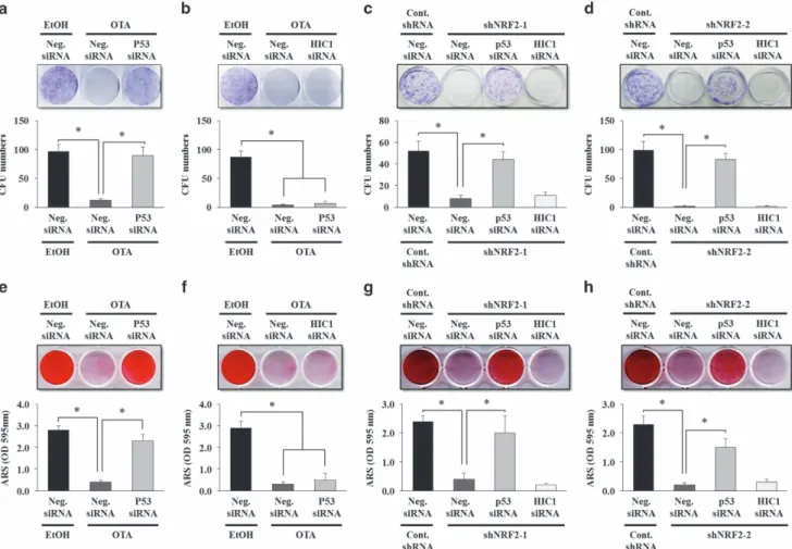
rescued by p53 RNA interference. We further examined
whether targeting p53 would rescue the suppressed
self-renewal and differentiation potentials in OTA-treated or
shNRF2-infected EP MSCs. Figure 6a shows that siRNA
targeting p53 rescued the colony-forming ability that was
decreased in OTA-treated EP MSCs, but this effect was not
observed for siRNA targeting HIC1 (Figures 6a and b). As
expected, in the shNRF2-infected EP MSCs, siRNA targeting
Figure 3 t-BHQ induces nuclear import of NRF2 and enhances the self-renewal capacity, but does not affect the osteogenic differentiation of EP MSCs. (a) EP MSCs were incubated in basal growth medium (DMEM-LG containing 10% FBS) in the presence of DMSO or t-BHQ (10μM) for 16 h. Cell lysates were prepared for the DMSO-treated or t-BHQ-treated EP MSCs, and then fractionated into nuclear and cytosolic extracts. The protein levels of NRF2 and phosphorylated NRF2 were analyzed by western blot analysis for the DMSO- or t-BHQ-treated EP MSCs. The LDH protein level was used as a loading control for cytosolic extracts, and the protein level of LAMIN-B was used as a loading control for nuclear extracts. The expression level ofβ-CATENIN was also used as a loading control for both cytosolic and nuclear extracts. DMSO was used as a vehicle of t-BHQ. (b) Immunofluorescence was performed to observe the nuclear and cytosolic localization of the NRF2 protein in the DMSO- or t-BHQ (10μM)-treated EP MSCs. The nuclei were stained with DAPI, and NRF2 was stained with Alexa Fluor 568 (Yellow)-conjugated secondary antibody. The images were obtained using confocal microscopy. Scale bar= 100 μm. (c) The mRNA expression levels of HO-1 and NQO-1 were also analyzed with qRT-PCR. *Po0.05 compared with control DMSO-treated MSCs. (d) A cell proliferation assay was performed to determine the proliferative capacities of DMSO- or t-BHQ (10μM)-treated EP MSCs using an EZ-Cytox Kit. Each experiment was performed in triplicate (n= 3). (e) EP-MSCs (1 × 10³cells per well in 100-mm dishes) treated with DMSO or t-BHQ (10μM) were incubated in basal growth medium for 12 days. The
colony-forming abilities were compared between EP-MSCs treated with DMSO or t-BHQ using CV staining, and the numbers of colony-colony-forming cells were counted in triplicate by three observers (n= 3). *Po0.05 compared with DMSO-treated EP-MSCs. (f) EP-MSCs (8 × 10⁴cells per well in 12-well plates) treated with DMSO or t-BHQ (10
μM) were incubated in osteogenic medium for 10 days. Alizarin red S staining was performed to detect mineral deposition at day 10. For quantitative analysis, absorbance was measured at 595 nm following destaining with 10% cetylpyridinium for 30 min. *Po0.05 compared with DMSO-treated EP MSCs
NRF2 regulates SIRT1 to maintain MSC stemness DS Yoonet al
p53 significantly enhanced the colony-forming ability that was
suppressed by shRNA targeting NRF2, whereas targeting
HIC1 did not affect the self-renewal capacity (Figures 6c and
d). The osteogenic potential suppressed by OTA treatment
was also rescued by siRNA targeting p53. Alizarin red S
staining showed that suppressed calcification by OTA
treatment was rescued in p53-targeting siRNA-transfected
EP MSCs (but not in HIC1-targeting siRNA-transfected EP
MSCs) (Figures 6e and f). Similarly, in shNRF2-infected EP
MSCs, targeting p53 using siRNA yielded the same results to
those of the CFU-F assay and alizarin red S staining
(Figures 6g and h). Accordingly, p53 may be an important
target in NRF2-mediated SIRT1 regulation for the
mainte-nance of the self-renewal and osteogenic differentiation
capacities during prolonged MSC culture.
The self-renewal and osteogenic potentials of LP MSCs
can be reactivated by t-BHQ treatment. Finally, we tested
whether the decreased self-renewal and osteogenic
poten-tials of LP MSCs can be reactivated by inducing the nuclear
import of cytosolic NRF2. Application of t-BHQ to LP MSCs
increased the mRNA expression of HO-1, NQO-1, and
SIRT1,
and
decreased
mRNA
expression
of
p53
(Figures 7a and b). We also found that the p53 protein level
was higher in LP-MSCs than EP MSCs, but the HIC1 protein
level was not affected by prolonged cell passages. t-BHQ
treatment in LP MSCs increased the protein level of p-NRF2,
resulting in decreased p53 and increased SIRT1 (Figure 7c).
We also confirmed the nuclear localization of NRF2 in
t-BHQ-treated LP MSCs (Figure 7d). The self-renewal and
multipotency of MSCs decreased during prolonged cell
passages. Surprisingly, reactivating the NRF2–SIRT1 axis
by applying t-BHQ to LP MSCs enhanced the colony-forming
ability of the cells (Figure 7e). Furthermore, LP MSCs
pretreated with t-BHQ for
∼ 4–7 days before osteogenic
differentiation
were
successfully
differentiated
to
the
Figure 4 Rescue patterns of suppressed osteogenic potential upon the removal of OTA or t-BHQ, and the association with NRF2 nuclear localization and stemness genes. (a) EP MSCs (8 × 10⁴cells per well in 12-well plates) treated with EtOH or OTA (10μM) were incubated in osteogenic medium for 10 days. In addition, OTA application was
stopped on day 7 of osteogenic differentiation in another group. Alizarin red S staining was performed to detect mineral deposition at days 3, 7, and 10. For quantitative analysis, absorbance was measured at 595 nm following destaining with 10% cetylpyridinium for 30 min. *Po0.05 compared with EtOH- or OTA-treated EP MSCs. (b) Similarly, EP MSCs (8 × 10⁴cells per well in 12-well plates) treated with DMSO or t-BHQ (10μM) were incubated in osteogenic medium for 10 days. In addition, t-BHQ application was stopped on day 7 of osteogenic differentiation in another group. Alizarin red S staining was performed to detect mineral deposition at days 3, 7, and 10. *Po0.05 compared with DMSO- or t-BHQ-treated EP MSCs. (c and d) EP MSCs were incubated in basal growth medium (DMEM-LG containing 10% FBS) in the presence of EtOH/DMSO or OTA (10μM)/t-BHQ (10 μM) for 16 h. The mRNA expression levels of NRF2, SIRT1, SOX2, OCT3/4, and NANOG were also analyzed by qRT-PCR. *Po0.05 compared with control EtOH/DMSO-treated MSCs. (e and f) EtOH/DMSO or OTA (10μM)/t-BHQ (10 μM)-treated EP MSCs differentiated to the osteogenic lineage, and cell pellets in each condition were harvested at each stage to prepare cell lysates. The protein levels of NRF2, phosphorylated NRF2, RUNX2, and SIRT1 were analyzed by western blot analysis. The protein level ofβ-CATENIN was used as a loading control
NRF2 regulates SIRT1 to maintain MSC stemness DS Yoonet al
osteogenic lineage (Figures 7f and g). These results suggest
that the NRF2–SIRT1 axis is an important target in the
maintenance of the self-renewal and differentiation potencies
of MSCs as well as in rejuvenating long-term MSC cultures
in vitro before animal studies and clinical applications.
Discussion
Our study showed that the inhibition or induction of NRF2
nuclear localization affected cell proliferation and the
colony-forming ability of EP MSCs. Interestingly, the in vitro
osteo-genic potential of MSCs was decreased by treatment with both
t-BHQ (a NRF2 activator) and OTA (a NRF2 inhibitor). These
results indicate that NRF2 activity is essential for maintaining
the self-renewal and osteogenic potentials of MSCs during
ex vivo cultivation. Because the sustained nuclear localization
of NRF2 also blocked MSC differentiation to the osteogenic
lineage, it is possible that sustained nuclear NRF2 maintains
an undifferentiated state of MSCs; the undifferentiated state
can be maintained in hypoxic environments, mimicked by
NRF2/hypoxia-inducible factor-1α (HIF-1α) signaling.
12,38Furthermore, oxidative stress induces cellular senescence of
human MSCs.
39Accordingly, we suggest that sustained NRF2
activation protects MSCs against oxidative stress during
ex vivo cultivation,
39resulting in the maintenance of
MSC stemness. NRF2 negatively regulates osteogenic
Figure 5 NRF2-mediated SIRT1 regulation occurs in a p53-dependent manner in EP MSCs. (a) EP MSCs were incubated in basal growth medium (DMEM-LG containing 10% FBS) in the presence of EtOH, 1μM OTA, and 10 μM OTA for 16 h. The mRNA expression levels of HO-1, NQO-1, and SIRT1 were also analyzed by qRT-PCR. *Po0.05 compared with control EtOH-treated MSCs. (b) The protein levels of NRF2, phosphorylated NRF2, SIRT1, HIC1, and p53 were analyzed by western blot analysis. The protein level ofβ-CATENIN was used as a loading control. (c) The mRNA expression levels of HO-1, NQO-1, and SIRT1 were also analyzed in EP MSCs infected with nontargeting shRNA, shRNF2-1, or shNRF2-2 by qRT-PCR. *Po0.05 compared with nontargeting shRNA-infected MSCs. (d) Similarly, the protein levels of NRF2, phosphorylated NRF2, SIRT1, HIC1, and p53 were analyzed in EP MSCs infected with nontargeting shRNA, shRNF2-1, or shNRF2-2 by western blot analysis. (e) siRNA targeting p53 was transfected in the OTA-treated EP MSCs. The protein levels of NRF2, phosphorylated NRF2, SIRT1, and p53 were analyzed in EtOH-, OTA (10μM)-, or siRNA targeting p53 plus OTA (10μM)-treated EP MSCs. (f) siRNA targeting HIC1 was transfected in the OTA-treated EP MSCs. The protein levels of NRF2, phosphorylated NRF2, SIRT1, and HIC1 were analyzed in EtOH-, OTA (10μM)-, or siRNA targeting HIC1 plus OTA (10 μM)-treated EP MSCs. (g) siRNA targeting p53 or HIC1 was transfected with shNRF2-1-infected EP MSCs. The protein levels of NRF2, phosphorylated NRF2, SIRT1, HIC1, and p53 were analyzed in the EP MSCs with nontargeting shRNA, shNRF2-1 alone, shNRF2-1 plus siRNA targeting p53, or shNRF2-1 plus siRNA targeting HIC1. (h) siRNA targeting p53 or HIC1 was transfected with shNRF2-2-infected EP MSCs. The protein levels of NRF2, phosphorylated NRF2, SIRT1, HIC1, and p53 were analyzed in the EP MSCs with nontargeting shRNA, shNRF2-2 alone, shNRF2-2 plus siRNA targeting p53, or shNRF2-2 plus siRNA targeting HIC1. The protein level ofβ-CATENIN was used as a loading control. NS, no significance; shC, nontargeting shRNA; sh-1, shNRF2-1; sh-2, shNRF2-2
NRF2 regulates SIRT1 to maintain MSC stemness DS Yoonet al
differentiation
40as well as osteoclastic differentiation.
41In
addition, there are conflicting results regarding the role of
NRF2 in osteogenic differentiation. Human periodontal
liga-ment cells can be efficiently differentiated to the osteogenic
lineage by increasing NRF2 levels in nuclear extracts.
42NRF2-knockout mice show a significant deficit in postnatal
bone acquisition.
43Taken together, NRF2 is a key factor in
MSC maintenance and osteogenesis; when NRF2 is lacking,
MSCs cannot self-renew and differentiate to the osteogenic
lineage.
NRF2 also has important roles in the chondrogenic and
adipogenic differentiation of MSCs. NRF2 is a negative
regulator of cellular differentiation in a chondrocytic cell line,
ATDC5. NRF2 overexpression decreases mRNA expression
of type II collagen in ATDC5 cells.
44However, sulforaphane
(SFN), another NRF2 activator, can suppress gene
expres-sion related to osteoarthritis and block cartilage destruction.
45The protein level of nuclear Nrf2 also continuously decreases
during adipogenic differentiation in ST2 cells, a bone
marrow-derived MSC cell line.
46This expression pattern is required
for normal adipocyte differentiation of 3T3L1 cells and is
associated with increased oxidative stress levels that can
facilitate differentiation processes.
47SFN can also suppress
the adipogenic differentiation of 3T3L1 cells.
48In contrast,
OTA inhibits the adipogenic differentiation of adipose-derived
MSCs.
49This is consistent with our results regarding the
Figure 6 RNA interference of p53 rescues the self-renewal and osteogenic potentials that are suppressed in OTA-treated and shNRF2-infected EP MSCs. The siRNAs targeting p53 (a) or HIC1 (b) were transfected in EP MSCs, and the cells were plated at 1 × 10³cells per well in 100-mm dishes. After 24 h, OTA (10μM) was treated with the
siRNA-transfected cells, and the cells were incubated in basal growth medium for 12 days. After 12 days, CV staining was performed, and the numbers of colony-forming cells were counted in triplicate by three observers (n= 4). *Po0.05 compared with EtOH-treated EP MSCs transfected with negative siRNA or OTA-treated EP MSCs transfected with negative siRNA. Similarly, the siRNAs targeting p53 or HIC1 were transfected in shNRF2-1 (c)- or shNRF2-2 (d)-infected EP MSCs, and the cells were plated at 1 × 10³cells per well in 100 mm dishes and incubated in basal growth medium for 12 days. After 12 days, CV staining was performed, and the numbers of colony-forming cells were counted in triplicate by three observers (n= 3). *Po0.05 compared with nontargeting shRNA-infected EP MSCs transfected with negative siRNA or shNRF2-infected EP-MSCs transfected with negative siRNA. The siRNAs targeting p53 (e) or HIC1 (f) were transfected in EP MSCs, and the cells were plated at 8 × 10⁴cells per well in 12-well plates. After 24 h, OTA
(10μM) was treated with the siRNA-transfected cells, and the cells were incubated in osteogenic medium for 10 days. After 10 days, alizarin red S staining was performed to detect mineral deposition. For quantitative analysis, absorbance was measured at 595 nm following destaining with 10% cetylpyridinium for 30 min. *Po0.05 compared with EtOH-treated EP MSCs transfected with negative siRNA or OTA-treated EP MSCs transfected with negative siRNA (n= 4). Similarly, the siRNAs targeting p53 or HIC1 were transfected in shNRF2-1 (g)- or shNRF2-2 (h)-infected EP MSCs, and the cells were incubated in osteogenic medium for 10 days. After 10 days, alizarin red S staining was performed to detect mineral deposition. For quantitative analysis, absorbance was measured at 595 nm following destaining with 10% cetylpyridinium for 30 min (n= 3). *Po0.05 compared with nontargeting shRNA-infected EP MSCs transfected with negative siRNA or shNRF2-infected EP MSCs transfected with negative siRNA. Cont.shRNA, nontargeting shRNA; Neg.siRNA, nontargeting siRNA
NRF2 regulates SIRT1 to maintain MSC stemness DS Yoonet al
osteogenic differentiation of MSCs. Taken together, NRF2
may be indispensible for the maintenance of the MSC
self-renewal capacity as well as the multi-differentiation potential to
the osteogenic, chondrogenic, and adipogenic lineages.
SIRT1 has been a recent focus of research as a regulator of
MSC stemness.
50It is required for the long-term cultivation of
MSCs,
51and its overexpression ameliorates cellular
senes-cence by reversing aged MSCs, resulting in an increase in cell
proliferation.
52SIRT1 regulates the osteogenic and
chondro-genic differentiation of MSCs by deacetylating
β-catenin.
53For
the maintenance of stemness, SIRT1 positively regulates the
self-renewal and multipotency of human MSCs by
deacetylat-ing the lysine residue of SOX2.
32SIRT1 is known to induce
deacetylation of NRF2 that subsequently decreases
NRF2-dependent gene transcription.
54However, it is thought that
NRF2 and SIRT1 have similar functions in adult stem
cells because the loss of NRF2 in the intestinal stem cells
of
Drosophila
induces
ROS
levels
and
age-related
degeneration.
55Similarly, the loss of SIRT1 is also related to
an increase in ROS levels in hematopoietic stem and
Figure 7 NRF2 activation by t-BHQ enhances the self-renewal capacity and osteogenic potential of LP MSCs. (a) LP MSCs were grown in basal growth medium (DMEM-LG containing 10% FBS) in the presence of t-BHQ (10μM) for 7 days. The mRNA expression levels of HO-1, NQO-1, (b) SIRT1, and p53 were also analyzed by qRT-PCR. *Po0.05 compared with control EtOH-treated MSCs. (c) Similarly, LP MSCs were grown in basal growth medium in the presence of t-BHQ for 7 days before the western blot analysis. The protein levels of NRF2, phosphorylated NRF2, SIRT1, HIC1, and p53 were analyzed, and the protein level ofβ-CATENIN was used as a loading control. (d) Immunofluorescence was performed to observe the nuclear and cytosolic localization of NRF2 in the EP or LP MSCs or t-BHQ- (10μM)-pretreated LP MSCs. The nuclei were stained with DAPI, and NRF2 was stained with Alexa Fluor 568 (Yellow)-conjugated secondary antibody. The images were obtained using confocal microscopy. Scale bar= 100 μm. (e) EP MSCs, LP MSCs, or t-BHQ-pretreated LP MSCs were incubated in basal growth medium for 12 days. The colony-forming abilities were compared using CV staining, and the numbers of colony-forming cells were counted in triplicate by three observers (n= 4). *Po0.05 compared with EP or LP MSCs. (f) Before the osteogenic differentiation of LP MSCs, LP MSCs were pretreated with t-BHQ. At the starting point of differentiation, LP MSCs were not treated with t-BHQ. EP MSCs, LP MSCs, or t-BHQ-pretreated LP MSCs were plated at 8 × 10⁴cells per well in 12-well plates and maintained in osteogenic medium for 10 days. After 10 days, alizarin red S staining was performed to detect mineral deposition.
(g) For quantitative analysis, absorbance was measured at 595 nm following destaining with 10% cetylpyridinium for 30 min. *Po0.05 compared with EP or LP MSCs NRF2 regulates SIRT1 to maintain MSC stemness
DS Yoonet al
progenitor cells.
56These reports indicate that the interaction
between NRF2 and SIRT1 in adult stem cells may be involved
in defense against oxidative stress during prolonged cell
passages, preventing cellular aging and the loss of
multi-potency. p53 can directly bind to two SIRT1 promoter regions
and suppress the transcriptional activity of SIRT1,
35whereas
NRF2 can induce the degradation of p53 via the regulation of
Mdm2 expression.
57Based on these results, our study
showed that the p53 protein level increased and the SIRT1
protein level decreased in OTA-treated or shNRF2-infected
MSCs, and RNA interference of p53 in OTA-treated or
shNRF2-infected MSCs rescued the SIRT1 protein level as
well as the osteogenic differentiation potential. Furthermore,
t-BHQ rejuvenated LP MSCs by enhancing the SIRT1
expression level via NRF2-mediated p53 suppression
(Figure 8). Given the importance of these findings in MSCs,
additional studies will further increase our understanding of
the role of NRF2 in MSC stemness and will facilitate the
optimization of the application of NRF2 activators to MSCs.
Recent studies have reported the mechanism of NRF2
action and its new function, excluding its well-known role in
antioxidant response. Here, we reviewed the mechanism of
NRF2 action that has been reported in several types of stem
cells (Supplementary Table S1). In embryonic stem cells,
NRF2 acts as a regulator of the proteasome that regulates
self-renewal and pluripotency.
25In addition, NRF2 can
modulate self-renewal and quiescence by regulating CXCR4
in hematopoietic stem cells.
58Our study was the first to clarify
the molecular mechanisms involved in the regulation of MSCs
by NRF2 to maintain the self-renewal and osteogenic
potentials via SIRT1. Our findings provide mechanistic insight
into how NRF2 contributes to the maintenance of MSC
stemness. Thus, the conservation of NRF2 nuclear
localiza-tion is an important target for the prevenlocaliza-tion of cellular
senescence and the loss of multipotency during prolonged cell
passage of ex vivo cultured MSCs.
Materials and Methods
Cell culture, differentiation, and drug treatment. Bone marrow aspirates were obtained from the posterior iliac crests of seven adult donors,
with the approval of the institutional review board (IRB) of the Yonsei University College of Medicine. MSCs isolated from bone marrow were selected based on their ability to adhere to plastic cell culture dishes, and498% of the cultured cells were positive for CD90 and CD105, but negative for CD34 and CD45,59as previously described. MSCs were maintained in low-glucose Dulbecco’s modified Eagle’s medium (DMEM-LG; Invitrogen, Carlsbad, CA, USA) supplemented with 10% fetal bovine serum (FBS; Gibco, Grand Island, NY, USA) and 1% antibiotic–antimycotic solution (Invitrogen) at 37 °C in a 5% CO2atmosphere. EP MSCs (passages 1–3)
were replated at a density of 5000 cells/cm2, and the cells were subcultured when they were 80% confluent up to passages 7–10 (LP MSCs). To induce osteogenic differentiation, MSCs were seeded onto 12-well culture pates at a density of 8 × 10⁴
cells per well. The medium used for the osteogenic differentiation of MSCs has been described previously.5Alizarin red S staining was used to evaluate osteogenic differentiation. Briefly, after cells were fixed in ice-cold 70% ethanol, freshly prepared 3% alizarin red S solution (wt/vol) (Sigma, St. Louis, MO, USA) was added, and cells were incubated in the dark for 30 min. For quantitative analysis of alizarin red S, absorbance was detected at 595 nm following destaining with 10% cetylpyridinium chloride monohydrate (Sigma) for 20 min. OTA (Sigma) was dissolved in ethanol (EtOH) and used at a concentration of 1–10 μM in EP MSCs. t-BHQ (Sigma) was dissolved in dimethyl sulfoxide (DMSO) and used at a concentration of 1–10 μM in EP or LP MSCs.
Quantitative real-time PCR (qRT-PCR). Real-time PCR analysis was performed as described previously.32 Briefly, total RNA was isolated using an RNeasy Kit (Qiagen, Valencia, CA, USA), according to the manufacturer’s instructions. Total RNA (1μg) was reverse-transcribed using the Omniscript Kit (Qiagen). Primer sets were validated and purchased from Bioneer (Daejeon, South Korea; http://www.bioneer.co.kr/). The primers used were as follows: NRF2 (P164742), HO-1 (P133045), NQO-1 (P113225), SIRT1 (P293039), SOX2 (P200205), and NANOG (P255522). There are no validated primers for OCT3/4 (a) orβ-ACTIN. To obtain PCR products specific to OCT3/4(a), which acts as a transcription factor, the primer must include exon 1 in its recognition site;60thus, the following primers were designed: 5′-GCAAGCCCTCATTTCACCA-3′ (sense, NM_002701) and 5′-GCCCATCACCTCCACCAC-3′ (antisense). The following primers were designed forβ-ACTIN: 5′-GTCCTCTCCCAAGTCCACACA-3′ (sense, NM_001101.3) and 5′-GGGCACGAAGGCTCATCATTC-3′ (antisense). Mean cycle threshold values from triplicate (n= 3) measurements were used to calculate gene expression, with normalization toβ-ACTIN as an internal control.
Western blot analysis. MSCs were lysed in passive lysis buffer (Promega, Madison, WI, USA). Protein concentrations were determined using the Bio-Rad Protein Assay (Bio-Rad Laboratories, Inc., Hercules, CA, USA) and 30 mg of protein was analyzed by 10% sodium dodecyl sulfate-polyacrylamide gel electrophoresis (SDS-PAGE) (Sigma). Transferred membranes were blocked with 5% skim milk (BD, Sparks, MD, USA), and incubated for 10 h with antibodies against NRF2 (Santa Cruz Biotechnology, Santa Cruz, CA, USA), Figure 8 Proposed models of the effects of OTA or t-BHQ on the regulation and maintenance of MSC stemness via SIRT1. Blocking the nuclear import of NRF2 activates p53, which suppresses SIRT1 promoter activity, resulting in a loss of MSC stemness. Conversely, the protein level of p53 can be decreased by the phosphorylation and nuclear import of NRF2, resulting in the activation of SIRT1 transcription as well as the enhancement of MSC stemness
NRF2 regulates SIRT1 to maintain MSC stemness DS Yoonet al
phosphorylated-NRF2 (Abcam, Cambridge, UK), HDAC1 (Santa Cruz Biotechnol-ogy), HSP90 (Santa Cruz BiotechnolBiotechnol-ogy), LAMIN-B (Santa Cruz BiotechnolBiotechnol-ogy), LDH (Santa Cruz Biotechnology), RUNX2 (EMD Millipore, San Diego, CA, USA), SIRT1 (Santa Cruz Biotechnology), p53 (Santa Cruz Biotechnology), and HIC1 (Santa Cruz Biotechnology). Membranes were further probed with an antibody againstβ-ACTIN (Santa Cruz Biotechnology, Dallas, TX, USA) that served as a loading control.
Nuclear and cytosolic fractionation. EP, LP, undifferentiated, or differentiated MSCs were collected by trypsinization and washed with phosphate-buffered saline (PBS) two times before nuclear and cytosolic fractionation. Nuclear and cytoplasmic fractionation were conducted using the NE-PER Nuclear and Cytoplasmic Extraction Reagents Kit (Thermo Fisher Scientific, Rockford, IL, USA) according to the manufacturer’s instructions. Each separated protein was analyzed by western blot analysis.
Immunocytochemistry. EP, LP, undifferentiated, differentiated, OTA-, or t-BHQ-treated MSCs were seeded at 5000 cells/cm2on 4-well glass chamber slides
(Nalge Nunc International, Rochester, NY, USA), and the cells were incubated in a 5% CO2incubator at 37 °C. After an overnight incubation, the cells were washed
with PBS followed by fixation with 4% paraformaldehyde (Sigma) for 30 min. Permeabilization was accomplished with 1% Triton X-100 in PBS for 10 min followed by blocking for 1 h with 3% bovine serum albumin (BSA) in PBS. The cells were incubated with a 1 : 200 dilution of primary antibodies against NRF2 (Santa Cruz Biotechnology) overnight at 4 °C. After washing three times with PBS, the cells were incubated with fluorescein isothiocyanate (FITC) and phycoerythrin-conjugated secondary antibodies (Santa Cruz Biotechnology) or Alexa Fluor 568 (Yellow, Abcam) in a 1 : 5000 dilution in 3% BSA-containing PBS for 1 h at room temperature in the dark. The nuclei were stained with 4,6-diamidino-2-phenyindole (DAPI, Sigma) and then examined using a Zeiss LSM700 scanning laser confocal microscope (Zen 2011; Carl Zeiss MicroImaging GHBH, Jena, Germany). Proliferation assay. Cell proliferation was examined using an EZ-Cytox Kit (Daeil Lab Service, Seoul, Korea). OTA- or t-BHQ-treated EP-MSCs were seeded in 12-well culture plates at a density of 1 × 10⁴ cells per well. The cells were maintained in DMEM-LG for 7 days, and the cell culture media were replaced once a day during cell viability assay periods. Briefly, after washing cells in PBS, 10μl of EZ-Cytox (tetrazolium salts) solution was added to each well and incubated at 37 °C for 4 h. After incubation, the conditioned medium was transferred to 96-well plates. The absorbance was measured at 450 nm. All samples were tested in triplicate (n= 6).
CFU-F assay. EP, LP, OTA-, or t-BHQ-treated MSCs were seeded at 1 × 10³
cells in 100 mm culture dishes, and maintained in DMEM-LG supplemented with 20% FBS for 10–12 days. OTA-or t-BHQ-treated media were replaced every 2 days during colony formation. Subsequently, the cells were fixed in a 1 : 1 acetone/ methanol fixative, stained with a 20% crystal violet (CV) solution (Merck, Darmstadt, Germany) for 30 min in the dark, and washed in distilled water. The colony-forming ability of the stained cells was then evaluated and counted.
Establishment of NRF2-knockdown MSCs. To obtain lentiviral particles with shNRF2, HEK293T cells were seeded in 100 mm culture dishes at a density of 3 × 10⁶cells per dish. On the next day, the cells were transfected with lentiviral
particles with either a nontargeting shRNA expression plasmid (MISSION Plko.1-puro Empty Vector Control Plasmid DNA, Sigma) or two shNRF2 expression plasmids (NFE2L2 MISSION shRNA Stock, TRC numbers: TRCN0000007555 (shNRF-1), TRCN0000007558 (shNRF-2), Sigma) with the Delta 8.9 plasmid (gag, pol, and rev genes) and VSV-G (envelope plasmid, Sigma) using Lipofectamine 2000 (Invitrogen). After 6 h of transfection, the medium was replaced. The shRNA-transfected HEK293T cells were maintained for 2 days, and then the supernatants were collected and stored at− 70 °C. To knockdown NRF2 in EP-MSCs, the cells were seeded in 6-well plates at a density of 5 × 10⁴cells per well. After 48 h of infection, the medium was replaced with freshly prepared medium with 10μg/ml puromycin dihydrochloride (Sigma), and the cells were maintained for 7 days. The knockdown efficiency of the selected cells was analyzed by western blot analysis. Below is a list of shRNAs targeting NRF2 used in this study.
1. shNRF2-1 (region: 3′ UTR) 5′-CCGGGCTCCTACTGTGATGTGAAATCTCGAGATTTCACATCACAGTAGGAG CTTTTT-3′ 2. shNRF2-2 (region: CDS) 5′-CCGGCCGGCATTTCACTAAACACAACTCGAGTTGTGTTTAGTGAAATGCC GGTTTTT-3′
siRNA transfection. Scramble, HIC1, and p53 siRNAs were purchased from Bioneer (http://sirna.bioneer.co.kr/). The scramble-sense siRNA targeted the sequence 5′-CCUACGCCACCAAUUUCGU-3′, and the scramble-antisense siRNA targeted the sequence 5′-ACGAAAUUGGUGGCGUAGG-3′. HIC1-sense siRNA targeted the sequence 5′-AGACGAUGCUGGACACGAU (dTdT)-3′, and HIC1-antisense siRNA targeted the sequence 5′-AUCGUGUCCAGCAUCGUCU (dTdT)-3′. p53-sense siRNA targeted the sequence 5′-CACUACAACUACAUGUGUA (dTdT)-3′, and p53-antisense siRNA targeted the sequence 5′-UACACAUGUAGUUGUAGUG (dTdT)-3′. NRF2-sense siRNA targeted the sequence 5′-GAGACUACCAUGGUUCCAA(dTdT)-3′, and NRF2-antisense siRNA targeted the sequence 5′-UUGGAACCAUGGUAGUCUC (dTdT)-3′.
Briefly, EP MSCs treated with OTA (10μM) or infected with shRNA-1 or shRNA2 were plated to obtain 70–80% confluence in 6-well plates and transfected with 100 nM of HIC1, p53, or scramble (negative control) siRNA using Lipofectamine 2000 (Invitrogen). After 6 h of transfection, fresh medium was exchanged.
Statistical analysis. Statistical analysis was performed using one-way analysis of variance for multiple comparisons or Student’s t-tests for differences between two groups, and the data are expressed as means+S.D. Values of Po0.05 were considered statistically significant.
Conflict of Interest
The authors declare no conflict of interest.
Acknowledgements. This work (NRF-2015R1A2A2A01003876) was supported by the Mid-career Researcher Program through an NRF grant funded by the MEST.
1. Sethe S, Scutt A, Stolzing A. Aging of mesenchymal stem cells. Ageing Res Rev 2006; 5: 91–116.
2. Fehrer C, Lepperdinger G. Mesenchymal stem cell aging. Exp Gerontol 2005; 40: 926–930. 3. Schellenberg A, Lin Q, Schuler H, Koch CM, Joussen S, Denecke B et al. Replicative senescence of mesenchymal stem cells causes DNA-methylation changes which correlate with repressive histone marks. Aging (Albany NY) 2011; 3: 873–888.
4. Yoon DS, Kim YH, Jung HS, Paik S, Lee JW. Importance of Sox2 in maintenance of cell proliferation and multipotency of mesenchymal stem cells in low-density culture. Cell Prolif 2011; 44: 428–440.
5. Yoon DS, Kim YH, Lee S, Lee KM, Park KH, Jang Y et al. Interleukin-6 induces the lineage commitment of bone marrow-derived mesenchymal multipotent cells through down-regulation of Sox2 by osteogenic transcription factors. FASEB J 2014; 28: 3273–3286. 6. Zhu W, Chen J, Cong X, Hu S, Chen X. Hypoxia and serum deprivation-induced apoptosis in
mesenchymal stem cells. Stem Cells 2006; 24: 416–425.
7. Sart S, Song L, Li Y. Controlling redox status for stem cell survival, expansion, and differentiation. Oxid Med Cell Longev 2015; 2015: 105135.
8. Haque N, Rahman MT, Abu Kasim NH, Alabsi AM. Hypoxic culture conditions as a solution for mesenchymal stem cell based regenerative therapy. ScientificWorldJournal 2013; 2013: 632972.
9. Basciano L, Nemos C, Foliguet B, de Isla N, de Carvalho M, Tran N et al. Long term culture of mesenchymal stem cells in hypoxia promotes a genetic program maintaining their undifferentiated and multipotent status. BMC Cell Biol 2011; 12: 12.
10. Ejtehadifar M, Shamsasenjan K, Movassaghpour A, Akbarzadehlaleh P, Dehdilani N, Abbasi P et al. The effect of hypoxia on mesenchymal stem cell biology. Adv Pharm Bull 2015; 5: 141–149.
11. Lavrentieva A, Majore I, Kasper C, Hass R. Effects of hypoxic culture conditions on umbilical cord-derived human mesenchymal stem cells. Cell Commun Signal 2010; 8: 18. 12. Mohyeldin A, Garzon-Muvdi T, Quinones-Hinojosa A. Oxygen in stem cell biology: a critical
component of the stem cell niche. Cell Stem Cell 2010; 7: 150–161.
13. Holzwarth C, Vaegler M, Gieseke F, Pfister SM, Handgretinger R, Kerst G et al. Low physiologic oxygen tensions reduce proliferation and differentiation of human multipotent mesenchymal stromal cells. BMC Cell Biol 2010; 11: 11.
14. D'Ippolito G, Diabira S, Howard GA, Roos BA, Schiller PC. Low oxygen tension inhibits osteogenic differentiation and enhances stemness of human MIAMI cells. Bone 2006; 39: 513–522.
15. Fehrer C, Brunauer R, Laschober G, Unterluggauer H, Reitinger S, Kloss F et al. Reduced oxygen tension attenuates differentiation capacity of human mesenchymal stem cells and prolongs their lifespan. Aging Cell 2007; 6: 745–757.
NRF2 regulates SIRT1 to maintain MSC stemness DS Yoonet al
16. Zhu H, Zhang L, Itoh K, Yamamoto M, Ross D, Trush MA et al. Nrf2 controls bone marrow stromal cell susceptibility to oxidative and electrophilic stress. Free Radic Biol Med 2006; 41: 132–143.
17. Surh YJ, Kundu JK, Na HK. Nrf2 as a master redox switch in turning on the cellular signaling involved in the induction of cytoprotective genes by some chemopreventive phytochemicals. Planta Med 2008; 74: 1526–1539.
18. Itoh K, Chiba T, Takahashi S, Ishii T, Igarashi K, Katoh Y et al. An Nrf2/small Maf heterodimer mediates the induction of phase II detoxifying enzyme genes through antioxidant response elements. Biochem Biophys Res Commun 1997; 236: 313–322.
19. Itoh K, Wakabayashi N, Katoh Y, Ishii T, Igarashi K, Engel JD et al. Keap1 represses nuclear activation of antioxidant responsive elements by Nrf2 through binding to the amino-terminal Neh2 domain. Genes Dev 1999; 13: 76–86.
20. Kobayashi A, Kang MI, Okawa H, Ohtsuji M, Zenke Y, Chiba T et al. Oxidative stress sensor Keap1 functions as an adaptor for Cul3-based E3 ligase to regulate proteasomal degradation of Nrf2. Mol Cell Biol 2004; 24: 7130–7139.
21. Yamamoto T, Suzuki T, Kobayashi A, Wakabayashi J, Maher J, Motohashi H et al. Physiological significance of reactive cysteine residues of Keap1 in determining Nrf2 activity. Mol Cell Biol 2008; 28: 2758–2770.
22. Choo KB, Tai L, Hymavathee KS, Wong CY, Nguyen PN, Huang CJ et al. Oxidative stress-induced premature senescence in Wharton's jelly-derived mesenchymal stem cells. Int J Med Sci 2014; 11: 1201–1207.
23. Mohammadzadeh M, Halabian R, Gharehbaghian A, Amirizadeh N, Jahanian-Najafabadi A, Roushandeh AM et al. Nrf-2 overexpression in mesenchymal stem cells reduces oxidative stress-induced apoptosis and cytotoxicity. Cell Stress Chaperones 2012; 17: 553–565. 24. Jang J, Wang Y, Kim HS, Lalli MA, Kosik KS. Nrf2, a regulator of the proteasome, controls
self-renewal and pluripotency in human embryonic stem cells. Stem Cells 2014; 32: 2616–2625.
25. Huang HC, Nguyen T, Pickett CB. Regulation of the antioxidant response element by protein kinase C-mediated phosphorylation of NF-E2-related factor 2. Proc Natl Acad Sci USA 2000; 97: 12475–12480.
26. Limonciel A, Jennings P. A review of the evidence that ochratoxin A is an Nrf2 inhibitor: implications for nephrotoxicity and renal carcinogenicity. Toxins (Basel) 2014; 6: 371–379. 27. Li J, Johnson D, Calkins M, Wright L, Svendsen C, Johnson J. Stabilization of Nrf2 by tBHQ
confers protection against oxidative stress-induced cell death in human neural stem cells. Toxicol Sci 2005; 83: 313–328.
28. Boiani M, Scholer HR. Regulatory networks in embryo-derived pluripotent stem cells. Nat Rev Mol Cell Biol 2005; 6: 872–884.
29. Park SB, Seo KW, So AY, Seo MS, Yu KR, Kang SK et al. SOX2 has a crucial role in the lineage determination and proliferation of mesenchymal stem cells through Dickkopf-1 and c-MYC. Cell Death Differ 2012; 19: 534–545.
30. Pierantozzi E, Gava B, Manini I, Roviello F, Marotta G, Chiavarelli M et al. Pluripotency regulators in human mesenchymal stem cells: expression of NANOG but not of OCT-4 and SOX-2. Stem Cells Dev 2011; 20: 915–923.
31. Tsai CC, Su PF, Huang YF, Yew TL, Hung SC. Oct4 and Nanog directly regulate Dnmt1 to maintain self-renewal and undifferentiated state in mesenchymal stem cells. Mol Cell 2012; 47: 169–182.
32. Yoon DS, Choi Y, Jang Y, Lee M, Choi WJ, Kim SH et al. SIRT1 directly regulates SOX2 to maintain self-renewal and multipotency in bone marrow-derived mesenchymal stem cells. Stem Cells 2014; 32: 3219–3231.
33. Kwon HS, Ott M. The ups and downs of SIRT1. Trends Biochem Sci 2008; 33: 517–525. 34. Chen WY, Wang DH, Yen RC, Luo J, Gu W, Baylin SB. Tumor suppressor HIC1 directly
regulates SIRT1 to modulate p53-dependent DNA-damage responses. Cell 2005; 123: 437–448.
35. Nemoto S, Fergusson MM, Finkel T. Nutrient availability regulates SIRT1 through a forkhead-dependent pathway. Science 2004; 306: 2105–2108.
36. Zhu J, Wang H, Fan Y, Lin Y, Zhang L, Ji X et al. Targeting the NF-E2-related factor 2 pathway: a novel strategy for glioblastoma (review). Oncol Rep 2014; 32: 443–450. 37. Rotblat B, Melino G, Knight RA. NRF2 and p53: Januses in cancer? Oncotarget 2012; 3:
1272–1283.
38. Ji X, Wang H, Zhu J, Zhu L, Pan H, Li W et al. Knockdown of Nrf2 suppresses glioblastoma angiogenesis by inhibiting hypoxia-induced activation of HIF-1alpha. Int J Cancer 2014; 135: 574–584.
39. Brandl A, Meyer M, Bechmann V, Nerlich M, Angele P. Oxidative stress induces senescence in human mesenchymal stem cells. Exp Cell Res 2011; 317: 1541–1547.
40. Hinoi E, Fujimori S, Wang L, Hojo H, Uno K, Yoneda Y. Nrf2 negatively regulates osteoblast differentiation via interfering with Runx2-dependent transcriptional activation. J Biol Chem 2006; 281: 18015–18024.
41. Park CK, Lee Y, Kim KH, Lee ZH, Joo M, Kim HH. Nrf2 is a novel regulator of bone acquisition. Bone 2014; 63: 36–46.
42. Chung JH, Kim YS, Noh K, Lee YM, Chang SW, Kim EC. Deferoxamine promotes osteoblastic differentiation in human periodontal ligament cells via the nuclear factor erythroid 2-related factor-mediated antioxidant signaling pathway. J Periodontal Res 2014; 49: 563–573.
43. Kim JH, Singhal V, Biswal S, Thimmulappa RK, DiGirolamo DJ. Nrf2 is required for normal postnatal bone acquisition in mice. Bone Res 2014; 2: 14033.
44. Hinoi E, Takarada T, Fujimori S, Wang L, Iemata M, Uno K et al. Nuclear factor E2 p45-related factor 2 negatively regulates chondrogenesis. Bone 2007; 40: 337–344. 45. Davidson RK, Jupp O, de Ferrars R, Kay CD, Culley KL, Norton R et al. Sulforaphane
represses matrix-degrading proteases and protects cartilage from destruction in vitro and in vivo. Arthritis Rheum 2013; 65: 3130–3140.
46. Chartoumpekis DV, Ziros PG, Sykiotis GP, Zaravinos A, Psyrogiannis AI, Kyriazopoulou VE et al. Nrf2 activation diminishes during adipocyte differentiation of ST2 cells. Int J Mol Med 2011; 28: 823–828.
47. Vomhof-DeKrey EE, Picklo MJ. NAD(P)H:quinone oxidoreductase 1 activity reduces hypertrophy in 3T3-L1 adipocytes. Free Radic Biol Med 2012; 53: 690–700.
48. Schneider KS, Chan JY. Emerging role of Nrf2 in adipocytes and adipose biology. Adv Nutr 2013; 4: 62–66.
49. Lim S, Jang HJ, Kim JK, Kim JM, Park EH, Yang JH et al. Ochratoxin A inhibits adipogenesis through the extracellular signal-related kinases-peroxisome proliferator-activated receptor-gamma pathway in human adipose tissue-derived mesenchymal stem cells. Stem Cells Dev 2011; 20: 415–426.
50. Chen H, Liu X, Chen H, Cao J, Zhang L, Hu X et al. Role of SIRT1 and AMPK in mesenchymal stem cells differentiation. Ageing Res Rev 2014; 13: 55–64.
51. Yuan HF, Zhai C, Yan XL, Zhao DD, Wang JX, Zeng Q et al. SIRT1 is required for long-term growth of human mesenchymal stem cells. J Mol Med (Berl) 2012; 90: 389–400.
52. Chen H, Liu X, Zhu W, Chen H, Hu X, Jiang Z et al. SIRT1 ameliorates age-related senescence of mesenchymal stem cells via modulating telomere shelterin. Front Aging Neurosci 2014; 6: 103.
53. Simic P, Zainabadi K, Bell E, Sykes DB, Saez B, Lotinun S et al. SIRT1 regulates differentiation of mesenchymal stem cells by deacetylating beta-catenin. EMBO Mol Med 2013; 5: 430–440.
54. Kawai Y, Garduno L, Theodore M, Yang J, Arinze IJ. Acetylation-deacetylation of the transcription factor Nrf2 (nuclear factor erythroid 2-related factor 2) regulates its transcriptional activity and nucleocytoplasmic localization. J Biol Chem 2011; 286: 7629–7640.
55. Hochmuth CE, Biteau B, Bohmann D, Jasper H. Redox regulation by Keap1 and Nrf2 controls intestinal stem cell proliferation in Drosophila. Cell Stem Cell 2011; 8: 188–199. 56. Rimmele P, Bigarella CL, Liang R, Izac B, Dieguez-Gonzalez R, Barbet G et al. Aging-like
phenotype and defective lineage specification in SIRT1-deleted hematopoietic stem and progenitor cells. Stem Cell Reports 2014; 3: 44–59.
57. You A, Nam CW, Wakabayashi N, Yamamoto M, Kensler TW, Kwak MK. Transcription factor Nrf2 maintains the basal expression of Mdm2: an implication of the regulation of p53 signaling by Nrf2. Arch Biochem Biophys 2011; 507: 356–364.
58. Tsai JJ, Dudakov JA, Takahashi K, Shieh JH, Velardi E, Holland AM et al. Nrf2 regulates haematopoietic stem cell function. Nat Cell Biol 2013; 15: 309–316.
59. Lee S, Yoon DS, Paik S, Lee KM, Jang Y, Lee JW. microRNA-495 inhibits chondrogenic differentiation in human mesenchymal stem cells by targeting Sox9. Stem Cells Dev 2014; 23: 1798–1808.
60. Liedtke S, Stephan M, Kogler G. Oct4 expression revisited: potential pitfalls for data misinterpretation in stem cell research. Biol Chem 2008; 389: 845–850.
Cell Death and Disease is an open-access journal
published by Nature Publishing Group. This work is
licensed under a Creative Commons Attribution 4.0 International
License. The images or other third party material in this article are
included in the article’s Creative Commons license, unless indicated
otherwise in the credit line; if the material is not included under the
Creative Commons license, users will need to obtain permission from
the license holder to reproduce the material. To view a copy of this
license, visit http://creativecommons.org/licenses/by/4.0/
Supplementary Information accompanies this paper on Cell Death and Disease website (http://www.nature.com/cddis)
NRF2 regulates SIRT1 to maintain MSC stemness DS Yoonet al