Clinical application of liver stiffness measurement using
transient elastography in chronic liver disease from
longitudinal perspectives
Beom Kyung Kim, James Fung, Man-Fung Yuen, Seung Up Kim Beom Kyung Kim, Seung Up Kim, Department of Internal
Medicine, Institute of Gastroenterology, Yonsei University Col-lege of Medicine, Seoul 120-740, South Korea
Beom Kyung Kim, Seung Up Kim, Liver Cirrhosis Clinical Re-search Center, Seoul 120-740, South Korea
James Fung, Man-Fung Yuen, Department of Medicine, State Key Laboratory for Liver Research, the University of Hong Kong, Hong Kong, China
Author contributions: Kim BK and Kim SU contributed to the study idea, study design, literature search, manuscript writing and final revision of the article; Fung J and Yuen MF contributed to manuscript writing and final revision of the article.
Supported by Liver Cirrhosis Clinical Research Center, in part by a grant from the Korea Healthcare technology R and D project, Ministry of Health and Welfare, Republic of Korea, No A102065; and by the Yonsei Liver Blood Bank, in part by a grant from sanofi-aventis Korea
Correspondence to: Seung Up Kim, MD, Department of In-ternal Medicine, Yonsei University College of Medicine, 250 Seongsanno, Seodaemun-gu, Seoul 120-752,
South Korea. ksukorea@yuhs.ac
Telephone: +82-2-22281982 Fax: +82-2-3936884 Received: August 10, 2012 Revised: August 29, 2012 Accepted: September 28, 2012
Published online: March 28, 2013
Abstract
Accurate determination of the presence and degree of fibrosis in liver is of great importance, because the prognosis and management strategies for chronic liver disease depend mainly on these factors. To date, liver biopsy (LB) remains the “gold standard” for assess-ing the severity of liver fibrosis; however, LB is often limited by its invasiveness, sampling error, and intra/ inter-observer variability in histological interpretation. Furthermore, repeated LB examinations within a short time interval are indeed ineligible in a real clinical prac-tice. Thus, due to the pressing need for non-invasive surrogates for liver fibrosis, transient elastography (TE),
as a novel ultrasound based technology, has allowed a noninvasive measurement of liver stiffness and has gained in popularity over recent years. In the past few years, additional roles for transient TE beyond the initial purpose of a non-invasive surrogate for LB have included the prediction of the most two critical conse-quences of fibrosis progression: the development of portal hypertension-related complications and hepato-cellular carcinoma. This indicates that the role of tran-sient TE is not merely limited to reducing the need for LB, but transient TE can enable the establishment of tailored management strategies by providing more de-tailed prognostic information. In particular, under the concept in which the clinical course of liver fibrosis is dynamic and bidirectional, especially when appropriate intervention is commenced, transient TE can be used to track the dynamic changes in fibrotic burden during antiviral or antifibrotic treatment. This review discus-sed extended applications of transient TE in prediction of the development of real clinical endpoints from a longitudinal perspective.
© 2013 Baishideng. All rights reserved.
Key words: Liver stiffness; Transient elastography; Fi-broscan; Fibrosis; Longitudinal; Outcome
Kim BK, Fung J, Yuen MF, Kim SU. Clinical application of liver stiffness measurement using transient elastography in chronic liver disease from longitudinal perspectives. World J Gastroenter-ol 2013; 19(12): 1890-1900 Available from: URL: http://www. wjgnet.com/1007-9327/full/v19/i12/1890.htm DOI: http://dx.doi. org/10.3748/wjg.v19.i12.1890
INTRODUCTION
The prognosis and management of chronic liver disease (CLD) depend mainly on the amount and progression
REVIEW
of liver fibrosis, which is defined as the excessive accu-mulation of extracellular matrix proteins, resulting from chronic liver insults[1,2]. The initiation of its deposition is an important phase of CLD. As liver fibrosis eventually progresses without appropriate intervention, this process will lead to architectural change of the liver, followed by deterioration of liver function and hemodynamics, com-plications due to portal hypertension, and an increased tendency for hepatocarcinogenesis[3].
Thus, accurate determination of the presence and de-gree of liver fibrosis is of paramount importance in choos-ing treatment strategies, evaluatchoos-ing responses to treatment and the risks of developing liver-related complications, and predicting prognosis in patients with CLD. To assess the severity of liver fibrosis, liver biopsy (LB) remains the “gold standard”. However, LB is often limited by its invasiveness and rare, but serious, complications, including bleeding, pneumothorax, and procedure-related death[4,5]. Moreover, repeated LB examinations within a short time interval are impractical. Additionally, concerning the reliability of pathological examinations, not only sampling error inher-ent in the percutaneous approach, but also intra- and inter-observer variability in histological interpretation may still occur[6]. Even if the LB is performed by an experienced physician and interpreted by an expert pathologist, it has an up to 20% error rate in disease staging[7,8].
Ideally, a method of evaluating liver fibrosis should accurately determine the presence of significant fibrosis, and be readily available, highly reproducible, and widely applicable to liver diseases of various etiologies. Although LB does not fulfil all these criteria, it has remained the gold standard, likely due to the absence of a better alter-native. Recently, liver stiffness measurement using tran-sient elastography (TE) was introduced as a promising non-invasive method for assessment of liver fibrosis[9-15]. In many studies, TE proved to be a reliable and accurate surrogate for LB in terms of prediction of significant fibrosis or cirrhosis[8,16-19]. In a large-scale meta-analysis including 50 studies, the mean areas under the receiver operating characteristic curves (AUROCs) for the diagno-sis of significant fibrodiagno-sis and cirrhodiagno-sis were 0.84 and 0.94, respectively, with optimal cutoff values of 7.6 and 13.1 kPa, respectively[20].
Most studies to date have focused on assessing the performance of TE, reflected by AUROC, from a cross-sectional perspective, with reference to histological fi-brosis. However, because LB as a reference standard is imperfect, it may have only limited clinical implications in terms of increasing the AUROC of TE to 1 (i.e., perfect concordance with LB). Thus, additional roles for TE, namely prediction of long-term prognosis of the disease and monitoring clinical courses, have recently begun to attract attention. This indicates that the role of TE is not merely limited to lessening the frequency of unnecessary LB, but TE can also enable establishment of tailored management strategies by providing more detailed prog-nostic information[21]. In this regard, the “classical” end-points of “static” liver fibrosis in recent cross-sectional
studies on TE are shifting to the “real and solid” end-points of the development of clinical events related to liver fibrosis progression, including hepatic decompensa-tion, hepatocellular carcinoma (HCC), or liver-related death in a longitudinal study from a prospective cohort with long-term follow-up. Additionally, the performance of non-invasive methods is being judged and compared from this viewpoint.
In this article, we reviewed recent studies that focused on the prognostic value of TE for prediction of clini-cal end-points related to liver fibrosis progression, such as decompensation events, HCC development, or liver-related death, from a longitudinal perspective.
PREDICTION OF THE DEVELOPMENT OF
LIVER-RELATED COMPLICATIONS
Portal hypertension-related complications
The development of portal hypertension is a common consequence of fibrosis progression, leading to the for-mation of esophageal and gastric varices responsible for variceal bleeding, and other severe complications, such as portosystemic encephalopathy, spontaneous bacterial peritonitis and sepsis[22-24]. Measurement of the hepatic venous pressure gradient (HVPG) is the gold standard for portal hypertension assessment in patients with cir-rhosis; however, it is invasive and is routinely available only in experienced centers[25-29]. Although TE was initially proposed for assessment of liver fibrosis, a good correla-tion between TE values and HVPG has been reported, as well as the presence of esophageal varices, suggesting that it may be a valuable tool for the non-invasive evaluation of portal hypertension[30-32]. Subsequent studies have in-vestigated correlations between TE values and the hepatic decompensation due to increased portal hypertension. A significant correlation between TE values and portal hy-pertension, expressed as the HVPG, was reported by Viz-zutti et al[33] suggesting that TE may reflect a progressive rise in portal pressure due primarily to increased hepatic vascular resistance, caused by fibrillar extracellular matrix accumulation. Based on this concept, Foucher et al[34] first reported that cutoff values of 27.5, 37.5, 49.1, 53.7 and 62.7 kPa had > 90% negative predictive values for the presence of large esophageal varices (stage 2/3), Child-Pugh score B or C, past history of ascites, HCC and esophageal bleeding, respectively.
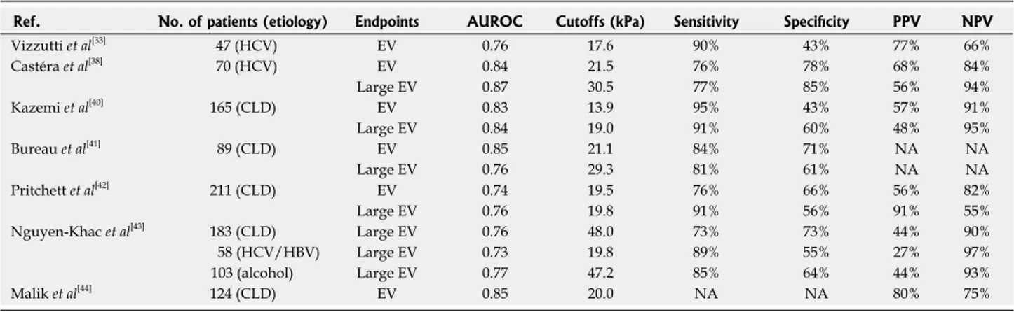
As variceal bleeding is a life-threatening complica-tion of portal hypertension, the relacomplica-tionship between TE values and the presence of esophageal varices has been investigated in several studies[35-40]. All demonstrated a sig-nificant correlation between TE values and the presence of esophageal varices and that TE values could predict the presence of large varices (more than grade 2)[38,40]. Table 1 summarizes reports of the relationship between TE values and esophageal varices[33,38,40-44].
Although TE can predict the presence of esophageal varices and consequently assist in selection of candidates for endoscopic screening or prophylactic treatment,
sever-al issues remain unresolved. First, the cutoff vsever-alues (range, 13.9-21.5 kPa) and performance of TE varied (AUROC range, 0.76-0.85) among studies[38-40]. Second, from data currently available, diagnostic performances of TE are acceptable for the prediction of esophageal varices, but far from satisfactory for screening cirrhotic patients with-out endoscopy confidently. Thus, Kim et al[45] recently proposed a novel prediction model [liver stiffness-spleen diameter to platelet ratio score (LSPS)] to address this is-sue, achieving higher accuracy using TE values and other parameters simultaneously that reflect portal hyperten-sion as constituent variables. Overall, this model had excellent diagnostic accuracy for the prediction of high-risk esophageal varices (HEV, AUROC = 0.953; negative predictive value 94.7%, positive predictive value 93.3%).
Beyond this cross-sectional analysis, a subsequent study by the same group recently showed that LSPS can be a reliable predictor of the development of variceal
bleeding[20]. In this prospective, longitudinal study analyz-ing 577 patients with hepatitis B virus-related cirrhosis, those with LSPS ≥ 5.5 had higher cumulative incidences of esophageal variceal bleeding during the follow-up pe-riod and LSPS score ≥ 6.5 was an independent risk factor of variceal bleeding among those with HEV, indicating that further prophylactic treatment such as endoscopic ligation in addition to a non-selective beta-blocker should be considered in these high-risk patients (Figure 1). In a similar context, Kim et al[46] stratified the risk of hepatic decompensation, such as ascites, hepatic encephalopathy, variceal hemorrhage, and deterioration of liver function to Child-Pugh class B or C, based upon three classes of TE values (TE value < 13, 13-18 and ≥ 18 kPa) in histologi-cally proven hepatitis B virus-related cirrhosis with well-preserved liver function and no history of decompensa-tion. In a multivariate analysis, patients with a TE value of 13-18 kPa [hazard ratio (HR), 4.547; P = 0.044] and ≥ 18 Ref. No. of patients (etiology) Endpoints AUROC Cutoffs (kPa) Sensitivity Specificity PPV NPV
Vizzutti et al[33] 47 (HCV) EV 0.76 17.6 90% 43% 77% 66% Castéra et al[38] 70 (HCV) EV 0.84 21.5 76% 78% 68% 84% Large EV 0.87 30.5 77% 85% 56% 94% Kazemi et al[40] 165 (CLD) EV 0.83 13.9 95% 43% 57% 91% Large EV 0.84 19.0 91% 60% 48% 95% Bureau et al[41] 89 (CLD) EV 0.85 21.1 84% 71% NA NA NA NA Large EV 0.76 29.3 81% 61% Pritchett et al[42] 211 (CLD) EV 0.74 19.5 76% 66% 56% 82% Large EV 0.76 19.8 91% 56% 91% 55% Nguyen-Khac et al[43] 183 (CLD) Large EV 0.76 48.0 73% 73% 44% 90% 58 (HCV/HBV) Large EV 0.73 19.8 89% 55% 27% 97% 103 (alcohol) Large EV 0.77 47.2 85% 64% 44% 93% Malik et al[44] 124 (CLD) EV 0.85 20.0 NA NA 80% 75%
Table 1 Diagnostic performance of transient elastography for prediction of esophageal varices or large esophageal varices
AUROC: Area under the receiver operating characteristic curve; PPV: Positive predictive value; NPV: Negative predictive value; CLD: Chronic liver disease; EV: Esophageal varix; HCV: Hepatitis C virus; HBV: Hepatitis B virus; NA: Not available.
Entire population (n = 577) Patients with LSPS ≥ 5.5 Patients with LSPS 3.5-5.5 Patients with LSPS < 3.5 0 1 2 3 4 yr No. at risk Patients with LSPS ≥ 5.5 107 76 51 33 18 Patients with LSPS 3.5-5.5 52 43 35 20 11 Patients with LSPS < 3.5 418 354 273 208 140 1.0 0.8 0.6 0.4 0.2 0.0 Cumulativ e EV bleeding risk
A B Patients with EVs (n = 214) Subgroup 2 Subgroup 1 Low-risk EVs 0 1 2 3 4 yr No. at risk Subgroup 2 88 61 41 28 16 Subgroup 1 62 53 41 28 19 Low-risk EVs 64 49 44 35 20 1.0 0.8 0.6 0.4 0.2 0.0 Cumulativ e EV bleeding risk
Figure 1 Cumulative incidences of variceal bleeding based on liver stiffness-spleen diameter to platelet ratio score values. A: The incidence of variceal bleeding increased significantly in association with higher liver stiffness-spleen diameter to platelet ratio score (LSPS) values (long-rank test, P < 0.001); B: In particu-lar, among patients with high risk esophageal varices (EV), the incidence of variceal bleeding was significantly higher in patient with LSPS 6.5 (subgroup 2) than those with LSPS < 6.5 (subgroup 1).
clinical role of TE in the noninvasive prediction of HCC development[52-56]. The first large prospective cohort study of 866 Japanese patients with chronic hepatitis C (CHC) tested whether TE can predict the future development of HCC[52]. During a mean follow-up of 3 years, 77 patients developed HCC. By multivariate analysis, together with age, male gender, and clinical cirrhosis, stratified TE value was identified as an independent risk factor for HCC development, with relative risks of 16.7, 20.0, 25.6 and 45.5 for TE values of 10-15, 15-20, 20-25 and > 25 kPa, respectively, vs an TE value of < 10 kPa as the reference and the cumulative incidence of HCC showed a step-wise increase according to stratified TE value (Figure 2A). Despite there being no histological analysis in relation to TE values and inclusion of patients with high alanine ami-notransferase (ALT) levels [> 5× upper limit of normal (ULN)] both of which can attenuate the accuracy of TE, this study confirmed that severity of liver fibrosis, reflect-ed by higher TE values, was closely associatreflect-ed with higher kPa (HR, 12.446; P < 0.001) showed independently higher
risks than patients with TE value < 13 kPa. HCC
Another promising area for the application of TE, other than portal hypertension-related decompensation events, is the prediction of HCC development. Unless HCC is diagnosed at an early stage, a poor prognosis is expected due to the limited treatment options[47-51]. Thus, early prediction of HCC development is of great importance, especially in high-risk patients. Among traditional risk factors, advanced liver fibrosis and cirrhosis is known to have a close association with risk of HCC develop-ment[47]. Thus, assessment of the severity of liver fibrosis at a given time point with subsequent monitoring of liver fibrosis progression by serial check-up is essential for ef-fective and optimized surveillance strategies for the early detection of HCC[3].
Recently, several Asian studies have investigated the
0 1 2 3
Years after enrollment No. at risk ≤ 10 kPa 511 501 479 427 10.1-15 kPa 142 130 111 94 15.1-20 kPa 79 76 63 51 20.1-25 kPa 47 41 36 29 > 25 kPa 87 76 54 41 1.0 0.9 0.8 0.7 0.6 0.5 0.4 0.3 0.2 0.1 0.0 Cumulativ e incidence P < 0.001 LSM > 25 kPa 20 < LSM ≤ 25 kPa 15 < LSM ≤ 20 kPa 10 < LSM ≤ 15 kPa LSM ≤ 10 kPa A B 0 1 2 3
Years after enrollment No. at risk ≤ 8 kPa 595 594 589 588 8-13 kPa 285 282 278 272 13-18 kPa 130 126 122 120 18-23 kPa 53 53 49 45 > 23 kPa 67 64 56 53 0.5 0.4 0.3 0.2 0.1 0.0 Cumulativ e incidence r ate P < 0.001 LSM > 23 kPa 18 < LSM ≤ 23 kPa 13 < LSM ≤ 18 kPa 8 < LSM ≤ 13 kPa LSM ≤ 8 kPa C Initial LSM Follow up LSM Group 4 Group 3 Group 2 Group 1 P < 0.001 13 kPa
Group 1 Group 2 Group 3 Group 4 (n = 598) (n = 71) (n = 34) (n = 119) Initial LSM ≤ 13 kPa > 13 kPa ≤ 13 kPa > 13 kPa Follow up LSM ≤ 13 kPa ≤ 13 kPa > 13 kPa > 13 kPa
7 6 5 4 3 2 1 0 Incidence r ate of HCC [% (person-year)] 0.44% 1.96% 2.05% 4.31%
Figure 2 Cumulative incidence of hepatocellular carcinoma development based on stratified transient elastography values in patients with chronic hepati-tis C (A, n = 866) and those with chronic hepatihepati-tis B (B, n = 1130). The cumulative incidences increased significantly in association with higher TE values (log-rank test, all P < 0.001). In particular, the overall incidence of HCC differed significantly among the four groups (C) (both initial and follow-up TE values ≤ 13 kPa (group 1), initial TE value > 13 kPa and follow-up TE value ≤ 13 kPa (group 2), initial TE value ≤ 13 kPa and up TE value > 13 kPa (group 3), and both initial and follow-up TE values > 13 kPa (grofollow-up 4) according to changing patterns of TE value during follow-follow-up (P < 0.001; Figure 2C). A: Cited from Masuzaki et al[52]; B and C: Cited
risk of HCC development and suggested a clinical role for TE in a longitudinal setting using HCC development as a solid clinical endpoint. Interestingly, in this study, even pa-tients with not so high level of TE (10-15 kPa) were still more subject to HCC development with an adjusted HR of 16.7, compared to those with a TE value < 10 kPa.
Another large Korean cohort study with 1130 patients with chronic hepatitis B (CHB) also confirmed the lon-gitudinal role of TE on HCC development[53]. Together with age, male gender, heavy alcohol consumption, lower serum albumin, and HBeAg positivity, stratified TE value was identified as an independent risk factor for HCC development, with relative risks of 3.07, 4.68, 5.55 and 6.60 for liver stiffness measurement (LSM) values of 8-13, 13-18, 18-23 and > 23 kPa, respectively, when compared with a LSM value of < 8 kPa as a reference (Figure 2B). In contrast to the Japanese study[52], several additional issues were further analyzed in this Korean study. First, when the diagnosis of cirrhosis showed discordant results between TE-based and clinical-based criteria, patients with cirrhosis based on TE were at a higher risk of HCC development than those with cirrhosis based on clinical criteria, indicating the superiority of TE for diagnosis of compensated liver cirrhosis. Second, patients with TE values below the cutoff level for cirrhosis, 8-13 kPa, had a higher relative risk of HCC development than those with LSM values < 8 kPa. Although this finding should be validated in large prospective studies, the issue of ex-pansion of the high-risk group for HCC surveillance to include those with significant fibrosis was raised by this study. Furthermore, when patients with available follow-up TE values were analyzed, the risk of HCC develop-ment changed according to the pattern of the changes in TE values, suggesting a potential role for serial mea-surements of TE as a dynamic monitoring tool for risk estimation of HCC development (Figure 2C). However, other confounding factors including lack of histological information, insensitive HBV DNA tests, and heteroge-neity in antiviral treatment should be noted when inter-preting these results. Recently, Chon et al[56] compared the performance of various noninvasive fibrosis prediction methods [aspartate aminotransferase-to-platelet ratio in-dex (ARRI), age-spleen-to-platelet ratio inin-dex (ASPRI), TE, LSPS, P2/MS and FIB-4] for prediction of HCC de-velopment in patients with CHB and concluded that TE and LSPS showed the best performance (AUROC = 0.789 and 0.788, respectively). Using multivariate analyses, TE and LSPS were identified as independent predictors of HCC development.
In another study[54] from Hong Kong, which followed up 528 patients with HBeAg negative CHB for a median length of 35 mo and identified seven patients with HCC development, the cumulative incidence of HCC was higher in patients with TE values ≥ 10 kPa than those with TE values < 10 kPa (9% vs 0%, respectively; P < 0.001), and the cumulative liver-related mortality was also higher in patients with TE values < 10 kPa compared with those with TE values ≥ 10 kPa (4% vs 0%,
respec-tively; P < 0.001). By multivariate analysis, only TE value was significantly associated with HCC development and liver-related mortality.
Similarly, Kim et al[55] investigated the prognostic role of TE in predicting the development of overall liver-related events (LREs), defined as development of HCC, hepatic decompensation, or liver-related mortality, among 128 patients with CHB showing histologically advanced liver fibrosis (≥ F3) and high viral loads (HBV DNA ≥ 2000 IU/mL) before starting nucleos(t)ide analogs. When the study population was stratified into two groups us-ing the optimal cutoff value (19 kPa), patients with TE values > 19 kPa were at significantly greater risk for LRE development than those with TE values ≤ 19 kPa (HR, 7.176; P = 0.001). Moreover, the incidence of LREs was similar in patients with F3 and F4 (22.2% vs 13.6%; P = 0.472); however, it differed significantly between patients with TE values ≤ 19 kPa and those with TE values > 19 kPa (6.9% vs 44.4%; P < 0.001), indicating the superior performance of TE to that of histology in prediction of LRE development.
Apart from predicting HCC development, the appli-cation of TE was validated in a study by Vergniol et al[57], in which 1457 patients with CHC were followed up; 5-year survival outcomes worsened as TE values increased. The prognostic values of TE were demonstrated to be statis-tically significant (P < 0.0001) after adjustment for other important factors, including treatment response, patient age, and estimates of necroinflammatory grade. For ex-ample, the 5-year overall survival was 96% in patients with TE value < 9.5 kPa, and 47% in patients with TE value > 40 kPa.
Overall, TE has shown the potential for a clinical role in predicting the development of portal hypertension-related hepatic decompensation and/or HCC and, in part, demonstrated superior performance to histology and other noninvasive tools[41,58-63]. This is most likely due to the wider dynamic range of TE values in the evaluation of liver cir-rhosis. In fact, as the stage of ‘‘cirrhosis’’ has to date been defined by histopathological evidence of one or two quali-tative categories (METAVIR stage F4 or ISHAK S5-S6), or more generally by the presence of so-called ‘‘regenerative’’ or ‘‘cirrhotic nodules’’, an interval scale cannot be used in this setting[64-66]. However, the degree of liver fibrosis may vary widely among patients in this category, and the risk of hepatic decompensation and HCC may not be uniform. Thus, in this regard, because TE value, expressed in kPa as a continuous variable, has a wide dynamic range within the cirrhotic stage from the cutoff level from non-cirrhosis (15-17 kPa) to the upper measurement limit of present de-vices (75 kPa), it would seem to be a more reasonable tool for detailed prognostication.
UTILITY OF TE IN THE SURGICAL
SETTING
Because TE values show significant correlations with portal hypertension and HCC development, prediction
of postoperative short-term outcomes, such as hepatic insufficiency, and long-term outcomes, such as recurrence or liver-related death using TE has been tested in several pilot studies[67-69]. Although further studies are required to validate these results, TE may facilitate stratification of patients undergoing curative resection according to dif-ferent prognoses.
In the first place, Kim et al[67] investigated whether preoperative TE values could predict the development of postoperative hepatic insufficiency after curative resec-tion of HCC. In this study, multivariate analyses revealed that a TE value > 25.6 kPa was the only predictor of postoperative insufficiency. The AUROC of 25.6 kPa was higher than that of indocyanine green R15, which is a popular method for assessment of preoperative func-tional reserve liver function (0.824 vs 0.620, respectively). Similar results were obtained in a subsequent investiga-tion by the same group[68]. In this study, the performance of TE was superior to that of diffusion-weighted mag-netic resonance imaging, which has also been shown to be a noninvasive fibrosis prediction tool for the assess-ment of liver fibrosis and the prediction of postoperative hepatic insufficiency.
Another issue is prediction of HCC recurrence after curative resection, that is, de novo recurrence in the back-ground liver with fibrotic burden, using preoperative TE. In an analysis of 133 patients who underwent preopera-tive TE and curapreopera-tive resection (HCC recurred in 62 pa-tients), TE was selected as an independent predictor of recurrence, whereas histological fibrosis status was not[69]. In the study, patients with preoperative TE values > 13.4 kPa were at a greater risk of recurrence, with an HR of 1.925 (P = 0.010). More specifically, when recurrence was stratified into early (< 2 years) and late (≥ 2 years), TE values were significantly related to late recurrence, thus supporting the hypothesis. These results suggest that pre-operative TE could reveal the potential influence of liver fibrosis on recurrence and explain multicentric carcino-genesis in a fibrotic liver. However, more data are needed to clarify this issue.
ROLE OF TE IN MONITORING FIBROTIC
BURDEN DURING ANTIVIRAL THERAPY
Recently, the concept of “cirrhosis” has changed from static and uncompromisingly progressive to rather dynamic and bidirectional, especially when treatment against the causative agent of tissue damage (i.e., antiviral agents against CHB or CHC and antifibrotic agents) can be introduced successfully at this stage of the disease. The ideal approach to evaluate histological outcomes du-ring antiviral therapy, such as fibrosis regression and ne-croinflammation stabilization, is serial LB examinations. However, this is impractical, primarily due to the inherent invasiveness of LB. Instead, because of the ease, safety, and rapidity of TE, it may be useful for monitoring the dynamic changes in liver fibrosis during antiviral or anti-fibrotic treatment. Indeed, several studies have reportedthe clinical usefulness of TE for monitoring potential fibrosis regression during antiviral treatment in patients with CHC and CHB[57,70-77].
Kim et al[71] analyzed 41 patients with CHB who re-ceived antiviral treatment using nucleos(t)ide analogs. To prevent the confounding effect of high ALT, pa-tients with high ALT levels more than 2× ULN, were excluded. Although ALT levels did not show a statisti-cally significant change during the first 12 or 24 mo of antiviral treatment, TE values decreased significantly, indicating potential fibrosis regression due to prolonged antiviral treatment. Indeed, fibrosis regression and stabi-lization of necroinflammation was noted in two patients with available paired LBs. Enomoto et al[70] reported the changes in LSM values during the first 12 mo of enteca-vir treatment in 20 patients. Median TE values decreased significantly from 11.2 to 7.8 kPa after 12 mo of treat-ment, and serum fibrosis markers, such as PIIINP and type Ⅳ collagen 7S domain, also decreased significantly. In one patient with available paired LBs, histological fi-brosis regression and stabilization of necroinflammation were noted. Although these studies suggest a role for TE for monitoring fibrosis regression due to prolonged an-tiviral treatment, the short duration of observation and small sample sizes with paired biopsies are major limita-tions of these studies.
Recently, data regarding a longer antiviral treatment duration (more than 3 years) have become available[72-74,78]. Fung et al[72] reported a significant decline in TE values from baseline after subsequent ALT normalization with 3-year treatment (n = 110, 7.8 to 6.1 kPa; P = 0.002). In this study, independent factors associated with a signifi-cant decline in TE value of ≥ 1 kPa included antiviral therapy and ALT levels at the follow-up time point. Another study by Andersen et al[78] also noted significant declines in TE values after a median antiviral treatment duration of 50.5 mo (n = 66), and concluded that pro-longed antiviral treatment in patients with CHB resulted in significant declines in TE values, suggesting regression of fibrosis in a majority of patients with advanced fibro-sis or cirrhofibro-sis.
Likewise, for patients with CHC, changes in TE va-lues during antiviral treatment have been investigated in several studies. Two prospective studies by Vergniol et al[57] and Ogawa et al[75] demonstrated that patients with CHC showing sustained virological responses to pegylated interferon-ribavirin combination therapy had significantly reduced TE values at the end of follow-up. Moreover, Ogawa et al[75] reported that patients with non-sustained virological responses, but with a biochemical response, showed a greater reduction in TE values than did those with a non-biochemical response. Subsequent studies reported similar results, suggesting that changes in TE values during antiviral treatment in patients with CHC may represent alterations in the severity of liver fibrosis[76,77]. However, it should be further confirmed whether the favorable changes in LSM values during or after antiviral treatment does have a significant influecne
on the long-term prognosis such as disease-specific survi-val in patients with CHC.
Taken together, TE value seems to decrease dur-ing and after antiviral therapy. However, without paired histological results through repeated LB, whether the reduction in TE values is closely correlated with regres-sion of liver fibrosis or improvement in necroinflamma-tory scores remains unclear. To clarify this, Lim et al[73] investigated patterns of TE values among patients who were treated with entecavir. In all subjects, the median TE value at baseline was 15.1 (range, 5.6-75.0) kPa and decreased significantly, to 8.8 (range, 3.0-33.8) kPa after 12 mo of therapy, and a decrease in TE values correlated significantly with increase in albumin, decrease in biliru-bin, decrease in ALT level, and decrease in aspartate ami-notransferase levels (all P < 0.05). However, among 15 patients with available paired LBs, decreases in TE values were correlated significantly with improved necroinflam-matory scores, but not with fibrosis regression. Similarly, Wong et al[74] insisted that the decline in absolute TE val-ues during antiviral treatment did not reflect the change in histologically assessed liver fibrosis, probably due to the confounding influence of ALT reduction caused by antiviral treatment.
However, regardless of whether TE values during antiviral treatment are due to fibrosis regression, activity stabilization, or both, changes in TE value during antiviral treatment can be translated into the overall response of chronically diseased liver to antiviral treatment from the viewpoint of its long-term clinical implications. Thus, it is more logical to investigate whether the decline in TE value can be used as a favorable predictor of long-term prognosis. Encouraging results were recently published by Jung et al[53] suggesting that the change in TE values in patients with CHB showed a significant correlation with differential future risk of HCC development. Additionally, Kim et al[79] insisted that changes in TE values were signifi-cantly associated with the difference risk of liver-related event occurrence, such as hepatic decompensation, HCC
development, and LREs (Figure 3). This would suggest that the assessment of overall background liver status us-ing TE may be an important end-point in the manage-ment of CHB and prediction of long-term outcomes. Further research is needed to evaluate the reproducibility of such findings in independent populations.
LIMITATIONS OF TE
Although TE has demonstrated reliable diagnostic accura-cy with excellent inter-observer and intra-observer agree-ment, additional space-occupying tissue abnormalities, such as edema and inflammation, cholestasis, and conges-tion may interfere with TE, regardless of the degree of liver fibrosis, because the liver is wrapped in a distensible, but non-elastic, envelope (Glisson’s capsule)[80].
First, the extent of histological necroinflammatory ac-tivity has been shown to influence TE results in patients with viral hepatitis, resulting in an overestimation of TE values that increases in parallel with the degree of necro-inflammatory score[81-85]. Consistent with these results, a risk of overestimation of TE values has been reported in cases of ALT flares in patients with acute viral hepatitis or CHB[86-92]. Thus, in such subjects, TE examinations should be delayed until ALT levels have stabilized. In this regard, several studies have investigated the optimal period (3 to 6 mo) for restoration of the reliability of TE values in patients with acute flares[88,91,93,94]. Furthermore, even mild to moderate elevalation in ALT can be asso-ciated with higher liver stiffness values, and may cause discrepancies between TE results and the actual underly-ing fibrosis. Apart from necroinflammation, extrahepatic cholestasis[95] and congestive heart failure[96-98] may also contribute to the overestimation of TE.
Additionally, the performance of TE may be limited in patients with a high body mass index (BMI), narrow intercostal space, or ascites[9]. Although TE reproduc-ibility has been shown to be excellent in terms of inter-observer and intra-inter-observer agreement, a high BMI (> 28 Initial LSM Follow up LSM Group 4 Group 3 Group 2 Group 1 P < 0.0001 11.6 kPa
Group 1 Group 2 Group 3 Group 4 (n = 39) (n = 24) (n = 8) (n = 28) Initial LSM < 11.6 kPa ≥ 11.6 kPa < 11.6 kPa ≥ 11.6 kPa Follow up LSM < 11.6 kPa < 11.6 kPa ≥ 11.6 kPa ≥ 11.6 kPa
12 10 8 6 4 2 0
Incidence of LREs [%
(person-year)] 1.22 2.10 2.89 11.05 A B Initial LSM Follow up LSM Group 4 Group 3 Group 2 Group 1 P < 0.0001 18.2 kPa
Group 1 Group 2 Group 3 Group 4 (n = 75) (n = 15) (n = 3) (n = 6) Initial LSM < 18.2 kPa ≥ 18.2 kPa < 18.2 kPa ≥ 18.2 kPa Follow up LSM < 18.2 kPa < 18.2 kPa ≥ 18.2 kPa ≥ 18.2 kPa
25 20 15 10 5 0
Incidence of LREs [%
(person-year)]
2.29
7.29
9.20
23.3
Figure 3 Incidence of liver-related events according to changes in transient elastography values after 6 mo of antiviral therapy. The overall incidence of liver-related events differed significantly among the four groups using TE value cutoffs of 11.6 kPa (A) and 18.2 kPa (B) (both P < 0.0001). Adapted from Kim et al[79].
kg/m2) and waist circumference were significantly associ-ated with TE failure[99]. These results emphasize the need for adequate operator training and for technological im-provements in specific patient populations, such as those with non-alcoholic fatty liver disease. For this, a new TE probe (the XL probe) was recently introduced to lessen the TE failure rate in obese patients; however, its efficacy should be further validated[100].
CONCLUSION
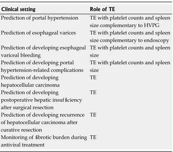
Over the past decade, significant progress has been made in the non-invasive assessment of liver fibrosis in patients with CLD. Of the methods now available, TE appears to be an excellent tool for assessing liver fibrosis, particu-larly for diagnosis of cirrhosis, and also has prognostic value from a longitudinal perspective. Although TE can-not completely obviate the need for invasive tests, such as LB, endoscopic examination for identification of varices, or HVPG, it represents an important non-invasive tool, enabling more efficient and tailored management strate-gies for patients with CLD (Table 2). We hope that other researchers will evaluate the usefullness of other similar techniques such as the measurement of spleen stiffness in comparison or in combination with TE in the future.
ACKNOWLEDGMENTS
The authors thank Mr. Dong-Su Jang, Research Assistant, Department of Anatomy, Yonsei University College of Medicine, Seoul, South Korea, for his help with the figures.
REFERENCES
1 Jang JW. Current status of liver diseases in Korea: liver cir-rhosis. Korean J Hepatol 2009; 15 Suppl 6: S40-S49 [PMID:
20037279 DOI: 10.3350/kjhep.2009.15.S6.S40]
2 Lok AS. Hepatitis B: liver fibrosis and hepatocellular car-cinoma. Gastroenterol Clin Biol 2009; 33: 911-915 [PMID: 19577871 DOI: 10.1016/j.gcb.2009.06.001]
3 Pungpapong S, Kim WR, Poterucha JJ. Natural history of hepatitis B virus infection: an update for clinicians. Mayo Clin Proc 2007; 82: 967-975 [PMID: 17673066 DOI: 10.4065/82.8.967]
4 McGill DB, Rakela J, Zinsmeister AR, Ott BJ. A 21-year experience with major hemorrhage after percutaneous liver biopsy. Gastroenterology 1990; 99: 1396-1400 [PMID: 2101588] 5 Jin SY. Role of liver biopsy in the assessment of hepatic
fibrosis--its utility and limitations. Korean J Hepatol 2007; 13: 138-145 [PMID: 17585187]
6 Regev A, Berho M, Jeffers LJ, Milikowski C, Molina EG, Pyr-sopoulos NT, Feng ZZ, Reddy KR, Schiff ER. Sampling error and intraobserver variation in liver biopsy in patients with chronic HCV infection. Am J Gastroenterol 2002; 97: 2614-2618 [PMID: 12385448 DOI: 10.1111/j.1572-0241.2002.06038.x] 7 Abraldes JG, Araujo IK, Turón F, Berzigotti A.
Diagnos-ing and monitorDiagnos-ing cirrhosis: Liver biopsy, hepatic ve-nous pressure gradient and elastography. Gastroenterol Hepatol 2012; 35: 488-495 [PMID: 22560536 DOI: 10.1016/ j.gastrohep.2012.02.010]
8 Castera L, Forns X, Alberti A. Non-invasive evaluation of liver fibrosis using transient elastography. J Hepatol 2008; 48: 835-847 [PMID: 18334275 DOI: 10.1016/j.jhep.2008.02.008] 9 Sandrin L, Fourquet B, Hasquenoph JM, Yon S, Fournier C,
Mal F, Christidis C, Ziol M, Poulet B, Kazemi F, Beaugrand M, Palau R. Transient elastography: a new noninvasive method for assessment of hepatic fibrosis. Ultrasound Med Biol 2003; 29: 1705-1713 [PMID: 14698338 DOI: 10.1016/j.ultr asmedbio.2003.07.001]
10 Talwalkar JA, Kurtz DM, Schoenleber SJ, West CP, Mon-tori VM. Ultrasound-based transient elastography for the detection of hepatic fibrosis: systematic review and meta-analysis. Clin Gastroenterol Hepatol 2007; 5: 1214-1220 [PMID: 17916549 DOI: 10.1016/j.cgh.2007.07.020]
11 Stauber RE, Lackner C. Noninvasive diagnosis of hepatic fibrosis in chronic hepatitis C. World J Gastroenterol 2007; 13: 4287-4294 [PMID: 17708599]
12 Kang JK, Cheong JY, Cho SW, Cho JH, Park JS, Kim YB, Kim DJ, Hwang SG, Yang JM, Park YN. Liver stiffness measurement for the diagnosis of hepatic fibrosis in pa-tients with chronic viral hepatitis. Korean J Hepatol 2007; 13: 521-529 [PMID: 18159150 DOI: 10.3350/kjhep.2007.13.4.521] 13 Jung HS, Kim YS, Kwon OS, Ku YS, Kim YK, Choi DJ, Kim
JH. Usefulness of liver stiffness measurement for predicting the presence of esophageal varices in patients with liver cir-rhosis. Korean J Hepatol 2008; 14: 342-350 [PMID: 18815457 DOI: 10.3350/kjhep.2008.14.3.342]
14 Kim SG, Kim YS, Jung SW, Kim HK, Jang JY, Moon JH, Kim HS, Lee JS, Lee MS, Shim CS, Kim BS. The usefulness of transient elastography to diagnose cirrhosis in patients with alcoholic liver disease. Korean J Hepatol 2009; 15: 42-51 [PMID: 19346784 DOI: 10.3350/kjhep.2009.15.1.42]
15 Kim SM, Sohn JH, Kim TY, Roh YW, Eun CS, Jeon YC, Han DS, Oh YH. Comparison of various noninvasive serum markers of liver fibrosis in chronic viral liver disease. Korean J Hepatol 2009; 15: 454-463 [PMID: 20037264 DOI: 10.3350/ kjhep.2009.15.4.454]
16 Scott DR, Levy MT. Liver transient elastography (Fibroscan): a place in the management algorithms of chronic viral hepatitis. Antivir Ther 2010; 15: 1-11 [PMID: 20167986 DOI: 10.3851/IMP1474]
17 Stebbing J, Farouk L, Panos G, Anderson M, Jiao LR, Man-dalia S, Bower M, Gazzard B, Nelson M. A meta-analysis of transient elastography for the detection of hepatic fibrosis. J Clin Gastroenterol 2010; 44: 214-219 [PMID: 19745758 DOI: 10.1097/MCG.0b013e3181b4af1f]
P- Reviewers Bener A S- Editor Song XX L- Editor Stewart GJ E- Editor Li JY
Clinical setting Role of TE
Prediction of portal hypertension TE with platelet counts and spleen size complementary to HVPG Prediction of esophageal varices TE with platelet counts and spleen
size complementary to endoscopy Prediction of developing esophageal
variceal bleeding
TE with platelet counts and spleen size
Prediction of developing portal hypertension-related complications
TE with platelet counts and spleen size
Prediction of developing hepatocellular carcinoma
TE Prediction of developing
postoperative hepatic insufficiency after surgical resection
TE Prediction of developing recurrence of hepatocellular carcinoma after curative resection
TE Monitoring of fibrotic burden during antiviral treatment
TE
Table 2 Proposal of application of transient elastography in each clinical setting
18 Han KH, Yoon KT. New diagnostic method for liver fibrosis and cirrhosis. Intervirology 2008; 51 Suppl 1: 11-16 [PMID: 18544943 DOI: 10.1159/000122594]
19 Smith JO, Sterling RK. Systematic review: non-invasive methods of fibrosis analysis in chronic hepatitis C. Aliment Pharmacol Ther 2009; 30: 557-576 [PMID: 19519733 DOI: 10.1111/j.1365-2036.2009.04062.x]
20 Kim BK, Kim do Y, Han KH, Park JY, Kim JK, Paik YH, Lee KS, Chon CY, Ahn SH. Risk assessment of esophageal variceal bleeding in B-viral liver cirrhosis by a liver stiffness measurement-based model. Am J Gastroenterol 2011; 106: 1654-1662, 1730 [PMID: 21691339]
21 Kim SU, Han KH, Ahn SH. Non-invasive assessment of liver fibrosis: time to move from cross-sectional studies to longitu-dinal ones. J Gastroenterol Hepatol 2010; 25: 1472-1473 [PMID: 20796140 DOI: 10.1111/j.1440-1746.2010.06432.x]
22 Addley J, Tham TC, Cash WJ. Use of portal pressure studies in the management of variceal haemorrhage. World J Gastro-intest Endosc 2012; 4: 281-289 [PMID: 22816007 DOI: 10.4253/ wjge.v4.i7.281]
23 Bosch J, García-Pagán JC. Complications of cirrhosis. I. Portal hypertension. J Hepatol 2000; 32: 141-156 [PMID: 10728801] 24 Suk KT, Kim CH, Park SH, Sung HT, Choi JY, Han KH,
Hong SH, Kim DY, Yoon JH, Kim YS, Baik GH, Kim JB, Kim DJ. Comparison of hepatic venous pressure gradient and two models of end-stage liver disease for predicting the survival in patients with decompensated liver cirrhosis. J Clin Gastro-enterol 2012; 46: 880-886 [PMID: 22810110]
25 Keiding S, Vilstrup H. Intrahepatic heterogeneity of hepatic venous pressure gradient in human cirrhosis. Scand J Gastro-enterol 2002; 37: 960-964 [PMID: 12229973]
26 Rincón D, Lo Iacono O, Tejedor M, Hernando A, Ripoll C, Catalina MV, Salcedo M, Matilla A, Senosiain M, Clemente G, Molinero LM, Albillos A, Bañares R. Prognostic value of he-patic venous pressure gradient in patients with compensated chronic hepatitis C-related cirrhosis. Scand J Gastroenterol 2012 Aug 8; Epub ahead of print [PMID: 22871085]
27 Pineda JA, Recio E, Camacho A, Macías J, Almodóvar C, González-Serrano M, Merino D, Tellez F, Ríos MJ, Rivero A. Liver stiffness as a predictor of esophageal varices requiring therapy in HIV/hepatitis C virus-coinfected patients with cir-rhosis. J Acquir Immune Defic Syndr 2009; 51: 445-449 [PMID: 19487952 DOI: 10.1097/QAI.0b013e3181acb675]
28 Wadhawan M, Dubey S, Sharma BC, Sarin SK, Sarin SK. He-patic venous pressure gradient in cirrhosis: correlation with the size of varices, bleeding, ascites, and child’s status. Dig Dis Sci 2006; 51: 2264-2269 [PMID: 17080245 DOI: 10.1007/ s10620-006-9310-2]
29 Kim MY, Baik SK, Suk KT, Yea CJ, Lee IY, Kim JW, Cha SH, Kim YJ, Um SH, Han KH. Measurement of hepatic venous pressure gradient in liver cirrhosis: relationship with the sta-tus of cirrhosis, varices, and ascites in Korea. Korean J Hepatol 2008; 14: 150-158 [PMID: 18617762]
30 Hobolth L, Bendtsen F, Møller S. Indications for portal pres-sure meapres-surement in chronic liver disease. Scand J Gastroen-terol 2012; 8-9: 887-892 [PMID: 22809270]
31 Bañares R, Catalina MV, Ripoll C, Rincón D. Prognostic markers in patients who have recovered from an acute vari-ceal bleeding: role of HVPG measurement. Dis Markers 2011; 31: 165-169 [PMID: 22045402]
32 Seijo S, Reverter E, Miquel R, Berzigotti A, Abraldes JG, Bosch J, García-Pagán JC. Role of hepatic vein catheterisa-tion and transient elastography in the diagnosis of idiopathic portal hypertension. Dig Liver Dis 2012; 44: 855-860 [PMID: 22721839]
33 Vizzutti F, Arena U, Romanelli RG, Rega L, Foschi M, Cola-grande S, Petrarca A, Moscarella S, Belli G, Zignego AL, Mar-ra F, Laffi G, Pinzani M. Liver stiffness measurement predicts severe portal hypertension in patients with HCV-related cir-rhosis. Hepatology 2007; 45: 1290-1297 [PMID: 17464971 DOI: 10.1002/hep.21665]
34 Foucher J, Chanteloup E, Vergniol J, Castéra L, Le Bail B,
Ad-houte X, Bertet J, Couzigou P, de Lédinghen V. Diagnosis of cirrhosis by transient elastography (FibroScan): a prospective study. Gut 2006; 55: 403-408 [PMID: 16020491 DOI: 10.1136/ gut.2005.069153]
35 Cho SB, Park KJ, Lee JS, Lee WS, Park CH, Joo YE, Kim HS, Choi SK, Rew JS, Kim SJ. Comparison of terlipressin and octreotide with variceal ligation for controlling acute esopha-geal variceal bleeding--a randomized prospective study. Ko-rean J Hepatol 2006; 12: 385-393 [PMID: 16998290]
36 Youn KH, Kim DJ. Prevention of variceal bleeding and mea-surement of hepatic vein pressure gradient. Korean J Hepatol 2006; 12: 464-468 [PMID: 16998301]
37 Seo YS, Um SH, Hyun JJ, Kim YH, Park S, Keum BR, Kim YS, Jeen YT, Lee HS, Chun HJ, Lee SW, Choi JH, Kim CD, Ryu HS. A prospective study comparing the efficacy of early administration of terlipressin and somatostatin for the control of acute variceal bleeding in patients with cirrhosis. Korean J Hepatol 2006; 12: 373-384 [PMID: 16998289]
38 Castéra L, Le Bail B, Roudot-Thoraval F, Bernard PH, Foucher J, Merrouche W, Couzigou P, de Lédinghen V. Early detection in routine clinical practice of cirrhosis and oesopha-geal varices in chronic hepatitis C: comparison of transient elastography (FibroScan) with standard laboratory tests and non-invasive scores. J Hepatol 2009; 50: 59-68 [PMID: 19013661 DOI: 10.1016/j.jhep.2008.08.018]
39 Lemoine M, Katsahian S, Ziol M, Nahon P, Ganne-Carrie N, Kazemi F, Grando-Lemaire V, Trinchet JC, Beaugrand M. Liver stiffness measurement as a predictive tool of clinically significant portal hypertension in patients with compensated hepatitis C virus or alcohol-related cirrhosis. Aliment Pharma-col Ther 2008; 28: 1102-1110 [PMID: 18691352 DOI: 10.1111/ j.1365-2036.2008.03825.x]
40 Kazemi F, Kettaneh A, N’kontchou G, Pinto E, Ganne-Carrie N, Trinchet JC, Beaugrand M. Liver stiffness measurement selects patients with cirrhosis at risk of bearing large oesoph-ageal varices. J Hepatol 2006; 45: 230-235 [PMID: 16797100 DOI: 10.1016/j.jhep.2006.04.006]
41 Bureau C, Metivier S, Peron JM, Selves J, Robic MA, Gour-raud PA, Rouquet O, Dupuis E, Alric L, Vinel JP. Transient elastography accurately predicts presence of significant portal hypertension in patients with chronic liver disease. Aliment Pharmacol Ther 2008; 27: 1261-1268 [PMID: 18397389 DOI: 10.1111/j.1365-2036.2008.03701.x]
42 Pritchett S, Cardenas A, Manning D, Curry M, Afdhal NH. The optimal cut-off for predicting large oesophageal varices using transient elastography is disease specific. J Viral Hepat 2011; 18: e75-e80 [PMID: 21040236 DOI: 10.1111/ j.1365-2893.2010.01375.x]
43 Nguyen-Khac E, Saint-Leger P, Tramier B, Coevoet H, Cap-ron D, Dupas JL. Noninvasive diagnosis of large esophageal varices by Fibroscan: strong influence of the cirrhosis etiolo-gy. Alcohol Clin Exp Res 2010; 34: 1146-1153 [PMID: 20477777] 44 Malik R, Lai M, Sadiq A, Farnan R, Mehta S, Nasser I,
Challies T, Schuppan D, Afdhal N. Comparison of tran-sient elastography, serum markers and clinical signs for the diagnosis of compensated cirrhosis. J Gastroenterol Hepatol 2010; 25: 1562-1568 [PMID: 20796156 DOI: 10.1111/ j.1440-1746.2010.06371.x]
45 Kim BK, Han KH, Park JY, Ahn SH, Kim JK, Paik YH, Lee KS, Chon CY, Kim do Y. A liver stiffness measurement-based, noninvasive prediction model for high-risk esopha-geal varices in B-viral liver cirrhosis. Am J Gastroenterol 2010; 105: 1382-1390 [PMID: 20087336 DOI: 10.1038/ajg.2009.750] 46 Kim BK, Park YN, Kim do Y, Park JY, Chon CY, Han KH,
Ahn SH. Risk assessment of development of hepatic decom-pensation in histologically proven hepatitis B viral cirrhosis using liver stiffness measurement. Digestion 2012; 85: 219-227 [PMID: 22414567 DOI: 10.1159/000335430]
47 Kudo M. Hepatocellular carcinoma 2009 and beyond: from the surveillance to molecular targeted therapy. Oncology 2008; 75 Suppl 1: 1-12 [PMID: 19092266 DOI: 10.1159/000181865] 48 El-Serag HB. Epidemiology of viral hepatitis and
hepato-cellular carcinoma. Gastroenterology 2012; 142: 1264-1273.e1 [PMID: 22537432]
49 Frenette C, Gish R. Targeted systemic therapies for hepa-tocellular carcinoma: clinical perspectives, challenges and implications. World J Gastroenterol 2012; 18: 498-506 [PMID: 22363115 DOI: 10.3748/wjg.v18.i6.498]
50 Bruix J, Sherman M. Management of hepatocellular carci-noma. Hepatology 2005; 42: 1208-1236 [PMID: 16250051 DOI: 10.1002/hep.20933]
51 Maggs JR, Suddle AR, Aluvihare V, Heneghan MA. Sys-tematic review: the role of liver transplantation in the man-agement of hepatocellular carcinoma. Aliment Pharmacol Ther 2012; 35: 1113-1134 [PMID: 22432733 DOI: 10.1111/ j.1365-2036.2012.05072.x]
52 Masuzaki R, Tateishi R, Yoshida H, Goto E, Sato T, Ohki T, Imamura J, Goto T, Kanai F, Kato N, Ikeda H, Shiina S, Kaw-abe T, Omata M. Prospective risk assessment for hepatocellu-lar carcinoma development in patients with chronic hepatitis C by transient elastography. Hepatology 2009; 49: 1954-1961 [PMID: 19434742 DOI: 10.1002/hep.22870]
53 Jung KS, Kim SU, Ahn SH, Park YN, Kim do Y, Park JY, Chon CY, Choi EH, Han KH. Risk assessment of hepatitis B virus-related hepatocellular carcinoma development using liver stiffness measurement (FibroScan). Hepatology 2011; 53: 885-894 [PMID: 21319193 DOI: 10.1002/hep.24121]
54 Fung J, Lai CL, Seto WK, Wong DK, Yuen MF. Prognostic significance of liver stiffness for hepatocellular carcinoma and mortality in HBeAg-negative chronic hepatitis B. J Viral Hepat 2011; 18: 738-744 [PMID: 20659306 DOI: 10.1111/ j.1365-2893.2010.01355.x]
55 Kim SU, Lee JH, Kim do Y, Ahn SH, Jung KS, Choi EH, Park YN, Han KH, Chon CY, Park JY. Prediction of liver-related events using fibroscan in chronic hepatitis B patients show-ing advanced liver fibrosis. PLoS One 2012; 7: e36676 [PMID: 22574212 DOI: 10.1371/journal.pone.0036676]
56 Chon YE, Jung ES, Park JY, Kim do Y, Ahn SH, Han KH, Chon CY, Jung KS, Kim SU. The accuracy of noninvasive methods in predicting the development of hepatocellular carcinoma and hepatic decompensation in patients with chronic hepatitis B. J Clin Gastroenterol 2012; 46: 518-525 [PMID: 22688146 DOI: 10.1097/MCG.0b013e31825079f1]
57 Vergniol J, Foucher J, Castéra L, Bernard PH, Tournan R, Ter-rebonne E, Chanteloup E, Merrouche W, Couzigou P, de Léd-inghen V. Changes of non-invasive markers and FibroScan values during HCV treatment. J Viral Hepat 2009; 16: 132-140 [PMID: 19175875 DOI: 10.1111/j.1365-2893.2008.01055.x] 58 Jung KS, Kim SU. Clinical applications of transient
elastogra-phy. Clin Mol Hepatol 2012; 18: 163-173 [PMID: 22893866 DOI: 10.3350/cmh.2012.18.2.163]
59 Poca M, Puente A, Graupera I, Villanueva C. Prognostic markers in patients with cirrhosis and portal hypertension who have not bled. Dis Markers 2011; 31: 147-154 [PMID: 22045400]
60 Robic MA, Procopet B, Métivier S, Péron JM, Selves J, Vinel JP, Bureau C. Liver stiffness accurately predicts portal hyper-tension related complications in patients with chronic liver disease: a prospective study. J Hepatol 2011; 55: 1017-1024 [PMID: 21354450 DOI: 10.1016/j.jhep.2011.01.051]
61 Pesce A, Scilletta R, Branca A, Nigro L, Montineri A, Larocca L, Fatuzzo F, Castaing M, Puleo S. Does transient elastography (FibroScan®) have a role in decision making in hepatocellular carcinoma? HPB (Oxford) 2012; 14: 403-408 [PMID: 22568417] 62 Motosugi U, Ichikawa T, Koshiishi T, Sano K, Morisaka H,
Ichikawa S, Enomoto N, Matsuda M, Fujii H, Araki T. Liver stiffness measured by magnetic resonance elastography as a risk factor for hepatocellular carcinoma: a preliminary case-control study. Eur Radiol 2013; 23: 156-162 [PMID: 22814828] 63 Masuzaki R, Tateishi R, Yoshida H, Arano T, Uchino K,
Enooku K, Goto E, Nakagawa H, Asaoka Y, Kondo Y, Goto T, Ikeda H, Shiina S, Omata M, Koike K. Assessment of disease progression in patients with transfusion-associated chronic hepatitis C using transient elastography. World J Gastroenterol
2012; 18: 1385-1390 [PMID: 22493553 DOI: 10.3748/wjg.v18. i12.1385]
64 Bedossa P, Poynard T. An algorithm for the grading of activ-ity in chronic hepatitis C. The METAVIR Cooperative Study Group. Hepatology 1996; 24: 289-293 [PMID: 8690394 DOI: 10.1002/hep.510240201]
65 Batts KP, Ludwig J. Chronic hepatitis. An update on termi-nology and reporting. Am J Surg Pathol 1995; 19: 1409-1417 [PMID: 7503362 DOI: 10.1097/00000478-199512000-00007] 66 Ishak K, Baptista A, Bianchi L, Callea F, De Groote J, Gudat
F, Denk H, Desmet V, Korb G, MacSween RN. Histological grading and staging of chronic hepatitis. J Hepatol 1995; 22: 696-699 [PMID: 7560864 DOI: 10.1016/0168-8278(95)80226-6] 67 Kim SU, Ahn SH, Park JY, Kim do Y, Chon CY, Choi JS, Kim
KS, Han KH. Prediction of postoperative hepatic insuffi-ciency by liver stiffness measurement (FibroScan((R))) before curative resection of hepatocellular carcinoma: a pilot study. Hepatol Int 2008; 2: 471-477 [PMID: 19669322 DOI: 10.1007/ s12072-008-9091-0]
68 Kim SU, Kim YC, Choi JS, Kim KS, Choi GH, Choi JS, Park JY, Kim do Y, Ahn SH, Choi EH, Park YN, Chon CY, Han KH, Kim MJ. Can preoperative diffusion-weighted MRI pre-dict postoperative hepatic insufficiency after curative resec-tion of HBV-related hepatocellular carcinoma? A pilot study. Magn Reson Imaging 2010; 28: 802-811 [PMID: 20395100 DOI: 10.1016/j.mri.2010.03.018]
69 Jung KS, Kim SU, Choi GH, Park JY, Park YN, Kim do Y, Ahn SH, Chon CY, Kim KS, Choi EH, Choi JS, Han KH. Pre-diction of recurrence after curative resection of hepatocellular carcinoma using liver stiffness measurement (FibroScan®). Ann Surg Oncol 2012; 19: 4278-4286 [PMID: 22752370] 70 Enomoto M, Mori M, Ogawa T, Fujii H, Kobayashi S, Iwai S,
Morikawa H, Tamori A, Sakaguchi H, Sawada A, Takeda S, Habu D, Shiomi S, Kawada N. Usefulness of transient elas-tography for assessment of liver fibrosis in chronic hepatitis B: Regression of liver stiffness during entecavir therapy. Hepatol Res 2010; 40: 853-861 [PMID: 20887589 DOI: 10.1111/j.1872-034X.2010.00687.x]
71 Kim SU, Park JY, Kim do Y, Ahn SH, Choi EH, Seok JY, Lee JM, Park YN, Chon CY, Han KH. Non-invasive assessment of changes in liver fibrosis via liver stiffness measurement in patients with chronic hepatitis B: impact of antiviral treat-ment on fibrosis regression. Hepatol Int 2010; 4: 673-680 [PMID: 21286337 DOI: 10.1007/s12072-010-9201-7]
72 Fung J, Lai CL, Wong DK, Seto WK, Hung I, Yuen MF. Sig-nificant changes in liver stiffness measurements in patients with chronic hepatitis B: 3-year follow-up study. J Viral Hepat 2011; 18: e200-e205 [PMID: 21692933 DOI: 10.1111/ j.1365-2893.2010.01428.x]
73 Lim SG, Cho SW, Lee YC, Jeon SJ, Lee MH, Cho YJ, Kim SS, Kim YB, Seok JY, Cheong JY, Kim JH. Changes in liver stiff-ness measurement during antiviral therapy in patients with chronic hepatitis B. Hepatogastroenterology 2011; 58: 539-545 [PMID: 21661428]
74 Wong GL, Wong VW, Choi PC, Chan AW, Chim AM, Yiu KK, Chu SH, Chan FK, Sung JJ, Chan HL. On-treatment mon-itoring of liver fibrosis with transient elastography in chronic hepatitis B patients. Antivir Ther 2011; 16: 165-172 [PMID: 21447865 DOI: 10.3851/IMP1726]
75 Ogawa E, Furusyo N, Toyoda K, Takeoka H, Maeda S, Hayashi J. The longitudinal quantitative assessment by transient elastography of chronic hepatitis C patients treated with pegylated interferon alpha-2b and ribavirin. Antivi-ral Res 2009; 83: 127-134 [PMID: 19443053 DOI: 10.1016/ j.antiviral.2009.04.002]
76 Wang JH, Changchien CS, Hung CH, Tung WC, Kee KM, Chen CH, Hu TH, Lee CM, Lu SN. Liver stiffness decrease after effective antiviral therapy in patients with chronic hepa-titis C: Longitudinal study using FibroScan. J Gastroenterol Hepatol 2010; 25: 964-969 [PMID: 20546451 DOI: 10.1111/ j.1440-1746.2009.06194.x]
M, Nakano T, Shimazaki H, Kobayashi K, Ichino N, Osak-abe K, Nishikawa T, Okumura A, Ishikawa T, Yoshioka K. Reduction of liver stiffness by interferon treatment in the pa-tients with chronic hepatitis C. Hepatol Res 2010; 40: 383-392 [PMID: 20236358 DOI: 10.1111/j.1872-034X.2009.00618.x] 78 Andersen ES, Weiland O, Leutscher P, Krarup H, Westin J,
Moessner B, Konopski Z, Frigstad SO, Kjær M, Christensen PB, Weis N. Low liver stiffness among cirrhotic patients with hepatitis B after prolonged treatment with nucleoside ana-logs. Scand J Gastroenterol 2011; 46: 760-766 [PMID: 21438780 DOI: 10.3109/00365521.2011.565068]
79 Kim SU, Oh HJ, Park YN, Park JY, Kim do Y, Ahn SH, Chon CY, Han KH. Sub-classification of cirrhosis and predic-tion of liver-related events using fibroscan in patients with HBV-related cirrhosis. J Hepatol 2012; 56: s34 [DOI: 10.1016/ S0168-8278(12)60092-0]
80 Bae RC, Cho HJ, Oh JT, Lee EK, Heo J, Shin KY, Park SY, Jeong MK, Jeon SW, Cho CM, Tak WY, Kweon YO. Clinical factors influencing liver stiffness as measured by transient elastography (Fibroscan) in patients with chronic liver dis-ease. Korean J Hepatol 2010; 16: 123-130 [PMID: 20606496 DOI: 10.3350/kjhep.2010.16.2.123]
81 Fraquelli M, Rigamonti C, Casazza G, Conte D, Donato MF, Ronchi G, Colombo M. Reproducibility of transient elastogra-phy in the evaluation of liver fibrosis in patients with chronic liver disease. Gut 2007; 56: 968-973 [PMID: 17255218 DOI: 10.1136/gut.2006.111302]
82 Arena U, Vizzutti F, Abraldes JG, Corti G, Stasi C, Moscarella S, Milani S, Lorefice E, Petrarca A, Romanelli RG, Laffi G, Bosch J, Marra F, Pinzani M. Reliability of transient elastogra-phy for the diagnosis of advanced fibrosis in chronic hepatitis C. Gut 2008; 57: 1288-1293 [PMID: 18448567 DOI: 10.1136/ gut.2008.149708]
83 Chan HL, Wong GL, Choi PC, Chan AW, Chim AM, Yiu KK, Chan FK, Sung JJ, Wong VW. Alanine aminotransferase-based algorithms of liver stiffness measurement by transient elastography (Fibroscan) for liver fibrosis in chronic hepatitis B. J Viral Hepat 2009; 16: 36-44 [PMID: 18673426 DOI: 10.1111/ j.1365-2893.2008.01037.x]
84 Taylor-Robinson SD, Cobbold JF, Thomas HC. Liver stiff-ness measurements in acute hepatitis B: implications for clini-cal practice. Eur J Gastroenterol Hepatol 2010; 22: 133-134 [PMID: 20061829 DOI: 10.1097/MEG.0b013e328334250a]
85 Lee da M, Moon EJ, Hwang JA, Lee MS, Cheong JY, Cho SW, Kim YB, Kim DJ, Hwang SG, Yang JM. Factors associated with liver stiffness in chronic liver disease. Korean J Hepatol 2009; 15: 464-473 [PMID: 20037265]
86 Viganò M, Massironi S, Lampertico P, Iavarone M, Paggi S, Pozzi R, Conte D, Colombo M. Transient elastography assess-ment of the liver stiffness dynamics during acute hepatitis B. Eur J Gastroenterol Hepatol 2010; 22: 180-184 [PMID: 19855283 DOI: 10.1097/MEG.0b013e328332d2fa]
87 Sagir A, Erhardt A, Schmitt M, Häussinger D. Transient elas-tography is unreliable for detection of cirrhosis in patients with acute liver damage. Hepatology 2008; 47: 592-595 [PMID: 18098325 DOI: 10.1002/hep.22056]
88 Wong GL, Wong VW, Choi PC, Chan AW, Chim AM, Yiu KK, Chan FK, Sung JJ, Chan HL. Increased liver stiff-ness measurement by transient elastography in severe acute exacerbation of chronic hepatitis B. J Gastroenterol Hepatol 2009; 24: 1002-1007 [PMID: 19457152 DOI: 10.1111/ j.1440-1746.2009.05779.x]
89 Arena U, Vizzutti F, Corti G, Ambu S, Stasi C, Bresci S,
Mos-carella S, Boddi V, Petrarca A, Laffi G, Marra F, Pinzani M. Acute viral hepatitis increases liver stiffness values measured by transient elastography. Hepatology 2008; 47: 380-384 [PMID: 18095306 DOI: 10.1002/hep.22007]
90 Kim SU, Kim JK, Park YN, Han KH. Discordance between liver biopsy and Fibroscan® in assessing liver fibrosis in chronic hepatitis b: risk factors and influence of necroinflam-mation. PLoS One 2012; 7: e32233 [PMID: 22384189 DOI: 10.1371/journal.pone.0032233]
91 Fung J, Lai CL, But D, Hsu A, Seto WK, Cheng C, Wong DK, Yuen MF. Reduction of liver stiffness following resolution of acute flares of chronic hepatitis B. Hepatol Int 2010; 4: 716-722 [PMID: 21286342 DOI: 10.1007/s12072-010-9189-z]
92 Coco B, Oliveri F, Maina AM, Ciccorossi P, Sacco R, Colom-batto P, Bonino F, Brunetto MR. Transient elastography: a new surrogate marker of liver fibrosis influenced by major changes of transaminases. J Viral Hepat 2007; 14: 360-369 [PMID: 17439526 DOI: 10.1111/j.1365-2893.2006.00811.x] 93 Oliveri F, Coco B, Ciccorossi P, Colombatto P, Romagnoli
V, Cherubini B, Bonino F, Brunetto MR. Liver stiffness in the hepatitis B virus carrier: a non-invasive marker of liver dis-ease influenced by the pattern of transaminases. World J Gas-troenterol 2008; 14: 6154-6162 [PMID: 18985805 DOI: 10.3748/ wjg.14.6154]
94 Park H, Kim SU, Kim D, Kim do Y, Ahn SH, Han KH, Chon CY, Park JY. Optimal time for restoring the reliabil-ity of liver stiffness measurement in patients with chronic hepatitis B experiencing acute exacerbation. J Clin Gastro-enterol 2012; 46: 602-607 [PMID: 22772739 DOI: 10.1097/ MCG.0b013e3182582a31]
95 Millonig G, Reimann FM, Friedrich S, Fonouni H, Mehrabi A, Büchler MW, Seitz HK, Mueller S. Extrahepatic cholestasis increases liver stiffness (FibroScan) irrespective of fibro-sis. Hepatology 2008; 48: 1718-1723 [PMID: 18836992 DOI: 10.1002/hep.22577]
96 Colli A, Pozzoni P, Berzuini A, Gerosa A, Canovi C, Molteni EE, Barbarini M, Bonino F, Prati D. Decompensated chronic heart failure: increased liver stiffness measured by means of transient elastography. Radiology 2010; 257: 872-878 [PMID: 20935077 DOI: 10.1148/radiol.10100013]
97 Hopper I, Kemp W, Porapakkham P, Sata Y, Condon E, Skiba M, Farber L, Porapakkham P, Williams TJ, Menahem S, Roberts S, Krum H. Impact of heart failure and changes to volume status on liver stiffness: non-invasive assessment us-ing transient elastography. Eur J Heart Fail 2012; 14: 621-627 [PMID: 22523374 DOI: 10.1093/eurjhf/hfs044]
98 Millonig G, Friedrich S, Adolf S, Fonouni H, Golriz M, Meh-rabi A, Stiefel P, Pöschl G, Büchler MW, Seitz HK, Mueller S. Liver stiffness is directly influenced by central venous pressure. J Hepatol 2010; 52: 206-210 [PMID: 20022130 DOI: 10.1016/j.jhep.2009.11.018]
99 Foucher J, Castéra L, Bernard PH, Adhoute X, Laharie D, Bertet J, Couzigou P, de Lédinghen V. Prevalence and factors associated with failure of liver stiffness measurement using FibroScan in a prospective study of 2114 examinations. Eur J Gastroenterol Hepatol 2006; 18: 411-412 [PMID: 16538113 DOI: 10.1097/00042737-200604000-00015]
100 de Lédinghen V, Wong VW, Vergniol J, Wong GL, Foucher J, Chu SH, Le Bail B, Choi PC, Chermak F, Yiu KK, Merrouche W, Chan HL. Diagnosis of liver fibrosis and cirrhosis using liver stiffness measurement: comparison between M and XL probe of FibroScan®. J Hepatol 2012; 56: 833-839 [PMID: 22173167 DOI: 10.1016/j.jhep.2011.10.017]
P- Reviewer Atta HM S- Editor Wen LL L- Editor A E- Editor Li JY