Robot-Assisted Radical Prostatectomy: Modified Ultradissection
Reduces pT2 Positive Surgical Margins on the Bladder Neck
Motoo Arakia,b, Wooju Jeonga, Sung Yul Parkc, Young Hoon Leea, Yasutomo Nasub, Hiromi Kumonb, Sung Joon Honga, and Koon Ho Rhaa*
a
-b
- c
-The purpose of this study was to compare the positive surgical margin (PSM) rates of 2 techniques of robot-assisted radical prostatectomy (RARP) for pT2 (localized) prostate cancer. A retrospective analysis was conducted of 361 RARP cases, performed from May 2005 to September 2008 by a single surgeon (KHR) at our institution (Yonsei University College of Medicine). In the conventional tech-nique, the bladder neck was transected first. In the modified ultradissection, the lateral border of the bladder neck was dissected and then the bladder neck was transected while the detrusor muscle of the bladder was well visualized. Perioperative characteristics and outcomes and PSM rates were analyzed retrospectively for pT2 patients (n=217), focusing on a comparison of those undergoing conventional (n=113) and modified ultradissection (n=104) techniques. There was no difference between the con-ventional and modified ultradissection group in mean age, BMI, PSA, prostate volume, biopsy Gleason score, and DʼAmico prognostic criteria distributions. The mean operative time was shorter ( <0.001) and the estimated blood loss was less ( <0.01) in the modified ultradissection group. The PSM rate for the bladder neck was significantly reduced by modified ultradissection, from 6.2オ to 0オ ( <0.05). In conclusion, modified ultradissection reduces the PSM rate for the bladder neck. Key words: robot-assisted radical prostatectomy, prostate cancer, surgery, surgical margin, technique
rostate cancer is the second most common can-cer in men worldwide. Localized disease is often managed with radical prostatectomy. The main objec-tive of radical prostatectomy (RP) is complete tumor resection with negative margins. A positive surgical margin (PSM) following RP is a significant risk factor for recurrence and correlates with decreased cancer-specific and overall survival [1-4]. Radical prostate-ctomy, when first introduced, was associated with
considerable perioperative morbidity, including blood loss and thromboembolic events. The adverse sequelae have, however, been reduced with the introduction of less invasive techniques.
Though the outcomes of laparoscopic and open
radical prostatectomy are similar [5], a significant barrier to the widespread implementation of laparo-scopic RP is the technical difficulty associated with the laparoscopic suturing of the urethrovesical anastomo-sis. Additionally, laparoscopic radical prostatectomy (LRP) has been criticized for a high incidence of PSM [6].
Robot-assisted radical prostatectomy (RARP)
P
CopyrightⒸ 2014 by Okayama University Medical School.
http ://escholarship.lib.okayama-u.ac.jp/amo/
Received May 8, 2013 ; accepted October 8, 2013.
*Corresponding author. Phone : +82ン2ン2228ン2320; Fax : +82ン2ン312ン2538 E-mail : KHRHA@yuhs.ac (K. H. Rha)
employing the da Vinci Surgical System (Intuitive Surgical, Sunnyvale, CA, USA) was first introduced in 2000 [7]. The da Vinci system is a master-slave robot that incorporates three-dimensional visualiza-tion, movement scaling and fully articulated wristed instrumentation. These factors allow all surgeons, even those with limited laparoscopic experience, to perform complex dissections and suturing. Despite these improvements, RARP is also associated with a higher rate of PSM than the open technique.
The most commonly reported sites for PSM in both
the open [1] and laparoscopic approaches [2, 8-10] are the apex, the posterolateral surface of the pros-tate, and the bladder neck (6-69オ, 6-54オ, 10-30オ respectively). The antegrade approach used for LRP/ RARP poses a challenge for bladder neck dissection
[11] and likely contributes to the higher PSM rate
reported for LRP/RARP compared to open surgery
[9, 10]. Comparison of PSM rates across studies is
difficult given the variation in patient demographics and clinicopathologic variables among the series; however, rates of PSM in organ-confined disease may be compared because PSMs are largely preventable in patients with organ-confined disease.
The da Vinci system was introduced to our practice
in July 2005. Since then, we have performed over 400 cases of RARP. This is the largest Asian series by a single surgeon (KHR) at a single institution. The initial cases were performed using the classic Vattikuti Institute prostatectomy technique [11]. However, bladder neck dissection with this technique is difficult in cases with large median lobes or a previous tran-surethral resection of the prostate (TURP). We sub-sequently modified this technique and optimized it for the robotic system. In this report, we detail our cur-rent technique and compare our results for pT2 dis-ease using the 2 methods.
Materials and Methods
Between July 2005 and
September 2008, RARP was performed in 361 patients at our institution. Case details and outcomes were retrospectively analyzed. We first implemented our modified technique with case 193 in November 2007. The first 361 consecutive RARP cases can therefore be divided into 2 groups: group 1, cases 1 to 192; and group 2, cases 193 to 361. Among them,
only pT2 disease patients (217 cases) were included in this analysis (113 conventional cases, 104 modified ultradissection cases). Yonsei University Severance Hospital institutional review board approval was obtained for this study. Patients provided written consent for participation in the study at admission, before surgery.
Our initial cases were
performed using the Vattikuti Institute prostatectomy technique [11]. Briefly, the endopelvic fascia was incised, the prostatic apex was mobilized, and the dorsal vein complex (DVC) was secured. The bladder neck was then circumferentially incised, exposing and transecting the vasa. The seminal vesicles were skel-etonized and transected. A posterior dissection was performed, preserving the neurovascular bundles (NVBs) in selected cases. The apex was then transected and the vesico-urethral anastomosis formed with two continuous sutures. The precise identifica-tion of the bladder neck in this technique is difficult, which may contribute to the increased risk of PSM
[11].
We modified the
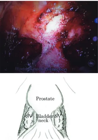
conventional technique to dissect out and identify the bladder neck more precisely. After developing the extraperitoneal space, the fat overlying the pubopro-static ligaments was removed. Prior to incising the endopelvic fascia or ligating the DVC, the lens was switched to 30° for the bladder neck dissection. Ultradissection of the bladder neck as described by Gaston . [12, 13] was performed in a modified manner. This technique is described in detail in our previous paper [14]. Briefly, the detrusor muscle fibers were identified and the lateral border of the bladder neck separated until the dissection reached the surface of the seminal vesicle (Fig. 1). Unlike the original method described by Gastonʼs group, the seminal vesicle was not dissected further. Following bilateral dissection of the bladder neck, the detrusor muscle was well visualized. Then, the bladder neck was transected. This technique allows bladder neck preservation even for a prostate with a large median lobe or a previous TURP. Following the bladder neck transection, the remainder of the procedure is similar to the conventional technique.
All specimens were
inked and sectioned in a standard manner. The base was shaved and submitted in 2 to 4 cassettes. The
apex was also shaved perpendicular to the axis of the urethra and sliced radially into 5 to 8 segments. The remaining prostate and seminal vesicles were serially sectioned in the transverse plane at 3- to 5- mm inter-vals. A PSM was defined according to a standard protocol, which is the presence of the tumor at the resection margin. All specimens were reviewed by one pathologist. In PSM cases, the location and the amount of cancer involvement were mapped and determined according to the method of Eichelberger and associ-ates [15]. All carcinomas were graded using the Gleason system and staged using the 2002 TNM sys-tem.
Descriptive results were
reported for all studied parameters. The chi-square test was used to compare the number of positive and
negative surgical margins in each group and the risk profiles for surgical margin positivity among the 2 groups. The chi-square test was also used to test the homogeneity of the Gleason score, the clinical stage and the pathologic stages of the 2 groups. Studentʼs
-test was used to compare the mean age, BMI, PSA,
prostate volume, biopsy Gleason score, operative time, and estimated blood loss (EBL) for the 2 groups. All statistical analysis tests were performed with Statistical Analysis Software 9.1 (SAS Institute, Cary, NC, USA).
Results
Mean follow-up times were 22.3 ± 9.9 (conventional) and 11.5±5.1 (modified ultradissection) months. Clinical characteristics were similar between the groups (Table 1). There was no difference between the conventional and modified ultradissection groups with respect to the mean age (62.4 and 63.0 years,
=0.56), BMI (24.6 and 24.1kg/M2, =0.17), PSA (8.3 and 8.2, =0.89), prostate volume (37.0 and 38.0, =0.70) or biopsy Gleason score (6.2 and 6.5,
=0.06). The DʼAmico prognostic criteria [16] distributions were similar. We also analyzed the risk Fig. 1 Bladder neck dissection. The picture shows a
well-pre-served bladder neck. The detrusor muscle fibers are identified and the lateral border of the bladder neck is exposed down to the semi-nal vesicles. Following bilateral dissection of the bladder neck, the bladder neck is transected. SV, seminal vesicle.
Table 1 Patient characteristics (pT2)
Conventional ultradissectionModified -value
Follow up (mos) 22.3±9.9 11.5±5.1
Age (yr) 62.4±8.2 63.0±7.6 0.56
BMI (kg/m2) 24.6±2.6 24.1±2.4 0.17
PSA (ng/mL) 8.3±4.3 8.2±4.8 0.89
Prostate Volume (cm3) 37.0±18.0 38.0±18.0 0.70
Biopsy Gleason Score 6.2±0.9 6.5±0.8 0.06
Dʼamico criteria N (%)
low risk 43 (38.1) 36 (34.6) 0.60
Intermediate risk 49 (43.4) 45 (43.3) 0.99
High risk 21 (18.6) 23 (22.1) 0.52
Risk profile for PSM N (%)
Low-risk profile* 47 (41.6) 45 (43.3) 0.80
High-risk profile† 66 (58.4) 59 (56.7) 0.80
Nerve sparing N (%) 109 (96) 94 (90) 0.07
N 113 104
Data are presented as means ± standard deviations or numbers of patients with percentages in parentheses.
PSM (positive surgical margin)
*PSA <10ng/mL, Gleason score <8, and 2 cores involved.
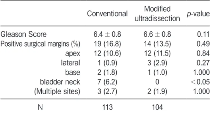
of PSM according to the risk profile described by Wieder and Soloway (Table 1) [1], and found no dif-ference between the 2 groups. Table 2 compares the pathologic and margin results in detail. The rate of PSM on the bladder neck was significantly reduced, from 6.2オ to 0 ( <0.05), by the modified ultradis-section. There was no change in the rate of PSM in the other areas. The overall rate of PSM in the modified ultradissection group was reduced; however, the difference was not statistically significant (16.8 and 13.5オ, =0.49). The PSA data need to be fol-lowed. Table 3 compares intraoperative and postop-erative data and complications. The mean operating time was shorter (218 and 195min, <0.001) and the mean estimated blood loss was less (381 and 282cc, Table 3 Intraoperative and postoperative data and complications
Conventional ultradissectionModified -value
Mean operative time (min) 218±49 195±38 <0.001
Mean blood loss (mL) 381±341 282±198 <0.01
Mean duration of catheter (d) 12.9±4.6 8.9±1.8 <0.0001
Mean hospital stay (d) 5.7±3.6 4.4±2.1 <0.01
Intraoperative complication
Rectal injury 1 (0.9) 2 (1.9) 0.61
Small bowel injury 0 1 (1.0) 0.48
Overall complications
*Medical complications 22 (19.5) 16 (15.4) 0.54
Deep vein thrombosis 0 0 NA
Pulmonary embolism 0 0 NA
Pneumonia 0 1 (1.0) 0.48
Other cardiopulmonary diseases 0 0 NA
Urinary tract infection 8 (7.1) 4 (3.8) 0.38
Orchiepididymitis 0 0 NA
Septicemia 0 0 NA
Duodenal ulcer 0 0 NA
Postoperative ileus 6 (5.3) 11 (10.6) 0.21
*Surgical complications
Conversion to open surgery 0 0 NA
Wound infection or hematoma 0 0 NA
Scar hernia 0 0 NA
Rectourethral fistula 0 0 NA
Hemorrhage 0 0 NA
Ureteral section 0 0 NA
Lymphocele 7 (6.2) 0 0.02
Urinary leakage (reintervention) 0 0 NA
Anastomotic stricture 1 (0.9) 0 1.000
N 113 104
Data are presented as means ± standard deviations or numbers of patients with percentages in parentheses. The overall complication rates were similar fot the conventional and ultradissection groups: 22 (19.5%) vs. 16 (15.4%), =0.54. The overall rates of medical complications were also similar: 14 (12.4%) vs. 16 (15.4%). =0.66.
Table 2 Pathologic features after RARP
Conventional ultradissectionModified -value
Gleason Score 6.4±0.8 6.6±0.8 0.11
Positive surgical margins (%) 19 (16.8) 14 (13.5) 0.49
apex 12 (10.6) 12 (11.5) 0.84 lateral 1 (0.9) 3 (2.9) 0.27 base 2 (1.8) 1 (1.0) 1.000 bladder neck 7 (6.2) 0 <0.05 (Multiple sites) 3 (2.7) 2 (1.9) 1.000 N 113 104
Data are presented as means ± standard deviations or numbers of patients with percentages in parentheses. The rate of positive sur-gical margin on the bladder neck was significantly reduced by modified ultradissection ( <0.05).
<0.01) in the modified ultradissection group (Table 3). The mean duration of catheter use (12.9 vs. 8.9 days, <0.0001) and the length of hospital stay (5.7 vs. 4.4 days, <0.01) were shorter in the modified ultradissection group. The overall complication rates were similar for the conventional and ultradissection groups: Clavien grade I-II complication, 18.6 and 15.4オ, respectively ( =0.53); Clavien grade III-V, 0.9 and 1.9オ, respectively ( =0.52). The incidences of rectal and small bowel injury were 1 (0.9オ) vs. 2 (1.9オ), =0.61 and 0 vs. 1 (1.0オ), =0.48, respec-tively. The overall rates of medical complications were similar: 14 (12.4オ) vs. 16 (15.4オ), =0.66. Most complications were minor, with the same pattern of occurrence in the 2 groups: urinary tract infection (UTI), 8 (7.1オ) vs. 4 (3.8オ), =0.38; ileus, 6 (5.3オ) vs. 11 (10.6オ), =0.21; pneumonia, 0 vs. 1 (1.0オ),
=0.48; and anastomotic stricture, 1 (0.9オ) vs. 0,
=1.0. The incidence of lymphoceles was signifi-cantly lower in the modified ultradissection group: 7 (6.2オ) vs. 0, =0.02. These results are listed in Table 3.
Discussion
The primary objective of radical prostatectomy is
the complete surgical resection of the cancerous tumor. A PSM is an independent risk factor for recurrence after radical prostatectomy [17-19]. The incidence of PSM in previously reported open prostatectomy series varied from 16オ to 46オ [18]. Its incidence in lap-aroscopic series has been 16-26オ [10, 20-22]. In the reported series for RARP, the overall PSM rate has varied between 6オ and 35.5オ [23, 24]. Vari-ations in clinicopathologic characteristics across techniques make direct comparisons difficult. PSM occurs either as the result of inadvertent entry into the prostate (iatrogenic) or by cutting across an extrapro-static tumor that extends beyond the limits of resec-tion (non-iatrogenic) [25]. PSM in patients with organ-confined disease (Stage pT2), therefore, is largely preventable.
In our series, the rate of PSM at the bladder neck
decreased significantly, from 6.2 to 0オ, with the implementation of modified ultradissection. Our initial cases were performed using the classic Vattikuti Institute prostatectomy technique (conventional tech-nique) [11], which complicates the identification of
the true bladder neck [11]. The dissection along the median border of the bladder neck is technically diffi-cult in cases with a large median lobe or a history of a prior TURP since the urethra is deviated from the midline. A large median lobe also increases the opera-tive time for the RARP due to the increased time required for posterior bladder neck and seminal vesi-cle dissection [26]. Finally, patients with larger prostates have significantly more blood loss (175 vs. 226mL).
Modified ultradissection required a significantly
shorter operative time (195 vs. 218min, <0.001) and resulted in less blood loss (282 vs. 381cc, <0.01) than the conventional technique. These improvements may be the result of not only the procedural improve-ments but also increased familiarity with the proce-dure and robotic system, as all ultradissection cases were performed after the conventional cases.
Clavien grades of complications did not differ
sta-tistically between the 2 groups. When we analyzed complications in detail, the only difference was the reduction in the incidence of lymphoceles in the ultra-dissection group (0 vs. 7 (6.2オ), =0.02). All were treated conservatively. The reason for the reduced rate of lymphoceles in the modified ultradissection is not clear. The use of lymphadenectomy in the ultra-dissection group was more extensive than that of the conventional group. It was performed bilaterally in the external iliac, obturator and infraobturator beds. Fatty tissue and lymph nodes medial to the genitofem-oral nerve from the iliac bifurcation to the inguinal ring were also removed. Symptoms vary depending on the size, site, and presence of infection. Significant lymphoceles can cause pelvic pain, voiding difficulty, leg edema, deep venous thrombosis, and hydroneph-rosis. Infected lymphoceles are often associated with febrile morbidity. The incidence of symptomatic lym-phoceles in an endoscopic extraperitoneal radical prostatectomy series was 2.5オ ( =45). In previous reports of LRP and RRP, the incidence of lymphoce-les has ranged from 0 to 11オ [27-31]. The incidence in the present study is within that range.
The main strength of this study is that it minimizes
the degree of variation of various clinical and patho-logic parameters. We retrospectively selected only pT2 disease in our series and analyzed the rate of PSM for each of the 2 techniques. Patient character-istics were also similar for the 2 groups. The risk
profiles for PSM as described by Wieder and Soloway were similar for the 2 groups [1]. Finally, the rate of nerve sparing was also similar (96オ vs. 90オ, =
0.07, Table 1).
The present study has several limitations. The data
were collected retrospectively. All surgery was per-formed by a single surgeon. Thus, these results may not have general applicability or reproducibility. Additionally, the surgeonʼs gain in experience over time likely contributed to the decrease in PSM rate; however, the decrease was greater than what would be expected from experience alone, given that the learning curve for this procedure is typically associ-ated with mastery at 50 cases [32]. Therefore, while the findings of this study are promising, they await confirmation in a prospective randomized trial.
References
1. Wieder JA and Soloway MS: Incidence, etiology, location, pre-vention and treatment of positive surgical margins after radical prostatectomy for prostate cancer. J Urol (1998) 160: 299-315. 2. Guillonneau B, el-Fettouh H, Baumert H, Cathelineau X, Doublet
JD, Fromont G and Vallancien G: Laparoscopic radical prosta-tectomy: oncological evaluation after 1,000 cases a Montsouris Institute. J Urol (2003) 169: 1261-1266.
3. Ward JF, Zincke H, Bergstralh EJ, Slezak JM, Myers RP and Blute ML: The impact of surgical approach (nerve bundle preserva-tion versus wide local excision) on surgical margins and biochemi-cal recurrence following radibiochemi-cal prostatectomy. J Urol (2004) 172: 1328-1332.
4. Yossepowitch O, Bjartell A, Eastham JA, Graefen M, Guillonneau BD, Karakiewicz PI, Montironi R and Montorsi F: Positive Surgical Margins in Radical Prostatectomy: Outlining the Problem and Its Long-Term Consequences. Eur Urol (2009) 55: 87-99.
5. Guillonneau B: To demonstrate the benefits of laparoscopic radical prostatectomy? Eur Urol (2006) 50: 1160-1161; discussion 1161-1162.
6. De La Rosette JJ, Abbou CC, Rassweiler J, Laguna MP and Schulman CC: Laparoscopic radical prostatectomy: a European virus with global potentials. Arch Esp Urol (2002) 55: 603-609. 7. Abbou CC, Hoznek A, Salomon L, Lobontiu A, Saint F, Cicco A,
Antiphon P and Chopin D: [Remote laparoscopic radical prostate-ctomy carried out with a robot. Report of a case]. Prog Urol (2000) 10: 520-523.
8. Serni S, Masieri L, Lapini A, Nesi G and Carini M: A low inci-dence of positive surgical margins in prostate cancer at high risk of extracapsular extension after a modified anterograde radical pros-tatectomy. BJU Int (2004) 93: 279-283.
9. Atug F, Castle EP, Srivastav SK, Burgess SV, Thomas R and Davis R: Positive surgical margins in robotic-assisted radical prostatectomy: impact of learning curve on oncologic outcomes. Eur Urol (2006) 49: 866-871.
10. Katz R, Salomon L, Hoznek A, de la Taille A, Antiphon P and Abbou CC: Positive surgical margins in laparoscopic radical prosta-tectomy: the impact of apical dissection, bladder neck remodeling
and nerve preservation. J Urol (2003) 169: 2049-2052.
11. Menon M, Tewari A and Peabody J: Vattikuti Institute prosta-tectomy: technique. J Urol (2003) 169: 2289-2292.
12. Mattei A, Naspro R, Annino F, Burke D, Guida R, Jr. and Gaston R: Tension and energy-free robotic-assisted laparoscopic radical prostatectomy with interfascial dissection of the neurovascular bundles. Eur Urol (2007) 52: 687-694.
13. Curto F, Benijts J, Pansadoro A, Barmoshe S, Hoepffner JL, Mugnier C, Piechaud T and Gaston R: Nerve sparing laparoscopic radical prostatectomy: our technique. Eur Urol (2006) 49: 344-352.
14. Jeong W, Araki M, Park SY, Lee YH, Kumon H, Hong SJ and Rha KH: Robot-assisted laparoscopic radical prostatectomy in the Asian population: modified port configuration and ultradissection. Int J Urol (2010) 17: 297-300.
15. Eichelberger LE, Koch MO, Daggy JK, Ulbright TM, Eble JN and Cheng L: Predicting tumor volume in radical prostatectomy speci-mens from patients with prostate cancer. Am J Clin Pathol (2003) 120: 386-391.
16. DʼAmico AV, Whittington R, Malkowicz SB, Schultz D, Blank K, Broderick GA, Tomaszewski JE, Renshaw AA, Kaplan I, Beard CJ and Wein A: Biochemical outcome after radical prostatectomy, external beam radiation therapy, or interstitial radiation therapy for clinically localized prostate cancer. JAMA (1998) 280: 969-974. 17. Blute ML, Bostwick DG, Bergstralh EJ, Slezak JM, Martin SK,
Amling CL and Zincke H: Anatomic site-specific positive margins in organ-confined prostate cancer and its impact on outcome after radical prostatectomy. Urology (1997) 50: 733-739.
18. Sofer M, Hamilton-Nelson KL, Schlesselman JJ and Soloway MS: Risk of positive margins and biochemical recurrence in relation to nerve-sparing radical prostatectomy. J Clin Oncol (2002) 20: 1853-1858.
19. Eastham JA, Kuroiwa K, Ohori M, Serio AM, Gorbonos A, Maru N, Vickers AJ, Slawin KM, Wheeler TM, Reuter VE and Scardino PT: Prognostic significance of location of positive margins in radi-cal prostatectomy specimens. Urology (2007) 70: 965-969. 20. Bollens R, Vanden Bossche M, Roumeguere T, Damoun A,
Ekane S, Hoffmann P, Zlotta AR and Schulman CC: Extraperi-toneal laparoscopic radical prostatectomy. Results after 50 cases. Eur Urol (2001) 40: 65-69.
21. Rassweiler J, Sentker L, Seemann O, Hatzinger M and Rumpelt HJ: Laparoscopic radical prostatectomy with the Heilbronn technique: an analysis of the first 180 cases. J Urol (2001) 166: 2101-2108.
22. Gill IS and Zippe CD: Laparoscopic radical prostatectomy: technique. Urol Clin North Am (2001) 28: 423-436.
23. Menon M, Shrivastava A, Sarle R, Hemal A and Tewari A: Vattikuti Institute Prostatectomy: a single-team experience of 100 cases. J Endourol (2003) 17: 785-790.
24. Ahlering TE, Skarecky D, Lee D and Clayman RV: Successful transfer of open surgical skills to a laparoscopic environment using a robotic interface: initial experience with laparoscopic radical prostatectomy. J Urol (2003) 170: 1738-1741.
25. Ahlering TE, Eichel L, Edwards RA, Lee DI and Skarecky DW: Robotic radical prostatectomy: a technique to reduce pT2 positive margins. Urology (2004) 64: 1224-1228.
26. Meeks JJ, Zhao L, Greco KA, Macejko A and Nadler RB: Impact of Prostate Median Lobe Anatomy on Robotic-Assisted Laparoscopic Prostatectomy. Urology (2009) 73: 323-327.
27. Noldus J, Hammerer P, Graefen M and Huland H: Surgical ther-apy for localized prostatic carcinoma. J Cancer Res Clin Oncol
(1997) 123: 180-184.
28. Lepor H and Kaci L: Contemporary evaluation of operative param-eters and complications related to open radical retropubic prostate-ctomy. Urology (2003) 62: 702-706.
29. Liatsikos E, Rabenalt R, Burchardt M, Backhaus MR, Do M, Dietel A, Wasserscheid J, Constantinides C, Kallidonis P, Truss MC, Herrmann TR, Ganzer R and Stolzenburg JU: Prevention and management of perioperative complications in laparoscopic and endoscopic radical prostatectomy. World J Urol (2008) 26: 571-580.
30. Remzi M, Klingler HC, Tinzl MV, Fong YK, Lodde M, Kiss B and
Marberger M: Morbidity of laparoscopic extraperitoneal versus transperitoneal radical prostatectomy verus open retropubic radical prostatectomy. Eur Urol (2005) 48: 83-89; discussion 89. 31. Guillonneau B, Rozet F, Cathelineau X, Lay F, Barret E, Doublet
JD, Baumert H and Vallancien G: Perioperative complications of laparoscopic radical prostatectomy: the Montsouris 3-year experi-ence. J Urol (2002) 167: 51-56.
32. Ou YC, Yang CR, Wang J, Yang CK, Cheng CL, Patel VR and Tewari AK: The learning curve for reducing complications of robotic-assisted laparoscopic radical prostatectomy by a single surgeon. BJU Int (2011) 108: 420-425.