93 P
Peeddiiaattrriiccss
Life-threatening Hemorrhage Caused by Mandibular Arteriovenous Malformation:
A Case Report
Dong Wook Kim, M.D.1, In Suk Sol, M.D.1, Min Jung Kim, M.D.1, Soo Yeon Kim, M.D.1, Jong Deok Kim, M.D.1, Bong Seok Choi, M.D.2, Yoon Hee Kim, M.D.1, Dong Joon Kim, M.D., Ph.D.3, Byung Moon Kim, M.D., Ph.D.3,
Seo Yeon Jung, M.D.4, Woong Nam, M.D., Ph.D.4, Kyung Won Kim, M.D., Ph.D.1*, Myung Hyun Sohn, M.D., Ph.D.1, Kyu-Earn Kim, M.D., Ph.D.1,5
Department of Pediatrics, Yonsei University College of Medicine, Seoul1, Department of Pediatrics, Kyungpook National University School of Medicine, Daegu2, Department of Radiology, Yonsei University College of Medicine, Seoul3, Department of Oral and Maxillofacial Surgery, Yonsei University College of Dentistry, Seoul4, Department of Medicine,
Sowha Children’s Hospital, Seoul5, Korea
Arteriovenous malformation (AVM) of the mandible is a rare vascular condition that can manifest as a wide range of symp-toms and, on rare occasions, cause fatal hemorrhage. The sympsymp-toms of mandibular AVM can range from soft tissue swelling and tooth mobility to severe hemorrhage. The recognition of early symptoms is crucial for the prevention of a fatal hemorrhage and for the proper diagnosis and treatment of mandibular AVM. For emergency hemostasis of a ruptured mandibular AVM, manual compression with gauze, topical thrombin, absorbable hemostat, suturing the lesion, and replanting the extracted tooth is recommended. Multiple treatment options for mandibular AVM are available, such as arter-ial embolization, venous embolization, direct surgical closure, and bone resection. A combination of treatment options should be considered in complicated cases. We report a case of a 10-year-old girl with a previous history of telangiectasia on the right cheek presented with cardiac arrest resulting from massive bleeding immediately after a tooth extraction.
Key Words: Arteriovenous Malformations, Mandible, Hemorrhage, Therapeutics
책임저자: 김 경 원
서울특별시 서대문구 연세로 50 연세대학교 의과대학 소아과학교실
Tel: 02-2228-2050, Fax: 02-393-9118, E-mail: [email protected]
접수일: 2017년 8월 18일, 1차 교정일: 2017년 8월 23일, 게재승인일: 2017년 10월 10일 Introduction
Arteriovenous malformation (AVM) of the mandible is a rare vascular malformation that can manifest in various symptoms such as gingival bleeding, dental loosening, swelling, mandibular pain, discoloration of overlying skin, and facial asymmetry1). Without recognition of the symptoms, it can sometimes cause massive bleeding, leading to a life-threatening situation.
We report a case of a 10-year-old girl who presented with a severe hemorrhage following tooth extraction. This case describes the management of massive bleeding from AVM, which was complicated by infection, with several interventions using various methods of
emboliza-tion, accompanied with surgical closure and administra-tion of systemic antibiotics for sepsis treatment due to infection at the hemorrhage site.
Case report
A 10-year-old female patient was referred to our hos-pital for torrential uncontrolled bleeding following a tooth extraction (#45, lower right 2nd premolar tooth) at home, which had resulted in cardiac arrest due to rapid hypovolemic shock. The patient had no past medical his-tory other than telangiectasia of the right cheek. On ini-tial assessment undertaken at a local hospital, pulseless electrical activity was checked. Her airway was secured
with endotracheal intubation, and cardiopulmonary resuscitation (CPR) was immediately initiated in addi-tion to an extensive transfusion of red blood cells. A return of spontaneous circulation was achieved after 30 minutes of CPR; however, bleeding from the tooth extraction site persisted. With direct compression, using oral gauze packing for hemostasis, the patient was trans-ferred to our hospital for evaluation and management of the ongoing bleeding.
On initial assessment at arrival to our hospital, the patient showed signs of severe hypovolemic shock: blood pressure was recorded as 55/33 mmHg, pulse rate was 128 beats per minute, and her body temperature was 35.0�C. Her mental status was comatose, and pupil size of 4-5 mm was observed, with sluggish light reflexes. Initial laboratory data showed severe metabolic acidosis, anemia, disseminated intravascular coagulation, and multiple organ damage: arterial blood gas analysis showed arterial blood gas analysis showed pH, 7.16; par-tial pressure of carbon dioxide, 21.8 mmHg; parpar-tial pres-sure of oxygen, 110.9 mmHg; base excess, -21 mmol/L; bicarbonate, 7.9 mmol/L; peripheral blood analysis showed white blood cell count, 18,920/mm3; hemoglo-bin, 4.8 g/dL; platelet, 105,000/mm3; lactate, 18 mmol/L; aspartate transaminase/alanine transaminase, 1,076/1,050 IU/L; prothrombin time, 28.5 seconds; activated partial thromboplastin time, 125.7 seconds; and d-dimer, 27,268 ng/mL. Inotropics and vasopressors were immediately started while intravascular volume resuscitation was ini-tiated, along with multiple transfusions of packed red blood cells and fresh frozen plasma.
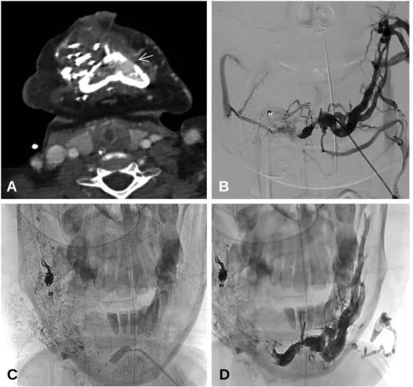
After the patient’s vital signs stabilized, investigation of the bleeding site began. Computed tomography (CT) angiography of the head and neck revealed a large AVM of the right mandible with focal bleeding points, and a subsequent emergency angiography revealed a right mandibular AVM feeding from multiple fine branches of the right facial artery and the internal maxillary artery (Fig. 1). Selective angiography and embolization of the right mandibular AVM with 25% and 33% N-butyl-2-cyanoacrylate (NBCA) was attempted, but complete embolization was difficult due to multiple finely separat-ed feseparat-eders and the unclear location of the hemorrhage site due to the external intraoral packing. Intraoral pack-ing was removed in an attempt to locate the hemorrhage site, and uncontrolled massive bleeding recurred, leading
to a second cardiac arrest. CPR and intravenous volume resuscitation was again performed, and intraoral packing was restored for hemostasis. The procedure was aborted, in consideration of the risks and benefits, and the patient’s unstable hemodynamic condition.
The patient was admitted into the intensive care unit for further observation and management. Multiple organ damage was evident because of multiple CPRs. The patient’s mental status remained comatose, a chest x-ray showed severe pulmonary edema, due to acute lung injury resulting from the multiple blood transfusions, and oliguria, due to acute kidney injury (AKI). Post-CPR care was initiated with induced hypothermia for 24 hours, followed with passive rewarming, lung-protective ventilator care for optimal oxygenation, inotropic agents for treating hypotension, continuous renal replacement therapy to treat AKI, and broad-spectrum antibiotics for prophylaxis in respect of ventilator-associated pneumo-nia and sepsis.
An initial blood culture showed a growth of multi-drug resistant Acinetobacter baumannii which was sensitive only to colistimethate. Oral cavity infection, associated with intraoral packing, was considered a possible source of infection because an oral examination had revealed a foul odor and a pus-like discharge from the packing site. For infection control, intravenous colistimethate was administered, and the oral packing was replaced with colistimethate-soaked gauze packing, which was sched-uled to be replaced every 7 days. During replacement of the oral gauze packing, hemodynamically significant bleeding of nearly 1 to 2 liters from the tooth extraction site persisted. On day 18 of hospitalization, the patient’s general condition deteriorated, with fever, hypotension, and C-reactive protein elevation, followed by a growth of Hafnia alvei on blood culture. Despite the use of antibiotics, clinical signs indicated sepsis progression, and since intraoral infection was a possible focus of sep-sis, treatment of the oral lesion was considered urgent to improve the patient’s clinical condition.
Therefore, a selective arterial glue embolization with 11.1% NBCA was performed on two separate occasions, at 7 day intervals, and an additional coil embolization was performed. Both embolization procedures resulted in reduced oral bleeding, which allowed enhanced visual access for oral cavity irrigation and decontamination, leading to the eradication of previously identified
pathogens, as confirmed in subsequent blood cultures. However, since minor bleeding persisted, additional direct intraoral bleeding control was considered another option. The endotracheal tube was removed and a tra-cheostomy tube was inserted to obtain a better surgical view of the bleeding sites. A bony defect on the tooth extraction site was packed with bone wax and Ostene. After direct packing, Floseal and thrombin reyon powder were used for additional hemostasis.
A follow-up CT angiography revealed a decreased degree of AVM in the right side of the mandible; howev-er, dilated venous structures in the contralateral left mandible had newly developed, due to transmitted
arteri-al pressure (Fig. 2A). Post-procedure intraorarteri-al examina-tion revealed persistent minor bleeding at the AVM site and a newly developing bleeding site around the left 1st bicuspid tooth. Since complex multiple arterial feeders present a major challenge for complete bleeding control, percutaneous venous embolization was scheduled for the patient’s fourth intervention. A percutaneous mandibular
venogram was obtained to visualize the large draining vein in relation to the AVM (Fig. 2B), and a balloon-assisted Onyx injection into the draining channel was performed (Fig. 2C, 2D). A final angiogram revealed markedly reduced AVM in the right mandible, and no remaining bleeding from either the right or the left
Fig. 1. Initial CT angiography and arterial angiography revealing mandibular AVM. (A) Axial view of CT angiography shows
the feeding artery (arrow) from the internal maxillary artery. (B) Axial view of CT angiography reveals venous pooling (circle) in the right mandible and cortical breach. (C, D) Initial arterial angiography reveals a right mandibular AVM feeding from multiple fine branches of the right facial artery (arrow) and the internal maxillary artery (arrow), (C, ante-rior view; D, lateral view).
CT: computed tomography, AVM: arteriovenous malformation
A
B
mandibular area was noted on visual inspection (Fig. 3). The patient’s neurologic status improved from comatose to drowsy throughout the four embolization procedures. Sepsis, which was suspected to have been caused through intraoral infection, was resolved with the use of intravenous antibiotics, and oral irrigation and decontamination. The patient was transferred to the gen-eral ward for further care and rehabilitation. After 1 year, the patient’s cognitive and motor functions have signifi-cantly improved. She responds to visual stimulus and auditory commands, and is now able to maintain head control and a sitting position without back support.
Throughout the treatment courses, teeth that were near the extracted tooth, namely #43, #44, #46, were highly mobile due to severe mucosal injury. Due to the risk of
spontaneous extraction and subsequent bleeding, those teeth with high mobility were extracted by surgical team with caution in between the treatment courses. Other teeth that still had some integrity (#34-42), were fixed with wire splint for augmentation. Six months after treat-ment, #47 tooth, also known as the wisdom tooth, began to erupt with some minor bleeding. However, no addi-tional bleeding occurred along with the progression of tooth eruption and follow-up CT angiography also showed no sign of recurrence. A 1-year follow-up intrao-ral exam still showed no sign of bleeding around #47 tooth and mucosa around it, and CT angiography of the head and neck also showed no residual AVM or recur-rence so far.
Fig. 2. Venous congestion on follow-up CT and venogram showing venous embolization process. (A) On a follow-up CT,
newly developed dilated venous structures (arrow) in the contralateral left mandible appear due to transmitted arterial pressure. (B) Venogram shows a single, large draining vein of the AVM. (C) The balloon-assisted method was used for accessing venous outflow tract. (D) Onyx injection into AVM nidus via draining veins was performed.
CT: computed tomography, AVM: arteriovenous malformation
A
B
Discussion
Mandibular AVMs are rare vascular malformations that can lead to massive blood loss after tooth extraction, without recognition of the condition2). Half of all intraosseous AVMs occur in the maxillofacial region and are extremely rare in the mandible3). AVM in the maxilla and mandible constitute 4-5% of all craniofacial AVMs and intraosseous AVMs of the mandible accounts for approximately 0.5-1% of all vascular malformations4). Without clear clinical signs, mandibular AVMs can remain unrecognized for several years. Symptoms reported by patients include soft tissue swelling, pain, tooth mobility, discoloration of the mucosal surface and overlying skin, paresthesia, and periodontal bleeding5). As described in our case, some patients might not dis-play any of the above described symptoms, sometimes leading to extreme hemorrhage after an unplanned tooth extraction.
Diagnoses of mandibular AVMs are usually made using radiographic investigations. On panoramic radi-ographs, mandibular lucency or ‘soap bubble’ appear-ances may occur6). CT angiography can be useful in assessing the extent of the lesion and the origin of the feeding vessels. Nevertheless, the diagnostic tool of choice is arterial angiography, which provides direct visualization of feeder arteries, draining veins, and the collateral flow of the AVM7). In our case, a diagnosis of
AVM was made with CT angiography, which revealed a large AVM with focal bleeding points in the right mandible and, furthermore, arterial angiography revealed feeder arteries from the internal maxillary artery and the facial artery. Panoramic radiographs could not be acquired due to the patient’s specific situation.
There is no consensus on the ideal treatment for mandibular AVMs, and several treatment options are available. Surgical options including curettage, cryosurgery, surgical devascularization through ligation of major feeding vessels, and en bloc resections are available8). Multiple endovascular approaches, including transarterial, transvenous, and percutaneous, using differ-ent embolizing materials, such as polyvinyl alcohol, glue, alcohol, ethibloc, coils, and balloons, have also been used in treatment of mandibular AVMs2,8). In a case report by Jung et al.9), a 14-year-old patient with mandibular AVM was treated by combined treatment of transarterial glue embolization, transvenous embolization, and percutaneous coil insertion. Also, in a more recent case report by Kaderbhai et al.4), treatment efficacy of surgical resection was shown, as two patients were treated with segmental resection of mandible in addition to transarterial emboliza-tion. Furthermore, in another recent case report by Spreafico et al.10), a 23-year-old patient with intraosseous and subcutaneous AVM in mandible was treated by intraosseous glue injection and surgical resection of sub-cutaneous AVM. As shown by previous case reports, there are various treatment options of AVM, and often times
Fig. 3. Comparison of initial arterial angiography and final angiography. (A) Initial arterial angiography reveals a mandibular
arteriovenous malformation (AVM) with multiple feeder arteries (arrows). (B) Final arterial angiography shows the obliterated right mandibular AVM (circle). AVM: arteriovenous malformation
more than one approach is required for complete cure. Transarterial embolization, one of the most commonly used method for treatment, can result in a permanent cure by a single treatment. However, as described in our case, the presence of multiple arterial feeders may pose difficulties for comprehensive treatment11), and in such case venous embolization can be helpful. Our patient’s AVM, following three separate arterial embolization interventions and a single direct surgical hemostasis intervention, was completely cured after a single percu-taneous venous embolization. Venous embolization of a single large draining varix can be undertaken using either direct puncture or the transvenous method, and it can be useful when transarterial embolization fails12). Additionally, to minimalize the bleeding risk from high-flow AVMs caused by outhigh-flow obstruction, transarterial embolization of the main arterial feeder before emboliz-ing the venous varix should be considered12).
Late complications of embolization, such as soft tissue infection, have been known to occur in other treatment groups2). However, our patient’s situation was complicat-ed due to suspectcomplicat-ed infection of the oral mucosa during the early course of treatment, and we suspected that local infection of the oral mucosa led to sepsis. Oral bacteria and periodontal infections have been proposed as possi-ble risk factors for systemic diseases, and due to the anatomical proximity of the periodontal biofilm to the gingival blood stream, periodontal pockets may act as reservoirs for microbial pathogens and their products, such as inflammatory mediators and immunocomplex-es13). The likelihood of systemic illness caused through local infection is increased in cases of impaired host resistance, as was shown in our case. A high prevalence of staphylococci, Pseudomonas, and Acinetobacter in saliva samples from hospitalized patients has been reported13). The dissemination of such oral microorgan-isms into the systemic circulation occurs when physical barriers, such as surface epithelium, are breached because of trauma, hypoxemia, or immunodeficiency14). The patient in our case had severely impaired general condition due to multiple cardiopulmonary resuscitations and we think it consequently lead to easier propagation of local infection into systemic infection. When oral infection is suspected after the rupture of an AVM, due to trauma or any other insults, using pathogen-specific antibiotics according to culture results would be the most
effective treatment. However, in cases of difficult-to-treat oral infection with unresolved underlying AVM, early prophylactic use of antibiotics to cover the usual oral cavity pathogens, especially in patients with an unstable clinical condition, may be beneficial.
The patient in our case had undergone repeated transarterial embolization and direct surgical hemostasis without achieving complete bleeding control. In cases of AVMs with multiple complex arterial feeders, different methods for hemostasis, such as percutaneous venous embolization, should be considered. Additionally, in cases of severe hemorrhage which may lead to shock and hypoxemia, the likelihood of systemic illness including sepsis increases, and proper use of antibiotics for infec-tion control should be considered even at the early stage.
When tooth extraction is identified as the cause of mandibular AVM hemorrhage, methods of emergency hemostasis include local compression of the alveolar bone using fingers or gauze packing15). If these methods fail to stop the bleeding, applying topical thrombin, absorbable hemostat, or suturing the lesion may be required15). However, replanting the extracted tooth into the socket would be best for urgent hemostasis.
In conclusion, intraosseous mandibular AVMs are rare conditions that can lead to fatal bleeding without a recog-nition of predisposing symptoms. In the treatment of mandibular AVM, combined methods of transarterial embolization, followed by percutaneous venous emboliza-tion, can be an effective strategy. Additionally, clinicians should be aware of the possibility of local infection during the course of treatment of mandibular AVM.
Conflict of Interest
The authors declare that there is no conflict of interest regarding the publication of this article.
References
01. van den Akker HP, Kuiper L, Peeters FL. Embolization of an arteriovenous malformation of the mandible. J Oral Maxillofac Surg. 1987;45:255-60.
02. Churojana A, Khumtong R, Songsaeng D, Chongkolwatana C, Suthipongchai S. Life-threatening arteriovenous malfor-mation of the maxillomandibular region and treatment
out-comes. Interv Neuroradiol. 2012;18:49-59.
03. Seehra J, Horner K, Coulthard P. Arteriovenous malfor-mation of the mandible: a case report. Br Dent J. 2006; 201:25-7.
04. Kaderbhai J, Breik O, Heggie AA, Penington AJ. High-flow paediatric mandibular arteriovenous malformations: case reports and a review of current management. Int J Oral Maxillofac Surg. 2017;46:1650-5.
05. Fan X, Zhang Z, Zhang C, Tang Y, Hu Y, Mao Q, et al. Direct-puncture embolization of intraosseous arteriove-nous malformation of jaws. J Oral Maxillofac Surg. 2002;60:890-7.
06. Ferre´s-Amat E, Prats-Armengol J, Maura-Solivellas I, Ferre´-Amat E, Mareque-Bueno J, Ferre´s-Padro´ E. Gingival bleeding of a high-flow mandibular arteriovenous malfor-mation in a child with 8-year follow-up. Case Rep Pediatr. 2015;2015:745718.
07. Dunfee BL, Sakai O, Pistey R, Gohel A. Radiologic and pathologic characteristics of benign and malignant lesions of the mandible. Radiographics. 2006;26:1751-68. 08. Cohen JE, Gomori JM, Grigoriadis S, Sibly Z, Rajz G.
Complete and persistent occlusion of arteriovenous mal-formations of the mandible after endovascular emboliza-tion. Neurol Res. 2009;31:467-71.
09. Jung MS, Ryu DM, Kim EJ, Oh JH. A treatment of
atreiovenous malformation on mandible. J Korean Assoc Oral Maxillofac Surg. 2007;33:69-75.
10. Spreafico R, Sordo L, Bellotto R, Schipano M, Rescaldani A, Parmigiani F. Arterio-venous malformation of the mandible. case report and review of literature. Acta Otorhinolaryngol Ital. 2016;36:333-6.
11. Chiras J, Hassine D, Goudot P, Meder JF, Guilbert JF, Bories J. Treatment of arteriovenous malformations of the mandible by arterial and venous embolization. AJNR Am J Neuroradiol. 1990;11:1191-4.
12. Beek FJ, ten Broek FW, van Schaik JP, Mali WP. Transvenous embolisation of an arteriovenous malforma-tion of the mandible via a femoral approach. Pediatr Radiol. 1997;27:855-7.
13. Vieira Colombo AP, Magalhn˜es CB, Hartenbach FA, Martins do Souto R, Maciel da Silva-Boghossian C Periodontal-disease-associated biofilm: a reservoir for pathogens of medical importance. Microb Pathog. 2016; 94:27-34.
14. Li X, Kolltveit KM, Tronstad L, Olsen I. Systemic dis-eases caused by oral infection. Clin Microbiol Rev. 2000; 13:547-58.
15. Roberts G, Scully C, Shotts R. ABC of oral health. dental emergencies. BMJ. 2000;321:559-62.