A THESIS
FOR THE DEGREE OF DOCTOR OF PHYSIOLOGY
Characterization and expression analysis
of C-type lectins from abalone,
Haliotis discus discus
Ning Wang
Department of Marine Biotechnology
GRADUATE SCHOOL
CHEJU NATIONAL UNIVERSITY
Characterization and expression analysis of C-type
lectins from abalone, Haliotis discus discus
Ning Wang
(Supervised by Professor Jehee Lee)
A thesis submitted in partial fulfillment of the requirement
for the degree of Doctor of Philosophy
2008 . 02 .
This thesis has been examined and approved
...
Thesis director,
Gi-Young Kim Prof. of Marine Life Science
...
You-Jin Jeon, Prof. of Marine Biotechnology
...
Moon-Soo Heo, Prof. of Marine Biotechnology
...
In-Kyu Yeo, Prof. of Marine Biotechnology
...
Jehee Lee, Prof. of Marine Biotechnology
...
Date
Department of Marine Biotechnology
GRADUATE SCHOOL
요 약 문
렉틴은 세포 표면의 탄수화물 부위에 가역적으로 결합할 수 있고 특이적으로 인식할 수 있는 단백질 또는 당단백질이다. 새로운 활성들을 지니는 많은 중요한 렉틴들이 무척추동물에서 발견되었다. 무척추동물들에서 렉틴은 비자기 항원의 인지를 위해 기본적으로 사용되고, 박테리아 식균작용과 캡슐화를 할 수 있게 해준다. 이 연구에서는 비브리오 응집과 진주형성에 관계된 2개의 유전자를 까막전복의 EST cDNA library 탐색을 통해 확인하였다. 그리고 전복의 CLHd 와 perlucin complete cDNA 대해서 처음으로 연구되어졌다. 다른 종들로부터 기존에 알려진 서열의 유사성을 확인하기 위해 NCBI 의 blastX 프로그램을 이용하였다. 전복에서 생리적 작용과 그들의 기능 분석을 위해서 분자 구조의 분석, in vivo 상에서의 유전자 전사조절, 그리고 in vitro 상에서 단백질 발현, 관계된 활성 측정에 대해 조사되어졌다. CLHd 유전자의 complete cDNA 염기서열은 151 아미노산을 암호화하는 508 bp 였다. CLHd는 연체동물과 어류로부터 밝혀진 C-type 렉틴들을 가지는 탄수화물 인식 도메인에 높은 유사성을 나타냈다. In vivo 상에서, 전복에 Vibrio alginolyticus 를 주입했을 때 소화관에서 CLHd의 관계된 발현 레벨이 의미있는 증가를 나타냈다. V. alginolyticus를 주입하고 12시간 후에 그 mRNA의 유도가 시작됬고, 24시간째에 발현률이 최고에 도달했다. 시간이 진행됨에 따라, challenge 테스트 48시간째부터 CLHd의 mRNA 전사량은 control에 비해 감소하였다. 전복에 V. alginolyticus를 주입했을 때 CLHd의 요구는 의미있는 증가를 나타냈다. In vitro 상에서 재조합 CLHd 단백질은 calcium에 의존하여 V. alginolyticus 에 특이적으로 결합하였다. CLHd 는 전복에서 미생물들이 침입했을 때 이를 억류시켰고 퍼지는 것을 저해하였다. V. alginolyticus에 선택적으로 결합하는 CLHd는 전복에서 내부방어와 식균작용의 증대를 위해 자기, 비자기 인식 하는데 없어서는 안될 의미를 제공할 수 있다. CLHd의 특성분석을 통해 전복 질병 조절을 위한 강한 면역 분자를 제공하고 전복의 면역 지식을 넓혀 주었다. CLHd는 Vibrio의 주입에 의해 높게 유도되어졌고 이를 토대로 전복 양식장의 환경 모니터링을 위한 마커 개발과 양식장
관리에 이용 되어질 수 있다. Vibrio 응집에도 효과적이기 때문에 CLHd의 많은
양이 존재하면 전복의 질병 저항력을 향상 시킬 수 있다. CLHd의 강한 발현을
위한 전복의 유전자 선별 또는 전복의 형질전환 육종은 Vibrio 감염 으로부터
어린 전복들을 구할 수 있는 가능성을 제공한다.
전복 perlucin 유전자의 full length cDNA는 신호서열 22 아미노산과 mature protein 129아미노산을 암호화하는 1038 bp 로 구성되어졌고 greenlip 전복의 perlucin 단백질과 55 % 의 상동성을 나타냈다. 진단된 perlucin의 예측된 분자량은 15 KDa 이고 isoelectric point는 5.7로 확인됐다. 발현된 재조합 perlucin은 calcium carbonate 침전 활성과 calcium carbonate 결정화 활성이 측정 되어졌다. 재조합 perlucin의 첨가로 인해 calcium carbonate 침전이 촉진 되어졌다. 연체동물의 진주층 성장은 aragonite crystal의 calcium carbonate 축적과 형성에 의존된다. Calcium carbonate 침전 상에서 perlucin의 positive 효과는 전복에서 진주 형성 촉진에 직접적으로 나타난다. Calcium carbonate 결정화 활성에서 결정들은 처음에 예리한 모퉁이에 둥글게 형성됬고 4°C에서 24시간이상 처리한 결과 육면체를 형성하지 않았다. 점차적으로, calcite 크리스탈 표면은 울퉁불퉁하게 됐고 예리한 모퉁이들이 모두 사라졌다. Perlucin을 가지고 배양 72시간 후, 육면체에 가까운 크리스탈이 형성됬고 크리스탈 윗쪽에 작은 사방면체 돌기가 생성됐다. Perlucin은 예리한 모퉁이에 calcium carbonate 크리스탈이 점차적으로 둥근 형태가 되었지만 동시에 모든 모퉁이들이 그렇게 된 것은 아니다. 이 perlucin은 특이적으로 calcite 크리스탈을 인지할 수 있고 점차적으로 calcite 크리스탈의 예리한 모서리의 성질을 변화시킨다. 그 calcium carbonate의 형태적 변형은 진주와 껍질의 최종 구조에 영향을 주고 성장 속도와 적응에 역시 영향을 미친다. 본 연구에서
perlucin은 ab-plane 상에서 calcium carbonate 크리스탈의 성장을
촉진시켰고calcite 크리스탈이 네개의 모퉁이에 모든 단계가 완성됐다.
ab-plane에서 크리스탈의 형성은 저해되어졌고 그 후 c-axis에서 성장이
갖고 있는 전형적인 C-type lectin의 형태이다. perlucin의 RT-PCR 수행시 mantle, 아가미와 소화관에서 발현이 확인되었다. 아가미와 소화관은 외부 환경으로부터 전복내의 환경수 교환과 먹이 섭취에 상시적으로 관련되어 있으므로 이들 기관은 해양환경내 병원성 미생물의 침입에 대한 방어 영역을 형성하기 위하여 병원성 감염에 대한 민감한 감수성과 체액성 면역 반응 분자들이 필요하다. 종합적으로, perlucin 유전자는 병원성 미생물과 해로운 유기물에 대하여 면역 방어에 관여하는 물질로 추정된다. 침입한 기생충 또는 물질의 배설이 되지 않는 경우, perlucin은 비자기 항원의 비활성 수단으로 불필요한 물질들의 진주층으로의 축적을 위해 분비될 것이다. 불필요한 입자들로 인한 진주층의 성장으로 화려한 진주가 생성되는 것이다. Perlucin은 전복의 진주 형성과 내부 nacrein 성장에 관여한다. 그러나, C-type lectin에 비교하여 전복에서 perlucin 유전자의 비자기 인식과 질병 내성에 대한 더 많은 조사가 필요할 것으로 생각된다.
CONTENTS
요 약 문...Ⅰ CONTENTS...Ⅳ LIST OF FIGURES...Ⅵ LIST OF TABLES...Ⅷ 1 . Intro duc ti o n . . . 1
Part I. A nove l C- type le c ti n f rom abalo ne , Ha l io t is d i s c us d i s c us , agglutinates Vibrio alginolyticus...6
Abstract...6
2. Materials and methods...7
2.1 Materials...7
2.2 Molecular cloning and sequence analysis...7
2.3 CLHd mRNA transcription tissue distribution and induction by Vibrio challenge...9
2.4 In vitro protein expression and purification...11
2.5 Bacterial agglutination assay...13
2.6 Hemocytes agglutinating assay...14
3. Result...14
3.1 EST cDNA library screening...14
3.2. Sequence analysis of the full length CLHd...17
3.3. CLHd mRNA expression induced by V. alginolyticus challenge...22
3.4. Recombinant expression of CLHd in E.coli...26
3.5. Bacterial agglutinating activity of CLHd...28
3.6. Hemocyte agglutinating activity of CLHd...30
Part II. Recombinant perlucin nucleates the growth of calcium carbonate crystals: molecular cloning and characterization of pearl formation gene from disk
abalone, Haliotis discus discus...35
Abstract...35
2. Materials and Methods ...36
2.1 Materials...36
2.2 Molecular cloning and sequencing...36
2.3 Protein expression and purification...38
2.4 Protein activity assay...38
2.5 RT-PCR...40
3. Result...40
3.1 Perlucin cDNA Sequence and analysis...40
3.2 Protein expression and purification...48
3.3 Recombinant perlucin activity...50
3.4 Tissue distribution of perlucin mRNA transcription...52
4. Discussion...54
Summary...62
References...65
LIST OF FIGURES
Figure 1. The nucleotide sequence of the cDNA clones and deduced amino acid sequence of CLHd from abalone, Haliotis discus discus...18 Figure 2. The E. coli codon usage analysis of the CLHd coding sequence...19 Figure 3. Multi sequence alignment of the CRD in CLHd and homologous C-type lectins...21 Figure 4. Tissue distribution of CLHd mRNA transcript in abalone...23 Figure 5. The mRNA temporal transcript profile of CLHd induced by V. alginolyticus challenge...24 Figure 6. The relative CLHd mRNA expression in the digestive tract of abalone after V. alginolyticus injection...25 Figure 7. SDS-PAGE gel analyses of recombinant CLHd...27 Figure 8. The agglutination of V. alginolyticus induced by CLHd...29 Figure 9. The nucleotide sequence of the cDNA clones and deduced amino acid sequence of perlucin from abalone, Haliotis discus discus...41 Figure 10. The E. coli codon usage analysis of the perlucin coding sequence...43 Figure 11. Alignment of the amino acid sequence of perlucin from Haliotis discus discus and the homologous sequence from Haliotis laevigata...44 Figure 12. Comparison of carbohydrate recognition domains from C-type lectins homologous to perlucin...45 Figure 13. SDS-PAGE gel analysis of MBP fusion perlucin...48 Figure 14. Recording of Calcium carbonate precipitation with or without perlucin in super saturated solution...50 Figure 15. Light microscopic images of crystals grown in presence or absence of recombinant perlucin...51
Figure 16. Tissue distribution of perlucin transcript revealed by RT-PCR in abalone H. discus discus...53 Figure 17. SEM images of allotropic calcium carbonate crystals precipitated from saturated calcium carbonate solutions...57 Figure 18. The illustration of kink sites...59
LIST OF TABLES
Table 1. Oligonucleotide primers used in the part I experiments...8 Table 2. C-type lectins in normalized cDNA library of abalone (Haliotis discus discus)...16 Table 3. Oligonucleotide primers used in the part II experiments...37
1. Introduction
Lectin
The term lectin, from the Latin legere meaning to pick out, choose or select, was coined by Boyd (Boyd and Shapleigh, 1954). Lectin is a protein or glycoprotein substance, usually of plant origin (mostly of seeds), of non-immunoglobulin nature, capable of specific recognition of and reversible binding to carbohydrate moieties on the surface of cells, on cytoplasmic and nuclear structures, and in extracellular matrix in cells or tissues. Generally, lectins are classified into Calnexin, Galectins (S-type), L-type, I-type, P-type, R-type on structural basis (Dodd and Drickamer, 2001). Calnexin, L-type, P-type are located mostly intracellularly, in luminal compartments. They function in the trafficking, sorting, and targeting of glycoproteins in the secretory and other pathways. C-type lectins, galectins, I-type lectin and R-type lectin are found in lectins that function largely outside the cell and are either secreted or localized to the plasma membrane (Wight, 2002).
Being as the most sensitive recognition molecule to detect the carbohydrate variation in cellular environment, lectins have done great contributions to human beings in biomedical events including blood type grouping, lymphocyte activation and proliferation, drug delivery targeting and cancer or virus disease diagnosis. When cancer or virus infected cells, the abnormal change of carbohydrates on the cell membrane commonly happened. Thus the detection and quantification of glycosylation changes and carbohydrate variations provided an easy way in serological diagnosis of cancer or virus diseases (Nowell, 1960; Judd, 1980; Bird, 1989; Schumacher et al., 1994; Brooks et al., 1996; Kilpatrick, 1999; Lehr, 2000; Khan et al., 2002; Wirth et al., 2002a; Wirth et al., 2002b; Gabor et al., 2004; Kakeji et al., 2006).
Lectins exist in all animal phyla and many lectins with novel activity were isolated from invertebrate animals (Okino et al., 1995; Vazquez,1997; Denis et al., 2003; Alpuche et al., 2005). The best known and utilized animal lectin was identified from snail Helix pomotia (Brooks et al., 1996; Brooks, 2000; Sanchez et al., 2006). However, most of the known lectin were identified from terrestrial invertebrate animals. The marine invertebrate animals are becoming more and more popular as a source for novel lectins' isolation (Bretting et al., 1983; Kouzuma et eal., 2003; Holanda et al., 2005; Yu et al., 2006).
Lectins in marine invertebrates
In marine invertebrate animals, lectins are mostly employed to distinguish the non-self antigens, enhance or potentiate the bacterial elimination by binding to specific carbohydrate residues present on the surface of pathogens such as lipopolyssachride, peptidoglycan from bacteria, and β-1, 3-glucan from fungi (Boswell, and Bayne, 1985; Chu, 1988; Fisher and Dinuzzo, 1991). Particularly, C-type lectins such as bacterial agglutinin and opsonin, have been demonstrated that they play the important roles in encapsulation and phagocytosis of potential pathogens in animals (Sminia et al., 1974; Loker et al., 1982; Renwrantz, 1983; Feng, 1988; Adema et al., 1992; Olafsen et al., 1992; Iwanaga et al., 1998; Tunkijjanukij and Olafsen, 1998). Mollusks lacks an adaptive immune system and it have to depend on its innate immunity against the invading pathogenic microbes (Loker et al., 2004). The fundamental of these innate immune responses is to recognize the conserved microbial motifs by pattern recognition proteins (PAMPs), and then to trigger the clearance of the invaders (Blalock, 1984; Medzhitov et al., 2000; Ottaviani, 2006). The two main innate immune responses are phagocytosis and encapsulation. Therefore, two genes, CLHd and perlucin, relative to these two responses are studied in this study.
CLHd
In the first part of the present study, a novel C-type lectin gene from Haliotis
discus discus (CLHd) with Vibrio agglutinating activity was identified using cDNA
library screening method. It contains highly conserved carbohydrate recognition domain, however CLHd does not specifically resemble any particular gene. Since it is from Haliotis discus discus, we designated it as CLHd. In vivo, the demand of CLHd was significantly increased when abalones were challenged by Vibrio species. The in vitro expressed recombinant CLHd successfully agglutinated Vibrio
alginolyticas, the major causative pathogen for mass mortality of juvenile abalones.
These suggest that CLHd behaved as an important bacterial agglutinin in abalone against the bacterial infection.
Perlucin
Perlucin is a functional C-type lectin and well known as pearl formation protein in abalone (Mann, 1988). When abalones can not eject the invasion parasites or particles, perlucin will be secreted to regulate nacre entomb the offending entity as a means of inactivation the non-self materials and finally produce the beautiful pearls. The interest of pearl industry, material science and medical treatment for bone repairing formation have driven great attention to pearl’s constitution and biogenic mechanism (Fritz et al., 1994; Stupp and Braun, 1997; Fritz and Morse, 1998; Kaplan, 1998; Sellinger et al., 1998; Westbroek and Marin, 1998; Kamat et al., 2000; Rubner, 2003; Tang, Kotov et al. 2003; Lamghari, Berland et al. 2001; Zhang and Zhang, 2006). Known as mother of pearl, nacre is mainly composed of calcium carbonate crystals in the form of aragonite tablets arranged in a continuous parallel lamina by sheets of organic matrix. The organic matrix separates the aragonite layer in 0.5 µm thick, which is comparable to the wavelength of visible light and results in iridescent appearance of pearl. Compared with the pure aragonite crystal, the
interplay of organic and mineral phase dramatically improved the material toughness and resilience of nacre (Goldberg, 2001). Scientists believe that the functional molecules of nacre formation and pearl production should be present in nacre organic matrix, which may guide the nucleation and growth of calcium carbonate crystals at ambient conditions and associate the aragonite tablet with organic matrix in its peculiar structure. Many candidate proteins have been isolated from nacre in different mollusk species, such as mucoperlin of mussel, Pinna Nobilis, nacrein of oyster
Pinctada fucata, 8 KD protein of abalone, H. rufescens and perlucin of abalone, H. laevigata (Mann et al., 2000; Marin et al., 2005; Miyamoto et al., 2005; Fu et al.,
2005; Tacheuchi and Endo, 2005). Perlucin is originally isolated from nacre of abalone inner shell and improved to promote calcium carbonate precipitation at ambient conditions, nucleate the calcium carbonate crystallization and modify the morphology of calcium carbonate crystals therefore it is not only a constitutive protein of nacre but also an important functional molecule regulating shell and pearl formation (Weiss et al., 2000; Blank, et al., 2003). More important, perlucin is the only one functional C-type lectin, which contains a highly conserved carbohydrate recognition domain suggesting that it should play some important roles in specific glycocalyx recognition of abalone (Mann et al., 2000). However, its specific physiological role in abalone is still not clear. Previous studies of perlucin are confined to the protein level and as far as we know, an investigation at a molecular level is anticipated.
In this study, both the perlucin and the CLHd of abalone (Haliotis discus
discus) were studied using molecular methods. Their full length cDNA were isolated
from the expression sequence tag cDNA library of disk abalone, Haliotis discus
expression and recombinant protein activity assay, the function of these two lectins were illustrated.
Abalone is a large herbivorous marine snail valued as a highly palatable seafood, widely cultured in Australia, China, Japan, Korea, Mexico, South Africa, and the United States (Gordon and Cook, 2001). In 2002, the world cultured abalone production was 8696 metric tons and the total value of the production was estimated at approximately US$ 0.8 billion (Gordon and Cook, 2004). However, since 2000, the mass mortality of abalone reared in grow-out ponds, settlement failure of larvae in the nursery ponds and post-larvae abalone infected by Vibrio species , displayed a pattern of sudden collapse or ‘crash’ in survival and forced many abalone farms to close (Lee et al., 2001; Cai et al., 2006; Cheng et al., 2004). The characterization of genes relative to abalone innate immunity and understanding of abalone self defense system against disease definitely will promote the management strategy of the abalone disease control and develop the healthy abalone farming system independent of the antibiotic treatments.
Part I
A novel C-type lectin from abalone, Haliotis discus discus, agglutinates
Vibrio alginolyticus
AbstractOwning to its specific binding to carbohydrate, lectins playe important roles in pathogen recognition and clearance in invertebrates. In this study, a novel C-type lectin (designated CLHd) gene was isolated from abalone, Haliotis discus discus, cDNA library. The complete cDNA sequence of the CLHd gene is 508 base pairs in length and encodes 151 amino acids. CLHd shares a highly conserved carbohydrate recognition domain with C-type lectins from mollusk and fish. However, the difference in functional amino acid residues relative to ligand binding suggested that CLHd possesses a novel specificity to carbohydrate recognition. Semi-quantitative reverse transcriptase polymerase chain reaction detected its mRNA expression in healthy and bacterial challenged abalones. CLHd mRNA transcription was up-regulated by Vibrio alginolyticus challenge and reached the maximum expression at 24 hr post the bacterial injection. For the understanding its biological activity, the recombinant CLHd gene was constructed and expressed in Escherichia coli. The recombinant CLHd specifically agglutinated V. alginolyticus at a concentration of 50 µg/ml in a calcium dependant way. Both the gene expression analysis and recombinant protein activity assay suggest that CLHd is an important immune gene involved in the recognition and elimination of pathogens in abalones.
Key words: C-type lectin; Bacterial challenge; Protein expression; Bacterial agglutination; Innate immune.
2. Materials and methods 2.1 Materials
Two-year old female abalones, Haliotis discus discus (n=15), was purchased from an abalone farm. They were cultured in flat-bottomed rectangular tanks (50 L) of aerated and sand-filtered seawater at 18-20 °C with daily twice feeding. Maximum 10 abalones per tank were kept undisturbed for one week in order to acclimatize to their environment before the experimental processing.
2.2 Molecular cloning and sequence analysis
The abalone (H. discus discus) cDNA library was synthesized by isolating total RNA from the whole abalone and using a cDNA library construction kit (CreatorTM, SMARTTM, Clontech, USA). For the purpose of isolation the rare transcript genes, the cDNA library was normalized using a Trimmer-Direct cDNA normalization kit (Evrogen, Russia).
After referring to single-pass random sequencing results and the sequence assembly program (http://pbil.univ-lyon1.fr/cap3.php) analysis reports, the candidate clone was selected for further cloning. All the primers used in the study of part I were listed in Table 1. Sequence analysis, ORF detection, signal peptide prediction, N/O glycosylation site and rare codon analysis were performed using the DNAssit 2.1, webpage server (http://www.ncbi.nlm.nih.gov/BLAST/), (http://www.cbs.dtu.dk/services/ SignalP/), (http://www.cbs.dtu.dk/services/NetNGlyc/), (http://www.Cbs.dtu.dk/services/ NetOGlyc/) and (http://www.faculty.ucr.edu/~mmaduro/codonusage/usage.htm),i
Primer name Sequence
RT-PCR CLHd primer (Forward) TCTGCTGTACCCTGTTGTTGCTGT RT-PCR CLHd primer (Reverse) TCGTCCATATCCAGCTTGACTCCA RT-PCR actin primer (Forward) GACTCTGGTGATGGTGTCACCCA RT-PCR actin primer (Reverse) ATCTCCTTCTGCATTCTGTCGGC Recombination primer (Forward) GAGAGAGAATTCTCAAGGTGTAGAGGCGGA Recombination primer (Reverse) GAGAGAAAGCTTTTACATTTTACAT
ATGAAATTGGCGA
2.3. Expression of CLHd mRNA in tissue distribution and in induction by Vibrio challenge
Three healthy abalones as a control were sacrificed and the tissues were immediately snap-frozen in liquid nitrogen and stored at -70 °C. The total mRNA was extracted from different tissues, including gill, mantle, digestive tract, muscle, and hemocytes.
In the bacterial challenge group, 1.0 ml live V. alginolyticus (O.D.600 = 1.0) was collected by centrifuge at 3000 rpm for 10 min and then resuspended in 100 µl of 0.1 M phosphate buffered saline (PBS). Fifty microlitre bacterial suspension or PBS, was injected into the adductor muscle of two-year old abalones. The abalones were sacrificed at each sampling time point ( 12 hr, 24 hr, 48 hr post the injection).
The RNA isolation was manipulated using TRI ReagentTM (Sigma, USA) followed the manufacturer's protocol. Except hemocytes, a 100 µg of each tissue sample was sliced into the pieces small as possible in a short time. In a 50 ml conical tube, a 1.5 ml of Tri Reagent was added and then the sample was homogenized completely till no visible particles. Through 5 min of standing in room temperature and 15 min of chloroform precipitation, all the samples were centrifuged (12000 ×g) at 4 °C for 25 min. Subsequently, 0.6 ml isopropanol were mixed with the collected pellet from the centrifuge. After a 15 min incubation in room temperature, the total RNA were separated from tissue samples by a 25 min of centrifuge (12000 ×g) at 4 °C. Finally, the total RNA of samples were washed one time using a 1.5 ml of cold ethanol. The remain pellet in each tube was dried for 5~10 min. Finally, the extracted RNA were dissolved in nuclease free water for the cDNA synthesis. The total RNA concentration was determined by spectrophotometer at the wave length of 280 nm.
The mRNA expression of CLHd in different tissues of healthy and bacteria challenged abalones were measured by semi-quantitative RT-PCR. Totally, 5 µg of RNA from each tissue sample was used for cDNA synthesis following the manual of the superscript III cDNA PCR synthesis kit (Invitrogen, USA). Briefly, RNA from each sample were incubated with 1 µl of 50 µM oligo (dT)12-18, 2 µl of 10 mM
dNTP mix and a appropriate amount of DEPC water (adjust the total volume to 10 µl) for 5 min at 65 °C. After chilling on the ice for 2 min, 4 µl of 25 mM MgCl2,
2 µl of the 10 × reverse transcriptase buffer, 2 µl of dithiothreitol (0.1 M, DTT), 1 µl of RNaseOUT (40 U/µl) and 1 µl of SuperScriptTM III reverse transcriptase (200 U/µl) were mixed gently with the products from the first step and collected by brief centrifuge. Then the mix was incubated for 1 h at 50 °C. The PCR reaction was terminated by adjusting the temperature to 85 °C for 5 min. Finally, 1 µl of RNase H was added to each tube and was incubated for 20 min at 37°C to remove the remain template RNA, which may interfere with the further PCR reaction. Then the target cDNA amplification was performed using two specific primers from the CLHd coding sequence. A 492 bp fragment of the actin sequence was amplified as an internal control. The PCR condition was optimized by going through several pilot PCR reactions. Finally, the PCR condition followed this protocol: one cycle of 94 °C for 2 min, 30 cycles ( or 24 cycles for actin) of 94 °C for 30 sec, 53.5°C for 30 sec, 72 °C for 30 sec and one cycle of 72 °C for 5 min. The RT-PCR products were separated on agarose gels, stained by ethidium bromide, and then detected under ultraviolet light. The amplified PCR fragments densities were measured using Quantity One software (BioRAD, USA). The relative mRNA levels of CLHd in abalone samples were expressed as the ratio to that of actin RNA. Statistical analysis was determined using one-way analysis of variance (ANOVA) followed by Tukey’s
test in sigma plot 10.0 (Sigma, USA). Data were represented as the mean ± SEM (n=3), and the differences were considered significant at P <0.05.
2.4. In vitro protein expression and purification
2.4-1 Cloning and construction of the recombinant CLHd
Based on the CLHd full length cDNA sequence, specific PCR primers were designed to amplify the mature protein coding region of the CLHd gene. In a total of 50 µl of PCR reaction, 5 units of Ex Taq polymerase (Takara Korea Biomedical Inc., Korea), 5 µl of 10X Ex Taq buffer, 4 µl of 2.5 mM dNTP, 50 ng of template and 50 pmol of each primer were used. After initial incubation at 94 °C for 2 min, 25 cycles were carried out with 30 sec denaturation at 94 °C, 30 sec of annealing at 55 °C, and 30 sec of elongation at 72 °C, followed by a final extension at 72 °C for 5 min. The PCR product was analysed using 1 % agarose gel and ethidium bromide staining. Thereafter, it was purified by the AccuprepTM gel purification kit (Bioneer Co., Korea) and digested with Eco RI and Hind III restriction enzymes. The expression vector, pMAL-c2X, was digested with the same restriction enzymes as the PCR product and dephosphorylated with calf intestine phosphatase (NEB, USA) according to the relative protocol. Thereafter the vector and the PCR product was purified by a 1 % agarose gel using the Qiaex-II gel purification kit (QIAGEN Inc., USA). Ligation was carried out at 16 °C, overnight with 100 ng of pMAL-c2X vector, 70 ng of PCR product, 1 µl of 10X ligation buffer and 0.5 µl 1X T4 DNA ligase (Takara Korea Biochemical Inc., Korea).
2.4-2 sub-cloning and transformation to the host cell for protein expression
The competent cells of E. coli (DH10b) were prepared using calcium chloride method. The ligated product was mixed with 10 times of competent E. coli cells for 30 min on ices and then the DNA / E. coli cell mixture was transferred into a water bath in 40 °C for 2 min heat shock. After a recovery of 2 min, 80 µl of the
DNA/E. coli cells mixture was plated on LB ampicillin plates. Large amounts of colonies were grew up in the plates after overnight incubation at 37 °C. Randomly, the candidate colonies were selected to confirm the integration of target DNA insert. The cracking plasmid DNA was compared with vector in size using gel electrophoresis. The plasmid containing correct recombinant DNA inserts were send out for sequencing to ensure the fidelity of all the cloning work. Subsequently, the plasmid were extracted again. And then they were transformed into competent cells of E. coli (Rosseta-gami DE3) for the protein over expression.
2.4-3 over expression of the recombinant CLHd
A 5 ml starter culture was inoculated into 100 ml of Luria broth with 100 µl ampicillin (100 mg/ml) and 10 mM glucose (2 % final concentration) and kept at 37 ºC with continuous rotary shaking at 180 rpm till density approached OD600 =
0.5~0.8. Then protein expression was induced by adding 0.5 mM isopropyl-beta-D-thiogalactopyranoside (IPTG). After 6 hr of induction at 37 ºC, the cells were harvested by centrifugation at 3500 rpm for 30 min at 4 ºC. Subsequently, all the cells were resuspended in a 6 ml column buffer (Tri-HCl, pH 7.4 + NaCl) and stored in -70 ºC overnight.
2.4-4 The purification of the recombinant CLHd
After thawing, the bacterial cells were placed in an ice-water bath and sonicated six times in short pulses of 10 sec. Having centrifuged at 9000x g for 30 min at 4 °C, the supernatant was diluted with a 1:5 column buffer. The pMAL protein fusion and purification system was followed. In brief, amylose resin was poured into a 1 x 5 cm column and washed with 8 x column volumes of column buffer. The diluted crude extract was loaded at a flow rate of 1 ml/hr. The column was then washed with 12 x column volumes of column buffer and the fusion protein was eluted with elution buffer (column buffer + 10 mM maltose). The elute was collected in 500 µl
fractions. The purified recombinant CLHd was subjected to 12 % SDS-polyacrylamide gel electrophoresis at 30 mA for 50 min. The protein separation was visualized by staining with coomassie brilliant blue R-250 (Sigma, USA). Molecular weight protein standards (Unstained Plus, Biorad, USA) were used to determine the target protein size. Through the amylose resin column, protein was purified and used for activity assay.
2.4-5 Quantification of the protein concentration
Protein concentration was measured by the Bradford method (Bradford, 1976). A 50 mg coomasie blue G-250 (Sigma, USA) was dissolved in 20 ml of 95 % ethanol. With the addition of 50 ml of 85 % phosphoric acid (ortho-phosphoric-acid)-cation nasty stuff and distilled water, the final volume of coomasie blue solution were made up to 500 ml. The completely dissolved solution was filtered through a whatman No. 1 filter paper to remove the redundant particles. Then it is ready for the measurement of protein concentration. Although it can be stored in a amber bottle for four weeks principally, all the coomasie blue G-250 solution used in this study were prepared within one week. The standard absorbance curve was attained through the measurement of a 1 mg/ml of bovine serum albumin (BSA) solution in series dilution. The formular between the absorbance and protein concentration was finally constructed as : Y= (23.63x-0.048) / 5. Every protein sample was measured 2~60 min after the addition of the coomasie blue G-250 solution.
2.5. Bacterial agglutination assay
The bacterial recognition of CLHd was assessed by a bacterial agglutination test following a similar method described in (Maruyama et al., 1975; Tunkijjanukij and Olafsen, 1998; Yang et al., 2007). The gram negative non-pathogenic E. coli (DH5α), gram negative pathogenic V. alginolyticus and gram positive Bacillus subtilis were suspended in a Tris buffered saline (TBS) (50 mM Tris–HCl, 100 mM NaCl, pH
7.5) at 5.0 × 10⁹ cells ml-1. A 10 µl of bacteria suspension was added to 40 µl of recombinant CLHd in series dilution (5, 25, 50 and 100 µg/ml) by TBS containing 10 mM CaCl2 or only TBS calcium chloride solution as a control. The mixtures
were incubated overnight at 4 ºC. To determine whether the agglutination was calcium dependent, the V. alginolyticus was incubated with CLHd in TBS-EDTA solution (50 mM Tris–HCl; 100 mM NaCl; 1 mM EDTA, pH 7.5) under the same condition described above. Cells were observed in a light microscopy.
2.6 Hemocyte agglutination assay
The hemocyte agglutination assay was manipulated in the similar method described in (Tunkijjanukij etal.,1998; Yang etal., 2006) with a little modification. Fresh blood sample were taken out from human (‘o’ blood type) or horse by syringe preloaded sodium citrate (1/9 volume). Erythrocytes were separated from plasma by centrifuge at 2500 rpm within 10 minutes. Then we washed the erythrocytes three times in phosphate buffered saline (PBS) (pH 7.4, 0.19 M NaCl; 1.01 mM NaH2PO4; 2.35
mM Na2HPO4) and suspended at 2 % in the same buffer (v/v). Four microliter of 25
% glutaraldehyde was carefully added to per milliliter erythrocyte suspensions, incubated for 15 min and washed three times with PBS before use or storage at 4 °C. On 96 well plate we add 20 µl 2 % blood sample and mixed with 60 µl PBS as control or 30 µl CLHd (0.6mg/ml) supplemented by 30 µl TBS calcium solution (50 mM Tris–HCl, pH 7.5, 100 mM NaCl, and 10 mM CaCl2). Hemocyte
agglutination was observed at room temperature under a microscope.
3. Results
3.1 EST cDNA library screening
Sequencing of the abalone normalized cDNA library yielded more than 8000 EST sequences. With the assistance of sequence assembly software, all the sequence were
arranged as contig or singleton of genes. Nine candidate clones homologous to C-type lectin genes were detected according to their expect value in BlastX analysis (Table 2). Since their identity with known gene sequence are not high (40~55 %), the sequence analysis is manipulated to ensure the selected clone with high integrity in functional cis-elements.
Table 2. C-type lectins in Normalized cDNA library of abalone (Haliotis discus discus).
* labelled the clones used in the following study Homologous Gene NCBI
Access No.
Clone number
Full
size ORF size
E-Value (Blast X) (in 2007.11) Perlucin (Haliotis discus discus) P82596 K431 523bp 381bp 2e-45 53-C11* (Perlucin) 1129bp 453bp 3e-40 50-E11* (CLHd) 526bp 453bp 6e-31 K413 808bp 495bp 2e-18 15-E07 656bp 495bp 3e-17 42-C04 1025bp 486bp 1e-11 Galactose-specific C-type lectin (Aedes aegypti) XP_001657848 65-G10 615bp 465bp 7e-19 C-type lectin A
(Chlamys farreri) ABB71672
69-G04 644bp 495bp 2e-17
3.2 Sequence analysis of the full length CLHd
BlastX analysis detected a 508 bp clone which was homologous to C-type lectins and thereby it was designated as CLHd, since it was from H. discus. The nucleotide sequence and deduced amino acid sequence are shown in Fig. 1.
The complete cDNA sequence of CLHd consisted of a 5’-untranslated region (UTR) of 11 bp, an open reading frame of 456 bp encoding 152 amino acid residues, a 3’ UTR of 29 bp and a polyA tail. In N terminal, a putative cleavage site was predicted after the position 22 (GAEG-SRC) by webpage server (http://www. cbs.dtu.dk/services/SignalP/). The deduced mature CLHd protein had a theoretical mass of 15 KDa and an isoelectric point of 9.1. In order to optimize the in vitro protein expression of recombinant CLHd, the glycosylation site and translation codon usage of CLHd was analyzed using the relative computer server. The coding sequence of CLHd contained six araginine condons, which were in low frequency (< 0.1 %) of E. coli usage. In the CLHd mature protein sequence, no significant glycosylation site was predicted through the computer software analysis.
TCTTCCCCAAG ATG AAA ACG TTC TGC TGT ACC CTG TTG TTG CTG TTG 47
M K T F C C T L L L L L 12
GGT TGT GCC CTC CAC CGT GGT GCT GAA GGT TCA AGG TGT AGA GGC 92
G C A L H R G A E G S R C R G 27
GGA TTT CAC AAG CAC GGA GGT TCC TGC TAT TGG TTC TCC AAC ATA 137
G F H K H G G S C Y W F S N I 42
CGG GGA ACA TTT GCC GAG GCA AGA TCA ATA TGC CGC TTC CTT GGG 182
R G T F A E A R S I C R F L G 57
AGT GAC CTT GCA TCC ATC ACC AGT GCA GCT GAA GAT GTC TTT ATC 227
S D L A S I T S A A E D V F I 72
AGA GGG TAT GCG ACC CAG AGA GGC AAA GCT AAG GTT TAC TAC CTG 272
R G Y A T Q R G K A K V Y Y L 87
GGA GGC GCC GAC CTC GGT CTG GAG TCA AGC TGG ATA TGG ACG AGG 317
G G A D L G L E S S W I W T R 102
AAC AAA CCC TTT ACT TTC ACC AAC TGG GGA TCT GGG CAA CCT GGA 362
N K P F T F T N W G S G Q P G 117
AAC TCA AAA AAT AAT GAA CAC TGT CTT GCA CTT CAA AGT AGT GAT 407
N S K N N E H C L A L Q S S D 132
GGA TAT CGT TGG CAT GAT TAC AAC TGT GAT TTT ATC GCC AAT TTC 452
G Y R W H D Y N C D F I A N F 147
ATA TGT AAA ATG TAA CTCGATCAATTAAACTATATCTGCAGGACAAAAAAAAAAAA 508
I C K M 151
Figure 1. The nucleotide and deduced amino acid sequence of CLHd from abalone, Haliotis discus discus. The nucleotides of untranslated region are in italics. Amino acid residues of predicted signal peptide are underlined. The nucleotides and amino acids are numbered along the right margin.Figure 2. The E. coli codon usage analysis of the CLHd coding sequence. Note the araginine codon in the deduced amino acid residue list, the condon usage frequence of the position 24 (AGG), 26 (AGA), 50 (AGA), 73 (AGA), 79 (AGA) and 102 (AGG) are lower than 0.1 %.
3.2. Homology comparison and characterization of CLHd
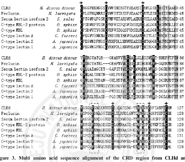
When the CLHd sequence was blasted against the database of national center of biotechnology information (NCBI), the comparatively low expect value of its alignment with some C-type lectins indicated its homology to this group protein in the view of molecular structure. The multiple alignments with the homologous proteins showed that CLHd was highly conserved in six cysteine residues and the carbohydrate recognition domain (CRD) (Fig. 3).
In most typical C-type lectins, there were several signature sequences: 4 disulfide-bonded cysteine residues, which defined the CRD, and 2 additional cysteine residues at the amino terminus (Drickamer, 1993). In CRD regions, there are in total 23 conserved residues, including 6 cysteine residues, 4 tryptophan residues and 4 glycine residues (Kolatkar and Weis, 1996; Sujatha, and Balaji, 2004). The "QPD" and the invariant "WND" motif near the C-terminal region (boxed by broken lines in Fig. 3) are extremely important in polysaccharide recognition and the calcium binding of lectins. Mostly, the "QPD" motif means that galactose binding, if it is substituted with "EPN," has specificity for mannose (Iobsts and Drickamer, 1994). CLHd exhibited highest amino acid sequence similarity (48 %) with perlucin in the CRD region. However, the full amino acid sequence of CLHd shared not more than 40% similarity with that of any homologous C-type lectin.
Figure 3. Multi amino acid sequence alignment of the CRD region from CLHd and its homologous C-type lectins. Six highly conserved cystine residues were labeled by asterisks; two motifs for the ligand binding were boxed using broken lines. All the conserved amino acid residues were shaded for the homologous comparison. All the referred sequences are from NCBI: C-type lectin 2 from Anguilla japonica, BAC54021; C-type lectin 1 from Anguilla japonica, BAC54022; C-type MBL-2 protein from Oncorhynchus mykiss, CAJ14130; C-type mannose-binding lectin from
Oncorhynchus mykiss, AAM21196; Perlucin from Haliotis laevigata, P82596; C-type
3.3. CLHd mRNA expression induced by V. alginolyticus challenge
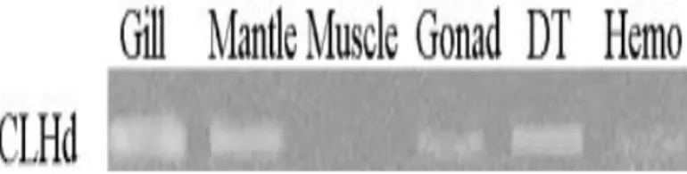
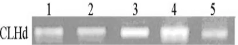
The CLHd mRNA tissue distribution in healthy abalone and its induction pattern in abalone challenged by V. alginolyticus were determined using semi-quantitative PCR. In the healthy tissue, CLHd expression was mainly detected in the gill, mantle, and digestive tract, subordinately in the gonad and hemocytes, but with no signal in muscle (Fig. 4). In V. alginolyticus challenged abalones, the relative expression level of CLHd was significantly up-regulated in the digestive tract (Fig. 5). At 12 hr post
V. alginolyticus injection, the induction of mRNA was initiated and reached
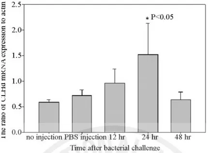
maximum expression at 24 hr. As the time progressed, mRNA transcription of CLHd dropped to the control value at 48 hr post the Vibrio challenge. All the gel electrophoresis results were quantitatively measured using Quantity One software (BioRAD, USA). At 24 hr post Vibrio challenge, it showed 2.5 times more CLHd mRNA expression than the control group and significant up-regulation in one-way analysis of variance (P <0.05) (Fig. 6).
Figure 4. Tissue distribution of CLHd mRNA transcript in abalone. All the PCR products were run 1.2% agarose gel electrophoresis. Actin was selected as internal control; DT: digestive tract; Hemo: hemocytes.
Figure 5. The mRNA temporal transcript profile of CLHd induced by V. alginolyticus challenge. 1: control; 2: PBS injection; 3: 12 hr post Vibrio injection; 4: 24 hr post Vibrio injection; 5: 48 hr post Vibrio injection.
Figure 6. The relative CLHd mRNA expression in the digestive tract of abalone after V. alginolyticus injection. Semi-quantitative PCR was repeated three times. The CLHd levels were expressed as the ratio between CLHd mRNA and the mRNA of actin (internal control) in the same sample. Data were presented as means ± SEM. Asterisk indicates statistically significant differences (p <0.05 by Tukey test, n=3).
3.4. Recombinant expression of CLHd in E.coli
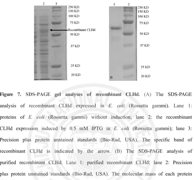
With the aim of characterization the function of CLHd, the coding sequence of CLHd was ligated into a pMal-c2X protein expression system (NEB, USA). The recombinant protein was expressed in E. coli (Rosetta-gammi DE3). In the pMal-c2X expression system, it provided the fusion MBP in the N terminal, which might enhance the solubility of recombinant CLHd and make the protein purification in a easy way. After IPTG induction of 3 hr, the whole cell lysate, purified CLHd and control E. coli cell lysate were analyzed by SDS-PAGE. A distinct band with a molecular weight of ~ 60 KDa was shown (Fig 6), which is consistent with the predicted size of MBP fusion CLHd (57.5 KDa).
Figure 7. SDS-PAGE gel analyses of recombinant CLHd. (A) The SDS-PAGE analysis of recombinant CLHd expressed in E. coli (Rossetta gammi). Lane 1:
proteins of E. coli (Rossetta gammi) without induction; lane 2: the recombinant CLHd expression induced by 0.5 mM IPTG in E. coli (Rossetta gammi); lane 3: Precision plus protein unstained standards (Bio-Rad, USA). The specific band of recombinant CLHd is indicated by the arrow. (B) The SDS-PAGE analysis of purified recombinant CLHd. Lane 1: purified recombinant CLHd; lane 2: Precision plus protein unstained standards (Bio-Rad, USA). The molecular mass of each protein marker band is labeled in the right side of the acrylamide gel.
3.5. Bacterial agglutination activity of CLHd
The gram negative no-pathogenic E. coli (DH5α), pathogenic gram negative V.
alginolyticus and gram positive B. subtilis were selected to test CLHd bacterial
agglutination activity. The incubation with a Tris buffer or MBP solution did not induce any visible agglutination. Only when the addition of recombinant CLHd reached a concentration higher than 50 µg/ml, was obvious agglutination observed in
V. alginolyticus solution (Fig. 8B) but not in E. coli and B. subtilis. Even when the
latter two strains of bacteria were incubated with 100 µg/ml of recombinant CLHd, they still did not show any agglutination. The addition of EDTA and no-calcium ion supplementation inhibited the agglutination of V. alginolyticus by CLHd in the same experiment conditions.
Figure 8. The agglutination of V. alginolyticus induced by CLHd. (A) the control V. alginolyticus with pure TBS buffer; (B) the V. alginolyticus incubated with 50 µg/ml
3.6 Hemocyte agglutination activity of CLHd
No visible agglutination was detected in the "O" type human blood cells or horse hemocytes, even when the addition of CLHd reached a concentration of 100 µg/µl and the incubation time was extended for several hours.
4. Discussion
One of the most intriguing characteristics of the immune system is its competence to recognize and respond to pathogens with considerable specificity. It can be postulated that the need for the organisms to recognize potential pathogens and distinguish between “"self”" and “"non-self”" arose at the time of the appearance of the first multicellular organisms and has played an essential role in the maintenance of their homeostasis. In vertebrates, the recognition of pathogens is carried out by two distinct types of immune receptors. The adaptive immune system is characterized by the generation of antibodies and T-cell receptors which have the capacity to recognize an innumerable repertoire of allogenic molecules. On the other hand, innate immunity relies on a restricted number of genotypically encoded molecules such as toll-like receptors (TLRs), integrins, nucleotide oligomerisation domain molecules (NODs) and scavenger receptors (known as pattern recognition receptors, PRRs) to recognize and respond to pathogens or their pathogen-associated molecular patterns (PAMPs) (Medzhitov, 2001; Zipfel and Felix, 2005). Pattern recognition receptors can be secreted proteins present in the body fluids. They can also be expressed on the cell surface or expressed in the cytoplasm. Functionally, pattern recognition receptors can be involved in: i) opsonization of bacteria and viruses for phagocytosis or activation of the lectin pathways of complement; ii) uptake of pathogens by phagocytes and dendritic cells; or iii) triggering of the signaling pathways that result in the induction of transcription of a variety of immune response genes, such as the
antimicrobial peptides and inflammatory cytokines (Fraser et al., 1998; Stahl and Ezekowitz, 1998; Buffer et al., 2003).
Invertebrates do not have antibodies and therefore they have to rely on their innate immune response to efficiently protect themselves from a variety of pathogens (Vazquez, 1997; Loker et al., 2004). Recognition of nonself materials in innate immune is mediated by PRPs, which could recognize and bind to different molecules on the surface of invading microorganisms and then trigger a series of immune responses.
There are six sub-groups of PRP in invertebrate animals, peptidoglycan recognition protein (PGRP), thioester-containing protein (TEP), gram-negative binding protein (GNBP), mulidomain scavenger receptor (SCR), C-type lectin (CTL), and galectin (GALE) (Christophides et al., 2002). C-type animal lectins represent an important recognition mechanism for extensive oligosaccharides and proteins in the serum and extracellular matrix, and for proteins and lipids on the surface of cells (Drickamer, 1988). C-type lectins in invertebrates are proved to be involved in various biological responses, for instance activation of the prophenoloxidase system, antibacterial activity , promotion of phagocytosis, and nodule formation (Suzuki et al., 1990; Tateno et al., 1998; Koizumi et al., 1999; Yu et al., 1999; Yu and Kanost, 2000; Yu et al., 2005; Luo et al., 2005). The most important one is its contribution in invertebrate immunity that C-type lectin may recognize and bind to terminal sugars on glycoproteins and glycolipids, and distinguish nonself antigens and initiate the clearance of invaders (Iwanaga and Kawabata, 1999; Yu and Kanost, 2003). In mollusks, the main mechanisms of neutralization and elimination of potential pathogens are phagocytosis and encapsulation (Sminia et al., 1974; Loker et al., 1982; Feng, 1988; Adema et al., 1992). These cellular defense reactions are closely to the activity of humoral factors and are intermediated by lectins such as
agglutinatins, opsonins, and lysins (Boswell and Bayne, 1985; Chu, 1988; Fisher and Dinuzzo, 1991; Ottaviani et al., 2004).
In the present study, one C-type lectin with novel Vibrio agglutinating activity was identified from abalone, H. discus discus. Unlike the traditional ways of lectin identification, the screening of 8000 EST cDNA library clones may detect all the C-type lectin genes in one time including some novel lectins, which can not be isolated using protein purification and sequencing method owing to its low translation level or unknown polysaccharide binding specificity. For example, CLHd is a novel lectin, which does not agglutinate human being blood cells or horse hemocytes as the other lectins from mollusk (Yakovleva, et al., 2001).
In the view of its molecular structure, the highly conserved CRD and six disulfide bond cysteines classified CLHd as a C-type lectin structurally. The 508 bp nucleotide cDNA of CLHd may encode 151 amino acid premature proteins. The signal peptide in its N terminal indicates that the mature CLHd will be transferred out of cell and function in intercellular communication. CLHd does not share high amino acid identity with any particular gene. However, in the CRD region, CLHd shows high conservation. The presence of one homologous CRD and six highly conserved cysteines suggest CLHd as a C-type lectin structurally and provide Ca2+ dependent sugar-recognition activity to initiate a broad range of biological processes, such as adhesion and pathogen neutralization. In the functional residues for ligand binding, CLHd shows QPG at the residues of 97-99, which is different with the typical "QPD" or "EPN", and the amino acid resides of 136-138 is not the invariant "WND" but "WHD". All these structural differences of CLHd with its known homologues suggest that CLHd is a novel C-type lectin.
In healthy abalones, CLHd mRNA transcript was mainly distributed in the digestive tract, gill and mantle. These three tissues are involved in a continuous
water exchange and food uptake from the outside environment to abalones and were more susceptible to pathogen infection. The tissue distribution of CLHd suggested it might offer the initial response to an invasion of marine pathogenic micro-organisms. The direct injection of V. alginolyticus significantly increased the CLHd mRNA expression and attained the maximum transcript level in the digestive tract of abalones within 24 hr. Therefore, CLHd is inducible and sensitive to Vibrio infection. The abrupt increase of CLHd mRNA transcription can be a dangerous signal indicating the pathogenic infection in abalones.
To understand the specific biological activity of CLHd, it was expressed heterologously in E. coli and the bacterial agglutination assay tested. The recombinant CLHd specifically agglutinated V. alginolyticus in a calcium dependent way. The dependence of calcium indicated CLHd was functionally a typical C-type lectin.
Basically, the bacterial agglutination immobilized the invading Vibrio and inhibited its further tissue invasion. Through its CRD, CLHd may selectively bind to the conserved carbohydrate residues on the surface of invading cells and thereby discriminated the pathogens in an efficient way. This provided the self and non-self-recognition necessary for the activation of abalone complement system, since abalone lacks immunoglobulins (Coombe. etal., 1984). Although the specific role of CLHd in innate immunity still need to be further investigated, the lectin pathway has been well established in its homologous C-type lectins such as mannose binding lectin and ficolin (Ofek and Sharon, 1988). After lectins bind to the non-self antigens, the lectin complement pathway is activated and then it will trigger the opsonization of pathogens, chemotaxis and activation of cytokines, and direct killing of pathogens (Teiz, 2002; Sodhi and Kesherwani, 2007). Taken all together, CLHd was an important agglutinin in abalones for the recognition and clearance of Vibrio pathogens.
The characterization of CLHd enriched the knowledge of abalone immunity and provided a powerful immune molecule for abalone disease control. Since it is highly inducible by Vibrio infection, in future applications, CLHd can be a useful marker for the monitoring of abalone farming environment and developing the husbandry management. Being as an effective Vibrio agglutinin, the presence of large amount of CLHd may definitely enhance the disease resistance of abalone. Gene selection or transgenic breeding of abalones with intensive expression of CLHd will provide a promising way to save juvenile abalones from the infection of Vibrio disease.
Part II
Recombinant perlucin nucleates the growth of calcium carbonate crystals: molecular cloning and characterization of pearl formation gene from disk abalone,
Haliotis discus discus
1. Abstract
Perlucin was well known as the pearl formation protein in abalone and a good research model of bio-mineralization and pearl formation. In this study, we isolated perlucin gene by screening disk abalone (H. discus discus) cDNA library. The full length cDNA of abalone (H. discus discus) perlucin gene consisted of 1038 bp nucleotide encoding putative signal peptide of 22 amino acids and a mature protein of 129 amino acids, which shared 55 % identity with greenlip abalone perlucin gene. The coding sequence was inserted into pMal-c2X expression vector and expressed the recombinant protein in Escherichia coli (Rosetta-gammi DE3). In protein activity assay, the in vitro expressed perlucin successfully promoted calcium carbonate precipitation and directed crystal morphological modification in super saturated calcium carbonate solution. Reverse transcription polymerase chain reaction (RT-PCR) results showed that perlucin gene was expressed in gill, mantle, and digestive tract, where directly confront outside environment. It implies perlucin may play some protective roles against invasive pathogenic micro-organisms like other similar C-type lectins. Further characterization of perlucin in non-self recognition is promising and anticipated.
Key words: Perlucin; Abalone (Haliotis discus discus); Protein expression; Escherichia
2. Materials and Methodology 2.1 Materials
Healthy adult abalones, H. discus discus ranging from 8 to 9 cm in body size (wet body weight at about 90 g) were purchased from abalone farms. To ensure the abalone was in natural growth condition, all the abalone were dissected on site and stored in liquid nitrogen immediately.
2.2 Molecular cloning and sequencing
The abalone (H. discus discus) cDNA library was synthesized by isolating total RNA from a whole abalone and using a cDNA library construction kit (CreatorTM, SMARTTM, Clontech, USA). Referred to single-pass random sequencing result and sequence assembly program (http://pbil.univ-lyon1.fr/cap3.php) analysis report, we chose the candidate clone of perlucin from our abalone cDNA library. Then we designed specific internal primers and completed its full length cDNA sequencing (Table 3). ORF detection, signal peptide prediction, N/O-glycosylation site and rare codon analysis were performed by DNAssit 2.1, webpage server (http://www. cbs.dtu. dk/services/NetNGlyc/), (http://www.Cbs.dtu.dk/services/NetOGlyc/) and (http://www. faculty.ucr.edu/~mmaduro/codonusage/usage.htm), respectively.
Primer name Sequence
Internal sequencing primer (Forward) ATATGAGGTGTGGTGTGC RT-PCR perlucin primer (Forward) GCTGCTCACATTTGCATCATTGC RT-PCR perlucin primer (Reverse) GTTGGTGTACGTCATGGCCTTGTT RT-PCR actin primer (Forward) GACTCTGGTGATGGTGTCACCCA RT-PCR actin primer (Reverse) ATCTCCTTCTGCATTCTGTCGGC Recombination primer (Forward) GAGAGAGAATTCTGTCCTCTGGGATTCTTTCG Recombination primer (Reverse) GAGAGAAAGCTTTTAAAGGTTCTTCTCACAGATAAAGTTTAAG
2.3 Protein expression and purification
Based on the perlucin full length cDNA sequence, specific PCR primers were designed to amplify the mature protein coding region of the perlucin gene. PCR products were digested by restriction endonucleases and ligated to expression vector pMAL-c2X under the instruction of kit manual (NEB, USA). Finally, the recombinant plasmids were transformed into E. coli (Rosetta-gammi DE3) for protein expression. A volume containing 5 ml of starter culture was inoculated into 100 ml Luria broth with 100 µl ampicillin (100 mg/ml) and 10 mM glucose (2 % final concentration) and kept at 37 ºC with continuous rotary shaking at 180 rpm till density approached OD600 = 0.5~0.8. Then protein expression was induced by adding 0.5 mM IPTG.
After 6 hr induction at 37 ºC, the cells were harvested by centrifugation at 3500 rpm for 30 min at 4 ºC. Subsequently, all the cells were resuspended in 6 ml column buffer and stored in -70 ºC overnight. After thawing, the bacterial cells were placed in an ice-water bath and sonicated. Having centrifuged at 9000 x g for 30 min at 4 ºC, the supernatant was loaded into a maltose binding resin column. Through amylose resin columns, proteins were purified and used for activity assays. Sodium dodecyl sulfate polyacrylamide gel electrophoresis (SDS PAGE) was performed under reducing condition (with 2-ME) to analysis the expression and purification of the recombinant perlucin. The gel (8 × 10 cm and 1.5 mm thick) was subjected to electrophoresis at 30 mA for 50 min. Molecular weight protein standards (Unstained Plus, Biorad, USA) were used. The gel was stained with coommasie brilliant blue R-250 (Sigma, USA). Protein concentration was calculated by Bradford method (Bradford, 1976).
2.4 Protein activity assay
To make sure the success of cloning and expression, here we tested our recombinant perlucin in two ways as mainly used in previous researches (Samata et
al., 1999 Weiss et al., 2000; Zhang et al., 2003; Suzuki et al., 2004). In pMal-c2X expression system, it provided the fusion MBP in N terminal, which might enhance the solubility of recombinant perlucin and simplify the protein purification. Since the recombinant perlucin has more attractive interests in further application, for example, color modification of pearls by recombinant perlucin gene transfer, the N terminal MBP fusion perlucin was directly tested by calcium carbonate precipitation and crystallization.
2.4-1 Calcium carbonate precipitation test
A calcium carbonate precipitation experiment was performed according to previous work with a few modifications (Morales et al., 1996). Briefly, 20 mM NaHCO3 was
adjusted to pH 8.70 by adding 1 N HCl and 1 N NaOH and supplemented with 0.5 M KCl to keep the total ionic strength close to that of seawater. Then 6 ml 20 mM CaCl2 was immediately mixed with the same volume NaHCO3 solution in 50 ml
conical tubes. Fifty microlitre 1 mg/ml maltose binding protein fusion perlucin (MBP-perlucin) was added to the calcium carbonate solution. As the control, distilled water or protein buffer solution (1.0 M Tris-HCl, pH 7.4 and 200 mM NaCl) was added into the calcium carbonate super saturated solution. The pH variation was determined by istek (720P model) pH meter every ten seconds.
2.4-2 Calcium carbonate crystallization test
About 30 ml of 100 mM NaHCO3 was added dropwise under permanent stirring
to 120 ml of a 40 mM CaCl2 solution (Blank, et al. 2003). As soon as the solution
became turbid, the addition of CaCl2 was stopped and the pH was adjusted to 8.2
with 1 N NaOH. If the pH value of the crystallization solution is too low (less than 7.7) or too high, it will cause pH drift during incubation (Walters et al. 1997). Thus we chose pH 8.2 for the crystallization test after several pilot experiments. The saturated calcium carbonate solution was then filtered using a 0.22 µm filter. 50 µl