저작자표시-비영리-변경금지 2.0 대한민국 이용자는 아래의 조건을 따르는 경우에 한하여 자유롭게 l 이 저작물을 복제, 배포, 전송, 전시, 공연 및 방송할 수 있습니다. 다음과 같은 조건을 따라야 합니다: l 귀하는, 이 저작물의 재이용이나 배포의 경우, 이 저작물에 적용된 이용허락조건 을 명확하게 나타내어야 합니다. l 저작권자로부터 별도의 허가를 받으면 이러한 조건들은 적용되지 않습니다. 저작권법에 따른 이용자의 권리는 위의 내용에 의하여 영향을 받지 않습니다. 이것은 이용허락규약(Legal Code)을 이해하기 쉽게 요약한 것입니다. Disclaimer 저작자표시. 귀하는 원저작자를 표시하여야 합니다. 비영리. 귀하는 이 저작물을 영리 목적으로 이용할 수 없습니다. 변경금지. 귀하는 이 저작물을 개작, 변형 또는 가공할 수 없습니다.
A Master’s Thesis
Inhibitory effect and action mechanism of
diphlorethohydroxycarmalol (DPHC) on the production of
interleukin-6 in lipopolysaccharide (LPS)-stimulated
murine macrophage RAW 264.7 cells
Na-Jin Kang
Department of Biomedicine & Drug Development
Graduate School
Jeju National University
LPS로 자극된 RAW 264.7에서
diphlorethohydroxycarmalol (DPHC)에 의한
IL-6 생성 억제효과 및 작용기전 연구
지도교수 유 은 숙
강 나 진
이 논문을 이학 석사학위 논문으로 제출함
2014 년 2 월
강나진의 이학 석사 학위 논문을 인준함
제주대학교 대학원
2014 년 2 월
Inhibitory effect and action mechanism of
diphlorethohydroxycarmalol (DPHC) on the production of
interleukin-6 in lipopolysaccharide (LPS)-stimulated
murine macrophage RAW 264.7 cells
Na-Jin Kang
(Supervised by professor Eun-Sook Yoo)
A thesis submitted in partial fulfillment of the requirement for the
degree of master of science in Biomedicine & Drug Development
Department of Biomedicine & Drug Develpment
Graduate School
Jeju National University
i
CONTENTS
CONTENTS...i LIST OF FIGURES...iv 1. ABSTRACT………...1 2. INTRODUCTION.……….23. MATERIALS AND METHODS………...6
3.1. Reagents………...6
3.2. Isolation of DPHC………7
3.3. Cell culture………..7
3.4. Cell viability………9
3.5. The enzyme-linked immunosorbent assay (ELISA)………....…9
3.6. Western blot analysis………..10
3.7. Confocal laser scanning microscopy analysis………11
ii
4. RESULTS………...…12
4.1. Anti-inflammatory effect of DPHC………..…….12
4.1.1 Effect of DPHC on cell viability in LPS-stimulated RAW 264.7 cells………….12
4.1.2 Time-course production of NO and inflammatory cytokines in LPS-stimulated RAW 264.7 cells………....14
4.1.3. Effect of DPHC on NO and PGE2 production in LPS-stimulated RAW 264.7 cells………19
4.1.4. Effect of DPHC on IL-6 and TNF-α production in LPS stimulated RAW 264.7 cells………...………22
4.2. Effect of DPHC on the signaling pathway of LPS………..…27
4.2.1. Effect of DPHC on iNOS and COX-2 expression and production in LPS-stimulated RAW 264.7 cells……….27
4.2.2. Effect of signaling inhibitors on IL-6 production in LPS-stimulated RAW 264.7 cells………29
4.2.3. Effect of DPHC on the phosphorylation of MAPKs in LPS-stimulated RAW264.7 cells………32
iii
264.7 cells……….……..34
4.2.5. Effect of DPHC on the phosphorylation of NF-κB in LPS-stimulated RAW 264.7 cells………36
5. DISCUSSION………...….41
iv
LIST OF FIGURES
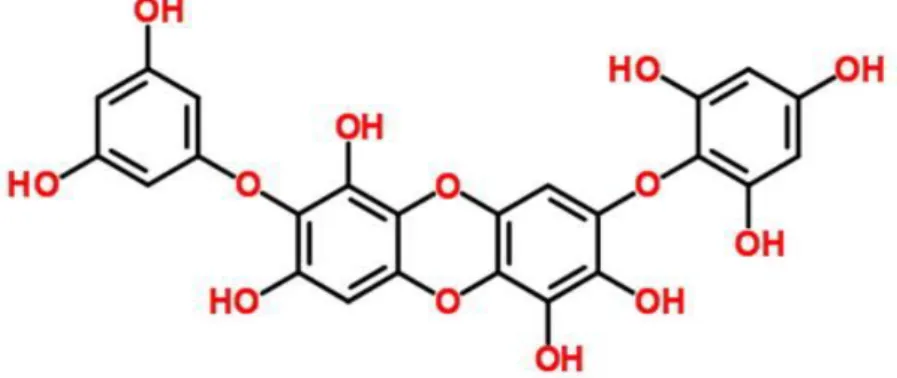
Figure 1. Chemical structure of diphlorethohydroxycarmalol (DPHC)………..…
8
Figure 2. Effect of diphlorethohydroxycarmalol (DPHC) on the cell viability in the LPS-stimulated RAW264.7 murine macrophages………..…
13
Figure 3. Nitric oxide (NO) production in LPS-stimulated RAW264.7 macrophages……….
15
Figure 4. PGE2 production in LPS-stimulated RAW264.7 macrophages………
16
Figure 5. The production of IL-6 in LPS-stimulated RAW264.7 macrophages………
17
Figure 6. The production of TNF-α in LPS-stimulated RAW264.7 macrophages………..
18
Figure 7. Effect of diphlorethohydroxycarmalol (DPHC) on the nitric oxide (NO) production in the LPS-stimulated RAW264.7 murine macrophages………..
20
Figure 8. Effect of diphlorethohydroxycarmalol (DPHC) on the Prostaglandin E2 (PGE2) production in the LPS-stimulated RAW264.7 murine macrophages……….………
21
Figure 9. Effect of diphlorethohydroxycarmalol (DPHC) on the Tumor necrosis factor- α (TNF-α) production in the LPS-stimulated RAW264.7 murine macrophages………..
23
Figure 10. Effect of diphlorethohydroxycarmalol (DPHC) on the Interleukin-6 (IL-6) production in the LPS-stimulated RAW264.7 murine macrophages……….
25
Figure 11. Effect of diphlorethohydroxycarmalol (DPHC) on Expression of iNOS and COX-2 in LPS-stimulated RAWCOX-264.7 murine macrophages………..
28
Figure 12. Effect of NF-κB inhibitors (PDCT, TPCK, parthenolide), MAPK inhibitors (SP, PD, SB 203580), and STAT inhibitors (AG490, JI, EGCG, fludarabine) on the Interleukin-6 (IL-6) production in the LPS-stimulated RAW264.7 murine
v
macrophages……….
30
Figure 13. Effect of diphlorethohydroxycarmalol (DPHC) on expression of MAPKs phosphorylation in LPS-stimulated RAW264.7 macrophages………..
33
Figure 14. Effect of diphlorethohydroxycarmalol (DPHC) on expression of STAT1 (Tyr701, Ser727), STAT3, STAT5 (Tyr694) phosphorylation in LPS-stimulated RAW264.7 macrophages……….
35
Figure 15. Effect of DPHC on the NF-κB phosphorylation in LPS-stimulated RAW264.7 macrophages……….
37
Figure 16. Effect of DPHC on the NF-κB phosphorylation in LPS-stimulated RAW264.7 macrophages……….
38
Figure 17. Effect of DPHC on the NF-κB phosphorylation in LPS-stimulated RAW264.7 macrophages……….
39
Figure 18. Inhibitory effect and action mechanism of diphlorethohydroxycarmalol (DPHC) on the production of interleukin-6 in lipopolysaccharide (LPS)-stimulated murine macrophage RAW 264.7 cells……….
40
1
1. Abstract
Inflammatory response is regulated and mediated by various immune cells, such as monocyte and macrophage, and especially macrophages are important players that are closely related to the initiation, propagation, and resolution of inflammation. Interleukin-6 (IL-6) is a representative pro-inflammatory cytokine generated in stimulated macrophages and may cause deleterious inflammatory states. Diphlorethohydroxycarmalol (DPHC) is a phlorotannin compound isolated from Ishigeokamuarae, a brown algae. This study was conducted to investigate the anti-inflammatory effect and action mechanism of DPHC in the lipopolysaccharide (LPS) stimulated RAW 264.7 murine macrophages. The experimental results showed that DPHC inhibited LPS-induced IL-6 production in a dose dependent manner at concentration of 12.5, 25, 50, and 100 μM. DPHC also suppressed the phosphorylation and the nuclear translocation of NF-κB (p65 and p50 subunits), a central signaling molecule in LPS induced inflammation process. Furthermore, the known inhibitors of NF-κB (pyrrolidinedithiocarbamate; PDTC, N-tosyl-L-phenylalaninechloromethyl ketone; TPCK, parthenolide) strongly inhibited IL-6 production in this system. However, DPHC had no or only minimal effects on the mitogen activated protein kinase pathways (JNK, ERK and P-38) and STAT1, 3, and 5 pathways activated by LPS. In conclusion, the present study demonstrates that DPHC could inhibit IL-6 production through the suppression of NF-κB activation and is suggested as a potential candidate for treating inflammatory diseases.
2
2. Introduction
Inflammation is known to be caused by various stimuli, such as bacteria, toxins, and other pathogens, which is usually accompanied with injures, irritation, infection of tissues. In addition, according to recent reports, inflammation is closely related to tumor progression. Inflammatory response is mediated by various immune cells, such as monocytes and macrophages. These inflammatory cells produce many chemokines and cytokines, which influence progression, growth regulation, and migration of tumor organ. Especially, macrophages play important roles in inflammation processes, such as initiation, propagation, and resolution (Lawrence, Willoughby & Gilroy 2002, Ben-Neriah, Karin 2011, Ariel, Timor 2013, Zhang, Mosser 2008, Coussens, Werb 2002). Activated macrophages secrete various inflammatory mediators and kill intracellular pathogens (Mosser 2003).
Cytokines are small proteins with molecular weights of 8 to 40,000 d, and are pharmacologically active proteins that have autocrine and paracrine effects. Cytokines are related to multiple biological activities in the response of various diseases, infection, and inflammation. Some cytokines are called “pro-inflammatory cytokines” or “anti-inflammatory cytokines”, because they clearly facilitate inflammation, and or suppress the activation of pro-inflammatory cytokines, such as Tumor necrosis factor-α (TNF-α), interleukin-6 (IL-6) and prostaglandin E2 (PGE2) (Dinarello 2000, Opal, DePalo 2000, Coppack 2001).
IL-6 is a pleiotropic cytokine which plays central roles in pro-inflammatory and autoimmune diseases responses, and exacerbates various inflammatory states, including sepsis, rheumatoid arthritis and inflammatory bowel disease (Hohki et al. 2010, Hack et al. 1989, Atreya et al. 2000). IL-6 is generated in the macrophage, and involved in inflammation. Especially, LPS-stimulated macrophages produce a number of inflammatory mediators, such
3
as IL-6, MIP-1 beta, etc through TLR receptor (Takeda, Akira 2005, Martin, Dorf 1991). According to Fattori.E. and Cappelletti. M. et al, in the absence of IL-6, mice could not be mounted a normal inflammation to tissue damage by turpentine injection. According to the results, they concluded that IL-6 is essential mediator of inflammation (Fattori et al. 1994). Therefore, IL-6 is an important factor in preventing and treating inflammation.
TLR is a trans-membrane receptor that is one of critical components in the innate immune system and autoimmunization, and directly recognize and activate immune cells. TLR signaling pathway modulates Toll-interleukin 1 receptor (TIR) domain-containing adaptor proteins included myeloid differentiation primary response gene 88 (MyD88), TIR domain containing adaptor protein (TIRAP), and TIR-domain containing adapter-inducing interferon-β (TRIF). TLRs also recognize various pathogens and participate in cell signaling pathway (Akira, Takeda 2004, Takeda, Akira 2004, Aderem, Ulevitch 2000, Akira, Uematsu & Takeuchi 2006, Kawai, Akira 2010). TLR4, a member of TLRs family, recognizes lipopolysaccharide (LPS, endotoxin) and mediates LPS-induced inflammation (Hoshino et al. 1999, Bortoluci, Medzhitov 2010). LPS, an inducer of the innate immune response, binds to the Myeloid differentiation factor-2 (MD-2)/TLR4 complex, initiates downstream signaling pathway, and releases various inflammatory chemokines and cytokines including IL-6, inducible nitric oxide synthase (iNOS), TNF-α (Akira, Takeda 2004, Park et al. 2009, Kagan, Medzhitov 2006). In the downstream signaling pathway, intracellular adaptor proteins leads to two distinct signaling pathways, MyD88-dependent pathway that includes the mitogen-activated protein kinases (MAPK) [c-Jun N-terminal kinase (JNK), p38 and extracellular-signal related kinase (ERK)] and the NF-kappaB (NF-κB; p50/p65) complex, and MyD88-independent signaling pathway that produce type 1 interferons. These type 1 interferons regulate STAT1-containing transcription factors. The STAT3 and STAT5 are also induced by LPS treatments (Lu, Yeh & Ohashi 2008, Kawai, Akira 2006, Ohmori, Hamilton 2001b, Bode, Ehlting & Haussinger 2012, Yamaoka et al. 1998).
4
NF-κB is one of the most principal transcription factors which involves in gene induction of cellular proliferation. NF-κB is also a pathogen-derived inflammatory cytokine that serves as a central inflammatory mediator (Akira, Takeda 2004, Park et al. 2009, Kagan, Medzhitov 2006, Lu, Yeh & Ohashi 2008, Kawai, Akira 2006, Li, Verma 2002). When the stimulus is applied, NF-κB is activated through the activation of I kappa B-kinase (IKK) complex, which phosphorylates I kappa B-alpha (IκB-α) and p50/p65. Then IκB-α undergoes proteasomal degradation. NF-κB exists mainly as a heterodimer including the subunits p50/p65 of Rel family. The p65 transactivation domain is activated by phosphorylation, and then the phosphorylation, ubiquitination, and degradation of IκB-α release the p50/p65 heterodimer. The free p50/p65 complex then translocate from cytosol to nucleus, and finally controls promotor region of target genes, which induces various pro-inflammatory factors. According to Pan, Lai et al, the expression of iNOS and COX-2 is mediated via NF-κB in LPS-stimulated macrophage (Gloire, Legrand-Poels & Piette 2006, Baeuerle, Baltimore 1996, Aggarwal 2004, Pan et al. 2006).
The cytokine-activated Janus kinase (JAK)-signaling transducer and activator of transcription (STAT) pathway has an important role in immune system. JAK-STAT signaling pathway regulates the initiation, propagation, and inflammation in all cell types. The phosphorylation of STATs is divided into two types, tyrosine phosphorylation and serine phosphorylation. The tyrosine phosphorylation acts a switching signal to activate STATs that require for dimerization, nuclear translocation and DNA binding. The serine phosphorylation seems to be independent on the tyrosine phosphorylation (Shuai, Liu 2003, O'Shea, Murray 2008). Although JAK2 induced the phosphorylation of STAT1 tyrosine and STAT1 serine, STAT1 serine phosphorylation occurs independently of tyrosine phosphorylation. STAT1 serine 727 phosphorylation is induced STAT1-mediated gene activation to the max and IFN-gamma-induced transcriptional activity of STAT1 (Zhu et al. 1997, Shuai 2003, Decker, Kovarik 2000, Wen, Zhong & Darnell 1995). The phosphorylation of STAT1-S727A, STAT1
5
transactivation, domain is induced IFN-gamma-dependent genes and related to innate immunity (Varinou et al. 2003). In addition to, IL-6 is an obligatory role that regulates through the JAK-STAT signal pathway, the ensuing up-regulation of iNOS and COX-2, and the development of a cardioprotective phenotype (O'Shea, Murray 2008, Kalinski 2012, Dawn et al. 2004).
Diphlorethohydroxycarmalol (DPHC) is a phlorotannin compound isolated from Ishige okamuarae, a brown algae, collected from the coast of Jeju island. According to recent reports, DPHC has been determined to have various biological effects. DPHC alleviated both the increase of postprandial blood glucose levels in diabetic mice and induced factors of high glucose-induced-oxidative stress in human umbilical vein endothelial cells (Heo et al. 2009, Heo et al. 2010). Also, DPHC suppressed intracellular reactive oxygen species (ROS) induced by ultraviolet (UV)-B radiation, and had protective effects on DNA tail and cell damages (Heo et al. 2010). However, little information is available regarding the ability of DPHC on the inhibition of IL-6 production.
In the present study, we examined the inhibitory effect of DPHC on the production of IL-6, a pivotal cytokine in the inflammatory process and explored the action mechanism in LPS-stimulated RAW264.7 cells.
6
3. Materials and methods
3.1 Reagents (Chemicals and antibodies)
Lipopolysaccharide (LPS, E. coli 0111:B4) was purchased from Sigma-Aldrich Chemical Co. (St. Louis, MO). Fetal bovine serum (FBS) and Dulbecco’s modified Eagle’s medium (DMEM) were obtained from Invitrogen-GIBCO (Grand Island, NY, USA). Mouse IL-6 Duoset enzyme-linked immunosorbent assay (ELISA) kits and prostaglandin E2 parameter assay kit were obtained from R&D Systems (St. Louis, MO, USA). MAPKs (anti-phospho-ERK1/2, anti-(anti-phospho-ERK1/2, anti-phospho-JNK, anti-JNK), STATs [anti-Phospho-STAT1 (Tyr701, Ser727), Phospho-STAT3 (Tyr705), Phospho-STAT5 (Tyr694), STAT3, anti-STAT5)], NF-κB [anti-phospho-p65 (Ser536), anti-p65, anti-IκB-α], phospho-p38 MAP kinase (Thr180/Tyr182), and anti-p38 were purchased from Cell Signaling Technology (Beverly, MA, USA). NF-κB [anti-p50 (E-10)] was purchased from Santa Biotechnology, Inc. (Santa Cruz, CA, USA). Anti-STAT1was purchased from Becton Dickison (San Diego, CA, USA). β-actin was obtained from Sigma (St. Louis, MO, USA). DyLight 488 conjugated Donkey anti-Rabbit antibody was purchased from BioLegend Inc. (San Diego, CA, USA). All other reagents were reagent grade.
7 3.2 Isolation of DPHC
The shade dried whole plant of Ishige okamurae (800 g) was percolated with 70% aqueous ethanol under stirring at room temperature for 2 days. The filtrate was concentrated in a vacuum freeze-dryer, suspended in distilled water and partitioned with ethyl acetate. The ethyl acetate fraction was chromatographed over reversed phase silica gel with gradient solvent (H2O/MeOH) system to provide 4 fractions (1~4). The fraction 1 was chromatographed over Sephadex-LH 20 with CHCl3/MeOH (1/1) to provide 6 fractions (4-1'~4-6'). DPHC was obtained from the fraction 4-5', and its structure (Fig. 1) was confirmed by comparing the NMR spectral data with literatures (Heo et al. 2008, Zou et al. 2008).
3.3 Cell Culture
RAW 264.7 cells, the murine macrophage cell line, were obtained from the American Type Culture Collection (ATCC, Rockville, MD, USA). Cells were cultured in DMEM supplement with 10% (vol/vol) FBS, containing100 U/ml penicillin-streptomycin (GIBCO, Grand Island, NY, USA), and were maintained at subconfluence in 5% CO2 humidfied atmosphere at 37℃. Cells were counted with a hemocytometer.
8
9
3.4 Cell viability
Cell viability was determined by using EZ-CyTox (tetrazolium salts, WST-1) assays (Daeil Lab Inc, Seoul, Korea). RAW 264.7 cells were seeded at a density of 1.5x10⁵ cells/ml in 96-well culture plates and were maintained for 18 h an incubator. Culture medium was replaced by new medium, DMEM supplemented with 10% FBS and 100 U/ml penicillin-streptomycin. Cells were treated with LPS in the absence or presence of various concentrations of DPHC. After incubation for 24 h, 100 μL of culture supernatant from each well was abandoned and each well was treated with 5μL WST, which was further incubated for 2 h at 37℃ and 5% CO2 atmosphere. After the incubation, the light absorbance of each well was quantified using a VersaMax ELISA microplate reader (Molecular Devices, CA, USA) at 450 nm.
3.5 The enzyme-linked immunosorbent assay (ELISA)
IL-6 production in the supernatant was measured by ELISA kit according to the manufacturer’s instructions. Briefly, 100 μL of cell culture supernatants or standards in reagent diluent was added to each well of ELISA kit well plate and incubated for 2 h at room temperature. After washing, 100 μL of the detection antibody in reagent diluent was added to each well and incubated for 2 h at room temperature. After washing, 100 μL of streptavidin conjugated horseradish-peroxidase (HRP) was added and left at room temperature for 20 min. After washing, 100 μL of substrate solution was added to each well and left in dark place at room temperature for 20 min. Then 50 μL of stop solution (2 N sulfuric acid) was added. After the reaction was stopped, the optical density of each well was determined using a VersaMax ELISA microplate reader at 450 nm.
10
3.6 Western blot analysis
RAW 264.7 cells were treated to LPS with absence or presence of the elapsed concentration of DPHC. After the elapsed times of incubation, the cells were washed twice with cold phosphate buffered saline (PBS) and were lysed in a protein lysis buffer [50 mM Tris-HCl (pH 7.5), 150 mM NaCl, 1% Nonidet P-40, 2 mM EDTA, 1 mM EGTA, 1 mM NaVO3, 10 mM NaF, 1 mM dithiothreitol, 1 mM phenylmethylsulfonyl fluoride, 25 μg/ml a protinin, and 25 μg/ml leupeptin], and then the cells were collected. Then The cell lysates were isolated using centrifugation at 15,000 rpm (4℃ for 15 min). The protein concentration in the supernatants was measured by the Bradford assay (Bio-rad, Herculers, CA, USA) and all proteins were adjusted to equal protein content. Aliquots of the lysates (20~30 μg protein/lane) were separated on a NuPAGE 4-12% bis-Tris gel (Invitrogen, Carlsbad, CA, USA) and the separated proteins were transferred from the electrophoresis gel to the surface of polyvinylidenedifluoride (PVDF) membranes using an iBlot gel transfer device (Invitrogen). Then membranes were treated with 5% non-fat skim milk as a blocking solution, and incubated with primary antibodies (1:1,000) at 4℃ overnight. After incubating, the membranes were washed by Tween 20-Tris-buffered saline (TBST) several times and incubated with secondary HRP-linked anti-rabbit or anti-mouse IgG (1:5,000, Cell signaling, Santa Cruz Biotechnology; respectively) for 90 min at room temperature. After washing, immunoactive proteins were detected by the WEST-ZOL (plus) Western Blot Detection System (iNtRON Biotechnology, Gyeonggi, Korea).
11
3.7 Confocal laser scanning microscopy analysis
RAW 264.7 cells were plated onto coverslips at 6-well plate and incubated for 18 h, and then treated with LPS with the absence or presence of DPHC for various times. Then the cells were washed in PBS and fixed with 3.5% formaldehyde (PFA) in PBS for 30 min at room temperature. After washing with PBS, cells were treated with 0.1M glycine for 15 min and were permeabilized with PBS containing 0.1% Triton X-100 for 10 min. After several washings with PBS, cells were blocked in PBS containing 3% BSA and 0.1% Triton X-100. In order to react cells and antibody, primary antibody anti-NF-κB (phospho-p65, p50) diluted 1:200 was treated at 4℃, overnight. Then cells were washed with Tween 20-PBS (PBST), and treated with DyLight488 conjugated donkey anti-rabbit secondary antibody diluted 1:200, and then left at dark place and room temperature for 30 min. After several washing, coverslips were mounted to slides by using VECTASHIELD Mounting Media with DAPI (Vector Labs, Burlingame, CA, USA). The images were visualized using FV500 confocal microscopy (Olympus, Tokyo, Japan).
3.8 Statistical analysis
Statistical differences between the test and control groups were analyzed by Student’s t-test. Statistical results obtained from at least three independent experiments were presented as the mean and standard deviation (means ± SD). P-value < 0.05, <0.01, and <0.001 was considered to be statistically significant.
12
4. Results
4.1. Anti-inflammatory effect of DPHC
4.1.1. Effect of DPHC on cell viability in LPS-stimulated RAW 264.7 cells
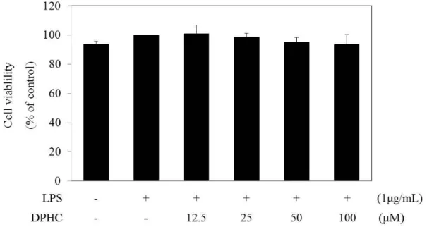
We measured the cell viability of LPS-induced RAW 264.7 cells at various concentrations (12.5, 25, 50, and 100 μM) of DPHC by the WST assay to exclude the possible suppression of inflammatory chemokine production by cytotoxic activity. RAW 264.7 cells were treated with various concentrations (12.5, 25, 50, and 100 μM) of DPHC for 24 h. As shown in Fig. 2, DPHC exhibited no effects on the cell viability.
13
Figure 2. Effect of diphlorethohydroxycarmalol (DPHC) on the cell viability in the LPS-stimulated RAW264.7 murine macrophages.
Cells (1.5 x 10⁵ cells/mL) were pre-incubated for 18 h. Cell viability was determined from the cells stimulated with LPS (1 µg/mL) in the presence or absence of DPHC for 24 h (12.5 to 100 µM). Cell viability was determined by the WST assay, and the results were expressed as percentage of surviving cells over positive cells (only LPS addition). Error bars indicates the mean ± S.D.
14
4.1.2.Time-course production of NO and inflammatory cytokines in LPS-stimulated RAW 264.7 cells
We measured the amount of NO and inflammatory cytokines at indicated times (0, 5, 15, 30, 60, and 120 min and 4, 6, 8, 10, 12, and 24 h) after LPS (1 μg/mL) treatment to characterize the time profile of NO and inflammatory cytokine production. NO production continuously increased from 6 h (0.030 μM) to 24 h (0.264 μM) after LPS-stimulation (Fig. 3). PGE2production continuously increased from 6 h (11,856.46 pg/mL) to 24 h (42,724.96 pg/mL) after LPS-stimulation (Fig. 4). IL-6 production continuously increased from 6 h (312.4 pg/mL) to 24 h (918.3 pg/mL) after LPS-stimulation (Fig. 5), and TNF-α production increased from 2 h (429.4 pg/mL) to 24 h (1,147.2 pg/mL) after LPS-stimulation (Fig. 6).
15
Figure 3. Nitric oxide (NO) production in LPS-stimulated RAW264.7 macrophages.
Cells (1.5 × 10⁵ cells/mL) were pre-incubated for 18 h, and cells were stimulated with LPS (1 µg/mL) for indicated times, respectively. NO production was determined from the culture supernatants of cells. The amount of NO was determined by using Griess method. The standard curve created by NaNO2 in culture medium. The measurement of NO was performed in triplicate. The error bars indicate standard deviation.
16
Figure 4. PGE2 production in LPS-stimulated RAW264.7 macrophages.
Cells (1.5 × 10⁵ cells/mL) were pre-incubated for 18 h, The cells were stimulated with LPS (1 µg/mL) for indicated times, respectively. The PGE2 production was determined from the culture supernatants of cells. The production of PGE2 was determined by ELISA. The measurement of PGE2 was performed in triplicate. The error bars indicate standard deviation.
17
Figure 5. The production of IL-6 in LPS-stimulated RAW264.7 macrophages.
Cells (1.5 × 10⁵ cells/mL) were pre-incubated for 18 h, and cells were stimulated with LPS (1 µg/mL) for different times, respectively. The IL-6 production was determined from the culture supernatants of cells. IL-6 production was determined by ELISA. The measurement of IL-6 was performed in triplicate. The error bars indicate standard deviation.
18
Figure 6. The production of TNF-α in LPS-stimulated RAW264.7 macrophages.
Cells (1.5 × 10⁵ cells/mL) were pre-incubated for 18 h, cells were stimulated with LPS (1 µg/mL) for different times, respectively. TNF-α production was determined from the culture supernatants of cells. TNF-α production was determined by ELISA. The measurement of TNF-α was performed in triplicate. The error bars indicate standard deviation.
19
4.1.3. Effect of DPHC on NO and PGE2 production in LPS-stimulated RAW 264.7 cells
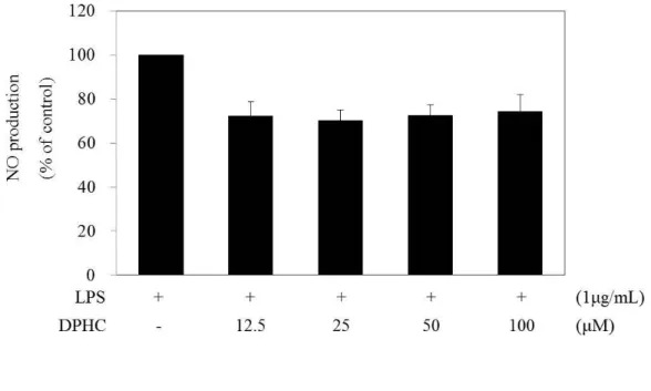
NO plays many regulatory functions in the development of inflammation and is synthesized from many cell types with inflammation (Coleman 2001, Esposito, Cuzzocrea 2007). NO production gradually increased with the indicated times after LPS treatment, and then reached a peak at 24 h (Fig. 3). We then confirmed the production of NO after 24 h of DPHC treatment. We examined NO production by Griess assay at 24 h to determine whether or not DPHC inhibited LPS-stimulated NO production. As shown in Fig. 7, LPS induced NO production. After 24 h of treatment, DPHC weakly reduced NO production in LPS-stimulated RAW 264.7 cells, and this result was no statistically significant. PGE2 also involves inflammatory mediators from LPS-stimulated resident macrophages, and the PGE2 -EP4 signaling pathway promotes immune inflammation (Dieter et al. 1999, Yao et al. 2009). PGE2 production gradually increased with the elapsed times after LPS treatment and then reached a peak at 24 h (Fig. 3). We then confirmed the production of PGE2 after 24 h of DPHC treatment. We examined PGE2production by ELISA to investigate whether or not DPHC inhibited LPS-stimulated PGE2 production. DPHC significantly reduced PGE2 production in a dose-dependent manner for 24 h in LPS-stimulated RAW 264.7 cells (Fig. 8).
20
Figure 7. Effect of diphlorethohydroxycarmalol (DPHC) on the nitric oxide (NO) production in the LPS-stimulated RAW264.7 murine macrophages.
Cells (1.5 x 10⁵ cells/mL) were pre-incubated with DMEM/10% FBS for 18 h and then NO production was determined from the culture supernatants of cells stimulated with LPS (1 µg/mL) in the presence of DPHC (12.5, 25, 50 , 100 µM) for 24 h. NO production was determined by the Griess reagent method. The measurement of NO was done in triplicate. Error bars indicate ± S.D.
21
Figure 8. Effect of diphlorethohydroxycarmalol (DPHC) on the Prostaglandin E2 (PGE2) production in the LPS-stimulated RAW264.7 murine macrophages.
Cells (1.5 x 10⁵ cells/mL) were pre-incubated with DMEM/10% FBS for 18 h and then PGE2production was determined from the culture supernatants of cells stimulated with LPS (1 µg/mL) in the presence of DPHC (12.5, 25, 50, 100 µM) for 24 h. PGE2 production was determined by ELISA. The measurement of PGE2 was performed in duplicate, and the results were expressed as percentage over positive cells. Error bars indicate ± S.D. *, P<0.05, **, P<0.01, significant when compared with LPS positive control.
22
4.1.4. Effect of DPHC on IL-6 and TNF-α production in LPS stimulated RAW 264.7 cells
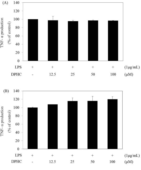
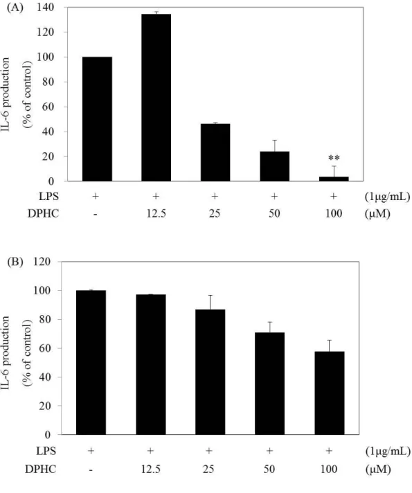
Endotoxin produces pro-inflammatory cytokines, such as IL-6 and TNF-α (Carlson et al. 1999). IL-6 mediate inflammation, infection, and metabolism, whereas TNF-α activates inflammatory and toxic cytokines (Scheller et al. 2011, Aggarwal 2003). IL-6 production gradually increased at 6 h and peaked at 24 h, and TNF-α production gradually increased from 1 h to 4~6 h and then peaked at 24 h (Figs. 5 and 6). We then confirmed the production of PGE2 after 24 h of DPHC treatment. We examined IL-6 and TNF-α production by ELISA for 6 h and 24 h, respectively, to determine whether or not DPHC inhibited the production of IL-6 and TNF-α. RAW 264.7 cells were treated with LPS with and without DPHC for 6 and 24 h, respectively. DPHC did not reduce TNF-α production in LPS-stimulated RAW 264.7 cells (Figs. 9A and 9B). DPHC inhibited IL-6 production in a dose-dependent manner at 6 and 24 h (Figs. 10A and 10B). The significant effect of DPHC after 24 h incubation is shown in Fig. 10A. The results suggest that DPHC primarily regulates the accumulation of IL-6 as a pro-inflammatory cytokine.
23
Figure 9. Effect of diphlorethohydroxycarmalol (DPHC) on the Tumor necrosis factor- α (TNF- α) production in the LPS-stimulated RAW264.7 murine macrophages.
(A) Cells (1.5 x 10⁵ cells/mL) were pre-incubated with DMEM/10% FBS for 18 h and then TNF-α production was determined from the culture supernatants of cells stimulated with LPS (1 µg/mL) in the presence of DPHC (12.5, 25, 50, 100 µM) for 24 h. TNF-α production was determined by ELISA. The measurement of TNF-α was done in duplicate, and the
24
results were expressed as percentage over positive cells. Error bars indicate ± S.D. (B) Cells (1.5 x 10⁵ cells/mL) were pre-incubated with DMEM/10% FBS for 18 h and then TNF-α production was determined from the culture supernatants of cells stimulated with LPS (1 µg/mL) in the presence of DPHC (12.5, 25, 50, 100 µM) for 6 h. TNF-α production was determined by ELISA. The measurement of TNF-α was performed in triplicate, and the results were expressed as percentage over positive cells. Error bars indicate ± S.D.
25
Figure 10. Effect of diphlorethohydroxycarmalol (DPHC) on the Interleukin-6 (IL-6) production in the LPS-stimulated RAW264.7 murine macrophages.
(A) Cells (1.5 x 10⁵ cells/mL) were pre-incubated with DMEM/10% FBS for 18 h and then IL-6 production was determined from the culture supernatants of cells stimulated with LPS (1 µg/mL) in the presence of DPHC (12.5, 25, 50, 100 µM) for 24 h. IL-6 production was determined by ELISA. The measurement of IL-6 was done in duplicate, and the results were expressed as percentage over positive cells. Error bars indicate ± S.D. **, P<0.01, significant
26
when compared with LPS positive control. (B) Cells (1.5 x 10⁵ cells/mL) were pre-incubated with DMEM/10% FBS for 18 h and then IL-6 production was determined from the culture supernatants of cells stimulated with LPS (1 µg/mL) in the presence of DPHC (12.5, 25, 50, 100 µM) for 6 h. IL-6 production was determined by ELISA. The measurement of IL-6 was performed in duplicate, and the results were expressed as percentage over positive cells. Error bars indicate ± S.D.
27
4.2 . Effect of DPHC on the signaling pathway of LPS
4.2.1. Effect of DPHC on iNOS and COX-2 expression and production in LPS-stimulated RAW 264.7 cells
Endotoxin (bacterial lipopolysaccharide) is the product of inducible NO synthase (iNOS) and inducible cyclooxygenase (COX-2) in monocytes and macrophages. iNOS and COX-2 disrupt the inflammatory mechanism in inflammatory cells (Murakami, Ohigashi 2007, Lazarov, Balutsov & Ianev 2000). Therefore, we therefore examined the effect of DPHC on the LPS-induced activity of iNOS and COX-2 at each time course (0, 5, 15, 30, 60, 120, 240, and 360 min) by western blot. When the RAW 264.7 cells were stimulated by LPS (1 μg/mL) for each time course (0, 5, 15, 30, 60, 120, 240, and 360min and 0, 3, 6, 9, 12, and 24 h), the expression levels of iNOS and COX-2began to increased at 360 min and then peaked at 24 h after LPS stimulation (data not shown). We used western blot to determine whether or not DPHC is affected by the down-regulation of iNOS and COX-2 protein levels. Various concentrations (12.5, 25, 50, and 100 µM) of DPHC lightly inhibited iNOS expression in the LPS-stimulated RAW 264.7 cells, and 100 µM of DPHC lightly inhibited COX expression in these cells (Fig. 11). These results suggest that DPHC slightly inhibits the expression of iNOS and COX-2 in LPS-stimulated RAW 264.7 cells.
28
Figure 11. Effect of diphlorethohydroxycarmalol (DPHC) on Expression of iNOS and COX-2 in LPS-stimulated RAW264.7 murine macrophages.
RAW264.7 cells (7.5 × 10⁵ cells/mL) were pre-incubated for 18 h. And cell were stimulated with LPS (1 µg/mL) and treated presence or absence of DPHC (12.5 to 100 µM) for 24 h. Then, expression of iNOS, COX-2 and β-actin proteins was determined by Western blotting of whole cell lysates with the indicated antibodies.
29
4.2.2. Effect of signaling inhibitors on IL-6 production in LPS-stimulated RAW 264.7 cells
We examined IL-6 production in the signaling inhibitors of mitogen-activated protein kinases (MAPKs; SP600125, PD098059, and SB203580) by ELISA to determine whether or not IL-6 production involves the generating mechanism (Assi et al. 2006, Alessi et al. 1995, Badger et al. 1996). Compared with the positive group of treated LPS, SP and SB 203580 were involved in IL-6 production (Fig. 12B).
Aside from the MAPK inhibitors, we examined whether or not STAT inhibitor suppresses IL-6 production. We used STAT inhibitors AG490, (-)-epigallocatechingallate (EGCG), and fludarabine. AG490 is Janus kinase (JAK) inhibitor, EGCG is a STAT inhibitor, and fludarabine is a STAT-1 inhibitor (Seo et al. 2009, Tedeschi, Suzuki & Menegazzi 2002, Frank, Mahajan & Ritz 1999). In this study, compared with the positive group of treated LPS, AG490 and JI were involved in IL-6 production (Fig. 12C).
We examined IL-6 production in the signaling inhibitors of NF-κB (pyrrolidinedithiocarbamate, PDTC; N-tosyl-L-phenylalaninechloromethyl ketone, TPCK; parthenolide) by ELISA to determine whether or not IL-6 production involves the generating mechanism. PDTC, TPCK, and parthenolide are strong NF-κB inhibitors (Wang et al. 2011, Ha et al. 2009, Saadane et al. 2007). Compared with the positive group of treated LPS, NF-κB inhibitors significantly reduced IL-6 production. NF-NF-κB is relevant to IL-6 production (Fig. 12A). However, compared with the result shown in Fig. 2A, the results in Fig. 12 showed that DPHC did not inhibit of MAPK mechanism but inhibited NF-κB mechanism. These results suggest that DPHC inhibits IL-6 production via the NF-κB signaling pathway.
30
Figure 12. Effect of NF-κB inhibitors (PDCT, TPCK, parthenolide), MAPK inhibitors (SP, PD, SB 203580), and STAT inhibitors (AG490, JI, EGCG, fludarabine) on the Interleukin-6 (IL-6) production in the LPS-stimulated RAW264.7 murine macrophages.
(A) Cells (1.5 x 10⁵ cells/mL) were pre-incubated with DMEM/10% FBS for 18 h and then IL-6 production was determined from the culture supernatants of cells stimulated with LPS (1 µg/mL) in the presence of NF-κB inhibitors (PDCT, TPCK, parthenolide) for 24 h (6.25 to 50 µM). IL-6 production was determined by ELISA. The measurement of IL-6 was performed in duplicate. Error bars indicate ± S.D. *, P<0.05, **, P<0.01, ***, P<0.001, significant when compared with LPS positive control. (B) Cells (1.5 x 10⁵ cells/mL) were pre-incubated with DMEM/10% FBS for 18 h and then IL-6 production was determined from the culture supernatants of cells stimulated with LPS (1 µg/mL) in the presence of MAPK inhibitors (SP, PD, SB 203580) for 24 h (5 to 20 µM). IL-6 production was determined by ELISA. The measurement of IL-6 was performed in duplicate. Error bars
31
indicate ± S.D. *, P<0.05, **, P<0.01, significant when compared with LPS positive control. (C) Cells (1.5 x 10⁵ cells/mL) were pre-incubated with DMEM/10% FBS for 18 h and then IL-6 production was determined from the culture supernatants of cells stimulated with LPS (1 µg/mL) in the presence of STAT inhibitors (AG490, JI, EGCG, fludarabine) for 24 h (5 to 20 µM). IL-6 production was determined by ELISA. The measurement of IL-6 was performed in triplicate. Error bars indicate ± S.D. **, P<0.01, significant when compared with LPS positive control.
32
4.2.3. Effect of DPHC on the phosphorylation of MAPKs in LPS-stimulated RAW 264.7 cells
The macrophage activates the MAPK signaling pathways (i.e., p38, JNK, ERK) by LPS stimulation (Meng, Lowell 1997, Qi, Shelhamer 2005). MAPK consists of p38 kinases, extracellular signal-regulated protein kinases (ERKs), and c-Jun N-terminal kinases/stress-activated protein kinases; in addition, it is related to the induction and progression of diseases including cancer, autoimmune diseases, and diabetes (Seger, Krebs 1995, Plotnikov et al. 2011). Therefore, we examined the effect of DPHC on the LPS-induced activity of MAPKs at each time course (0, 5, 15, 30, 60, 120, 240, and 360 min) by western blot. When the RAW 264.7 cells stimulated LPS (1 μg/mL) at each time course (0, 5, 15, 30, 60, 120, 240, and 360 min), the phosphorylation of MAPKs (p38, JNK, and ERK) increased at 15 min after LPS stimulation (data not shown). We then investigated whether or not DPHC affects the down-regulation of the MAPK signal pathway using western blot. DPHC (12.5 and 100 μM) had no effect on the LPS-stimulation activation of MAPKs (Fig. 13). These results suggest that DPHC does not affect the MAPK pathways.
33
Figure 13. Effect of diphlorethohydroxycarmalol (DPHC) on expression of MAPKs phosphorylation in LPS-stimulated RAW264.7 macrophages.
RAW264.7 cells (7.5 × 10⁵ cells/mL) were pre-incubated for 18 h. And cell were stimulated with LPS (1 µg/mL) and treated presence or absence of DPHC (12.5, 100 µM) for indicated time. Expression of MAPKs phosphorylation was determined by Western blotting of whole cell lysates with the indicated antibodies.
34
4.2.4. Effect of DPHC on the phosphorylation of STATs in LPS-stimulated RAW 264.7 cells
The activation of STATs, such as LPS-stimulated type-I interferon activation (Bode, Ehlting & Haussinger 2012, Yamaoka et al. 1998, Ehlting et al. 2011, Rhee et al. 2003, Ohmori, Hamilton 2001a), is also crucial to inflammatory response. Therefore, we measured the LPS-induced phosphorylation of STATs (STAT1, STAT3, and STAT5; data not shown) and determined the effect of DPHC on such phosphorylation. Samavati et al (2009) stimulated cells with LPS at each time course (0, 5, 15, and 30 min and 1, 2, 3, 4, and 6 h) and found that STAT1 and STAT3 were phosphorylated at 2 h. Hu et al (2012) stimulated cells with LPS at each time course (0 and 30 min and 1, 2, 3, and 4 h) and found that STAT5 was phosphorylated by LPS at 2 h. Thus, we investigated whether or not DPHC affect the down-regulation of the STAT signaling pathway by western blot. RAW 264.7 cells were treated with LPS or LPS (1 μg/mL) and DPHC (12.5 and 100 μM) for 120, 240, and 360 min. As shown in Fig. 14, DPHC weakly reduced STAT5 phosphorylation and total STAT5, and DPHC (12.5 and 100 μM) had no effect on the LPS-stimulated activation of STATs (STAT1, STAT3). These results suggest that DPHC weakly affect STAT5 pathway, and does not affect the STAT1 and STAT3 pathways .
35
Figure 14. Effect of diphlorethohydroxycarmalol (DPHC) on expression of STAT1 (Tyr701, Ser727), STAT3, STAT5 (Tyr694) phosphorylation in LPS-stimulated RAW264.7 macrophages.
RAW264.7 cells (7.5 × 10⁵ cells/mL) were pre-incubated for 18 h. And cell were stimulated with LPS (1 µg/mL) and treated presence or absence of DPHC (12.5, 100 µM) for indicated time. The phosphorylation of STAT1 (Tyr701, Ser727), STAT3, STAT5 (Tyr694) was determined by Western blotting of whole cell lysates with the indicated antibodies.
36
4.2.5. Effect of DPHC on the phosphorylation of NF-κB in LPS-stimulated RAW 264.7 cells
NF-κB is basic transcription factor and a central inflammatory mediator involved in the genetic induction of cellular proliferation, inflammatory cytokines, and pathogen-derived substances (Akira, Takeda 2004, Park et al. 2009, Kagan, Medzhitov 2006, Lu, Yeh & Ohashi 2008, Kawai, Akira 2006, Li, Verma 2002). When RAW 264.7cells were stimulated with LPS (1 μg/mL) at each time course (0, 5, 15, 30, 60, 120, 240, and 360 min), the activation of NF- κB (p65, IκB-α) significantly increased at 5, 60, and 240 min after LPS stimulation (data not shown). Thus, we investigated whether or not DPHC inhibits the phosphorylation of NF-κB at each time course (0, 5, 15, 30, 60, 120, 240, and 360 min) by western blot. DPHC (12.5 and 100 μM) inhibited the LPS-induced phosphorylation of NF- κB at 5, 15, 30, and 60 min and 6 h (Fig. 15).
We examined the activity of NF-κB (phospho-p65, p50) by confocal laser scanning microscopy to confirm whether or not DPHC affects nuclear translocation. DPHC inhibited the LPS-induced translocation of NF-κB (phosphor-p65, p50) at 30, 60, and 360 min (Figs. 16 and 17). These results suggest that DPHC inhibits IL-6 production via the NF-κB signaling pathway.
37
Figure 15. Effect of DPHC on the NF-κB phosphorylation in LPS-stimulated RAW264.7 macrophages.
(A) RAW264.7 cells (7.5 × 10⁵ cells/mL) were pre-incubated for 18 h. And cell were stimulated with LPS (1 µg/mL) and treated presence or absence of DPHC (12.5, 100 µM) for indicated time. Expression of NF-κB phosphorylation was determined by Western blotting of whole cell lysates with the indicated antibodies. (B) RAW264.7 cells (7.5 × 10⁵ cells/mL) were pre-incubated for 18 h. And cell were stimulated with LPS (1 µg/mL) and treated presence or absence of DPHC (12.5, 100 µM) for 2, 4, 6 h. Expression of NF-κB phosphorylation was determined by Western blotting of whole cell lysates with the indicated antibodies.
38
Figure 16. Effect of DPHC on the NF-κB phosphorylation in LPS-stimulated RAW264.7 macrophages.
Cells (2.0 x 10⁵ cells/mL) were pre-incubated with DMEM/10% FBS for 18 h. Then cells were stimulated with LPS (1 μg/mL) and treated presence or absence of DPHC (100 µM) for indicated time. Immunofluorescence stain of phospho-NF-κB was stained with DyLight488-conjugated 2nd antibody and the fluorescence was identified using confocal microscopy (FV500, OLYMPUS) and the images were acquired at constant PMT, gain, offset, magnification (40x oil immersion objective with zoom factor of 3.0) and resolution. These data are representative of three independent experiments.
39
Figure 17. Effect of DPHC on the NF-κB phosphorylation in LPS-stimulated RAW264.7 macrophages.
Cells (2.0 x 10⁵ cells/mL) were pre-incubated with DMEM/10% FBS for 18 h. Then cells were stimulated with LPS (1 μg/mL) and treated presence or absence of DPHC (100 µM) for indicated time. Immunofluorescence stain of total-NF-κB (p50) was stained with DyLight488-conjugated 2nd antibody and the fluorescence was identified using confocal microscopy (FV500, OLYMPUS) and the images were acquired at constant PMT, gain, offset, magnification (40x oil immersion objective with zoom factor of 3.0) and resolution. These data are representative of three independent experiments.
40
Figure 18. Inhibitory effect and action mechanism of diphlorethohydroxycarmalol (DPHC) on the production of interleukin-6 in lipopolysaccharide (LPS)-stimulated murine macrophage RAW 264.7 cells.
41
5. Discussion
In the present study, we investigated the effect of DPHC, isolated from the brown alga ishige okamurae on the production of inflammatory mediators, such as IL-6, TNF-α, NO, and PGE2, in LPS-stimulated murine macrophage RAW 264.7 cells. We also investigated the action mechanism of DPHC in these cells. We found that DPHC inhibits the production of IL-6 via the NF-κB signaling pathway in these cells.
DPHC isolated from I.okamurae protects against radiation-induced cell damage and high glucose-induced-oxidative stress (Heo et al. 2010, Heo et al. 2009). Moreover, DPHC increases prostaglandin E2 via the expression of COX-1 and COX-2 and attenuates UVB-induced cell damage by absorbing UVB rays and enhancing the antioxidant system in human HaCaT keratinocytes (Kang et al. 2012, Piao et al. 2013).
Inflammation is a complex of interaction with cells and factors that can arise in various tissues in response to autoimmune injury and infection. Inflammation presents in various diseases, such as stress, metabolic disorders, and obesity (Nathan 2002, Wellen, Hotamisligil 2005, Hotamisligil 2006). Various cytokines participate in inflammation (Hanada, Yoshimura 2002). For example, IL-6 mediates the hypoferremia of inflammation by inducing the synthesis of the iron regulatory hormone hepcidin in cultures of human and mouse liver cells (Nemeth et al. 2004). IL-6 is also target in molecular therapy because inhibition of IL-6 treats inflammatory diseases related to it, including rheumatoid arthritis, Castleman’s disease, juvenile idiopathic arthritis, Crohn’s disease, and other chronic inflammatory diseases (Nishimoto, Kishimoto 2004, Gabay 2006).
IL-6 regulation is especially important in regulating the acute-phase response in infection and injury (Heinrich et al. 2003). More specifically, the level of IL-6 is high in the plasma of
42
septic patients (Hack et al. 1989). Hohki and Ohguro et al. reported that IL-6 blockade is inhibited by the suppresses of inflammatory T-helper17 responses mediated via mechanisms from TNF blockade in autoimmune uveoretinitis; thus, IL-6 blockade may have a therapeutic effect on human ocular inflammation (Hohki et al. 2010). Therefore, we investigated whether or not DPHC inhibits IL-6 production in LPS-stimulated RAW 264.7 cells. As shown in Fig. 10, DPHC (25, 50, and 100 μM) strongly inhibited IL-6 production in LPS-induced RAW 264.7 cells in a dose-dependent manner (Fig. 10). In addition to IL-6 cytokines, TNF-α also is a crucial cytokine in inflammation and a central regulator of inflammation. TNF inhibition effectively treats rheumatoid arthritis and inflammatory disease (Esposito, Cuzzocrea 2009, Feldmann, Maini 2001). We also examined the effect of DPHC on TNF-α production in LPS-stimulated RAW 264.7 cells, but the results suggest that DPHC does not affect TNF-α production (Fig. 9). NO is an important biological mediator that is gaining wide recognition, and NO production and nitric oxide synthase (NOS) expression are important to the pathogenesis of various diseases, such as inflammation and immune disorders (Nussler, Billiar 1993). Human NOS is observed in monocytes or macrophages in patients with infectious or inflammatory disease (MacMicking, Xie & Nathan 1997). iNOS gene expression is controlled by NF-κB, JAK-STAT, activator protein-1 (AP-1) and signaling pathway. Prostaglandins are small molecular derivatives of arachidonic acid, and PGE2 is an essential homeostatic factor and a key mediator of immunophathology that supports local inflammation. Prostaglandins produce COX-2 (Kalinski 2012, Phipps, Stein & Roper 1991), which can be regulated rapidly and transiently by pro-inflammatory mediators and stimulators, such as cytokines, endotoxins, and growth factors (Surh et al. 2001). COX-2 is also controlled by the MAPK signaling pathway, which activates the transcription factors of COX-2, including AP-1 and NF-κB (Surh et al. 2001, Kleinert et al. 1998, Samardzic et al. 2001, Fukata, Abreu 2008, Wu, Meydani 2004, Subbaramaiah et al. 2000, Subbaramaiah, Dannenberg 2003). NO and PGE2 are factors related to inflammatory response; thus, NO, iNOS, PGE2, and COX-2 are potential molecular targets for
pre-43
inflammatory responses (Kang et al. 2013, Brooke et al. 2013). In this study, we confirmed that DPHC (12.5, 25, 50, and 100 μM) weakly inhibited NO and PGE2 production, and inflammatory factors, by LPS-induced RAW 264.7 cells (Figs. 7 and 8). DPHC (25, 50, and 100 μM) also weakly inhibited iNOS and COX-2 expression by LPS-induced RAW 264.7 cells (Fig. 11). Therefore, we focused on the inhibitory effect and action mechanism of DPHC on the production of IL-6 in LPS-stimulated murine macrophage RAW 264.7 cells.
We were examined the inhibitory mechanism of IL-6 production by DPHC in LPS-induced RAW 264.7 cells. LPS-LPS-induced IL-6 gene expression is mediated by various signaling pathways, including MAPK, STATs, and NF-κB (Sweet, Hume 1996, Olsnes, Olofsson & Aarstad 2011, Minogue, Barrett & Lynch 2012). Lycopene inhibits LPS-induced IL-6 production by suppressing the activation of ERK, p38, and NF-κB but has no effect on TNF-α. Theaflavin suppresses LPS-induced IL-6, monocyte chemoattractant protein-1, and intercellular adhesion molecule-1 production via the blockade of the NF-κB and MAPK signaling pathways in bone-marrow-derived macrophages isolated from ICR mice (Kim, Joo 2011, Feng, Ling & Duan 2010). We evaluated the effect of DPHC on the phosphorylation of MAPKs and STATs. Although several compounds or extracts inhibit IL-6 production via the MAPK and STATs signaling pathways, DPHC did not have any effect on MAPK and STATs phosphorylation in our experiments (Figs. 13 and 14). The cytokine-activated JAK-signaling transducer and activator of transcription (STAT) pathway considerably affect the immune system. The JAK-STAT signaling pathway regulates all cell type involved in initiation, propagation, and inflammation (Shuai, Liu 2003, O'Shea, Murray 2008). IL-6 is also required in the up-regulation of iNOS and COX-2 through the JAK-STAT signal pathway (O'Shea, Murray 2008, Kalinski 2012, Dawn et al. 2004). IL-6 production regulates the JAK-STAT mechanism, and JAK-STAT3 tyrosine phosphorylation is critical for IL-6 production in response to LPS and live bacteria (Samavati et al. 2009, Kimura et al. 2005). Thus, we examined whether or not STAT inhibitors (AG490, JI, EGCG, and fluoroadenine) inhibit
IL-44
6 cytokines. The results suggest that IL-6 production is related to STATs (Fig. 12). DPHC weakly inhibited iNOS protein in LPS-stimulated RAW 264.7 cells (Fig. 11). Although several reports determined that compound or extracts inhibited IL-6 production via MAPK, STATs signaling pathway, our findings showed that DPHC did not have any effect on MAPK, STATs phosphorylation in our experiments (Fig. 12 ~ 14).
We confirmed the effect of DPHC on the NF-κB signaling pathway, which activates IL-6 production by LPS. NF-κB is one of the basic transcription factors involved in the gene induction of cellular proliferation, inflammatory cytokines, and pathogen-derived substances (Li, Verma 2002).The products of NF-κB-driven genes, including IL-6, have been a major breakthrough in rheumatoid arthritis patients (NF-kB and its relevance to arthritis and inflammation). We tested the effect of DPHC on NF-κB phosphorylation and on nuclear translocation of p65 and p50 subunits in LPS-stimulated RAW 264.7 cells by western blot and confocal scanning microscopy to examine the inhibitory mechanism of IL-6 production for DPHC (Fig. 4). DPHC (12.5 and 100 μM) inhibited NF-κB phosphorylation at 5, 60, and 360 min, compared with that in the positive control group. DPHC also suppressed LPS-stimulated I-kappaB (IκB) degradation at 60 and 360 min but weakly inhibited it at 5 and 30 min; NF-κB phosphorylation was inhibited at 5, 15, 30, and 60, and 360 min (Fig. 15). DPHC inhibited the nuclear translocation of phosphor-p65 and p50 proteins (Figs. 16 and 17). The NF-κB p50 subunit may function as the promoter region of IL-6 with IκB-zeta (Yamamoto et al. 2004). Thus, our finding that DPHC inhibited p50 proteins by confocal scanning microscopy is similar (Fig. 17). We also found that NF-κB inhibitors (PDTC, TPCK, and parthenolide) and MAPK inhibitors (SP600125, PD098059, and SB203580), inhibited IL-6 cytokines (Fig. 12). However, DPHC strongly inhibited the NF-κB, and not the MAPK signal pathway (Figs. 13 and 15~17). These results suggest that the inhibitory effect of DPHC on IL-6 cytokine is exerted through the down-regulation of the NF-κB mechanism rather than the STAT or MAPK mechanism. Thus, the STAT mechanism is also
45 related to IL-6 production.
In conclusion, DPHC modulates inflammatory conditions in murine macrophage RAW 264.7 cells and strongly suppresses the production of IL-6 and weakly affect that of other inflammatory mediators. DPHC also strongly suppresses the NF-κB pathway and weakly affect STAT5 pathway. Thus, DPHC suppresses the production of IL-6 via the down-regulation of the NF-κB pathway in LPS-stimulated RAW 264.7 cells. These findings enhance the importance of DPHC as anti-inflammatory material in the treatment of inflammatory diseases (Fig. 18).
46
6. References
Aderem, A. & Ulevitch, R.J. 2000, "Toll-like receptors in the induction of the innate immune response", Nature, vol. 406, no. 6797, pp. 782-787.
Aggarwal, B.B. 2004, "Nuclear factor-kappaB: the enemy within", Cancer cell, vol. 6, no. 3, pp. 203-208.
Aggarwal, B.B. 2003, "Signalling pathways of the TNF superfamily: a double-edged sword", Nature reviews. Immunology, vol. 3, no. 9, pp. 745-756.
Akira, S. & Takeda, K. 2004, "Toll-like receptor signalling", Nature reviews. Immunology, vol. 4, no. 7, pp. 499-511.
Akira, S., Uematsu, S. & Takeuchi, O. 2006, "Pathogen recognition and innate immunity", Cell, vol. 124, no. 4, pp. 783-801.
Alessi, D.R., Cuenda, A., Cohen, P., Dudley, D.T. & Saltiel, A.R. 1995, "PD 098059 is a specific inhibitor of the activation of mitogen-activated protein kinase kinase in vitro and in vivo", The Journal of biological chemistry, vol. 270, no. 46, pp. 27489-27494.
47
Ariel, A. & Timor, O. 2013, "Hanging in the balance: endogenous anti-inflammatory mechanisms in tissue repair and fibrosis", The Journal of pathology, vol. 229, no. 2, pp. 250-263.
Assi, K., Pillai, R., Gomez-Munoz, A., Owen, D. & Salh, B. 2006, "The specific JNK inhibitor SP600125 targets tumor necrosis factor-alpha production and epithelial cell apoptosis in acute murine colitis", Immunology, vol. 118, no. 1, pp. 112-121.
Atreya, R., Mudter, J., Finotto, S., Mullberg, J., Jostock, T., Wirtz, S., Schutz, M., Bartsch, B., Holtmann, M., Becker, C., Strand, D., Czaja, J., Schlaak, J.F., Lehr, H.A., Autschbach, F., Schurmann, G., Nishimoto, N., Yoshizaki, K., Ito, H., Kishimoto, T., Galle, P.R., Rose-John, S. & Neurath, M.F. 2000, "Blockade of interleukin 6 trans signaling suppresses T-cell resistance against apoptosis in chronic intestinal inflammation: evidence in crohn disease and experimental colitis in vivo", Nature medicine, vol. 6, no. 5, pp. 583-588.
Badger, A.M., Bradbeer, J.N., Votta, B., Lee, J.C., Adams, J.L. & Griswold, D.E. 1996, "Pharmacological profile of SB 203580, a selective inhibitor of cytokine suppressive binding protein/p38 kinase, in animal models of arthritis, bone resorption, endotoxin shock and immune function", The Journal of pharmacology and experimental therapeutics, vol. 279, no. 3, pp. 1453-1461.
48
Baeuerle, P.A. & Baltimore, D. 1996, "NF-kappa B: ten years after", Cell, vol. 87, no. 1, pp. 13-20.
Ben-Neriah, Y. & Karin, M. 2011, "Inflammation meets cancer, with NF-kappaB as the matchmaker", Nature immunology, vol. 12, no. 8, pp. 715-723.
Bode, J.G., Ehlting, C. & Haussinger, D. 2012, "The macrophage response towards LPS and its control through the p38(MAPK)-STAT3 axis", Cellular signalling, vol. 24, no. 6, pp. 1185-1194.
Bortoluci, K.R. & Medzhitov, R. 2010, "Control of infection by pyroptosis and autophagy: role of TLR and NLR", Cellular and molecular life sciences : CMLS, vol. 67, no. 10, pp. 1643-1651.
Brooke, R.C., Sidhu, M., Sinha, A., Watson, R.E., Friedmann, P.S., Clough, G.F. & Rhodes, L.E. 2013, "Prostaglandin E2 and nitric oxide mediate the acute inflammatory (erythemal) response to topical 5-aminolaevulinic acid photodynamic therapy in human skin", The British journal of dermatology, vol. 169, no. 3, pp. 645-652.
Carlson, N.G., Wieggel, W.A., Chen, J., Bacchi, A., Rogers, S.W. & Gahring, L.C. 1999, "Inflammatory cytokines IL-1 alpha, IL-1 beta, IL-6, and TNF-alpha impart neuroprotection to an excitotoxin through distinct pathways", Journal of immunology (Baltimore, Md.: 1950), vol. 163, no. 7, pp. 3963-3968.
49
Coleman, J.W. 2001, "Nitric oxide in immunity and inflammation", International immunopharmacology, vol. 1, no. 8, pp. 1397-1406.
Coppack, S.W. 2001, "Pro-inflammatory cytokines and adipose tissue", The Proceedings of the Nutrition Society, vol. 60, no. 3, pp. 349-356.
Coussens, L.M. & Werb, Z. 2002, "Inflammation and cancer", Nature, vol. 420, no. 6917, pp. 860-867.
Dawn, B., Xuan, Y.T., Guo, Y., Rezazadeh, A., Stein, A.B., Hunt, G., Wu, W.J., Tan, W. & Bolli, R. 2004, "IL-6 plays an obligatory role in late preconditioning via JAK-STAT signaling and upregulation of iNOS and COX-2", Cardiovascular research, vol. 64, no. 1, pp. 61-71.
Decker, T. & Kovarik, P. 2000, "Serine phosphorylation of STATs", Oncogene, vol. 19, no. 21, pp. 2628-2637.
Dieter, P., Hempel, U., Kamionka, S., Kolada, A., Malessa, B., Fitzke, E. & Tran-Thi, T.A. 1999, "Prostaglandin E2 affects differently the release of inflammatory mediators from resident macrophages by LPS and muramyl tripeptides", Mediators of inflammation, vol. 8, no. 6, pp. 295-303.
50
Dinarello, C.A. 2000, "Proinflammatory cytokines", Chest, vol. 118, no. 2, pp. 503-508.
Ehlting, C., Ronkina, N., Bohmer, O., Albrecht, U., Bode, K.A., Lang, K.S., Kotlyarov, A., Radzioch, D., Gaestel, M., Haussinger, D. & Bode, J.G. 2011, "Distinct functions of the mitogen-activated protein kinase-activated protein (MAPKAP) kinases MK2 and MK3: MK2 mediates lipopolysaccharide-induced signal transducers and activators of transcription 3 (STAT3) activation by preventing negative regulatory effects of MK3", The Journal of biological chemistry, vol. 286, no. 27, pp. 24113-24124.
Esposito, E. & Cuzzocrea, S. 2009, "TNF-alpha as a therapeutic target in inflammatory diseases, ischemia-reperfusion injury and trauma", Current medicinal chemistry, vol. 16, no. 24, pp. 3152-3167.
Esposito, E. & Cuzzocrea, S. 2007, "The role of nitric oxide synthases in lung inflammation", Current opinion in investigational drugs (London, England : 2000), vol. 8, no. 11, pp. 899-909.
Fattori, E., Cappelletti, M., Costa, P., Sellitto, C., Cantoni, L., Carelli, M., Faggioni, R., Fantuzzi, G., Ghezzi, P. & Poli, V. 1994, "Defective inflammatory response in interleukin 6-deficient mice", The Journal of experimental medicine, vol. 180, no. 4, pp. 1243-1250.
51
Feldmann, M. & Maini, R.N. 2001, "Anti-TNF alpha therapy of rheumatoid arthritis: what have we learned?", Annual Review of Immunology, vol. 19, pp. 163-196.
Feng, D., Ling, W.H. & Duan, R.D. 2010, "Lycopene suppresses LPS-induced NO and IL-6 production by inhibiting the activation of ERK, p38MAPK, and NF-kappaB in macrophages", Inflammation research : official journal of the European Histamine Research Society ...[et al.], vol. 59, no. 2, pp. 115-121.
Frank, D.A., Mahajan, S. & Ritz, J. 1999, "Fludarabine-induced immunosuppression is associated with inhibition of STAT1 signaling", Nature medicine, vol. 5, no. 4, pp. 444-447.
Fukata, M. & Abreu, M.T. 2008, "Role of Toll-like receptors in gastrointestinal malignancies", Oncogene, vol. 27, no. 2, pp. 234-243.
Gabay, C. 2006, "Interleukin-6 and chronic inflammation", Arthritis research & therapy, vol. 8 Suppl 2, pp. S3.
Gloire, G., Legrand-Poels, S. & Piette, J. 2006, "NF-kappaB activation by reactive oxygen species: fifteen years later", Biochemical pharmacology, vol. 72, no. 11, pp. 1493-1505.
52
Ha, K.H., Byun, M.S., Choi, J., Jeong, J., Lee, K.J. & Jue, D.M. 2009, "N-tosyl-L-phenylalanine chloromethyl ketone inhibits NF-kappaB activation by blocking specific cysteine residues of IkappaB kinase beta and p65/RelA", Biochemistry, vol. 48, no. 30, pp. 7271-7278.
Hack, C.E., De Groot, E.R., Felt-Bersma, R.J., Nuijens, J.H., Strack Van Schijndel, R.J., Eerenberg-Belmer, A.J., Thijs, L.G. & Aarden, L.A. 1989, "Increased plasma levels of interleukin-6 in sepsis", Blood, vol. 74, no. 5, pp. 1704-1710.
Hanada, T. & Yoshimura, A. 2002, "Regulation of cytokine signaling and inflammation", Cytokine & growth factor reviews, vol. 13, no. 4-5, pp. 413-421.
Heinrich, P.C., Behrmann, I., Haan, S., Hermanns, H.M., Muller-Newen, G. & Schaper, F. 2003, "Principles of interleukin (IL)-6-type cytokine signalling and its regulation", The Biochemical journal, vol. 374, no. Pt 1, pp. 1-20.
Heo, S.J., Hwang, J.Y., Choi, J.I., Han, J.S., Kim, H.J. & Jeon, Y.J. 2009, "Diphlorethohydroxycarmalol isolated from Ishige okamurae, a brown algae, a potent alpha-glucosidase and alpha-amylase inhibitor, alleviates postprandial hyperglycemia in diabetic mice", European journal of pharmacology, vol. 615, no. 1-3, pp. 252-256.
Heo, S.J., Hwang, J.Y., Choi, J.I., Lee, S.H., Park, P.J., Kang, D.H., Oh, C., Kim, D.W., Han, J.S., Jeon, Y.J., Kim, H.J. & Choi, I.W. 2010, "Protective effect of
53
diphlorethohydroxycarmalol isolated from Ishige okamurae against high glucose-induced-oxidative stress in human umbilical vein endothelial cells", Food and chemical toxicology : an international journal published for the British Industrial Biological Research Association, vol. 48, no. 6, pp. 1448-1454.
Heo, S.J., Kim, J.P., Jung, W.K., Lee, N.H., Kang, H.S., Jun, E.M., Park, S.H., Kang, S.M., Lee, Y.J., Park, P.J. & Jeon, Y.J. 2008, "Identification of chemical structure and free radical scavenging activity of diphlorethohydroxycarmalol isolated from a brown alga, Ishige okamurae", Journal of microbiology and biotechnology, vol. 18, no. 4, pp. 676-681.
Heo, S.J., Ko, S.C., Kang, S.M., Cha, S.H., Lee, S.H., Kang, D.H., Jung, W.K., Affan, A., Oh, C. & Jeon, Y.J. 2010, "Inhibitory effect of diphlorethohydroxycarmalol on melanogenesis and its protective effect against UV-B radiation-induced cell damage", Food and chemical toxicology : an international journal published for the British Industrial Biological Research Association, vol. 48, no. 5, pp. 1355-1361.
Hohki, S., Ohguro, N., Haruta, H., Nakai, K., Terabe, F., Serada, S., Fujimoto, M., Nomura, S., Kawahata, H., Kishimoto, T. & Naka, T. 2010, "Blockade of interleukin-6 signaling suppresses experimental autoimmune uveoretinitis by the inhibition of inflammatory Th17 responses", Experimental eye research, vol. 91, no. 2, pp. 162-170.