저작자표시-비영리-변경금지 2.0 대한민국 이용자는 아래의 조건을 따르는 경우에 한하여 자유롭게 l 이 저작물을 복제, 배포, 전송, 전시, 공연 및 방송할 수 있습니다. 다음과 같은 조건을 따라야 합니다: l 귀하는, 이 저작물의 재이용이나 배포의 경우, 이 저작물에 적용된 이용허락조건 을 명확하게 나타내어야 합니다. l 저작권자로부터 별도의 허가를 받으면 이러한 조건들은 적용되지 않습니다. 저작권법에 따른 이용자의 권리는 위의 내용에 의하여 영향을 받지 않습니다. 이것은 이용허락규약(Legal Code)을 이해하기 쉽게 요약한 것입니다. Disclaimer 저작자표시. 귀하는 원저작자를 표시하여야 합니다. 비영리. 귀하는 이 저작물을 영리 목적으로 이용할 수 없습니다. 변경금지. 귀하는 이 저작물을 개작, 변형 또는 가공할 수 없습니다.
Doctoral Thesis of Philosophy
Therapeutic effects and its mechanism of
lithium in spinal cord injury
Department of Veterinary Medicine
GRADUATE SCHOOL
JEJU NATIONAL UNIVERSITY
Yonghoon Kim
CONTENTS
List of Abbreviation ---1
List of Figures ---2
List of Tables ---3
General Introduction ---4
References ---12
CHAPTER I:
A case of spinal cord injury in a dog
1 Abstract ---19
2 Introduction ---20
3 Case description ---21
4 Treatment ---28
5 Discussion ---29
References ---30
CHAPTER II:
Therapeutic trials of lithium, an inhibitor of glycogen synthase-3β
in rat spinal cord injury
1 Abstract ---33
2 Introduction ---
34
3 Materials and Methods ---
38
4 Results ---
44
5 Discussion ---
53
References ---
56
Abstract in Korean ---
62
List of Abbreviations
GSK-3β Glycogen synthase kinase-3β SCI Spinal cord injury
Nrf-2 Nuclear factor erythroid 2-related factor 2
HO-1 Heme oxygenase-1
DPI Day post-injury
Iba-1 Ionized calcium-binding protein-1
MASCIS Multicenter animal spinal cord injury study DNA Deoxyribonucleic acid
ROS Reactive oxygen species
EAE Experimental autoimmune encephalomyelitis MOG Myelin oligodendrocyte glycoprotein
NIH National institutes of health PBS Phosphate-buffered saline BBB Basso, Beattie, and Bresnahan SDS Sodium dodecyl sulfate
SLS Sodium lauryl sulfate H&E Hematoxylin and eosin CNS Central nervous system
List of Figures
Figure 1. Schematic illustrations of cross section of spinal cord in the vertebral column --- 4 Figure 2. Schematic illustrations of functional lesions of spinal
cord --- 5 Figure 3. Schematic illustrations of both mechanical and
inflammatory response-induced damage of spinal cord --- 8 Figure 4. A case of spinal cord injured dog --- 21 Figure 5. X-ray image of spinal cord injured dog. lateral view ---- 26 Figure 6. CT image of spinal cord injured dog --- 27 Figure 7. Schematic illustrations of relationship between GSK-3β
and Nrf-2 expression. --- 37
Figure 8. Schematic illustrations of behavioral evaluation of SCI -- 41
Figure 9. Locomotor outcomes as evaluated by Basso, Beattie, and Bresnahan (BBB) scoring --- 44
Figure 10. Locomotor outcomes as evaluated by modified the sciatic functional index test
---45
Figure 11.
Histological profiles of the spinal cords of
vehicle-treated and lithium- treated groups 4 day post
injury --- 47
Figure 12.
Ionized calcium-binding protein-1 (Iba-1) immunostaining of the core cord regions of vehicle-treated and
lithium-treated groups on Day 4 post-injury--- 49 Figure 13. Western blotting to detect GSK-3β in rats with SCI --- 51 Figure 14. Western blotting to detect nuclear factor Nrf-2 and
List of Tables
Table 1. Classification of acute spinal cord injury--- 7
Table 2. Animal models of spinal cord injury --- 9
Table 3. Classification of spinal cord injury and therapeutic substance in dog and cat --- 11
Table 4. Hematological changes of spinal cord injured dog --- 23
Table 5. Serum chemistry profile of spinal cord injured dog--- 24
General Introduction
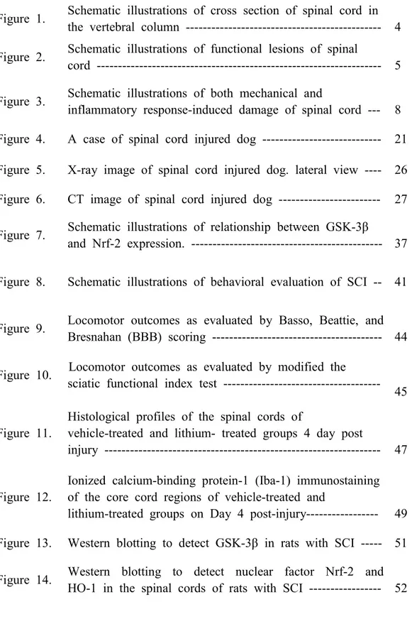
Spinal cord anatomyThe spinal cord is connected to the brain. It is the part of the central nervous system (CNS) located within the vertebral column, and it includes both sensory and motor nerves. It is also the main pathway for information between the brain and peripheral nervous system (Squire et al., 2013). It carries sensory information from the body and part of the head to the CNS via afferent fibers, and performs the initial processing of this information (Figure 1).
Figure 1. Schematic illustrations of cross section of spinal cord in the
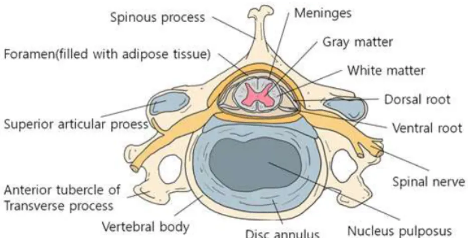
Axons of the motor neurons in the ventral horn project to the periphery to innervate skeletal and smooth muscles and mediate voluntary and involuntary reflexes. The spinal cord contains neurons whose descending axons mediate autonomic control of most visceral functions. The spinal cord is composed of central gray matter and peripheral whiter matter (Figure 2). Two systems are responsible for the transmission of motor function: the upper and lower motor neuron systems (Olby et al., 1999).
Figure 2. Schematic illustrations of functional lesions of spinal cord (Evans.
Classification of spinal cord injury
The spinal cord can be damaged by various primary injuries, including concussion (Vitale et al., 2007), shear (Griffin et al., 2009), laceration (Olby, 1999), intervertebral disk herniation (Griffin et al., 2009), vertebral injuries such as fractures, dislocations, and subluxations (Jeffery et al., 2010), penetrating injuries including gunshot and stab wounds (Dumont et al., 2001), and non-traumatic injuries including embolism (Abrhamson et al., 2002). Intervertebral disk herniation is classified into Hansen types Ⅰ (disk extrusion), Ⅱ (disk protrusion), and Ⅲ (nucleus pulposus extrusion) (Griffin et al., 2009, de Lahunta et al., 2009). Table 1 groups the various types of SCI.
Classification
Primary Injury
Concussion, Compression (Vitale et al. 2007)
Shear ( Griffin et al., 2009), Laceration ( Olby. 1999)
Contusion (Edward et al., 2012)
Intervertebral Disk Herniation
Hansen TypeⅠ -disk extrusion, Hansen TypeⅡ -disk protrusion
(Griffin et al., 2009)
Hansen TypeⅢ –nucleus pulposus extrusion
(De Lahunta et al., 2009)
Vertebral Injuries Fractures, Dislocations, Subluxations (Jeffery et al., 2010)
Penetrating Injuries Gunshot, Stab wounds (Dumont et al., 2001)
Non-traumatic Injuries Fibrocartilage emoli (Abrhamson et al., 2002)
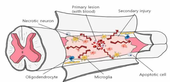
Irrespective of the primary injury type, secondary degeneration follows, leading to severe damage (Figure 3) (Tator et al., 1991; Olby, 1999, 2010; Dumont et al., 2001). The neuropathological outcome of SCI is characterized by edema, axonal degeneration, inflammatory cell infiltration, fibronectin exudation through the damaged blood–brain barrier, microglia activation, and reactive astrogliosis (Prewitt et al., 1997; Jung et al., 2003; Kim et al., 2003; Shin, 2007; Song et al., 2009).
Figure 3. Schematic illustrations of both mechanical and inflammatory
Models Methods
Contusion models Weight drop model (Basso et al., 1995) Weight drop impact sys.(Kim et al., 2017)
Compression models
Clip compression (Rivin et al., 1978) Balloon compression (Tarlov. 1953) Lamina to compress (Sun et al., 2017) Section models Transection (Basso et al., 1996)
Partial transection (Zeman et al., 1997)
Animal models of spinal cord injury
Many animal models have been developed to study the pathogenesis of SCI. Models based on mechanical impact include the New York University (NYU) impact device injury, Ohio State University (OSU) impact device injury, and clip-compression injury models (Kwon et al., 2002; Shin et al., 2013). Compression models include balloon compression (Tarlov, 1953) and lamina compression (Sun et al., 2017) models. Transection and partial transection have also been used to study SCI (Basso et al., 1996; Zeman et al., 1997). Basso et al. (1996) used a rat model of SCI in a preliminary study for the application of candidate therapeutic agents in dogs with SCI to study their therapeutic effects and mechanisms. Table 2 summarizes the animal models of spinal cord injury.
Occurrence of spinal cord injury in dogs and cats
In veterinary medicine, the incidence of spinal cord injury in dogs and cats is increasing, and emergency care is necessary for such injured animals. Because patients suffer from secondary insults after SCI, drugs are used to reduce neuronal cell death and glial activation. Opiate receptor antagonists and thyrotrophin-releasing hormones have been used to improve spinal cord blood flow (Faden et al., 1981, Pitts et al., 1989). The neuroprotective effects of minocycline (Cho et al., 2006) and anti-oxidant therapies (Hall, 1987; Gul et al., 2005; Kaplan, 2005; Baltzer et al., 2008) are being studied. Olfactory glial cells have been transplanted into the spinal cord (Jeffery et al., 2005). Table 3 summarizes SCI cases in dogs and cats and shows the clinical care and therapeutic agents used. In general, the therapeutic strategy aims to minimize secondary degeneration following SCI.
With the increase in SCI in dogs, case studies and therapeutic trials in rodent models are needed.
Type
Distinction
Initial Stabillization (Muir.1998) ABCs
(Airway, Breathing, Circulation) Prevention of Secondary Injury
(Platt et al., 2005)
Controversial use of methylprednisolone Calcium-Channel Antagonists
(Guha et al., 1985) Improve spinal cord blood flow 21-Aminosteroids (Hall. 1998) Alternative to methylprednisolone Opiate Receptor Antagonists
(Faden et al., 1981) Decrease spinal cord blood flow Thyrotrophin-Releasing Hormone
(Pitts et al., 1989) Improve spinal cord blood flow
Minocycline (Cho et al., 2006) Anti-inflammatory and neuroprotective effects
Intraspinal Olfactory Glial Transplantation
(Jeffery et al., 2005) Need further research
Anti-Oxidant Therapy
Vitamin E and selenium (Hall. 1987) Melatonin (Gul et al., 2005)
Resveratrol (Kaplan. 2005) N-actylcystenine (Baltzer et al., 2008)
Table 3. Classification of spinal cord injury and therapeutic substance in dog
Reference
Abrhamson C, Platt S, Stedman N. Tetraparesis in a cat with fibrocartilaginous emboli. J Am Anim Hosp Assoc 2002; 38(2): 153-157.
Baltzer W, McMichael M, Hosgood G et al. Randomized, blinded, placebo-controlled clinical trial of N-acetylcysteine in dogs with spinal cord trauma from acute intervertebral disc disease. Spine 2008; 33(13): 1397-1402.
Basso DM, Beattie MS, Bresnahan JC. A sensitive and reliable locomotor rating scale for open field testing in rats. J Neurotrauma 1995; 12: 1-21.
Basso DM, Beattie MS, Bresnahan JC. Graded histological and locomotor outcomes after spinal cord contusion using the NYU weight-drop device versus transection. Exp Neruol 1996; 139: 244-256.
Beattie MS. Inflammation and apoptosis: linked therapeutic targets in spinal cord injury. Trends Mol Med 2004; 10(12): 580-583.
Cho KO, La HO, Cho YJ et al. Minocyline attenuates white matter damage in a rat model of chronic cerebral hypoperfusion. J Neurosci Res 2006; 83(2):285-291.
De Lahunta A, Glass E. Small animal spinal cord disease, In: De Lahunta A, Glass E. eds. Veterinary Neuroanatomy and Clinical Neurology, 3rd edn. St. Louis, MO: Saunders 2009; 243-284.
Dumont R, Okonkwo D, Verma S, et al. Acute spinal cord injury, partⅠ: pathophysiologic mechanisms. Clin Neuropharm 2001; 24(5): 254-261.
Edward H, George A, Lisa M. Mechanisms of injury and emergency care of acute spinal cord injury in dogs and cats. J Vet Emerg Crit Care 2012; 22(2): 160-178.
Evans HE. Millers Anatomy of the Dog, 3th edn. Philadelphia: WB Saunders 1993.
Faden AI, Jacobs TP, Mougey E et al. Endorphins in experimental spinal cord injury: therapeutic effects of naloxone. Ann Neurol 1981; 10(4): 326-332.
Griffin J, Levine J, Kerwin S. Canine thoracolumbar intervertebral disk disease: pathophysiology, neurologic examination, and emergency medical therapy. Compend Contin Educ Pract Vet 2009; 31(3): E1-E13.
Guha A, Tator CH, Piper I. Increase in rat spinal cord blood flow with the calcium channel block, nimodipine. J Neurosurg 1985; 63(2): 250-259.
Gul S, Celik SE, Kalayci M et al. Dose-dependent neuroprotective effects of melatonin on experimental spinal cord injury in rats. Surg Neurol 2005; 64(4): 355-361.
Hall ED. Effects of the 21-aminosteroid U74006F on post-traumatic spinal cord ischemia in cats. J Neurosurg 1998; 68(3): 462-465.
Hall ED. Intensive anti-oxidant pretreatment retards motor nerve degeneration. Brain Res 1987; 413(1): 175-178.
Jeffery ND. Vertebral fracture and luxation in small animal. Vet Clin Small Anim 2010; 40(5): 809-828.
Jeffery N, Lakatos A, Franklin R. Autologous olfactory glial cell transplantation is reliable and safe in naturally occurring canine spinal cord injury. J Neurotrauma 2005; 22(11): 1282-1293.
Jung K, Min DS, Sim KB, Ahn M, Kim H, Cheong J, Shin T. Upregulation of phospholipase D1 in the spinal cords of rats with clip compression injury. Neurosci Lett 2003; 336: 126-130.
Kaplan S, Bisleri G, Morgan JA, Cheema FH, Oz MC. Resveratriol, a natural red wine polyphenol, redudes ischemia-reperfusion induced spinal cord injury. Ann Thorac Surg 2005; 80(6): 2242-2249.
Kim DH, Heo SD, Ahn MJ, Sim KB, Shin TK. Activation of embryonic intermediate filaments contributes to glial scar formation after spinal cord injury in rats. J Vet Sci 2003; 4: 109-112.
Kim H, Kim JW, Hyun JK, Pack I. Multimodal sensor-based weight drop spinal cord impact system for large animals. Spine J 2017; 9430(17): 30920-30928.
Kwon, B.K., Oxland, T.R., and Tetzlaff, W. (2002). Animal models used in spinal cord regeneration research. Spine (Phila Pa 1976) 27, 1504-1510.
Muir W. Shock. Compend Contin Educ Pract Vet 1998; 20(5): 549-563.
Olby N. Current concepts in the management of acute spinal cord injury. J Vet Intern Med 1999; 13(5): 399-407.
Olby N. The pathogenesis and treatment of acute spinal cord injuries in dogs. Vet Clin Small Anim 2010; 40(5): 791-807.
Pitts LH, Ross A, Chase GA et al. TRH analog YM-14673 improves outcome fellowing traumatic brain and spinal cord injury in rats: dose-response studies. Brain Res 1989; 486(2): 228-235.
Platt S, Abramson C, Garosi L. Administering corticosteroids in neurologic disease. Compend Contin Educ Pract Vet 2005; 210-220.
Prewitt CM, Niesman IR, Kane CJ< Houle JD. Activated macrophage/microglial cells can promote the regeneration of sensory axons into the injured spinal cord. Exp Neurol 1997; 148(2): 433-443.
Profyris C, Cheema SS, Zang D, Azari MF< Boyle K, Petratos S. Degenerative and regenerative mechanises governing spinal cord injury. Neurobiol Dis 2004; 15(3): 415-436.
Rivin AS, Tator CH. Effect of duration of acute spinal cord compression in a new acute model in the rat. Surg Neurol 1978; 10: 39-43.
Shin T, Ahn M, Moon C, Kim S, Sim KB. Alternatively activated macrophages in spinal cord injury and remission: another mechanism for repair? Mol Neurobiol 2013; 47: 1011-1019.
Squire, Larry Squire. Fundamental neuroscience (4th ed.). Amsterdam. Elsevier/Academic Press 2013; p.628.
Sun GD, Chen Y, Zhou ZG, Yang SX, Zhong C, Li ZZ. A progressive compression model of thoracic spinal cord injury in mice: function assessment and pathological changes in spinal cord. Neural Reqen Res 2017; 12(8): 1365-1374.
Tarlov IM, Klinger H, Vitale S. Spinal cord compression studiesⅠ. Experimental techniques to produce acute and gradual compression. AMA Archiv neurol Psych 1953; 70: 813-819.
Tator CH, Fehlings MG. Review of the secondary injury theory of acute spinal cord trauma with emphasis on vasculamechanisms. J Neurosurg 1991; 75(1): 15-26.
Vitale C, Coates J. Acute spinal cord injury. Stand Care: Emerg Crit Care Med 2007; 9.7:1-11
Zeman RJ, Zhang Y, Etlinger JD. Clenbuterol, a beta2-adrenoceptor agonist, reduces scoliosis due to partical transection of rat spinal cord. Am J Physiol 1997; 272(4 Pt 1): E712-E715.
CHAPTER I
1. Abstract
A 4-year-old male miniature poodle developed chronic progressive paraplegia of the hind limbs following an accident 2 years prior to presentation for treatment. All hematological parameters, including the complete blood count, were normal.
Neurological examination revealed that the dog was ambulatory, with severe ataxia of the hindlimbs. Radiological examination showed a fracture of the 10ththoracic vertebra in the lateral view. The bladder was expanded, and the gas shadow in the stomach was elevated. Computed tomography revealed a fracture in the 10th thoracic vertebra in the sagittal and coronal views, and lack of continuity of the thoracic vertebrae.
We diagnosed complete transection of the spinal cord, and postulated that connective tissue injury in the damaged spinal cord was followed by permanent paralysis. The hematological parameters were in the normal range, although there might have been transient changes immediately after the spinal cord injury (SCI). A therapeutic trial is recommended only within a few days after SCI in cases with potential for improvement.
Key words: paraplegia, Computed tomography, Fracture, Spinal cord
2. Introduction
Spinal cord injury (SCI) can result in complete or incomplete damage and can compromise the major functions of the spinal cord (e.g., proprioception, motor, nociception, and reflexes) (Edward et al., 2012). The principle mechanical forces involved in acute traumatic injury are concussion and compression (Vitale and Coates, 2007), shear (Griffin et al., 2009), laceration (Olby, 1999), and distraction (Dumont et al., 2001). Acute intervertebral disk herniation is a common acute SCI seen primarily in canine patients. The types of hernia are disk extrusion, disk protrusion (Griffin et al., 2009), and nucleus pulposus extrusion (De Lahunta et al., 2009). However, the most common cause of paraplegia in clinical practice is traumatic SCI, such as with fractures.
Spinal cord injury is characterized by both mechanical and inflammatory response-induced damage (Jung et al., 2003; Ahn et al., 2015) (Figure 3). The mechanical forces that affect the spinal cord at the time of injury may cause immediate tissue bursting. The neuropathological features of SCI include edema, axonal degeneration, inflammatory cell infiltration, and fibronectin exudation through the damaged blood–brain barrier (Jung et al., 2003; Shin et al., 2013). This report describes a case of SCI in a dog.
3. Case description of spinal cord injury in a dog
Case history
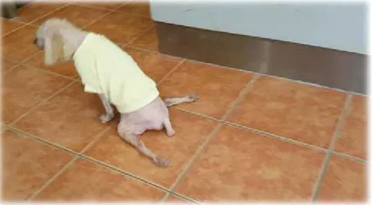
A 4-year-old male miniature poodle was evaluated because of chronic progressive paraplegia of the hind limbs (Figure 4). The dog had been in an accident 2 years earlier. Its body temperature was 38.7℃, and heart rate was 130 beats/minutes. This case was diagnosed based on the clinical signs and computed tomography (CT) obtained at Jeju National University Animal Hospital.
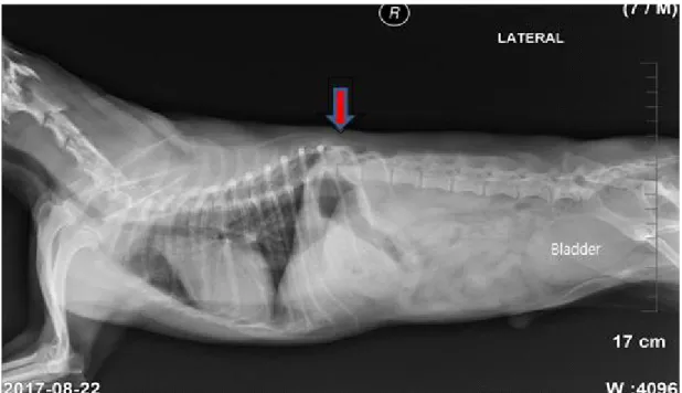
Figure 4. A case of spinal cord injured dog.
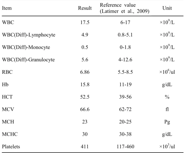
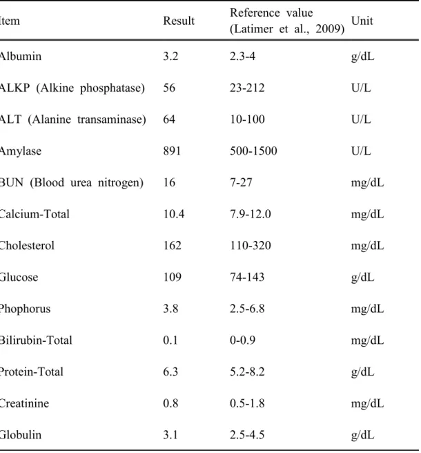
Hematological analysis and urinalysis
blood cell count (WBC) 17.5 × 10⁹/L and monocytes 0.5×10⁹/L (Table4). Biochemistry included normal blood urea nitrogen (BUN)16mg/dL, phosphorus 3.8mg/dL, and creatinine 0.8mg/dL. The specific gravity of the urine was 1.035, which was lower than normal. The urine sediments after centrifugation were normal.
Neurological examination
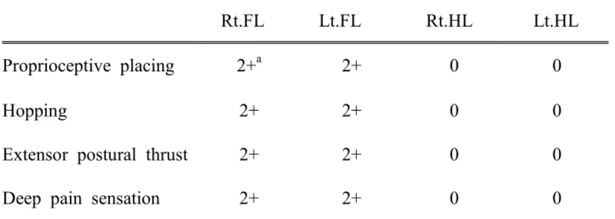
Neurological examination revealed that the dog was ambulatory, with severe ataxia of the hindlimbs. Proprioception was decreased in both hindlimbs, although the bilateral spinal reflexes were unremarkable. In hopping and extensor postural thrust, the forelimbs showed a normal response (2+), while the hindlimbs did not respond (0). Deep pain sensation was detected during palpation of the lumbar portion of the vertebral column (Table 6).
Radiological examination
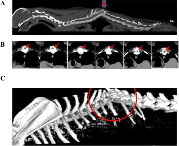
The lateral view showed a fracture of the 10ththoracic vertebra, with signs of ossification. The bladder was expanded, and the gas shadow in the stomach was elevated (Figure 5, arrow). CT revealed a fracture of the 10ththoracic vertebra in the sagittal and coronal views (Figure 6A, arrow), lack of continuity of the thoracic vertebrae, and a spinal cord shadow in the thoracic vertebral cavity (Figure 6B, arrow). There was severe torsion around the 10ththoracic vertebra (Figure 6C, circle).
Table 4. Hematological changes of spinal cord injured dog.
Item Result Reference value
(Latimer et al., 2009) Unit
WBC 17.5 6-17 ×109/L WBC(Diff)-Lymphocyte 4.9 0.8-5.1 ×10⁹/L WBC(Diff)-Monocyte 0.5 0-1.8 ×10⁹/L WBC(Diff)-Granulocyte 5.6 4-12.6 ×10⁹/L RBC 6.86 5.5-8.5 ×10⁶/ul Hb 15.8 11-19 g/dL HCT 52.5 39-56 % MCV 66.6 62-72 fl MCH 23 20-25 Pg MCHC 30 30-38 g/dL Platelets 411 117-460 ×103/ul
WBC: white blood cells, Diff: differential count, RBC: red blood cell, Hb: hemoglobin, HCT: hematocrit, MCV: mean corpuscular volume, MCH: mean corpuscular hemoglobin, MCHC: mean corpuscular hemoglobin concentration
Table 5. Serum chemistry profile of spinal cord injured dog.
Item Result Reference value
(Latimer et al., 2009) Unit
Albumin 3.2 2.3-4 g/dL
ALKP (Alkine phosphatase) 56 23-212 U/L ALT (Alanine transaminase) 64 10-100 U/L
Amylase 891 500-1500 U/L
BUN (Blood urea nitrogen) 16 7-27 mg/dL
Calcium-Total 10.4 7.9-12.0 mg/dL Cholesterol 162 110-320 mg/dL Glucose 109 74-143 g/dL Phophorus 3.8 2.5-6.8 mg/dL Bilirubin-Total 0.1 0-0.9 mg/dL Protein-Total 6.3 5.2-8.2 g/dL Creatinine 0.8 0.5-1.8 mg/dL Globulin 3.1 2.5-4.5 g/dL
Table 6. Neurological evaluation of spinal cord injured dog.
Rt.FL Lt.FL Rt.HL Lt.HL
Proprioceptive placing 2+a 2+ 0 0
Hopping 2+ 2+ 0 0
Extensor postural thrust 2+ 2+ 0 0
Deep pain sensation 2+ 2+ 0 0
Rt: Right, Lt: left, FL: forelimb, HL: hind limb
a
Absent 0, No reflex movement; Reduced 1+, Slow, decreased, reduced movement; Normal 2+, Brisk reflex action followed by relaxation; Increased 3+, Exaggerated, larger, brisker, reflex action; Clonus 4+, Increased, repetitive, sustained reflex action (Susan, 2010).
A
B
C
4. Treatment
Because the caregiver’s intention was symptomatic treatment rather than active treatment, eczema dermatitis treatment was performed, and lactulose 0.5 mL/kg (twice a day orally) was prescribed for smooth defecation. The caregiver was taught to perform bladder compression to prevent uremia.
5. Discussion
In most animals with spinal disease, the hematological analysis is unremarkable, although leukocytosis is a common finding (Nicholas and Simon, 2005), as seen in this case.
Paraplegia due to a vertebral fracture has been reported in dogs (Fitzmaurice, 2010). In this case, proprioceptive placing, hopping, extensor postural thrust, and deep pain sensation were normal in the forelimbs, while the hindlimbs were unresponsive. The spinal cord injury was caused by a thoracic fracture, which caused paraplegia. The radiological examination confirmed that the 10th thoracic vertebra was fractured, and we speculated that the fracture caused the paraplegia. Furthermore, the fracture caused bladder expansion due to the loss of reflexes for defecation and urination.
Following a vertebral fracture or chronic spinal cord injury, time is required for the growth of new tissues to protect the nerves in the spinal cord. In this case, there was no emergency care, diagnosis, or appropriate treatment at the time of the SCI.
Reference
Ahn M, Moon C, Park C, Kim J, Sim KB, Shin T. Transient activation of an adaptor protein, disabled-2, in rat spinal cord injury. Acta Histochem 2015; 117: 56-61.
Basso DM, Beattie MS, Bresnahan JC. Graded histological and locomotor outcomes after spinal cord contusion using the NYU weight-drop device versus transection. Exp Neruol 1996; 139: 244-256.
De Lahunta A, Glass E. Small animal spinal cord disease, In: De Lahunta A, Glass E. eds. Veterinary Neuroanatomy and Clinical Neurology, 3rd edn. St. Louis, MO: Saunders 2009; 243-284.
Dumont R, Okonkwo D, Verma S, et al. Acute spinal cord injury, partⅠ: pathophysiologic mechanisms. Clin Neuropharm 2001; 24(5): 254-261.
Edward ML. Hyperbaric oxygen therapy. partⅠ: history and principles. J Vet Emerg Crit Care 2010; 20(3): 284-288.
Fitzmaurice N. Small animal neurology. Saunders 2010; p21, p181-183.
Griffin J, Levine J, Kerwin S. Canine thoracolumbar intervertebral disk disease: pathophysiology, neurologic examination, and emergency medical therapy. Compend Contin Educ Pract Vet 2009; 31(3): E1-E13.
Jung K, Min DS, Sim KB, Ahn M, Kim H, Cheong J, Shin T. Upregulation of phospholipase D1 in the spinal cords of rats with clip compression injury. Neurosci Lett 2003; 336: 126-130.
Hall ED. Effects of the 21-aminosteroid U74006F on post-traumatic spinal cord ischemia in cats. J Neurosurg 1998; 68(3): 462-465.
Latimer KS, Mahaffery EA, Prasse KW. Duncan and Prasses Veterinary Laboratory Medicine: clinacal pathology. BlackwellPublishing 2009.
Nicholas JH, Simon J. Small Animal Spinal Disorders. Mosby 2005; p43.
Olby N. Current concepts in the management of acute spinal cord injury. J Vet Intern Med 1999; 13(5): 399-407.
Shin T, Ahn M, Moon C, Kim S, Sim KB. Alternatively activated macrophages in spinal cord injury and remission: another mechanism for repair? Mol Neurobiol 2013; 47: 1011-1019.
Vitale C, Coates J. Acute spinal cord injury. Stand Care: Emerg Crit Care Med 2007; 9.7:1-11
CHAPTER II
Therapeutic trials of lithium, an inhibitor of
glycogen synthase-3β in rat spinal cord injury
1. Abstract
Glycogen synthase kinase (GSK)-3β and related enzymes are associated with various forms of neuroinflammation, including spinal cord injury (SCI). Our aim was to evaluate whether lithium, a non-selective inhibitor of GSK-3β, ameliorated SCI progression, and also to analyze whether lithium affected the expression levels of two representative GSK-3β-associated molecules, nuclear factor erythroid 2-related factor 2 (Nrf-2) and heme oxygenase 1 (HO-1) (a target gene of Nrf-2). Intraperitoneal lithium chloride (80 mg/kg/day for 3 d) significantly improved locomotor function at 8 d post-injury (DPI); this was maintained until 14 DPI (p < 0.05). Western blotting showed significantly increased phosphorylation of GSK-3β (Ser9), Nrf-2, and the Nrf-2 target HO-1 in the spinal cords of lithium-treated animals. Fewer neuropathological changes (e.g., hemorrhage, inflammatory cell infiltration, and tissue loss) were observed in the spinal cords of the lithium-treated group compared with the vehicle-treated group. Microglial activation (evaluated by measuring the immunoreactivity of ionized calcium-binding protein 1 [Iba-1]) was also significantly reduced in the lithium-treated group. These findings suggest that GSK-3β becomes activated after SCI, and that a non-specific enzyme inhibitor, lithium, ameliorates rat SCI by increasing phosphorylation of GSK-3β and the associated molecules Nrf-2 and HO-1.
Key words: spinal cord injury, lithium, glycogen synthase kinase 3β,
2. Introduction
Spinal cord injury (SCI) is characterized by both mechanical and inflammatory response-induced damage (Jung et al., 2003; Ahn et al., 2015) (Figure 3). The mechanical forces that impact the spinal cord at the time of injury may cause immediate tissue bursting (Kwon et al., 2004). There are a variety of animal models of spinal cord injury, including those using weight-drop impact devices (Shin et al., 2013), the transection method (Donnelly and Popovich, 2008), and clip-compression injuries (Grimes and Jope, 2001; De et al., 2002; Ahn et al., 2017).
Clip compression, which results in the progressive recovery of hindlimb paralysis in rats (Fang et al., 2016), is regarded as a manual injury technique for spinal cords in the absence of digital analysis systems, such as the Multicenter Animal Spinal Cord Injury Study (MASCIS) impactor (Young, 2009). The neuropathological features of SCI include edema, axonal degeneration, inflammatory cell infiltration, and exudation of fibronectin through the damaged blood–brain barrier (Jung et al., 2003; Shin et al., 2013), Activated neutrophils, microglia, and macrophages synthesize free radicals that trigger apoptosis of neurons and glia by irreversibly oxidizing polyunsaturated fatty acids, proteins, and DNA (Donnelly and Popovich, 2008). An increase in the cellular antioxidant capacity or purging of reactive oxygen species (ROS) can protect against SCI.
Lithium, a non-selective inhibitor of glycogen synthase kinase (GSK)-3β, has been used as a mood stabilizer in patients with bipolar disorder, and possibly acts as a neuroprotectant (Grimes and Jope, 2001). Lithium has also been used to ameliorate chronic experimental autoimmune encephalomyelitis (EAE) in mice expressing anti-myelin oligodendrocyte glycoprotein (MOG) antibodies (De et al., 2002) and acute monophasic EAE in the rat (Ahn et al., 2017). In addition, lithium is used to protect neurons after SCI, reducing post-injury inflammation (Fang et al., 2016). Lithium promotes the production and release of neurotrophins, stimulates neurogenesis, enhances autophagy, and inhibits apoptosis (Young, 2009). However, any neuroprotective effect of lithium in terms of activating antioxidative systems, such as the nuclear factor erythroid 2-related factor (Nrf)-2/heme oxygenase (HO)-1 mechanism requires further study in models of SCI.
Oxidative stress-induced cell damage is attributable to an imbalance between reactive oxygen free radical production and the efficacy of the anti-oxidant system (Pratheeshkumar et al., 2014). An increase in cellular antioxidant capacity or ROS removal can ameliorate various diseases and injuries, including SCI (Jiang et al., 2016). Nrf-2 and its downstream target (HO-1), along with other antioxidant enzymes including superoxide dismutase and glutathione peroxidase play important roles in protecting various tissues and cells against oxidative stress both by regulating the expression levels of cytoprotective and antioxidant genes (Jiang et al., 2016) and reducing inflammation (Ahn et al., 2016). Normally, Nrf-2 is associated with the Kelch like-ECH-associated protein 1 (Keap-1) in the cytoplasm; upon stimulation,
transcription of various phase II and/or antioxidant enzyme genes (Chen et al., 2006). Targeting of Nrf-2/HO-1 after SCI suppresses oxidative stress and exerts a neuroprotective effect (Lv et al., 2015). However, any relationship between GSK-3β and Nrf-2 expression in the SCI rat model remains unclear.
In the present study, we investigated the neuroprotective effects of lithium, a non-selective inhibitor of GSK-3β, in rats where SCI was induced by clip compression. We explored the underlying protective molecular mechanisms via semi-quantitative analysis of Nrf-2 and HO-1 levels (Figure 4).
Figure 7. Schematic illustrations of relationship between GSK-3β and Nrf-2
3. MATERIALS AND METHODS
Animals
We used female Sprague–Dawley rats (200–250 g, 7–8 weeks of age) (OrientBio Inc., Kyunggido, Korea). All experimental procedures were conducted in accordance with the Guidelines for the Care and Use of Laboratory Animals of Jeju National University. The animal protocols also conformed to current international laws and policies national institutes of health (NIH) Guide for the Care and Use of Laboratory Animals, NIH Publication No. 85-23, 1985, revised 1996). Every effort was made to minimize the number of animals used and their suffering.
Surgical procedures
Clip compression injury was inflicted using a modification of previously published methods (Jung et al., 2003; Kim et al., 2003; Ahn et al., 2012). Animals were anesthetized via intramuscular injection of Zoletil®50 (Virbac,France) and subjected to laminectomy at T9/T10. Immediately there after, the spinal cord was compressed with a vascular clip (Stoelting, WoodDale, IL, USA) applied vertically to the exposed spinal cord at a nocclusion pressure of 15–20g for 1min. After compression, the muscles and skin layers were closed. Sham-operated control rats under went laminectomy only. Spinal cord tissues from the surgical sites were harvested either fixed in 4%(v/v) paraformaldehyde in phosphate-buffered saline (PBS, pH7.2) for histological examination or storedat−80°C prior to Western blot analysis.
Antibodies
We used a rabbit anti-Iba-1 (Wako Pure Chemical Industries, Ltd., Osaka, Japan) antibody to immunohistochemically stain the ramified microglia and macrophages of rat spinal cords, and for Western blotting. We employed monoclonal rabbit anti-phospho-GSK-3β (Ser9) (p-GSK-3β) and monoclonal rabbit anti-GSK-3β antibodies (Cell Signaling Technology, Beverly, MA, USA) to detect GSK-3β. Rabbit polyclonal anti-Nrf-2 and -HO-1 antibodies (Santa Cruz Biotechnology, Santa Cruz, CA) were used to stain the respective proteins. We also employed a mouse monoclonal anti-β-actin antibody (Sigma-Aldrich, St. Louis, MO, USA).
Lithium treatment
To assess the effects of lithium on SCI, rats were divided into the following three treatment groups (10 animals/group): sham control, vehicle, and lithium. To rapidly elevate the lithium level, the first dose of lithium chloride (Sigma-aldrich, USA) (80 mg/kg/day) was intraperitoneally injected into the lithium-treated group 30 min after surgery; identical doses were given on each of the next 3 d. Lithium is nontoxic to rats at this level; the serum levels are equivalent to those in human patients (De et al., 2002). As in our previous study, serum lithium concentrations were measured using a lithium assay kit (catalog number LI01ME; MG Metallogenics, Chiba, Japan) (Ahn et al., 2017). The body weights and behavioral features of all rats were checked
Behavioral tests and histological examination
Locomotor function after SCI was examined using the Basso, Beattie, and Bresnahan (BBB) rating scale (Figure 5A and Table 3) and modified the sciatic functional index test (Figure 5B). All evaluations were performed in a double-blinded manner; average scores were calculated for each group and used to compare the severity of hind-limb paralysis.
Spinal cords collected at various time points (0, 4, 7, and 14 d post-injury [DPI]) were perfused with 4% (v/v) paraformaldehyde in PBS, pH 7.2. The T8–T10 regions of the spinal cords, including the sites of injury, were collected and post-fixed in 10% (v/v) neutral-buffered formalin for 48 h. Subsequently, sagittal sections (5 µm thick) were stained with hematoxylin and eosin (H&E).
Figure 8. Schematic illustrations of behavioral evaluation of SCI. (A) BBB
(Basso-Beattie-Bresnahan) Scoring ( Basso et al., 1996) and (B) sciatic functional index test (Luis et al., 1982)
Immunohistochemistry
To assess early responses to treatment, we compared the microglial features of the vehicle- and lithium-treated groups at 4 DPI, as microglial reactions are prominent within the first week after SCI (Koshinaga and Whittemore, 1995; Dusart and Schwab, 1994; Loane and Byrnes, 2010). We immunostained the spinal cord for Iba-1 (a marker of activated cord microglia and macrophages) as described previously (Ahn et al., 2016). Briefly, after incubation with matched blocking serum (10% [v/v] normal goat serum in PBS; Vectastain® Elite ABC kit; Vector Laboratories, Burlingame, CA, USA), the samples were incubated with rabbit anti-Iba-1 (Iba-1; 1:800; Wako Pure Chemical Industries, Ltd. Osaka, Japan) for 1 h at room temperature (RT). After three washes in PBS, we proceeded as recommended by the manufacturer; the peroxidase reaction was developed using a diaminobenzidine substrate kit (Vector, USA).
Western blot analysis
We performed Western blotting as described previously (Kim et al., 2009). Briefly, spinal cord tissue was homogenized in TNN lysis buffer containing protease and phosphatase inhibitors (1 mM Na3VO4, 1mM phenyl
methane sulphonyl fluoride, 10μg/mL aprotinin, 10μg/mL leupeptin), centrifugedat 10,900g for 20 min at 4°C, and the supernatant was harvested. The cytosolic and nuclear fractions were separated using NE-PER® Nuclear and Cytoplasmic Extraction Reagents as recommended by the manufacturer (Thermo Scientific, Rockford, IL, USA). Proteins (40 μg) were subjected to
10% (w/v) sodium dodecyl (or lauryl) sulfate polyacrylamide gel electrophoresis (SDS-PAGE or SLS-PAGE) and transferred to nitrocellulose membranes (Schleicher and Schuell, Keene, NH, USA). The membranes were blocked by incubation with 5% (v/v) skim milk in Tris-buffered saline for 1 h and then incubated with primary antibodies (anti-p-GSK-3β, 1:1,000 dilution; anti-GSK-3β, 1:1,000 dilution; anti-Nrf-2, 1:1,000 dilution; and anti-HO-1, 1:1,000 dilution) for 2 h. After washing, the membranes were incubated with the appropriate secondary antibodies for 1 h. Bound antibodies were detected using a chemiluminescent substrate (in the WEST-one™ kit; iNtRON Biotech, Gyeonggi, Korea) according to the manufacturer’s instructions. After imaging, the membranes were stripped and reprobed using an anti-β-actin antibody (1:10,000 dilution). The optical density (OD)/mm2 of each band was measure d using Image J soft ware (NIH, Bethesda, MD, USA). To detect Iba-1,we used the Wes™ system (Proteinsimple, California, USA) as instructed by the simple western user manual (Chen et al., 2013). All electrophoresis and immunoblotting steps were performed using a fully automated capillary system.
Statistical analysis
All measurements are averages of three independent experiments. All values are presented as means ± standard error of the mean (SE). The results were analyzed using a one-way analysis of variance (ANOVA) followed by Student–Newman–Keuls post hoc testing for multiple comparisons. A p-value < 0.05 was considered to reflect statistical significance.
4. Result
Lithium-mediated behavioral changes
Locomotor function began to recover commencing on 3 DPI. By 8 DPI, the BBB scores was significantly higher in the lithium-treated group (10.4 ± 0.52, p < 0.05) compared with those of the vehicle-treated group (6.78 ± 0.55); the improvements were maintained until 14 DPI (Figure 7).
The foot printed images that the modified the sciatic functional index test showed very sliding foot image in both groups to 1 DPI. on day 3 post-injury, the images in the lithium-treated group was enhancer than that in the vehicle-treated group. This difference persisted until 14 DPI (Figure 8).
Figure 9. Locomotor outcomes as evaluated by Basso, Beattie, and Bresnahan
(BBB) scoring (n = 5/daily). The BBB scores were very low in both groups to 3 DPI; on Day 8 post-injury, the BBB score in the lithium-treated group was significantly higher than that in the vehicle-treated group. This difference persisted until 14 DPI. *p < 0.05 vs. vehicle-treated group.
After injury 1day 3day 7day 14day
Vehicle
Lithium
Figure 10. Locomotor outcomes as evaluated by modified the sciatic
functional index test (n = 5/daily). The foot printed images were very sliding in both groups to 1 DPI. On day 3 post-injury, the images in the lithium-treated group was enhanced than that in the vehicle-treated group. This difference persisted until 14 DPI.
Histological findings
The sham-operated group exhibited no mechanical change in the core region of the spinal cord (data not shown), as found previously (Jung et al., 2003; Ahn et al., 2012). The cords of vehicle-treated rats exhibited reduced cellularity and edema in longitudinal sections of core lesions (Fig. 1A). Inflammatory cells had infiltrated by 4 DPI (Fig. 2C). In contrast, severe edema and hemorrhage were evident in the lithium-treated group (Fig. 2B). Additionally, accumulation of round-type inflammatory cells (Fig. 2G, arrowheads); activated microglia; and small, round vacuoles were evident in the core regions of spinal cords of the lithium-treated group by 4 DPI (Fig. 2D).
Figure 11. Histological profiles of the spinal cords of vehicle-treated (A and
C) and lithium- treated groups (B and D) 4 DPI. (A and B) Low-magnification images of sagittal sections. (C and D) High-magnification images of the squares in A and B, respectively. A–D, hematoxylin and eosin staining. Scale bars: 100 μm.
Microglial reactions and infiltration of inflammatory cells
To assess microglial reactions and inflammatory cell infiltration, we immunohistochemically stained for Iba-1 and used a simple system to quantify the protein levels. Iba-1-positive microglial cells and macrophages were evident in all cord regions, including the white and gray matter (Fig. 3). Figure 3C illustrates a representative experiment; the data are displayed in pseudo-gel and electropherogram formats. By 4 DPI, the Iba-1 level in the lithium-treated group (25.78 ± 9.99%, relative OD) was significantly less than that in the vehicle-treated group (50.93 ± 6.39%, relative OD; p < 0.05 ) (Fig. 3C).
Figure 12. Ionized calcium-binding protein 1 (Iba-1) immunostaining of the
core cord regions of vehicle-treated (A) and lithium-treated groups (B) on Day 4 post-injury. Iba-1-positive microglial cells were evident in all regions, including the white and gray matter. Ramified microglial cells and many inflammatory cells immunostained for Iba-1 in the core cord regions of both groups. Bar graphs: semi-quantitative analysis of Iba-1 levels (the ~17 kDa protein) using a simple Western blotting system. Normalization was achieved by reprobing the membranes with an anti-β-actin antibody. Means ± SE (n = 5 per group) are shown. *p < 0.05 vs. the vehicle-treated group. Scale bars: 100 μm.
Lithium-mediated modulation of the levels of GSK-3β, Nrf-2, and HO-1 in the spinal cords of injured rats
We evaluated the GSK-3β phosphorylation status via Western blotting to explore whether lithium inhibited GSK-3 activity in the spinal cords of injured rats (n = 5 per group). Lithium significantly increased the p-GSK-3β expression level (relative OD values, 2.92 ± 0.44-fold; p < 0.05) compared with vehicle (1.00 ± 0.13-fold) at 4 DPI (Fig. 4A). We performed Western blotting to determine whether lithium influenced Nrf-2 levels in the cytosol and nucleus. Lithium significantly increased both the cytoplasmic Nrf-2 level (4.54 ± 1.76-fold, relative OD/mm2, p < 0.05) compared with the vehicle-treated group (0.21 ± 0.04-fold) and also the extent of nuclear translocation (3.23 ± 1.08-fold, p < 0.05) compared with the vehicle-treated group (0.14 ± 0.04-fold) (Fig. 5A). In addition, the HO-1 protein level in the lithium-treated group was significantly greater (3.42 ± 0.46-fold, p < 0.01) than that in the vehicle-treated group (1.00 ± 0.26-fold) (Fig. 5B), reflecting enhanced Nrf-2 expression and translocation in SCI rats given lithium.
Figure 13. Western blotting to detect glycogen synthase kinase (GSK)-3β in
rats with spinal cord injury (SCI). (A) Representative immunoblots of phosphorylated GSK (p-GSK)-3β (Ser9), total GSK-3β (~46 kDa), and β-actin (~45 kDa). Bar graphs: The p-GSK-3β level increased significantly in the spinal cords of lithium-treated rats. To quantify GSK-3β phosphorylation, the levels of the phosphorylated form were normalized to those of total GSK-3β. Normalization was achieved by reprobing the membranes with an anti-β-actin antibody. Means ± SEM (n = 5 per group) are shown. *p < 0.05 vs. the lithium-treated group.
Figure 14. Western blotting to detect nuclear factor erythroid 2-related factor
(Nrf)-2 and heme oxygenase (HO)-1 in the spinal cords of rats with SCI. (A and B) Representative immunoblots of Nrf-2 (~57 kDa), HO-1 (~32 kDa), and β-actin (~45 kDa). Bar graphs: Both the Nrf-2 and HO-1 levels increased significantly and Nrf-2 was translocated from the cytoplasm to the nucleus in the spinal cords of lithium-treated rats. Normalization was achieved by reprobing the membranes with an anti-β-actin antibody. Means ± SEM (n = 5 per group) are shown. *p < 0.05 vs. the lithium-treated group.
5. Discussion
Many types of spinal cord injury models have been developed using rodents (Shin et al., 2013). To obtain reliable data associated with impact power, computerized data analyses have been applied using the New York University impact device (Vijayaprakash and Sridharan, 2013), Ohio State University impact device (Koshinaga and Whittemore, 1995), and MASCIS impactor (Dusart and Schwab, 1994). Alternatively, for the present study, a clip compression technique was employed to induce spinal cord injury because there is a progressive improvement in BBB scores in rats following the use of this technique (Ahn et al., 2015; Kim et al., 2009; Basso et al., 2006). Additionally, the clip compression technique is an alternative choice for the induction of spinal cord injury in the absence of digital analysis systems, such as MASCIS (Vijayaprakash and Sridharan, 2013).
We first showed that lithium exerted significant anti-inflammatory and anti-oxidative effects by inhibiting inflammatory cell infiltration, microglial activation, and Nrf-2 translocation (thus enhancing HO-1 synthesis), significantly improving functional recovery in rats. Earlier, we showed that lithium treatment reduced the extent of the Iba-1-positive area of the spinal cord, and reduced the serum TNF-α level, in an experimental rat model of EAE (Ahn et al., 2017). Lithium suppresses both the level of circulating pro-inflammatory mediators and the number of CNS microglial cells, and enhances locomotor function in rats with SCI. A previous report indicated that GSK-3β, a negative regulator of Nrf-2, influenced the relative Nrf-2 proportions in the cytosol and nucleus (Dusart and Schwab, 1994). Here, we
accumulation, thus activating HO-1, in rats with SCI.
Lithium chloride reduces the disruption in the blood-spinal cord barrier and promotes the recovery of neurological function after SCI. This occurs partly due to decreases in the activation of endoplasmic reticulum stress, which plays an important role in SCI by inhibiting GSK-3β activation (Loane and Byrnes, 2010). In the present study, lithium inhibited GSK-3β phosphorylation and thus may have reduced inflammation in the spinal cord samples subjected to clip compression. Additionally, the regulation of GSK-3β activity is generally mediated by phosphorylation of the amino-terminal domain (at Ser9) by any of several kinases, including Akt, protein kinase A, and/or protein kinase C, which inactivate the enzyme (Kim et al., 2009). Furthermore, toll-like receptors mediate GSK-3β phosphorylation at Ser9 via the regulation of pro- and anti-inflammatory cytokines (Chen et al., 2013). Thus, it is possible that the inhibition of GSK-3β relieves the clip compression SCI in rats.
Various drugs exert antioxidative neuroprotective activities in the spinal cord by activating Nrf-2 such as asiatic acid, rosmarinic acid and resveratrol (Jiang et al., 2016; Shang et al., 2017; Wang et al., 2016; Kesherwani et al., 2013). In Wistar rats, carnosol protects against SCI-induced oxidative stress and inflammation by modulating nuclear factor-κB, cyclooxygenase-2, and Nrf-2 levels (Wang et al., 2016). Several drugs associated with Nrf-2 activation have been used to treat rat SCI; however, this is the first report to show that lithium-mediated inhibition of GSK-3β protects against SCI by regulating Nrf-2 translocation and subsequent activation of the HO-1 target gene. Recent evidence indicates that GSK-3β plays a critical role in regulating and degrading Nrf-2 in a Keap1-independent manner (Rada et al., 2011).
Furthermore, it was postulated that lithium exerts neuroprotective effects via the activation of Nrf-2 in spinal cord-injured rats.
In conclusion, our findings suggest that lithium ameliorates rat paralysis caused by SCI, and that the molecular mechanism involves inhibition of GSK-3β, increased nuclear translocation of Nrf-2, and subsequent upregulation of HO-1.
References
Ahn M, Kim J, Bang H, Moon J, Kim GO, Shin T. Hepatoprotective effects of allyl isothiocyanate against carbon tetrachloride-induced hepatotoxicity in rat. Chem Biol Interact 2016; 254: 102-108.
Ahn M, Kim J, Park C, Cho J, Jee Y, Jung K, Moon C, Shin T. Potential involvement of glycogen synthase kinase (GSK)-3beta in a rat model of multiple sclerosis: evidenced by lithium treatment. Anat Cell Biol 2017; 50: 48-59.
Ahn M, Lee C, Jung K, Kim H, Moon C, Sim KB, Shin T. Immunohistochemical study of arginase-1 in the spinal cords of rats with clip compression injury. Brain Res 2012; 1445: 11-19.
Ahn M, Moon C, Park C, Kim J, Sim KB, Shin T. Transient activation of an adaptor protein, disabled-2, in rat spinal cord injury. Acta Histochem 2015; 117: 56-61.
Basso DM, Beattie MS, Bresnahan JC. Graded histological and locomotor outcomes after spinal cord contusion using the NYU weight-drop device versus transection. Exp Neruol 1996; 139: 244-256.
Basso DM, Fisher LC, Anderson AJ, Jakeman LB, McTigue DM, Popovich PG. Basso Mouse Scale for locomotion detects differences in recovery after spinal cord injury in five common mouse strains. J Neurotrauma 2006; 23: 635-659.
Chen JQ, Heldman MR, Herrmann MA, Kedei N, Woo W, Blumberg PM, Goldsmith PK. Absolute quantitation of endogenous proteins with precision and accuracy using a capillary Western system. Anal Biochem 2013; 442: 97-103.
Chen XL, Dodd G, Thomas S, Zhang X, Wasserman MA, Rovin BH, Kunsch C. Activation of Nrf2/ARE pathway protects endothelial cells from oxidant injury and inhibits inflammatory gene expression. Am J Physiol Heart Circ Physiol 2006; 290: H1862-870.
De Sarno P, Li X, Jope RS. Regulation of Akt and glycogen synthase kinase-3 beta phosphorylation by sodium valproate and lithium. Neuropharmacology 2002; 43: 1158-1164.
Donnelly DJ, Popovich PG. Inflammation and its role in neuroprotection, axonal regeneration and functional recovery after spinal cord injury. Exp Neurol 2008; 209: 378-388.
Dusart I, Schwab ME. Secondary cell death and the inflammatory reaction after dorsal hemisection of the rat spinal cord. Eur J Neurosci 1994; 6: 712-724.
Fang XY, Zhang WM, Zhang CF, Wong WM, Li W, Wu W, Lin JH. Lithium accelerates functional motor recovery by improving remyelination of regenerating axons following ventral root avulsion and reimplantation. Neuroscience 2016; 329: 213-225.
Grimes CA, Jope RS. The multifaceted roles of glycogen synthase kinase 3beta in cellular signaling. Prog Neurobiol 2001; 65: 391-426.
Jiang W, Li M, He F, Bian Z, He Q, Wang X, Yao W, Zhu L. Neuroprotective effect of asiatic acid against spinal cord injury in rats. Life Sci 2016; 157: 45-51.
Jung K, Min DS, Sim KB, Ahn M, Kim H, Cheong J, Shin T. Upregulation of phospholipase D1 in the spinal cords of rats with clip compression injury. Neurosci Lett 2003; 336: 126-130.
Kesherwani V, Atif F, Yousuf S, Agrawal SK. Resveratrol protects spinal cord dorsal column from hypoxic injury by activating Nrf-2. Neuroscience 2013; 241: 80-88.
Kim DH, Heo SD, Ahn MJ, Sim KB, Shin TK. Activation of embryonic intermediate filaments contributes to glial scar formation after spinal cord injury in rats. J Vet Sci 2003; 4: 109-112.
Kim H, Moon C, Ahn M, Byun J, Lee Y, Kim MD, Matsumoto Y, Koh CS, Shin T. Heat shock protein 27 upregulation and phosphorylation in rat experimental autoimmune encephalomyelitis. Brain Res 2009; 1304: 155-163.
Koshinaga M, Whittemore SR. The temporal and spatial activation of microglia in fiber tracts undergoing anterograde and retrograde degeneration following spinal cord lesion. J Neurotrauma 1995; 12: 209-222.
Kwon BK, Tetzlaff W, Grauer JN, Beiner J, Vaccaro AR. Pathophysiology and pharmacologic treatment of acute spinal cord injury. Spine J 2004; 4: 451-464.
Loane DJ, Byrnes KR. Role of microglia in neurotrauma. Neurotherapeutics 2010; 7: 366-377.
Luis DE, William J, Richard J. An index of the functional condition of rat sciatic nerve based on measurements made from walking tracks. Exp Neurol 1982; 77: 634-643.
Lv R, Mao N, Wu J, Lu C, Ding M, Gu X, Wu Y, Shi Z. Neuroprotective effect of allicin in a rat model of acute spinal cord injury. Life Sci 2015; 143: 114-123.
Pratheeshkumar P, Son YO, Divya SP, Roy RV, Hitron JA, Wang L, Kim D, Dai J, Asha P, Zhang Z, Wang Y, Shi X. Luteolin inhibits Cr(VI)-induced malignant cell transformation of human lung epithelial cells by targeting ROS mediated multiple cell signaling pathways. Toxicol Appl Pharmacol 2014; 281: 230-241.
Rada P, Rojo AI, Chowdhry S, McMahon M, Hayes JD, Cuadrado A. SCF/{beta}-TrCP promotes glycogen synthase kinase 3-dependent degradation of the Nrf2 transcription factor in a Keap1-independent manner. Mol Cell Biol 2011; 31: 1121-1133.
Shang AJ, Yang Y, Wang HY, Tao BZ, Wang J, Wang ZF, Zhou DB. Spinal cord injury effectively ameliorated by neuroprotective effects of rosmarinic acid. Nutr Neurosci 2017; 20: 172-179.
Shin T, Ahn M, Moon C, Kim S, Sim KB. Alternatively activated macrophages in spinal cord injury and remission: another mechanism for repair? Mol Neurobiol 2013; 47: 1011-1019.
Vijayaprakash KM, Sridharan N. An experimental spinal cord injury rat model using customized impact device: A cost-effective approach. J Pharmacol Pharmacother 2013; 4: 211-213.
Wang ZH, Xie YX, Zhang JW, Qiu XH, Cheng AB, Tian L, Ma BY, Hou YB. Carnosol protects against spinal cord injury through Nrf-2 upregulation. J Recept Signal Transduct Res 2016; 36: 72-78.
Young W. Review of lithium effects on brain and blood. Cell Transplant 2009; 18: 951-975.
Abstract in Korean
척수손상모델에서 리튬의 치료 효과 및
그 기전에 대한 연구
(지도교수 : 신 태 균) 김 용 훈 제주대학교 일반대학원 수의학과 척수손상(SCI)은 기계적 및 염증반응에 의해 유발되는 손상을 특징으 로 하며, 병리학적으로 부종, 축삭변성, 염증세포침윤, Fibronectin의 삼출 등을 특징으로 한다. 임상에서 척수손상은 대부분 외상성 손상에 의해 발생하며, 내원한 증 례의 경우에도 밟힘에 의하여 후지마비 증상을 보였다. 혈액검사에서는 백 혈구증가증의 소견을 보였고, 신경검사에서는 Proprioception 등에서 후지 의 마비를 확인하였으며, 영상학적 검사에서는 Sagittal view에서 흉추10 번의 골절을 확인하였고 Coronal view에서 흉추10번의 척수강 내 붕괴가 확인되었다.Glycogen synthase kinase (GSK) -3β 및 이와 관련된 효소는 척수 손상 (spinal cord injury: SCI)을 비롯한 다양한 형태의 신경 염증과 관련 되어있다. 전통적으로 양극성 신경질병의 치료제로 쓰이는 리튬은 최근 여 러 연구에서 척수손상 후 신경원세포를 보호하고 신경 발생을 자극하며, 세 포사멸을 억제하는 것으로 알려졌다.
본 연구에서는 GSK-3β의 비선택적 억제제인 리튬을 이용하여 랫트의 압박성 척수손상모델에서 효능을 평가하였다. 또한 그 기전연구로 GSK-3β 관련 분자이며, 산화적 손상을 억제시켜 세포를 보호하는 것으로 알려져 있 는 nuclear factor erythroid 2-related factor-2 (Nrf-2) 및 그 표적 단 백질인 heme oxygenase-1 (HO-1)의 변화를 확인하였다. 랫트의 8-10번 흉추골을 절단하여 척수를 노출시킨 후, 혈관 클립을 이용하여 1분 동안 척 수를 압박하는 방법을 이용하여 척수손상 모델을 유도하였고, 수술 후 뒷다 리의 완전 마비가 유도된 랫트를 선별하여 대조군과 리튬 투여군으로 나누 어 행동학적 평가, 조직학적 평가, 면역조직화학적 평가, Western blot analysis을 진행하였다.
행동학적 평가에서는 Basso, Beattie, Bresnahan (BBB) score와 sciatic functional index test 방법을 변형하여 보행정도를 평가하였고, 대조군과 리튬 투여군 모두 손상 후 3일부터 회복하기 시작하여 손상 후 8 일부터는 대조군에 비해 리튬 투여군이 유의성 있게 행동학적 차이를 보였 으며 손상 후 14일까지 유지되었다 (p<0.05). 보행형태는 손상 직후에는 발 등을 끄는 형태의 보행을 나타내다가 리튬 투여 후 3일째부터 보행정도가 점차 발바닥에서 발가락으로 옮겨 보행하는 형태를 확인할 수 있었다. 조직학적 평가에서는 척수손상 후, 척수 조직 내 출혈, 염증세포의 침 윤 및 조직의 손실되었으나, 리튬을 투여한 경우에는 이러한 조직학적 소견 이 향상되었다. 이러한 조직학적 소견을 바탕으로 척수 조직 내 염증정도를 확인 할 수 있는 microglia의 활성정도를 ionized calcium-binding protein-1 (Iba-1) 항체를 이용하여 면역염색을 실시한 결과, 리튬을 투여 한 경우에 microglia 양성세포의 수가 감소되었을 뿐만 아니라, 웨스턴 블 럿을 이용하여 그 양을 측정한 경우에도 유의성 있게 감소되었음을 확인하 였다. 또한 리튬은 억제성 GSK-3β의 인산화와 Nrf-2의 핵내 발현을 증가 시켰고, 표적단백질은 HO-1의 발현도 증가시켰다. 이는 척수손상으로부터
산화적 손상을 리튬이 완화시켰음을 의미한다. 본 연구는 리튬이 척수손상이 유도된 랫트의 마비를 개선시켰고, 척 수손상에 의한 염증세포의 침윤 및 조직 내 출혈, 조직의 손실을 완화시켰 으며, GSK-3β의 억제, Nrf-2의 핵 전이 증가로 인한 표적 단백질인 HO-1 의 활성화를 통해 신경세포를 산화적 손상으로부터 보호하는 효과가 있었 다. 이러한 리튬은 척수손상의 치료에 보조적인 역할을 할 것으로 기대된 다. 주요어 : 척수손상, Lithium, Glycogen synthase kinase (GSK)-3β, nuclear factor erythroid 2-related factor 2(NrF-2), heme